HDAC1 antibody


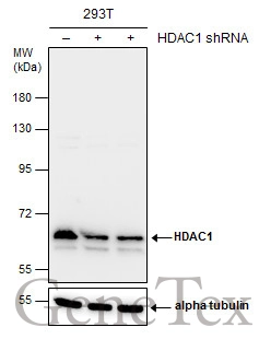
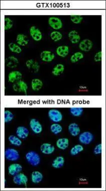
Non-transfected (–) and transfected (+) 293T whole cell extracts (30 μg) were separated by 7.5% SDS-PAGE, and the membrane was blotted with HDAC1 antibody (GTX100513) diluted at 1:4000. The HRP-conjugated anti-rabbit IgG antibody (GTX213110-01) was used to detect the primary antibody.
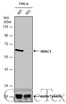
Wild-type (WT) and HDAC1 knockout (KO) HeLa cell extracts (30 μg) were separated by 10% SDS-PAGE, and the membrane was blotted with HDAC1 antibody (GTX100513) diluted at 1:500. The HRP-conjugated anti-rabbit IgG antibody (GTX213110-01) was used to detect the primary antibody.
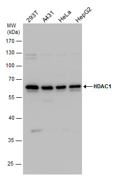
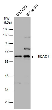
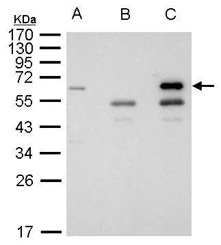
HDAC1 antibody detects HDAC1 protein by western blot analysis. Various whole cell extracts (30 μg) were separated by 10% SDS-PAGE, and the membrane was blotted with HDAC1 antibody (GTX100513) diluted by 1:1000. The HRP-conjugated anti-rabbit IgG antibody (GTX213110-01) was used to detect the primary antibody.
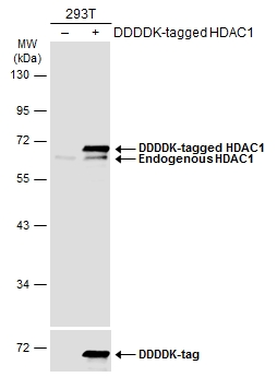
Non-transfected (–) and transfected (+) 293T whole cell extracts (30 μg) were separated by 10% SDS-PAGE, and the membrane was blotted with HDAC1 antibody (GTX100513) diluted at 1:1000. The HRP-conjugated anti-rabbit IgG antibody (GTX213110-01) was used to detect the primary antibody.
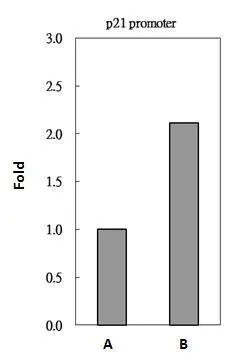
HDAC1 antibody immunoprecipitates HDAC1 protein-DNA in ChIP experiments. ChIP Sample: 293T whole cell lysate/extract A. 5 μg preimmune rabbit IgG B. 5 μg of HDAC1 antibody (GTX100513) The precipitated DNA was detected by PCR with primer set targeting to p21 promoter.
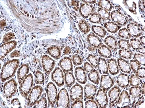
HDAC1 antibody detects HDAC1 protein at nucleus on mouse colon by immunohistochemical analysis.
Sample: Paraffin-embedded mouse colon.
HDAC1 antibody (GTX100513) dilution: 1:500.
Antigen Retrieval: Trilogy™ (EDTA based, pH 8.0) buffer, 15min
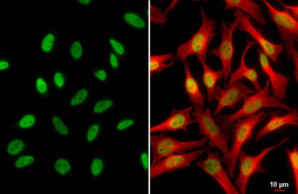
HDAC1 antibody detects HDAC1 protein at nucleus by immunofluorescent analysis.Sample: HeLa cells were fixed in 4% paraformaldehyde at RT for 15 min.Green: HDAC1 stained by HDAC1 antibody (GTX100513) diluted at 1:500.Red: alpha Tubulin, a cytoskeleton marker, stained by alpha Tubulin antibody [GT114] (GTX628802) diluted at 1:1000.Scale bar= 10μm.
Immunofluorescence analysis of paraformaldehyde-fixed A431, using HDAC1(GTX100513) antibody at 1:200 dilution.
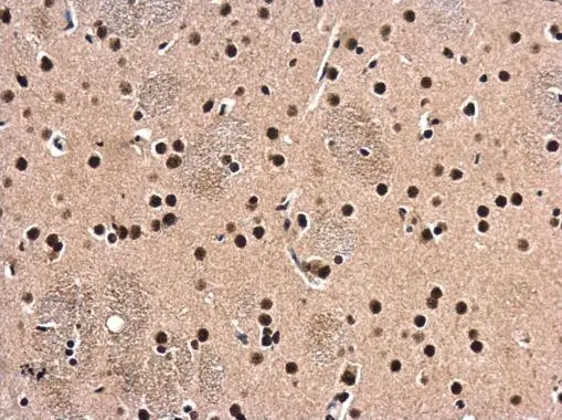
HDAC1 antibody detects HDAC1 protein at cytoplasm and nucleus in rat brain by immunohistochemical analysis.
Sample: Paraffin-embedded rat brain.
HDAC1 antibody (GTX100513) diluted at 1:500.
Antigen Retrieval: Citrate buffer, pH 6.0, 15 min
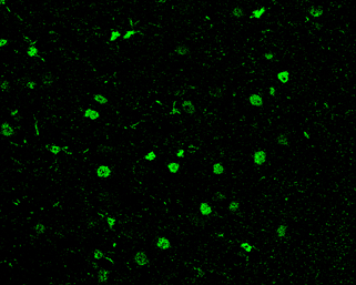
HDAC1 antibody detects HDAC1 protein at nucleus in rat brain by immunohistochemical analysis.
Sample: Paraffin-embedded rat brain.
Green: HDAC1 antibody (GTX100513) diluted at 1:200. The signal was developed using goat anti-rabbit IgG antibody (Dylight488) (GTX213110-04).
Antigen Retrieval: Citrate buffer, pH 6.0, 15 min
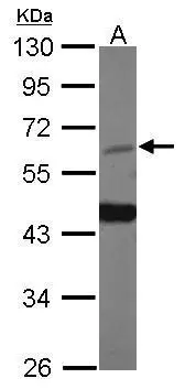
Various whole cell extracts (30 μg) were separated by 10% SDS-PAGE, and the membrane was blotted with HDAC1 antibody (GTX100513) diluted at 1:1000. The HRP-conjugated anti-rabbit IgG antibody (GTX213110-01) was used to detect the primary antibody.
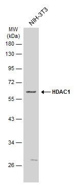
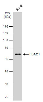
Whole cell extract (30 μg) was separated by 10% SDS-PAGE, and the membrane was blotted with HDAC1 antibody (GTX100513) diluted at 1:1000. The HRP-conjugated anti-rabbit IgG antibody (GTX213110-01) was used to detect the primary antibody.
HDAC1antibody immunoprecipitates HDAC1 protein in IP experiments. IP Sample: 1000 μg 293T whole cell lysate/extract A. 40 μg 293T whole cell lysate/extract B. Control with 2.5 μg of preimmune rabbit IgG C. Immunoprecipitation of HDAC1 protein by 2.5 μg of HDAC1 antibody (GTX100513) 10% SDS-PAGE The immunoprecipitated HDAC1 protein was detected by HDAC1 antibody (GTX100513) diluted at 1:1000. EasyBlot anti-rabbit IgG (GTX221666-01) was used as a secondary reagent.
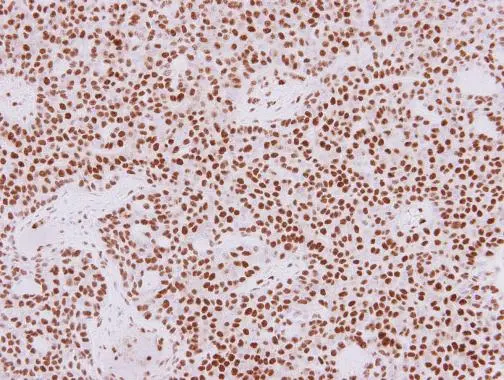
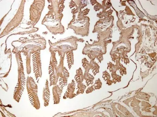
HDAC1 antibody detects HDAC1 protein at nucleus in human lung adenocarcinoma by immunohistochemical analysis.
Sample: Paraffin-embedded human lung adenocarcinoma.
HDAC1 antibody (GTX100513) diluted at 1:250.
Antigen Retrieval: Citrate buffer, pH 6.0, 15 min
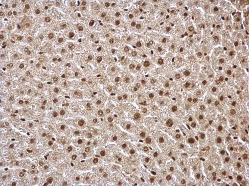
HDAC1 antibody detects HDAC1 protein at nucleus on mouse liver by immunohistochemical analysis.
Sample: Paraffin-embedded mouse liver.
HDAC1 antibody (GTX100513) dilution: 1:500.
Antigen Retrieval: Trilogy™ (EDTA based, pH 8.0) buffer, 15min
Whole cell extract (30 μg) was separated by 10% SDS-PAGE, and the membrane was blotted with HDAC1 antibody (GTX100513) diluted at 1:1000. The HRP-conjugated anti-rabbit IgG antibody (GTX213110-01) was used to detect the primary antibody.
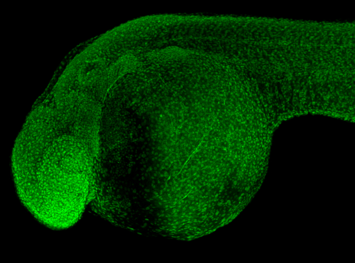
HDAC1 antibody detects Hdac1 protein on zebrafish by whole mount immunohistochemical analysis.
Sample: 2 days-post-fertilization zebrafish embryo.
HDAC1 antibody (GTX100513) dilution: 1:100.
Sample (30 μg of whole cell lysate)
A: zebrafish eye
10% SDS PAGE
GTX100513 diluted at 1:1000
Immunohistochemical analysis of paraffin-embedded zebrafish tissue, using HDAC1 antibody (GTX100513) at 1:300 dilution.
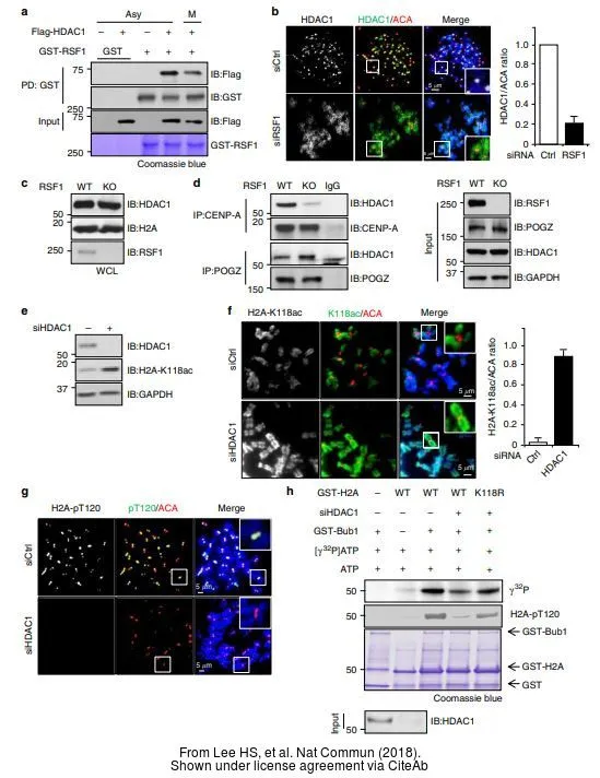
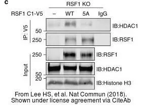
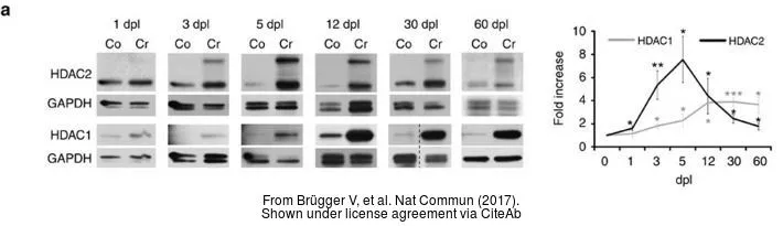
The data was published in the journal Nat Commun in 2017. PMID: 28139683
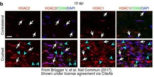
The data was published in the journal Nat Commun in 2017. PMID: 28139683
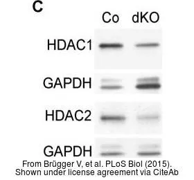
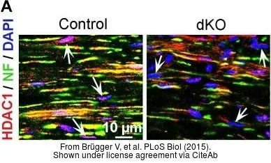
The data was published in the journal PLoS Biol in 2015.PMID: 26406915
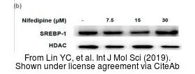
The data was published in the journal Int J Mol Sci in 2019.PMID: 30934807
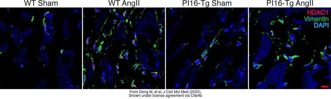
The data was published in the journal J Cell Mol Med in 2020.PMID: 32227584
-
HostRabbit
-
ClonalityPolyclonal
-
IsotypeIgG
-
ApplicationsWB ICC/IF IHC-P IHC-Fr IHC-Wm IP ChIP assay
-
ReactivityHuman, Mouse, Rat, Zebrafish