Normal Tissue Gallery- GTX04438

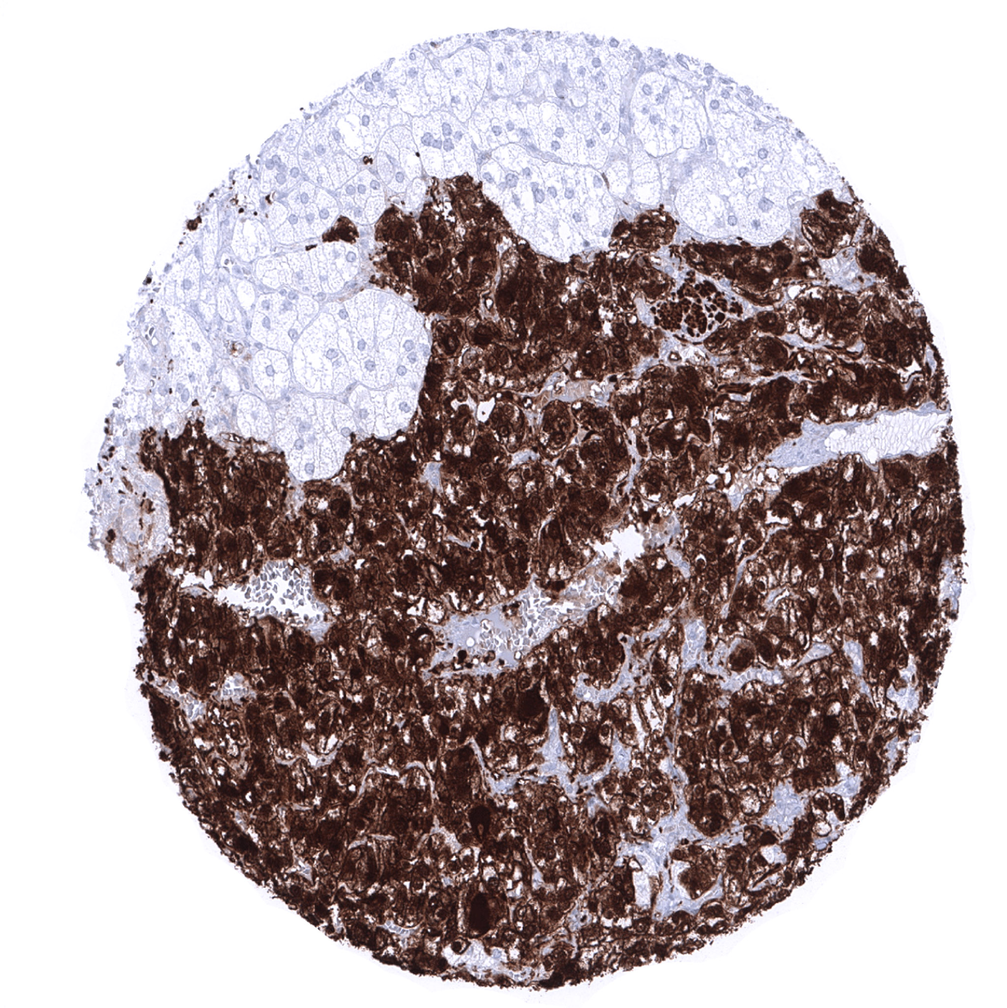
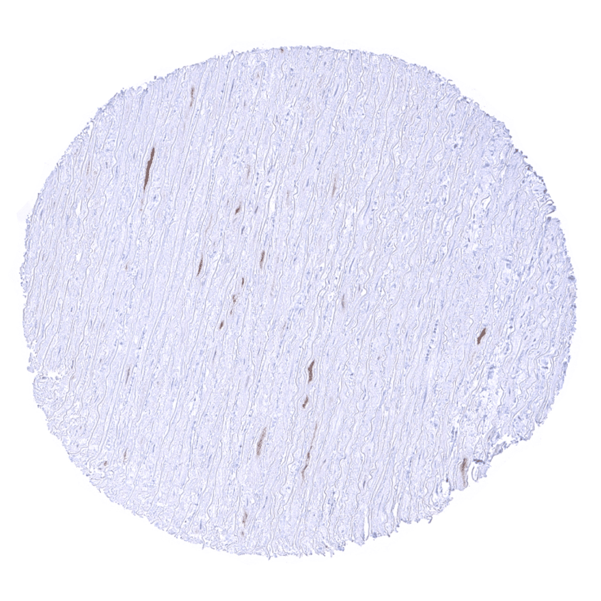
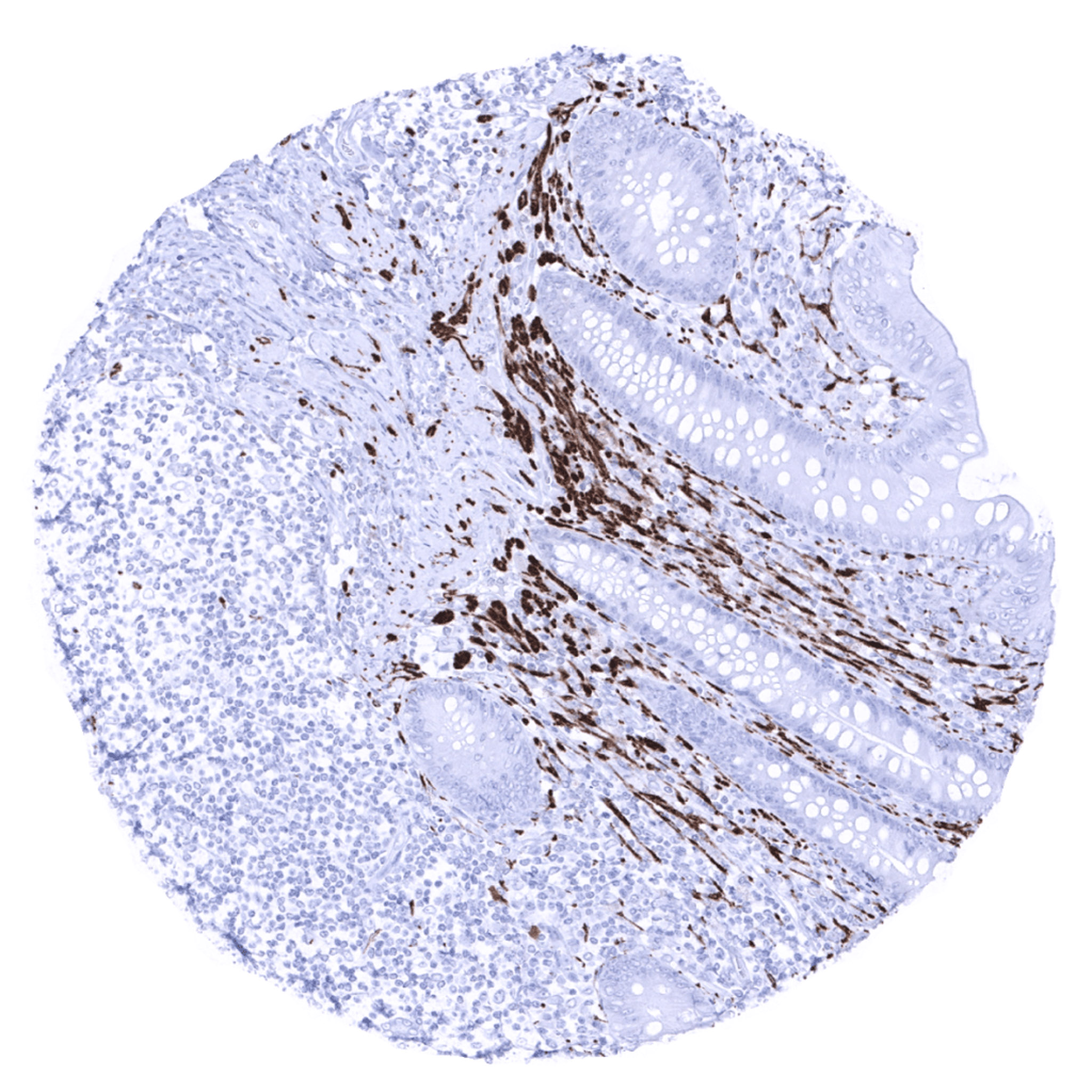
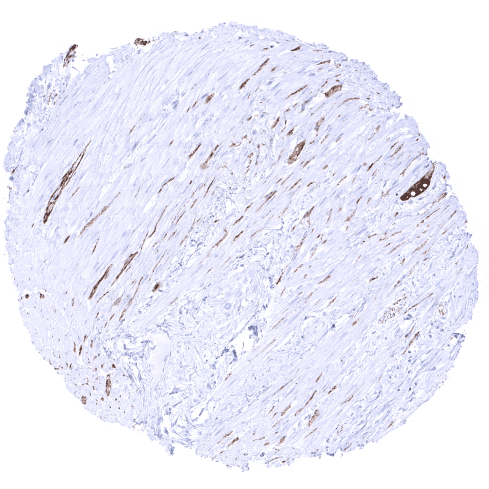
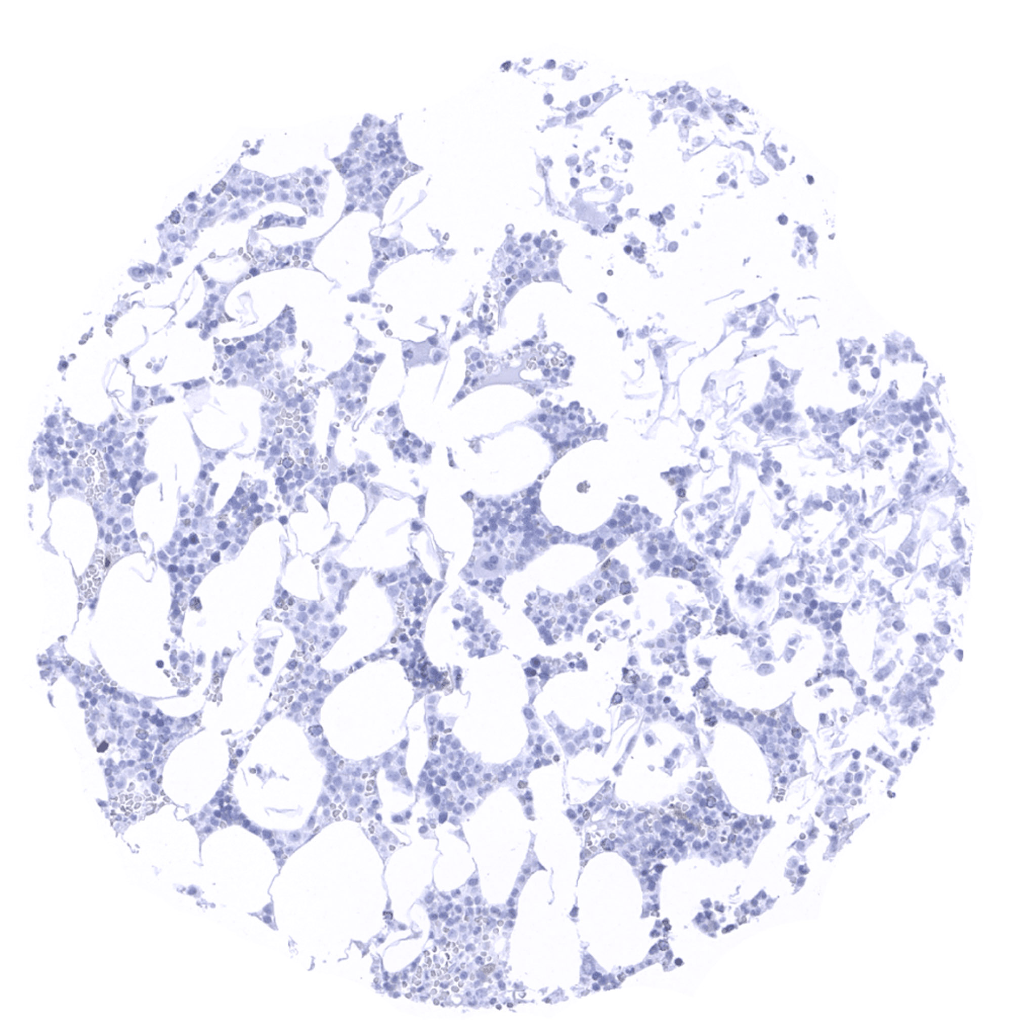
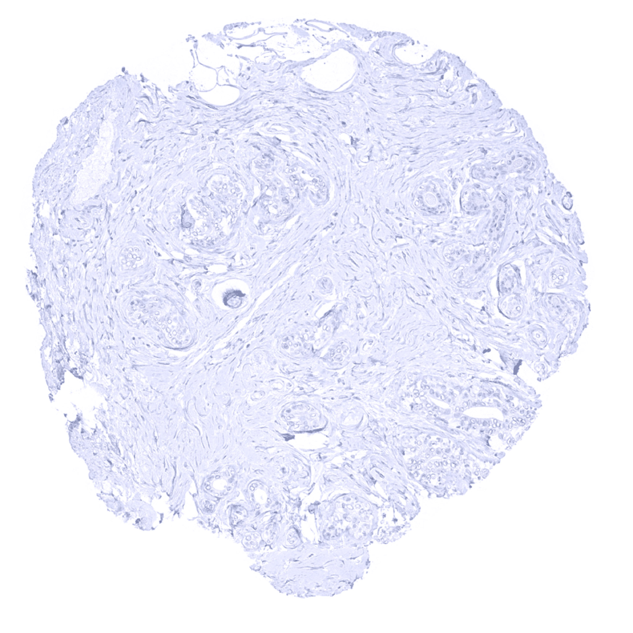
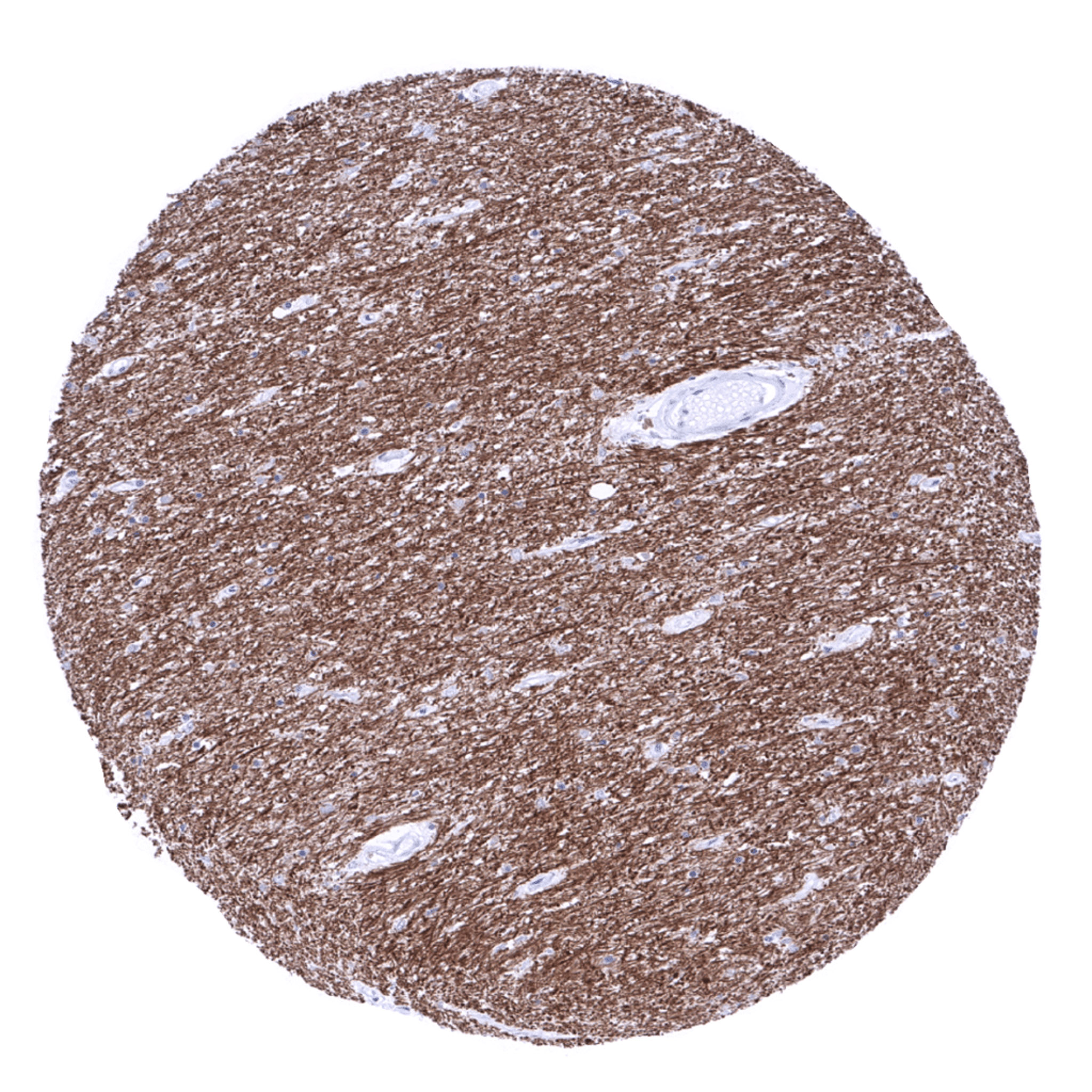
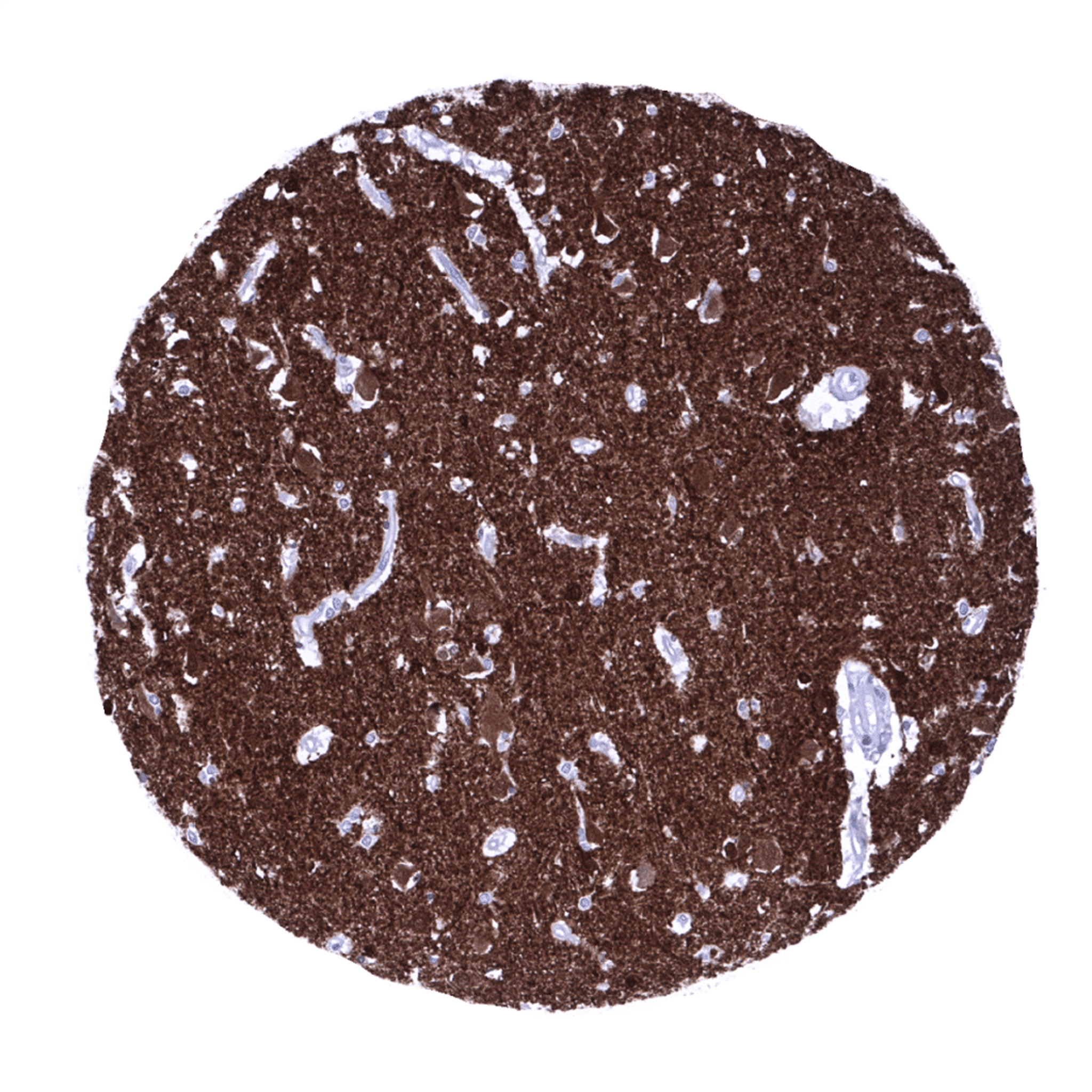
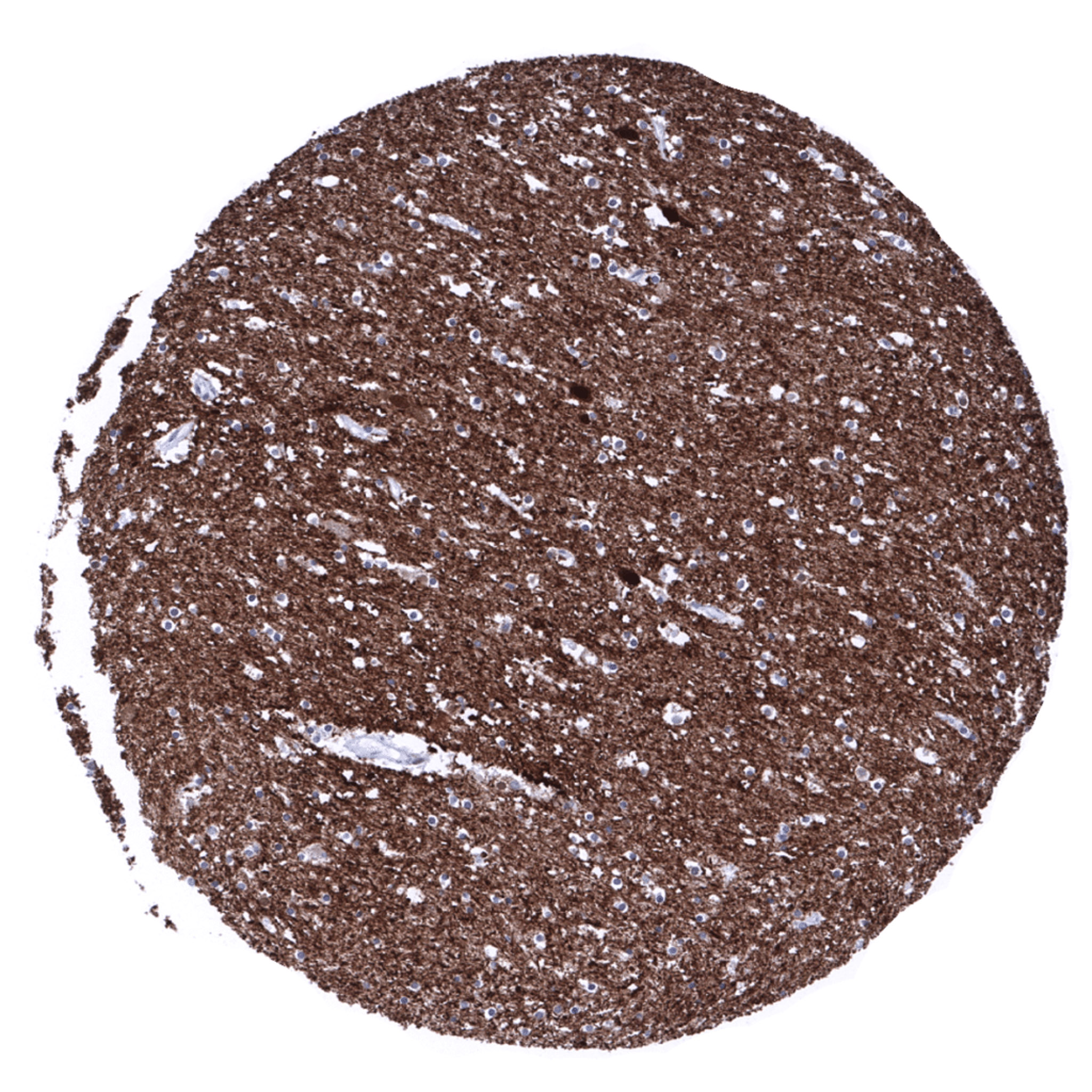
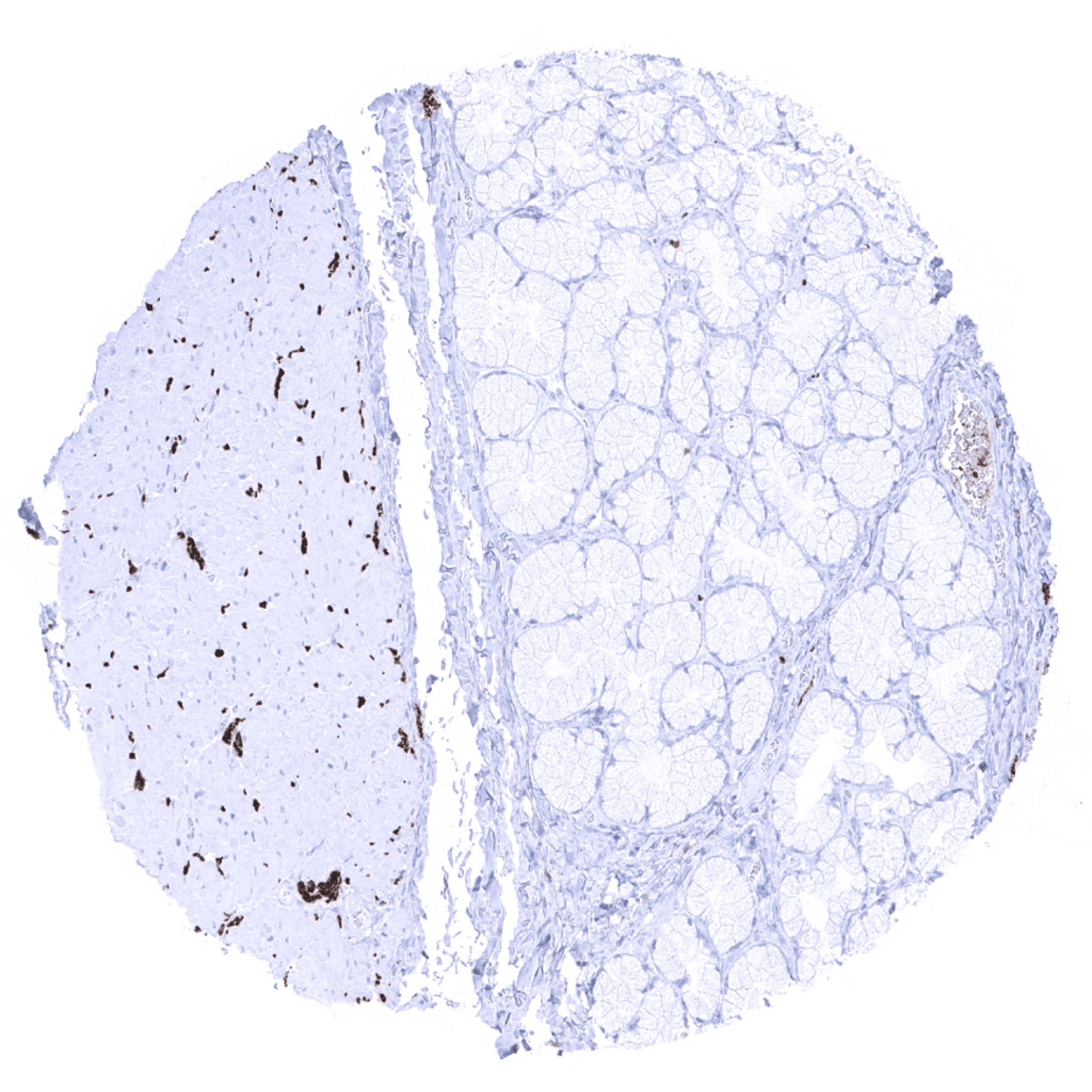
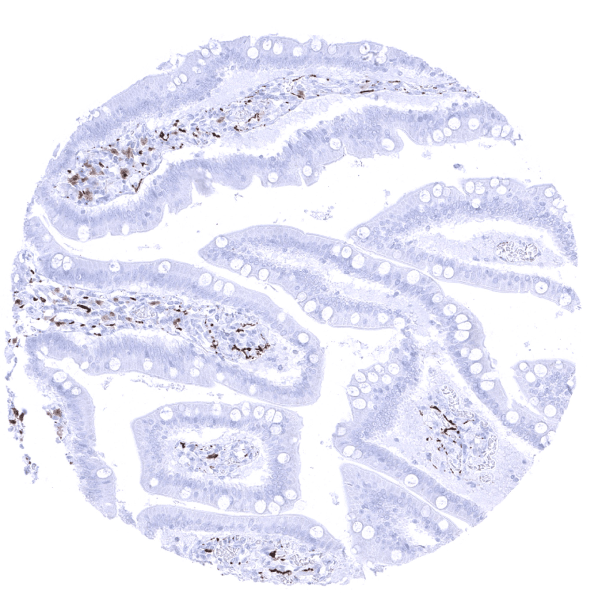
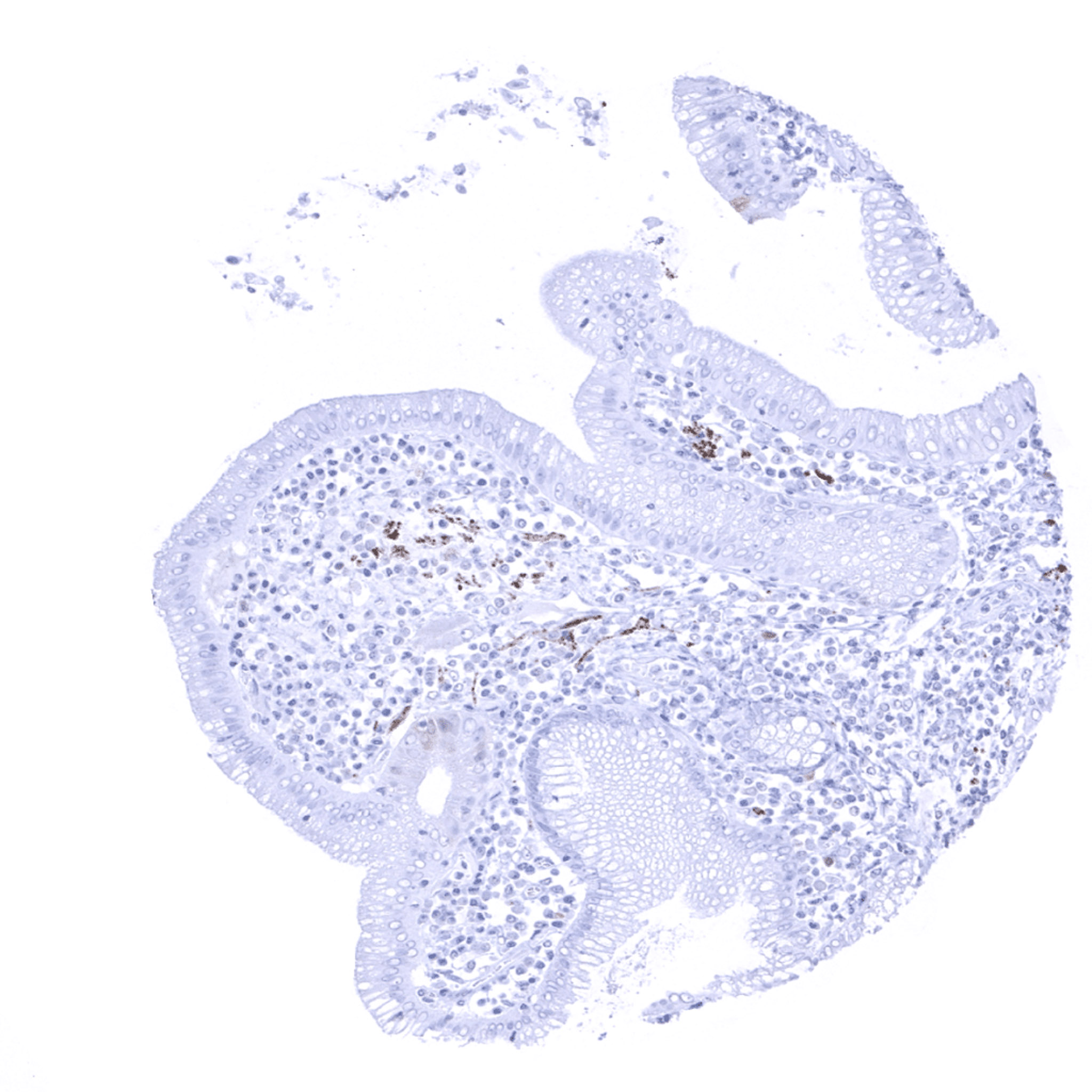
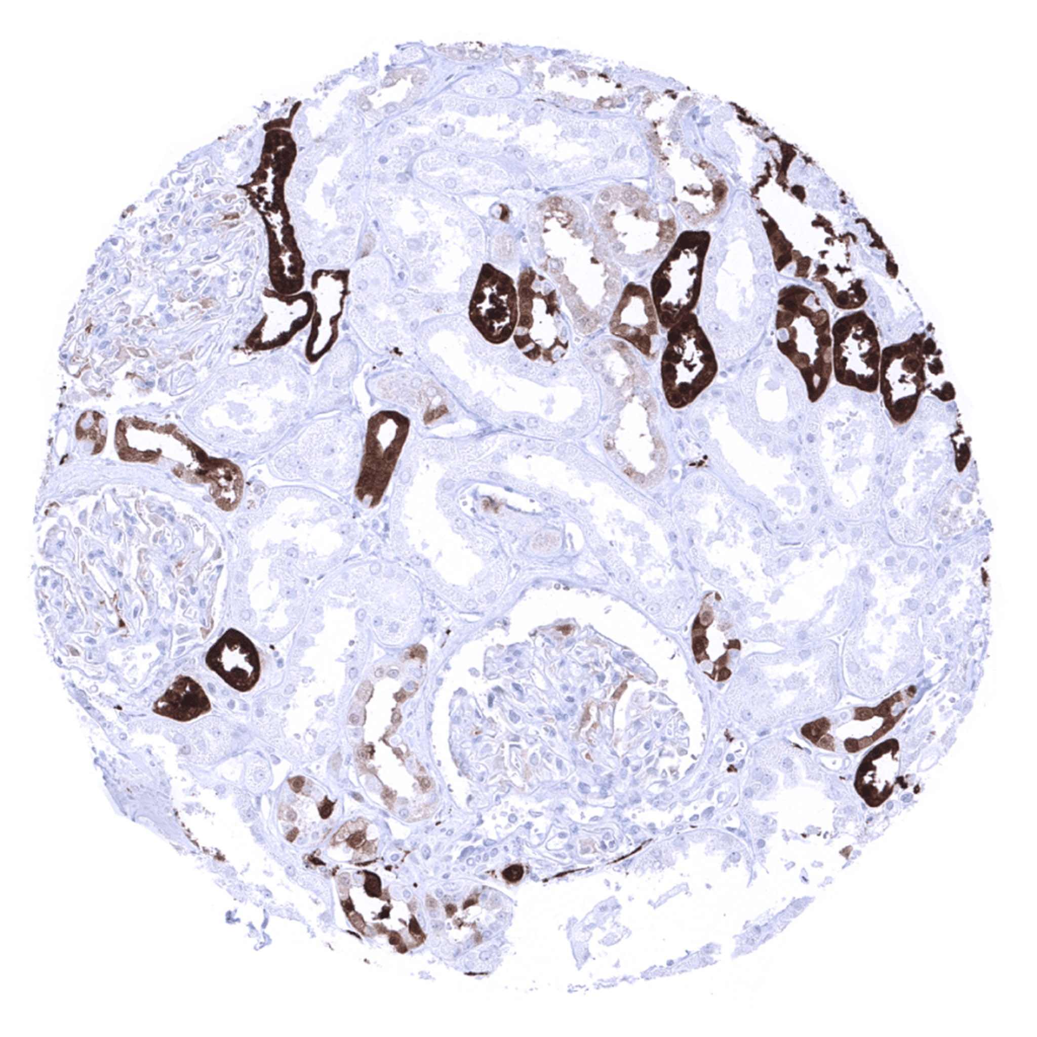
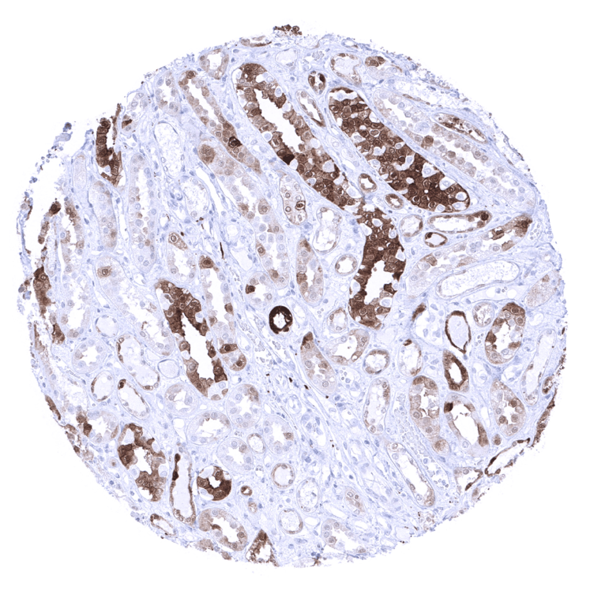
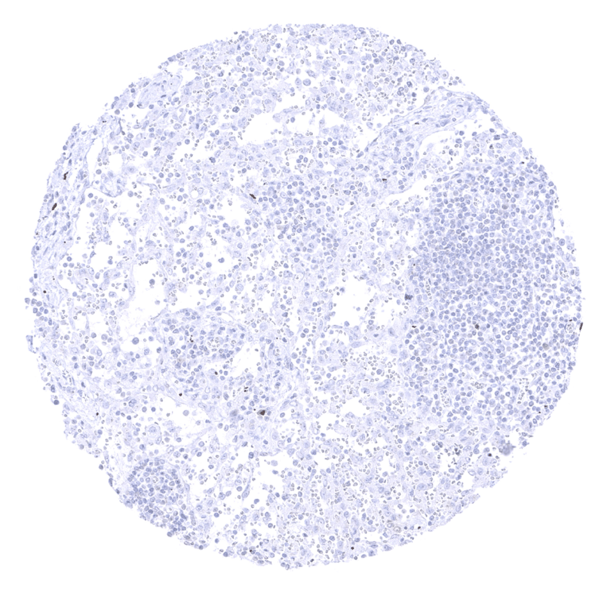
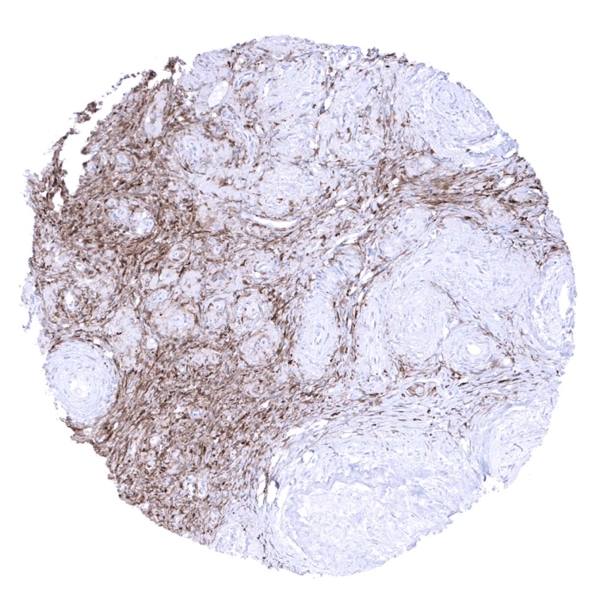
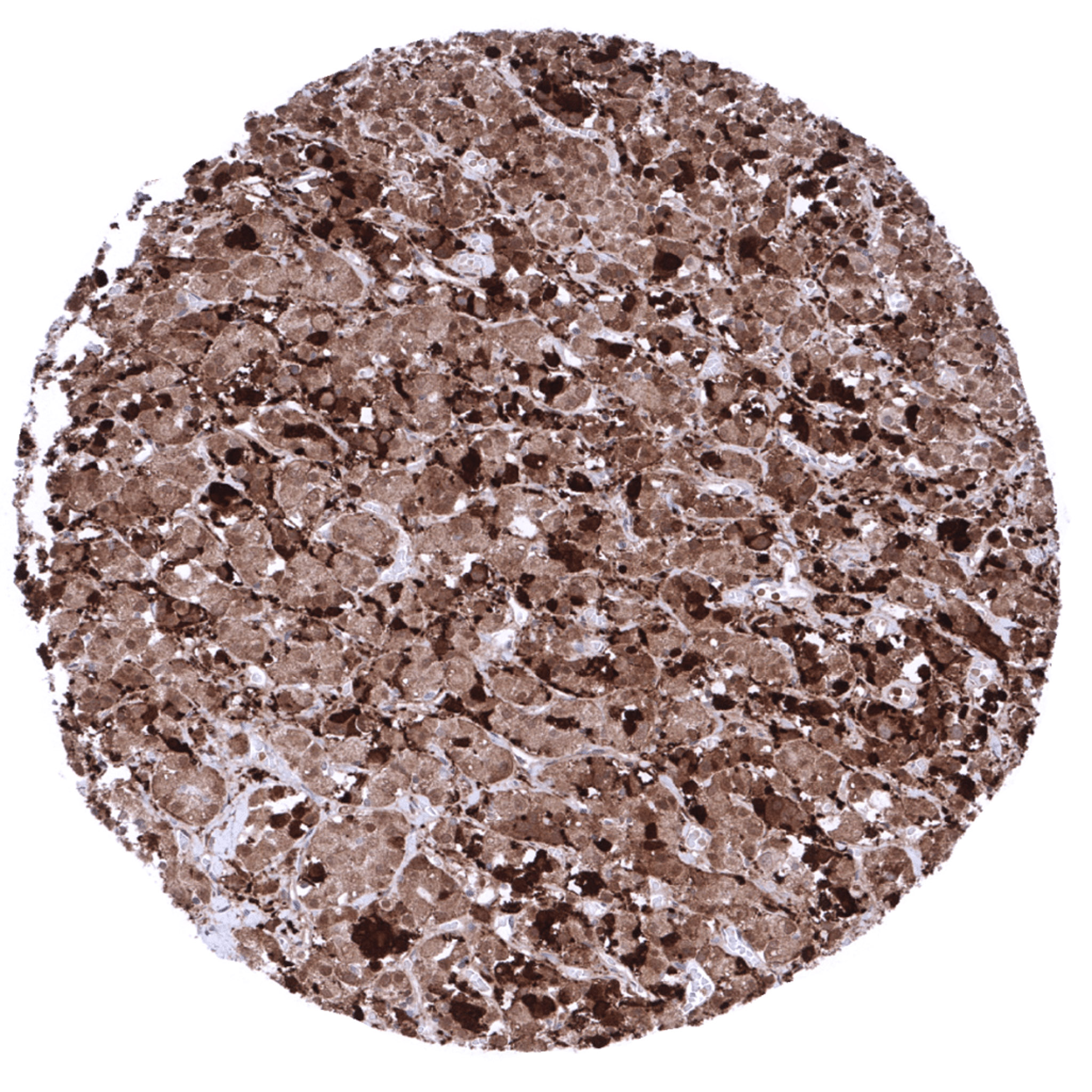
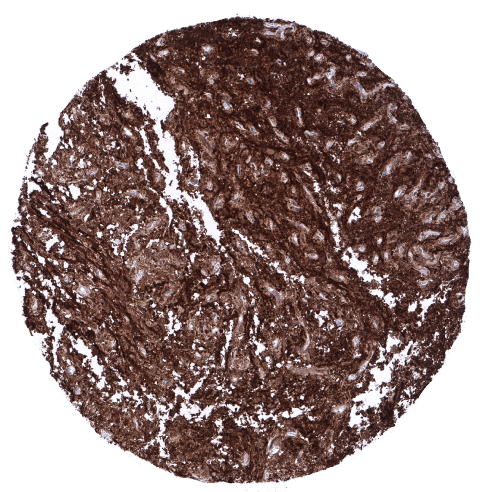
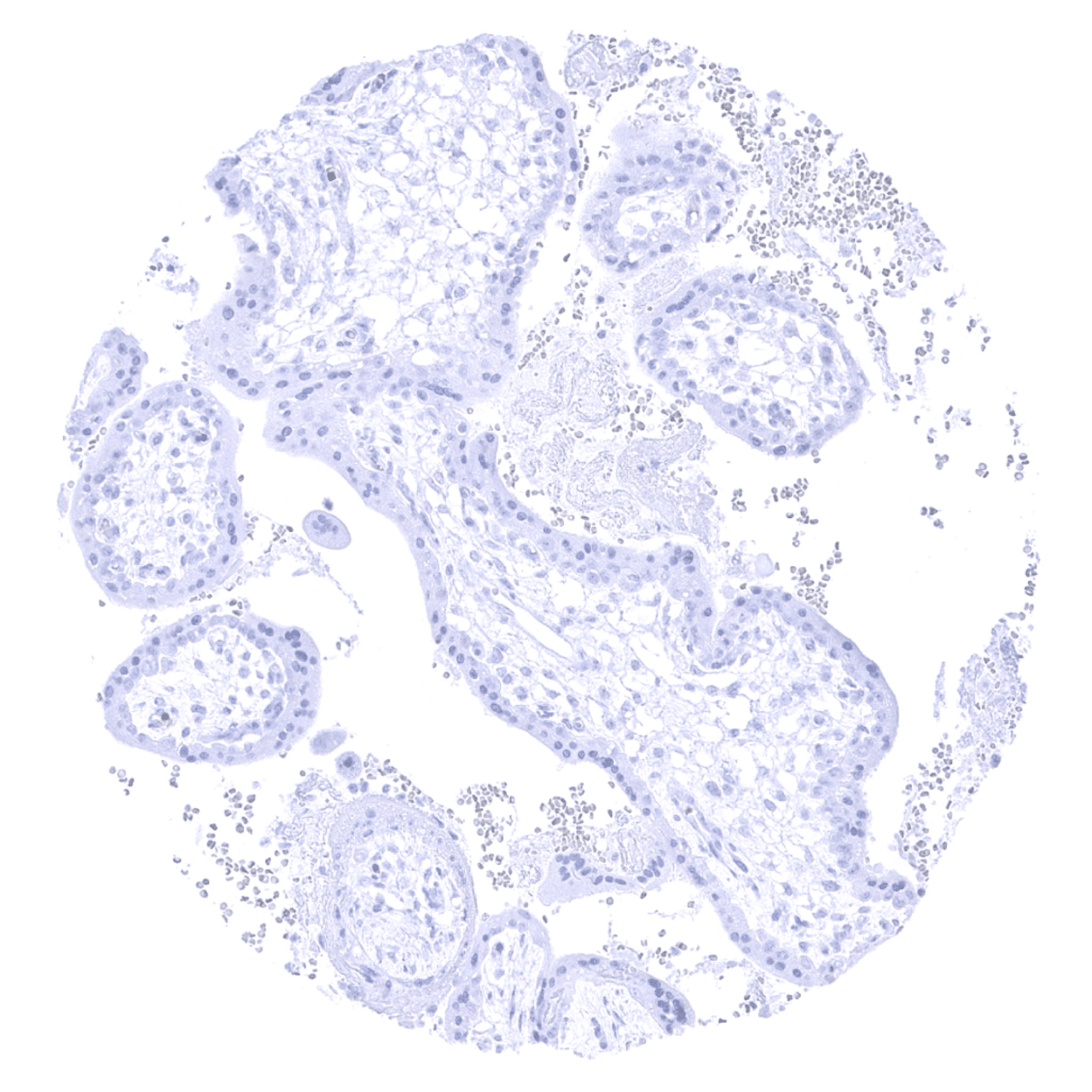
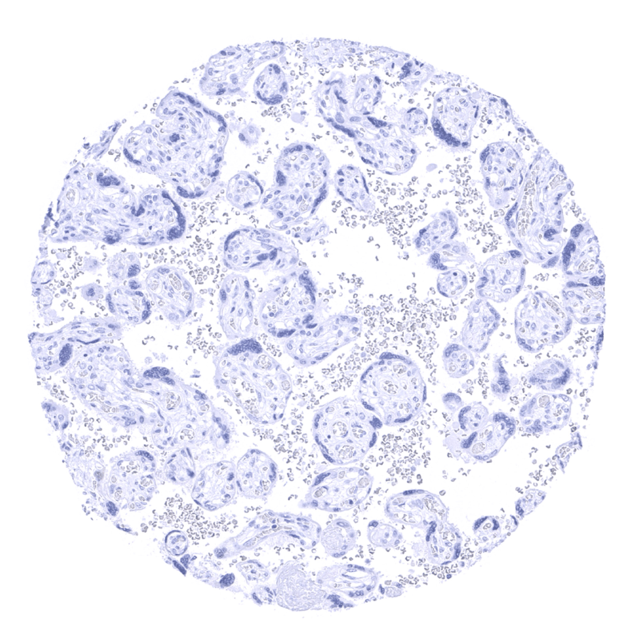
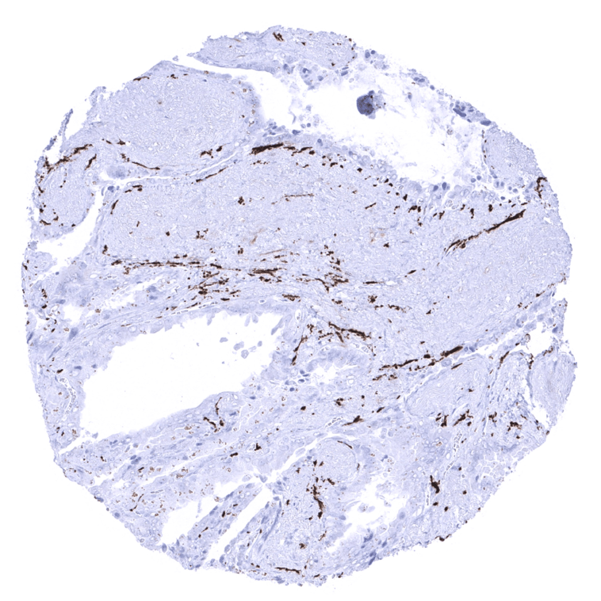
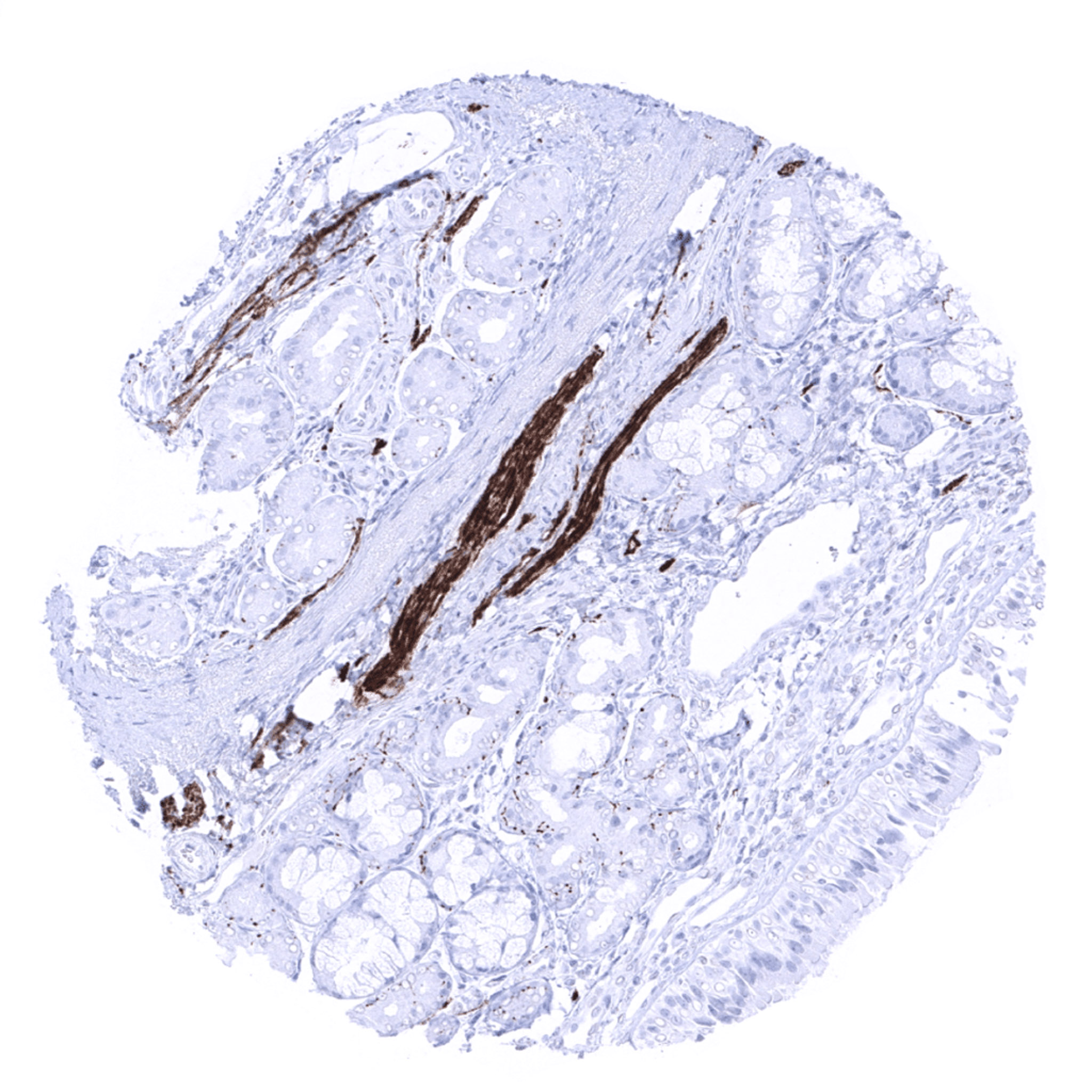
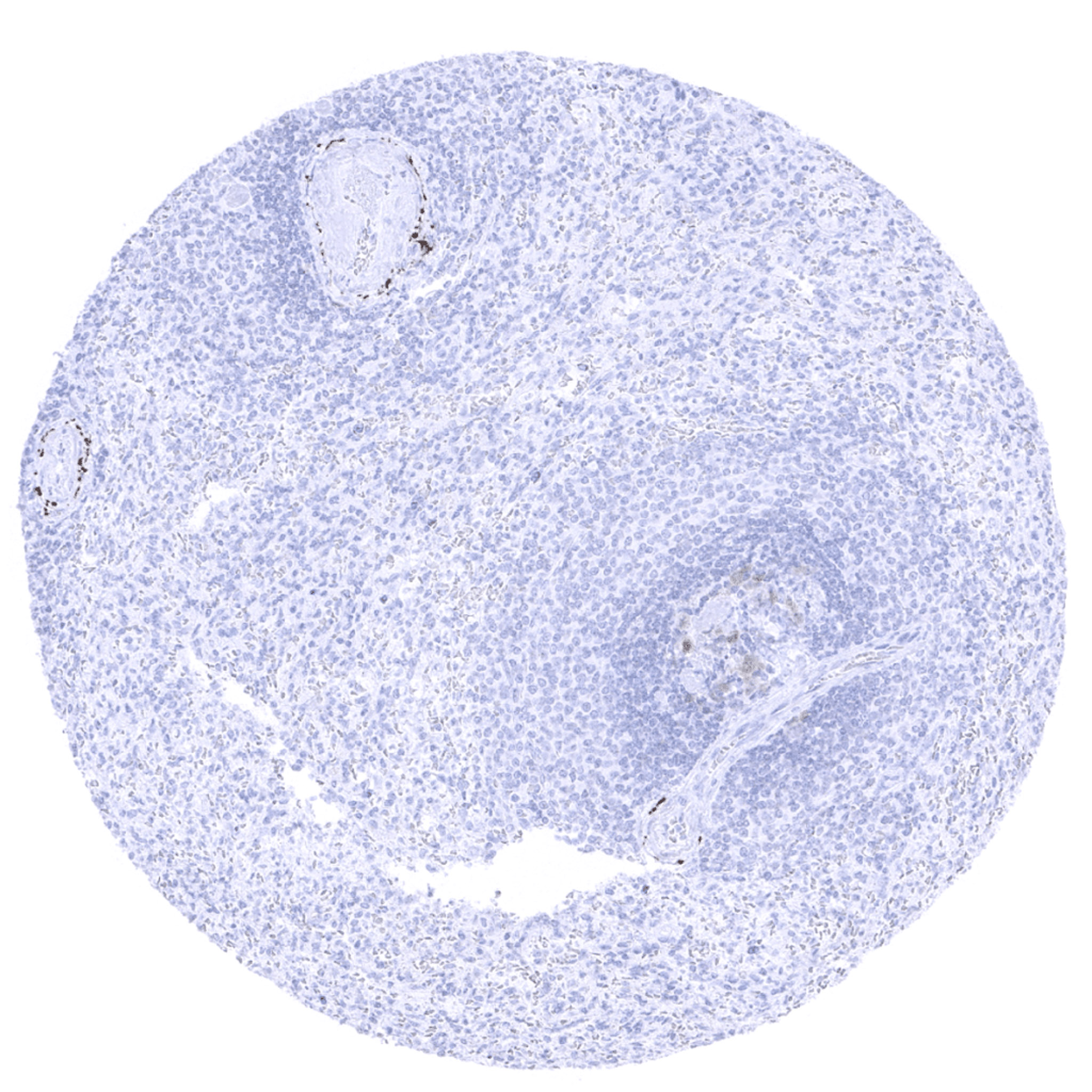
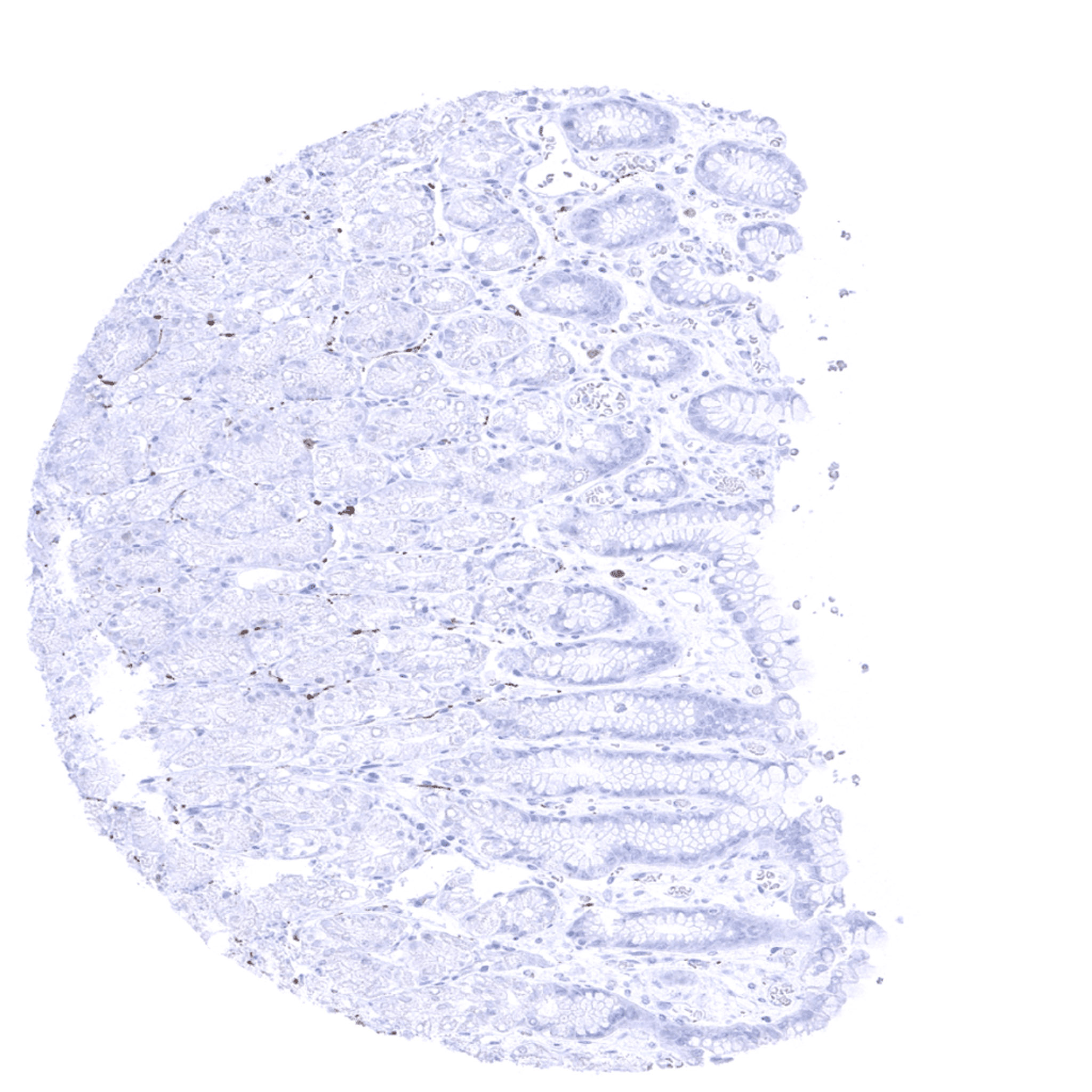
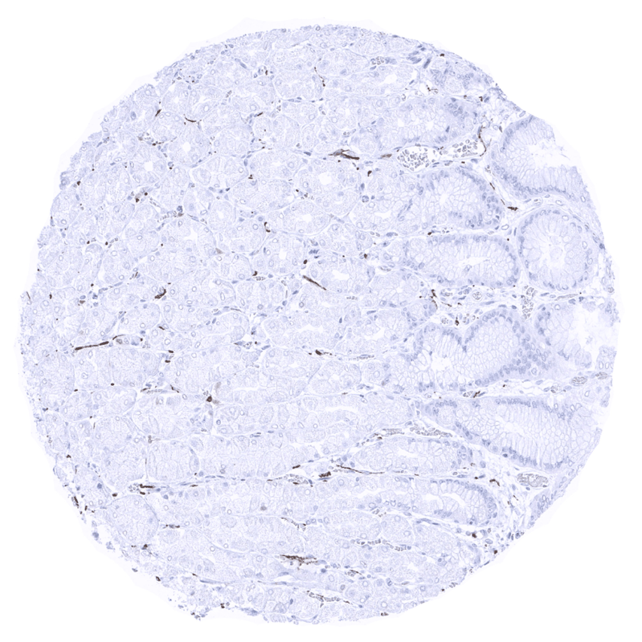
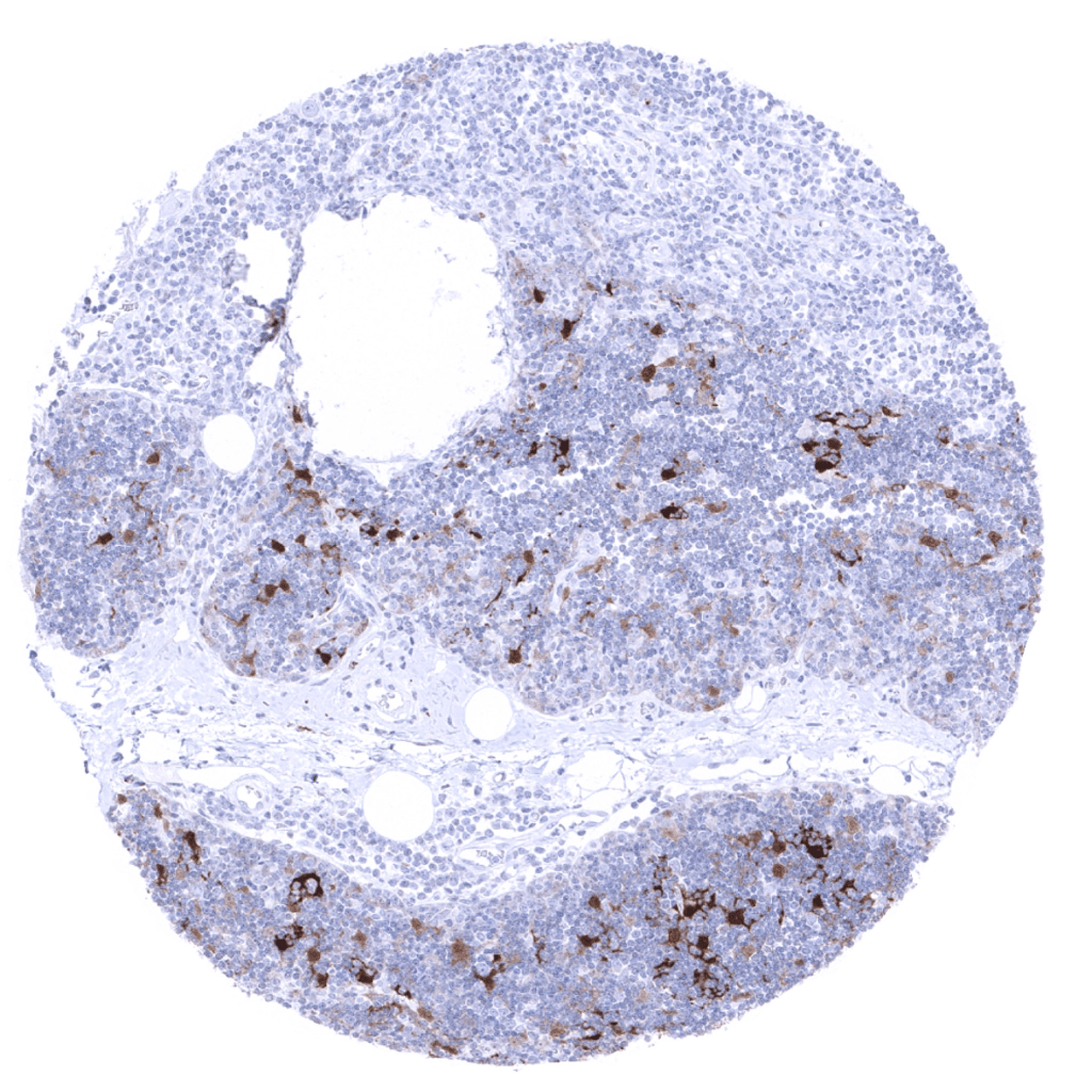
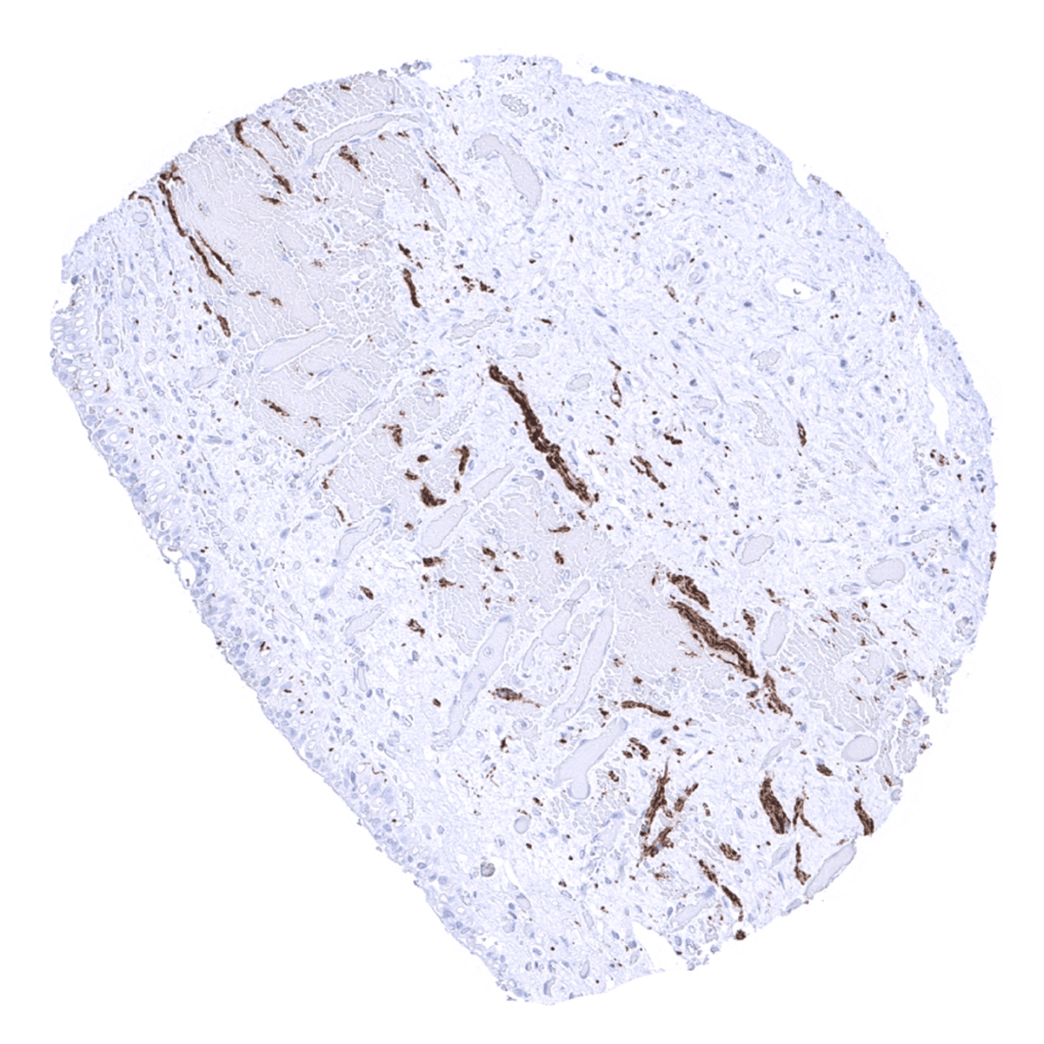
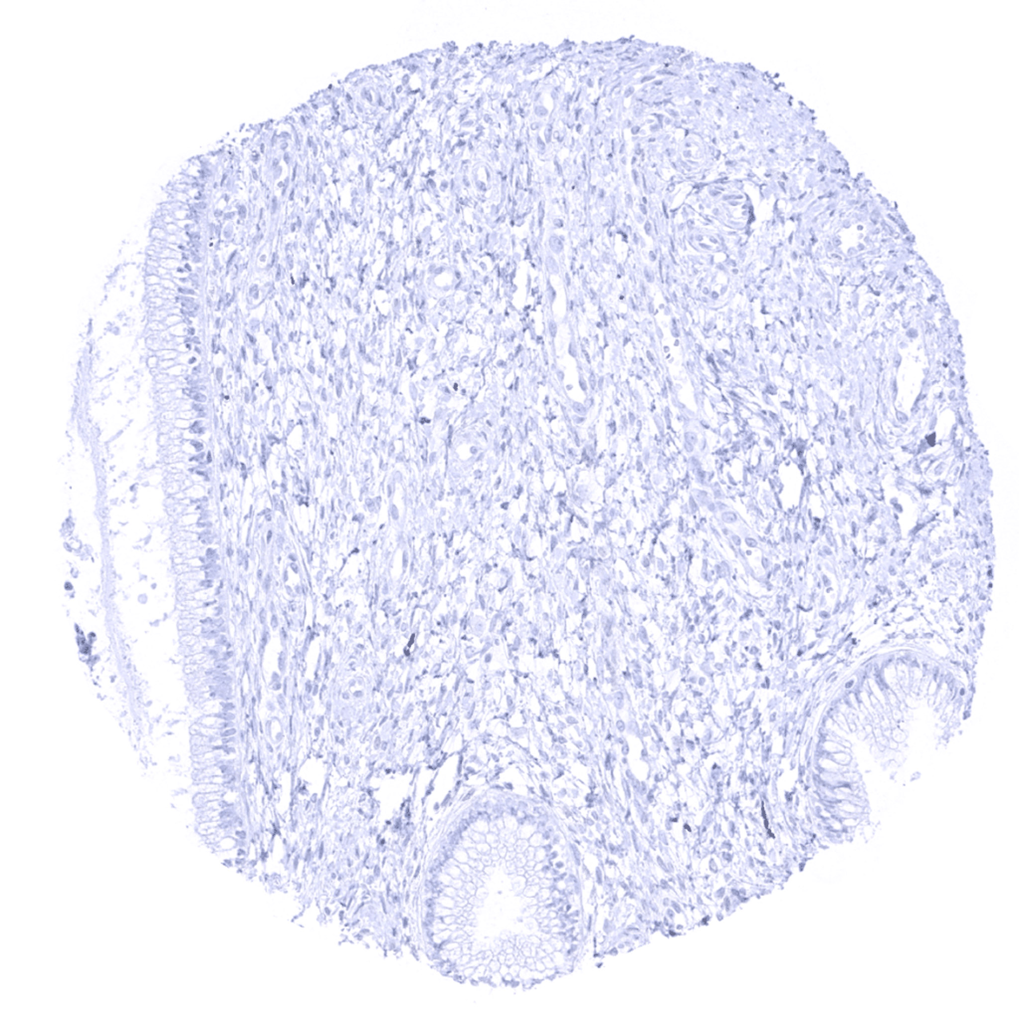
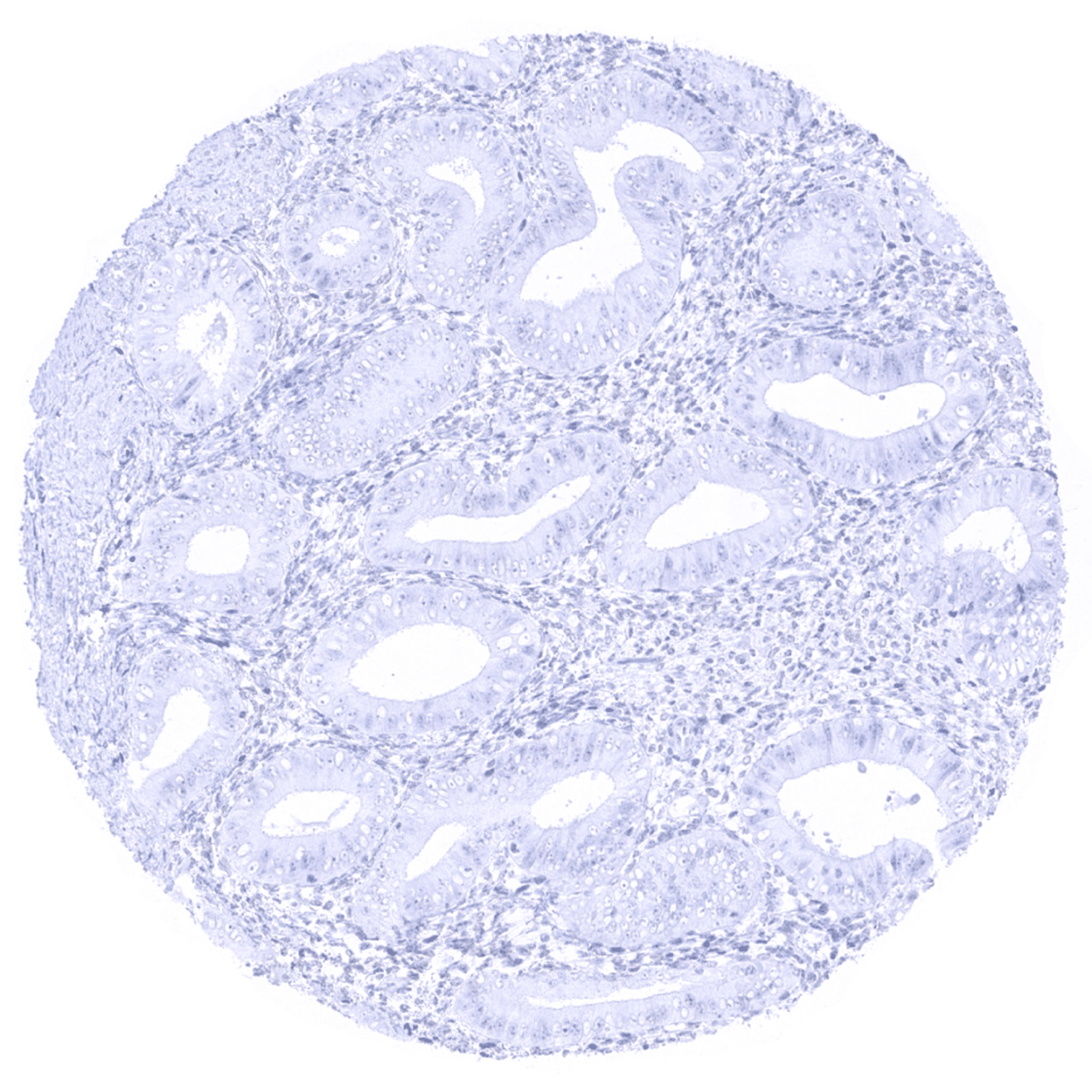
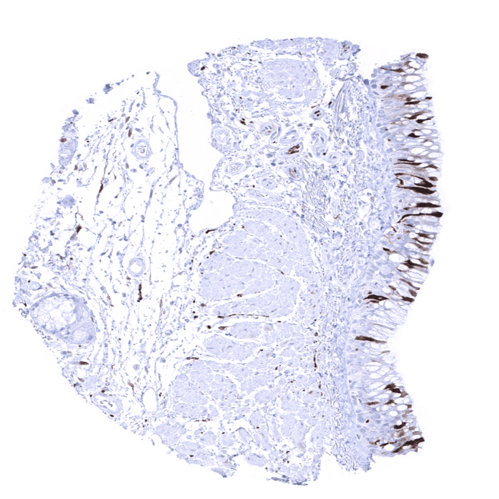
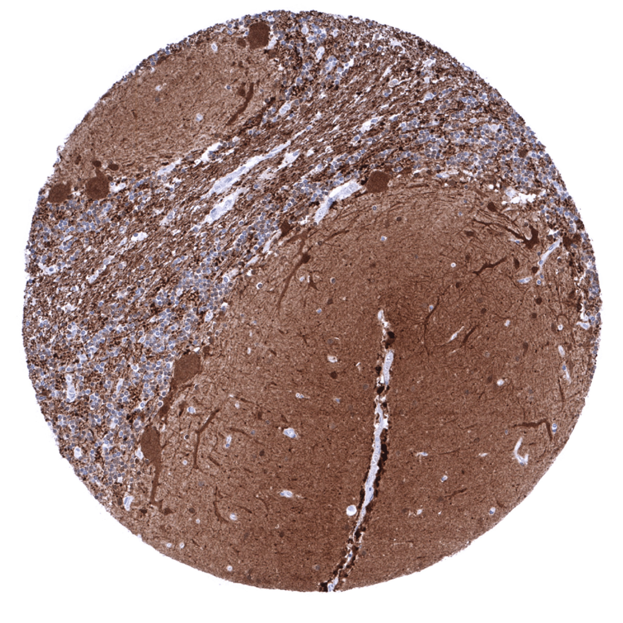
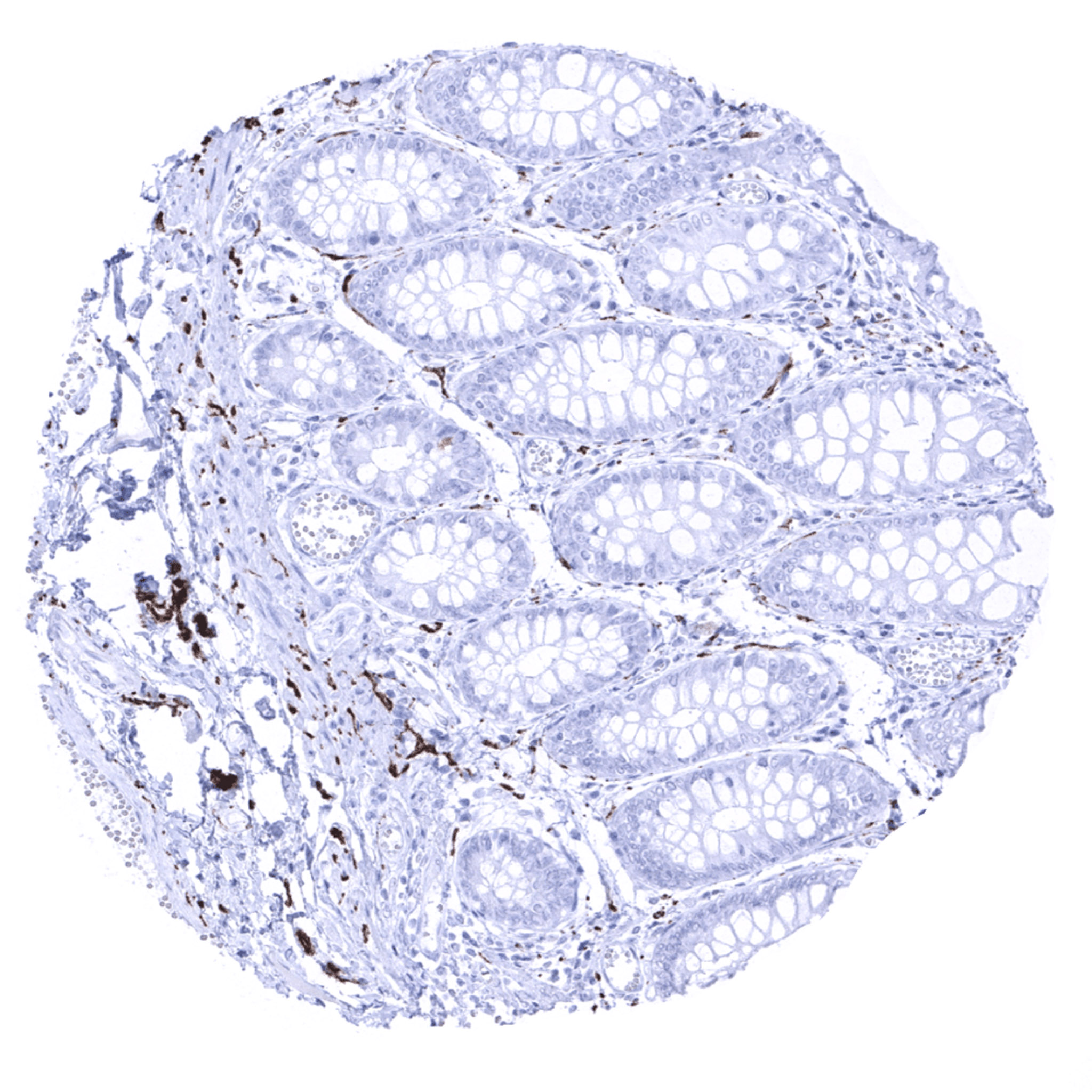
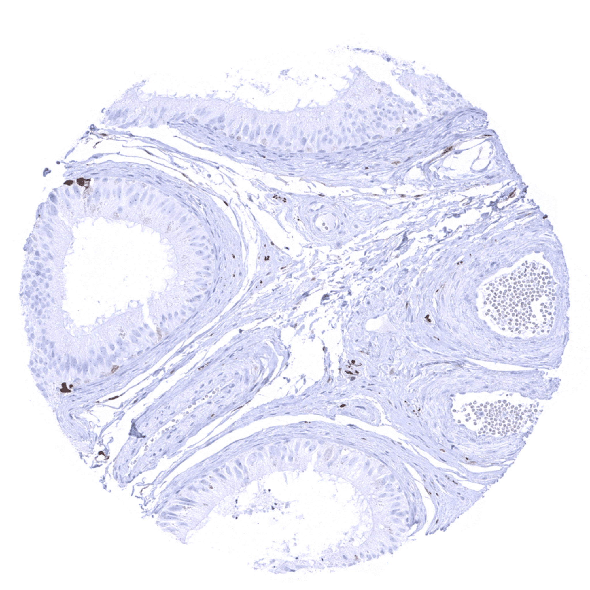
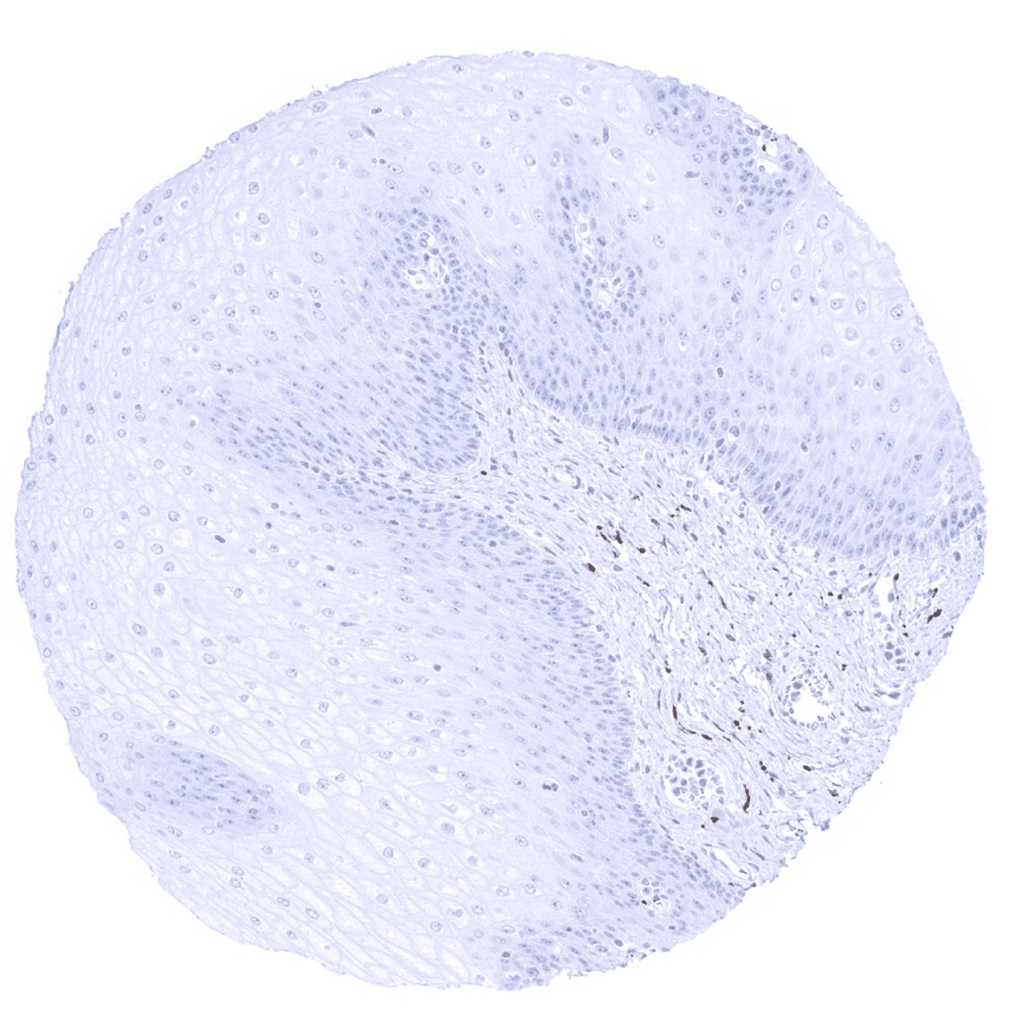
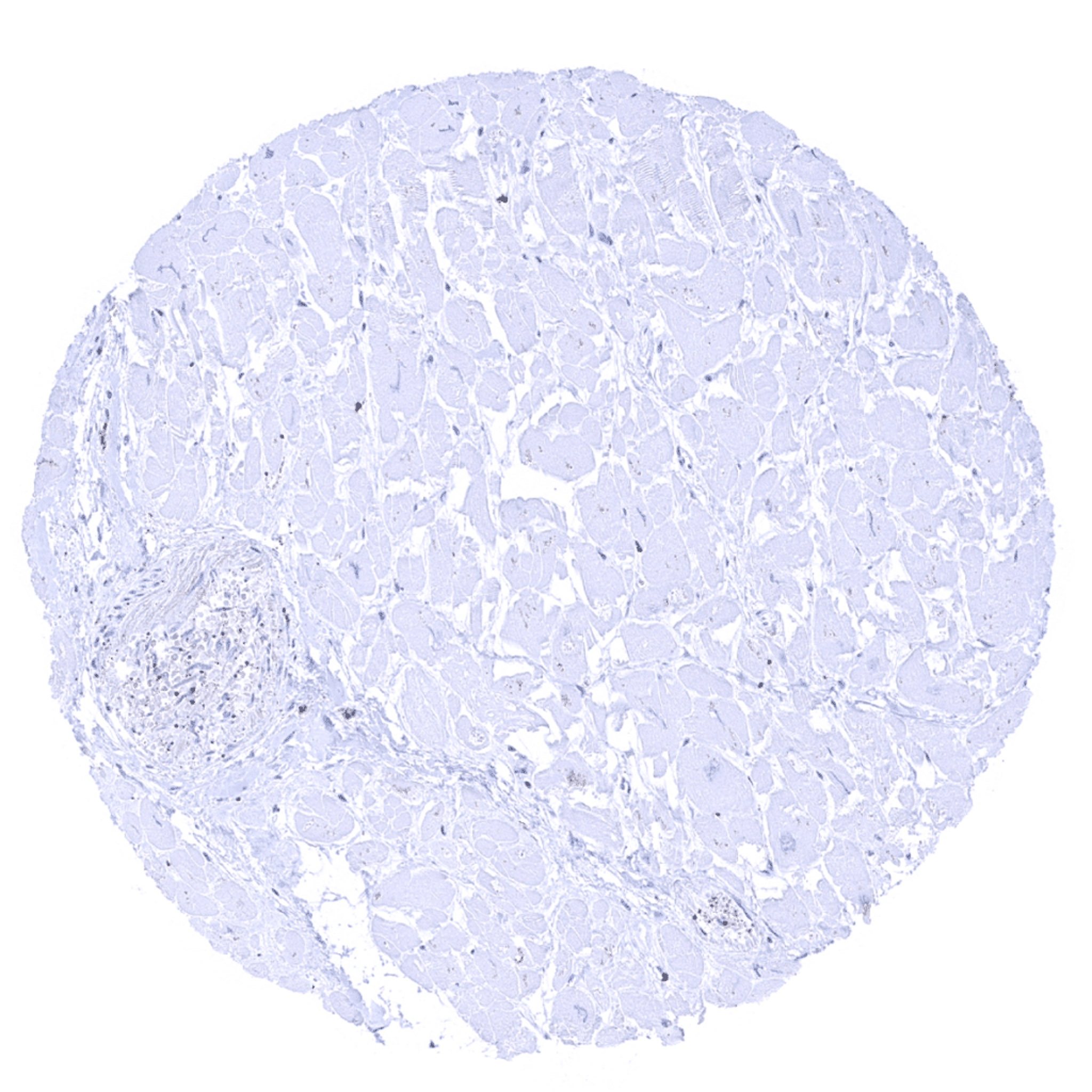
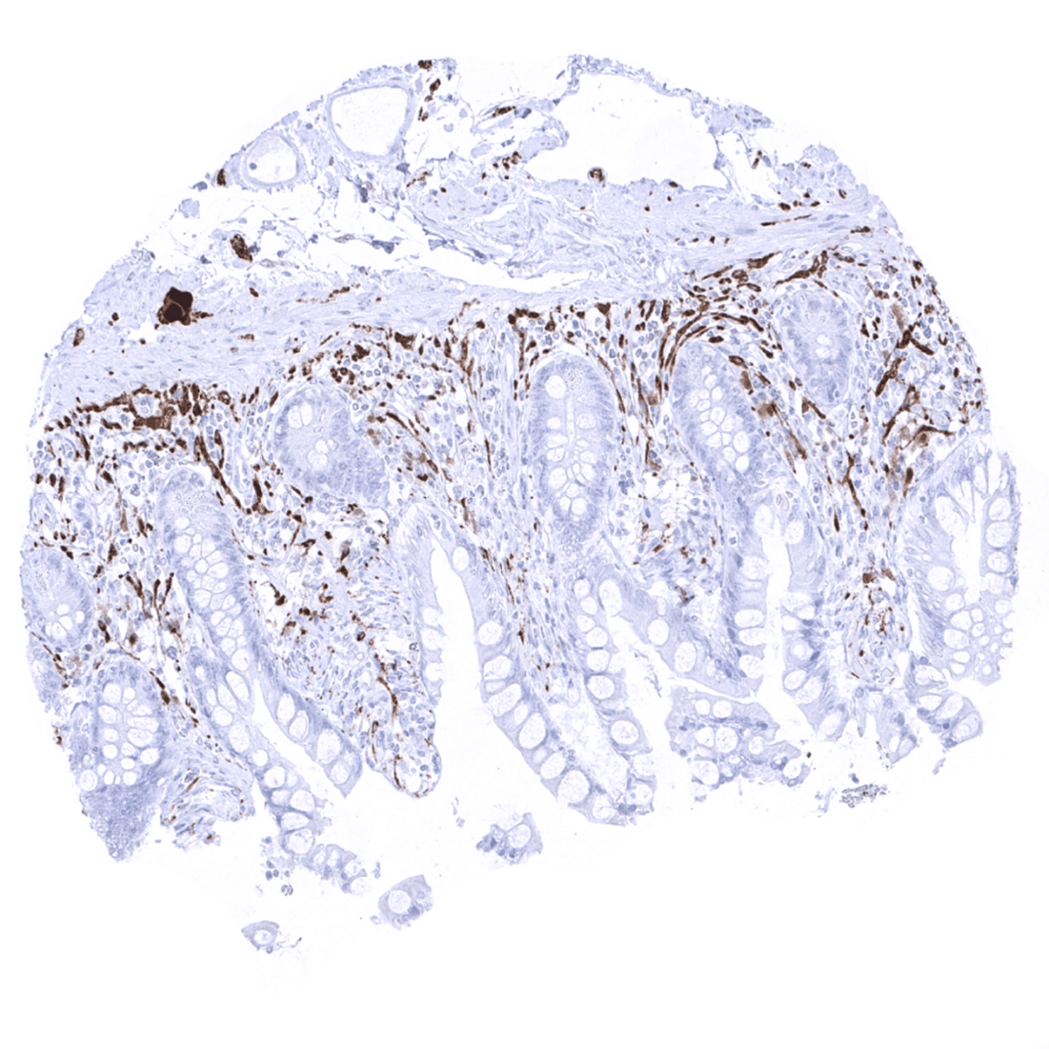
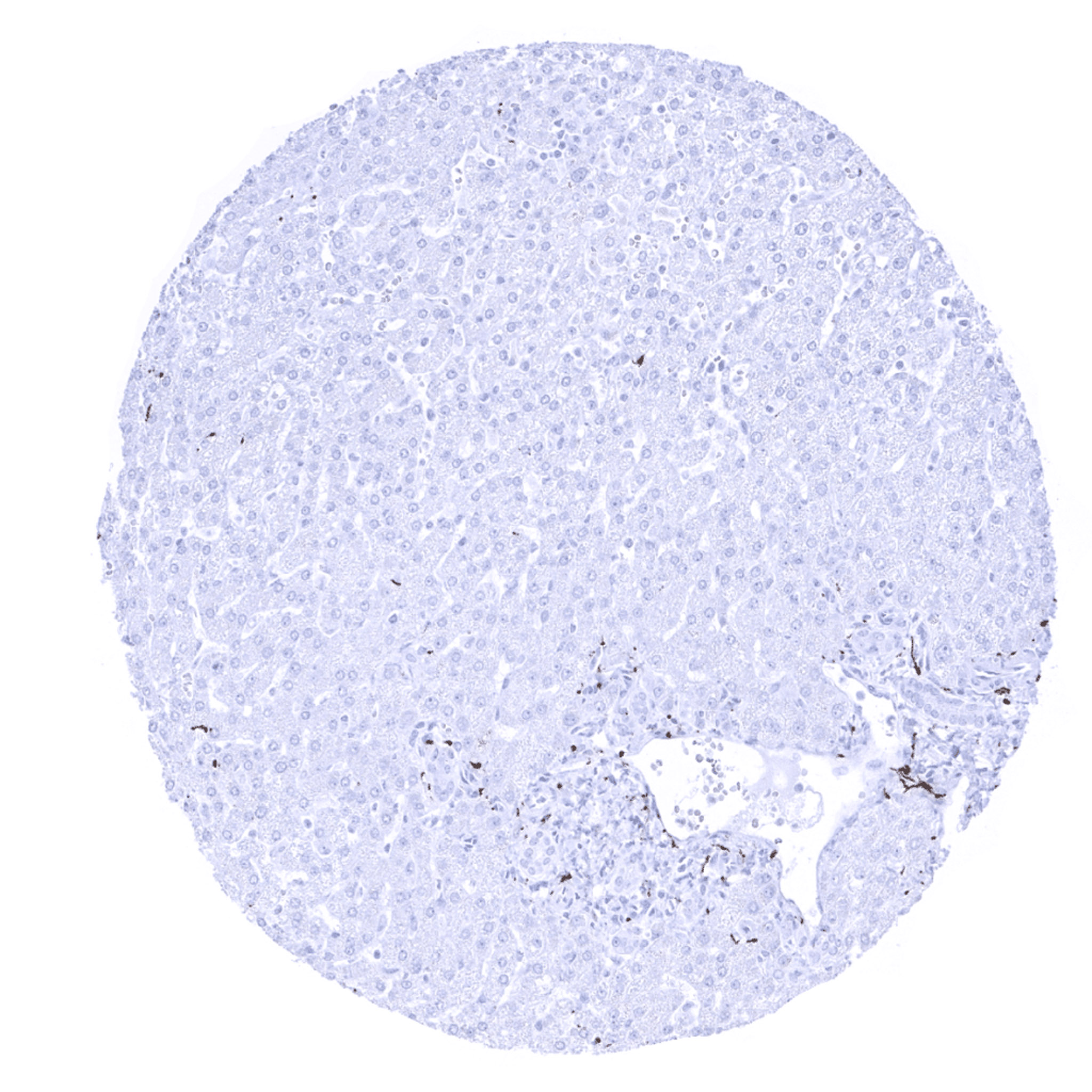
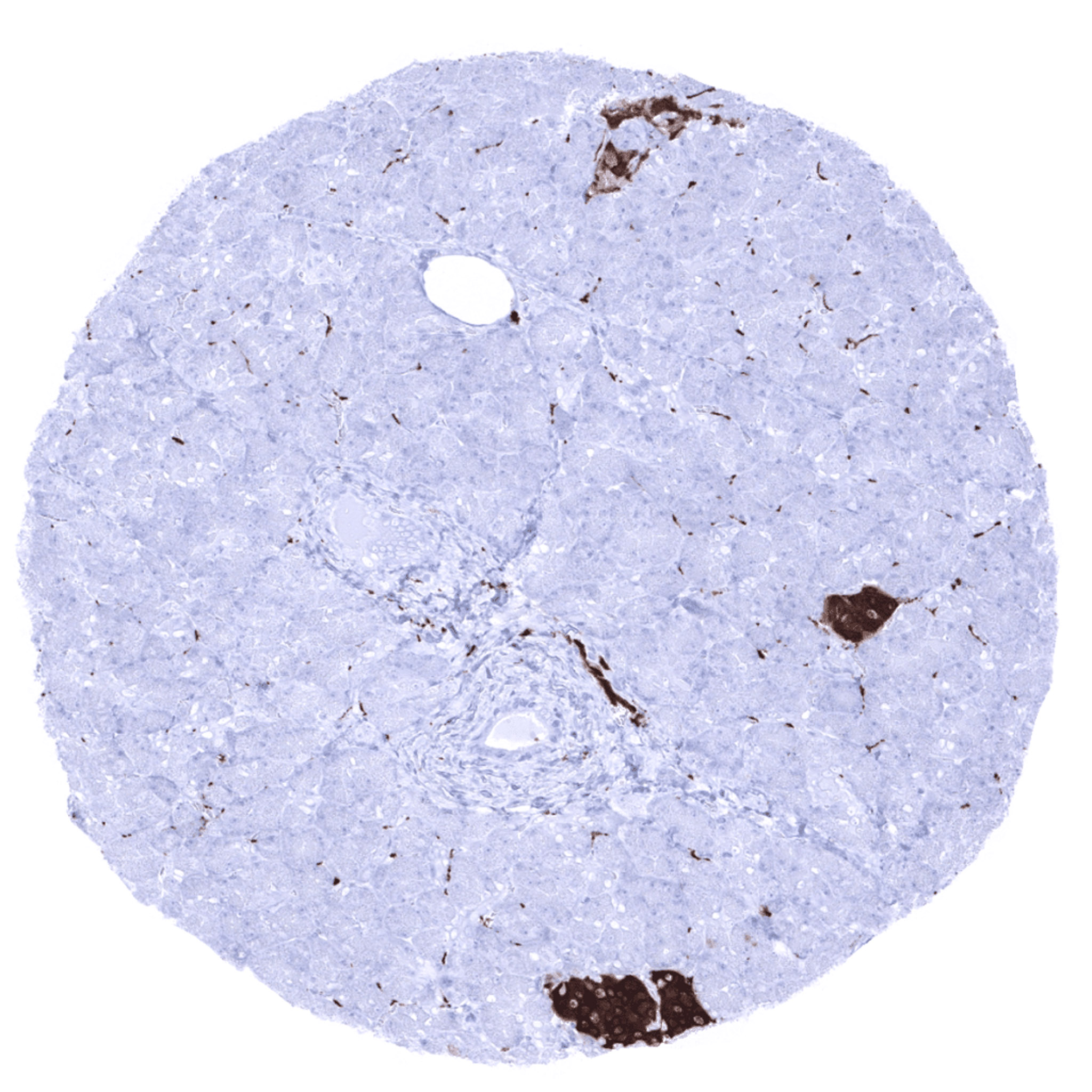
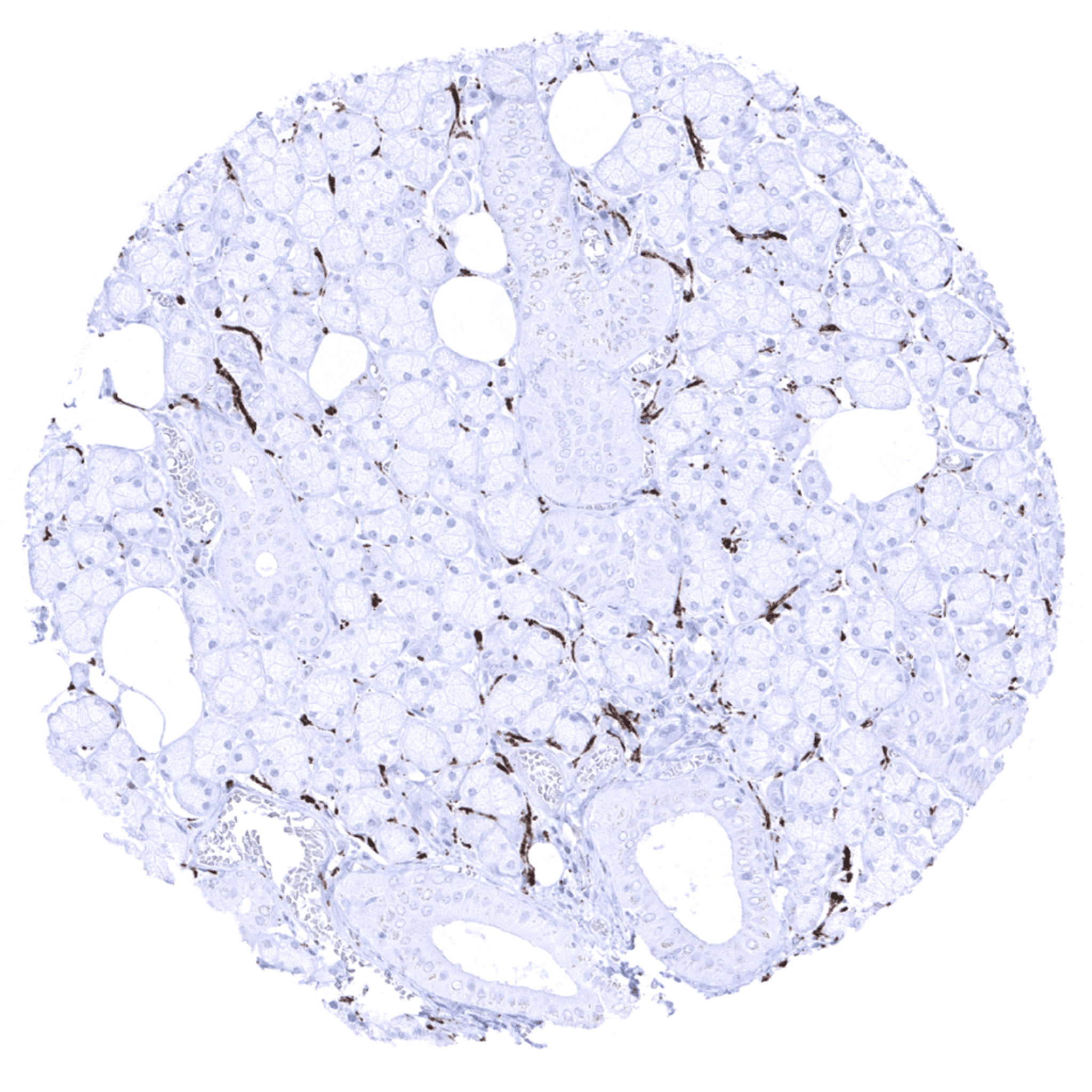
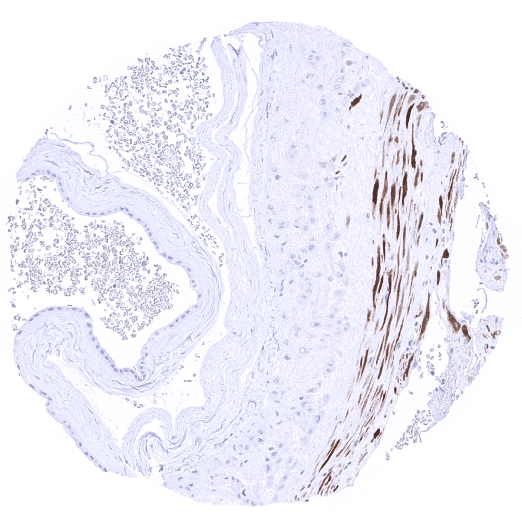
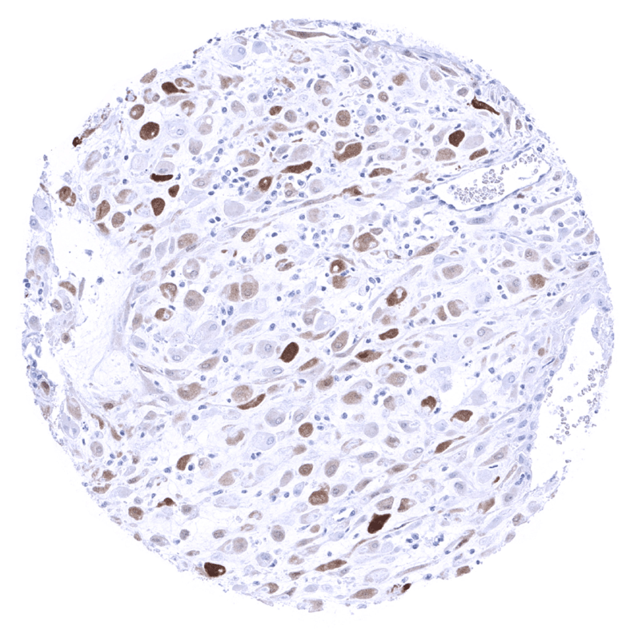
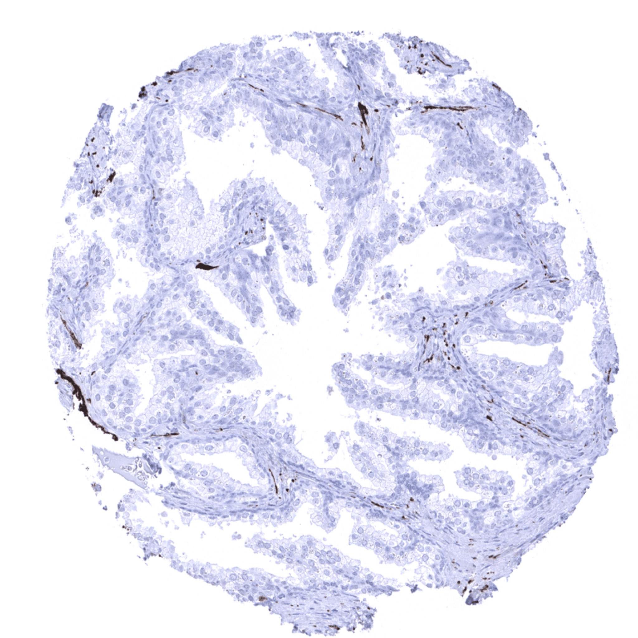
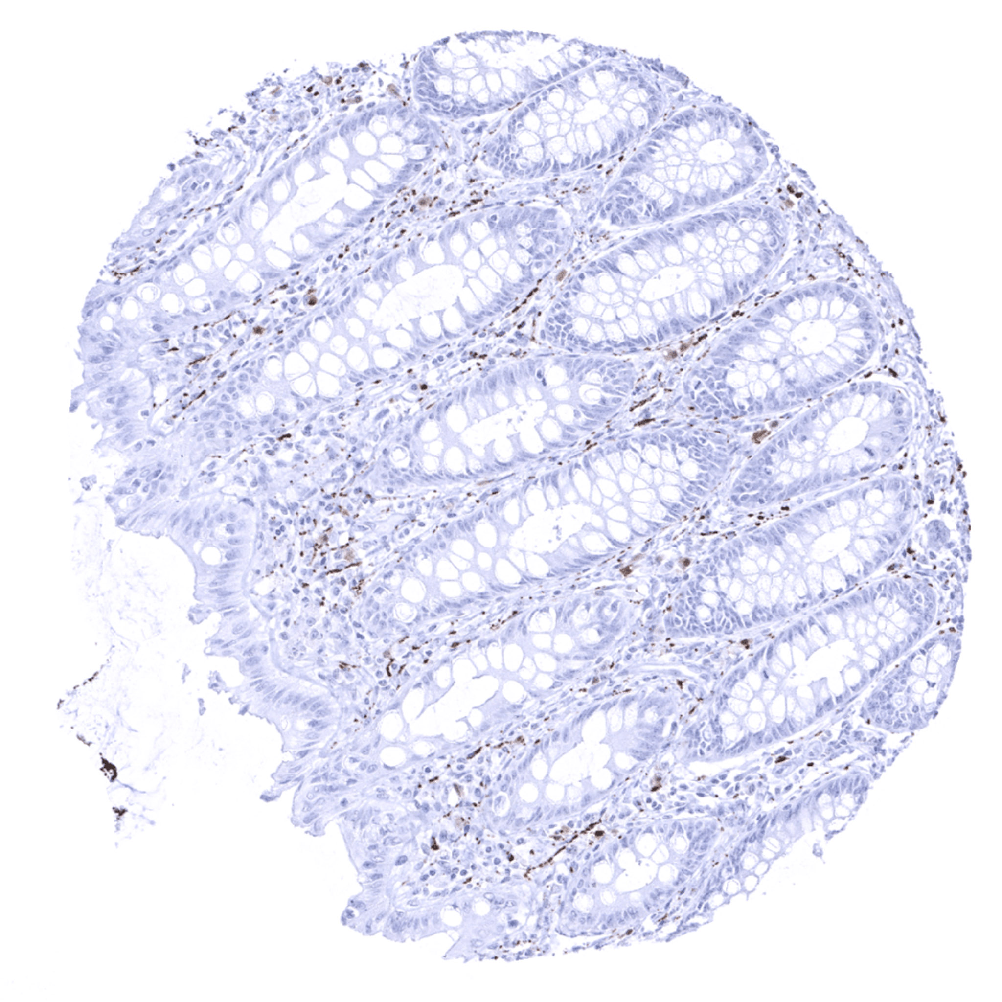
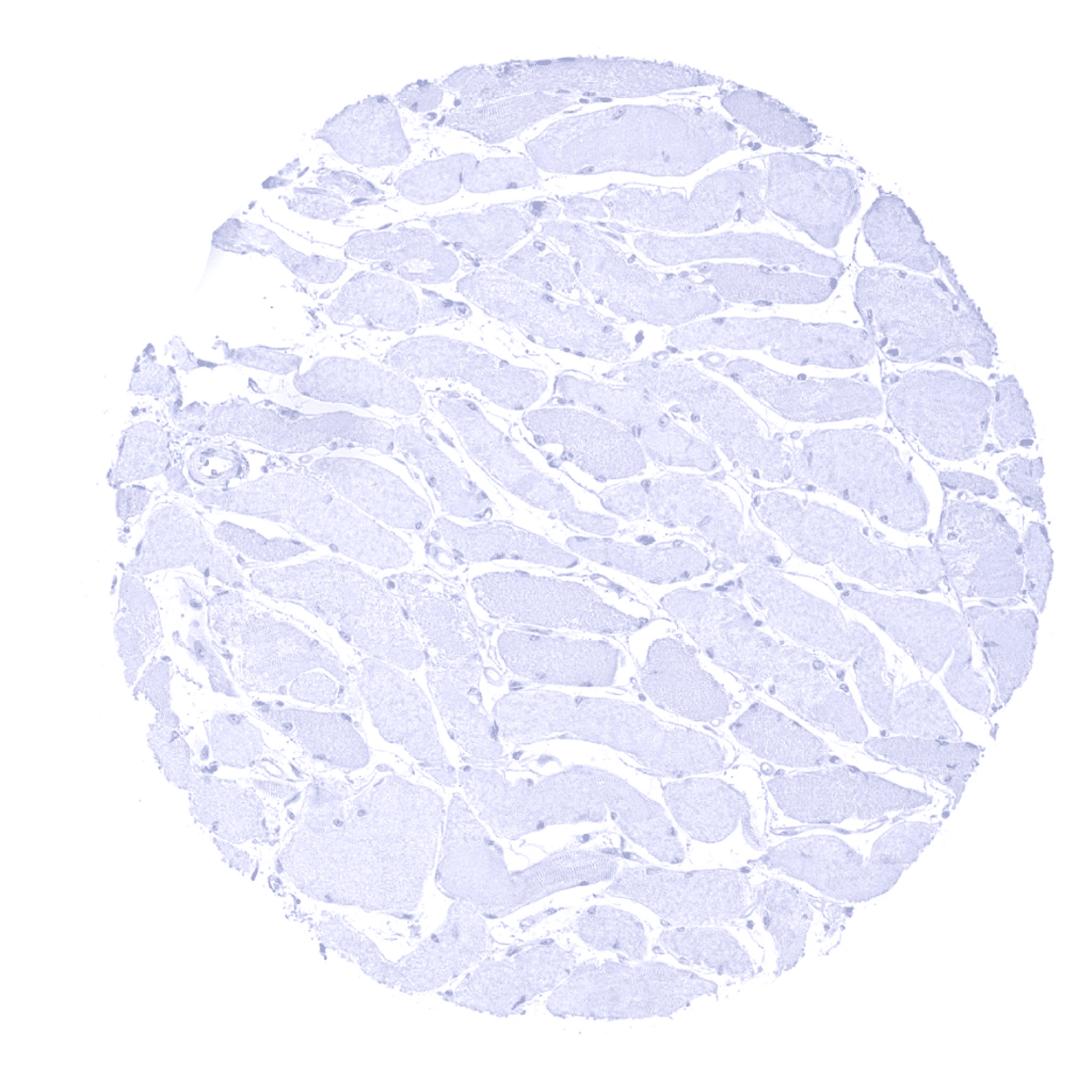
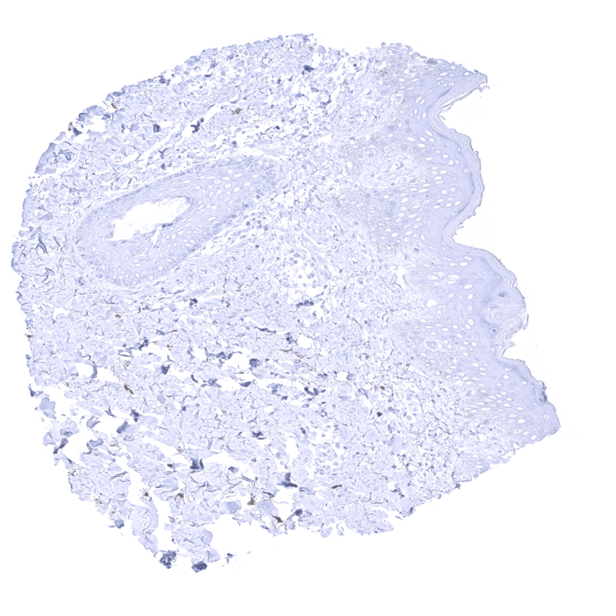
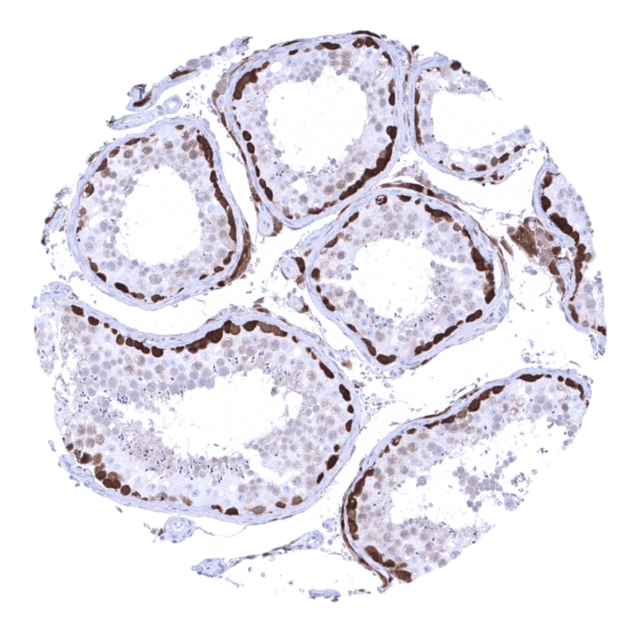
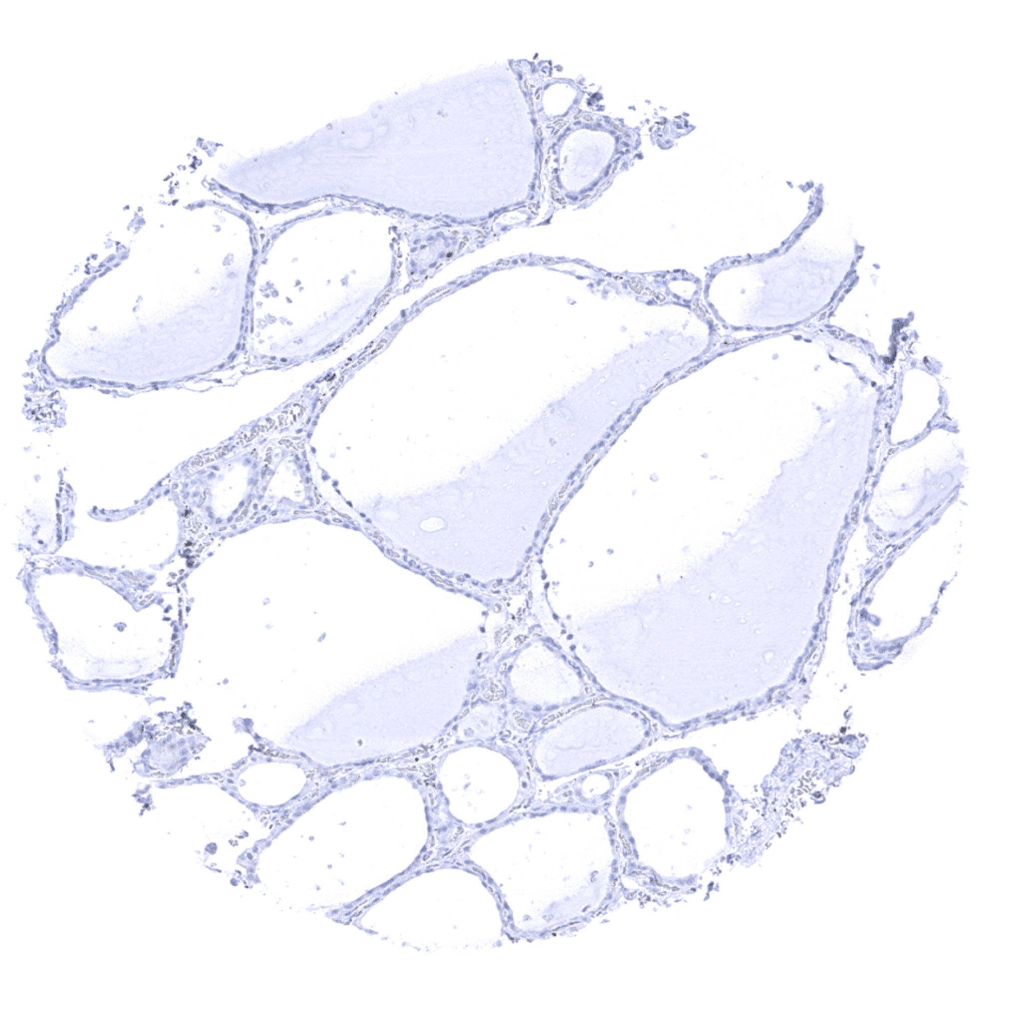
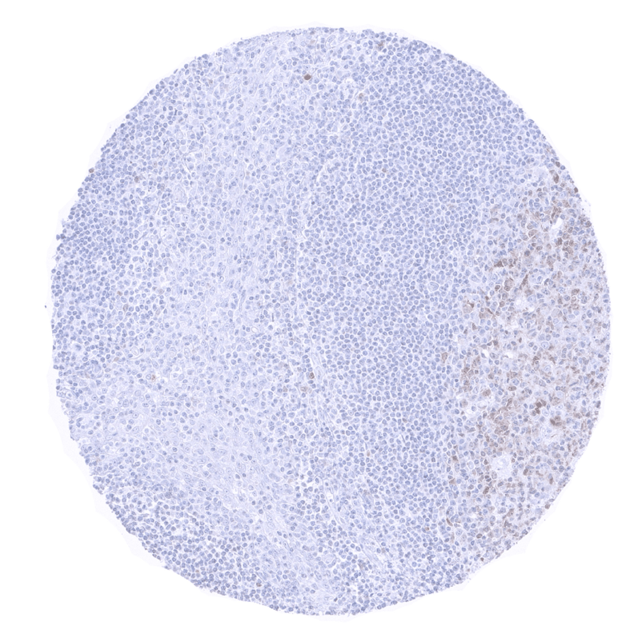
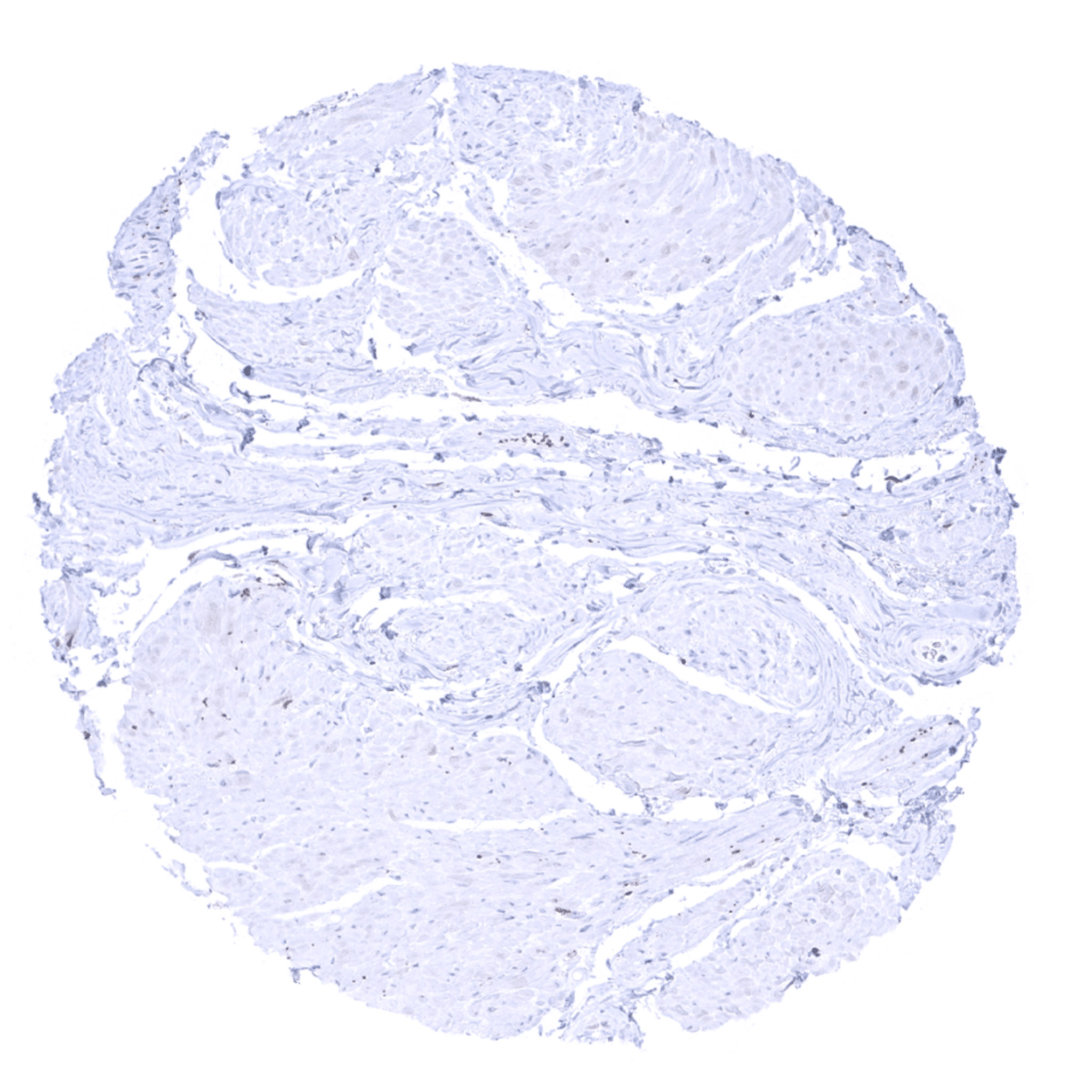
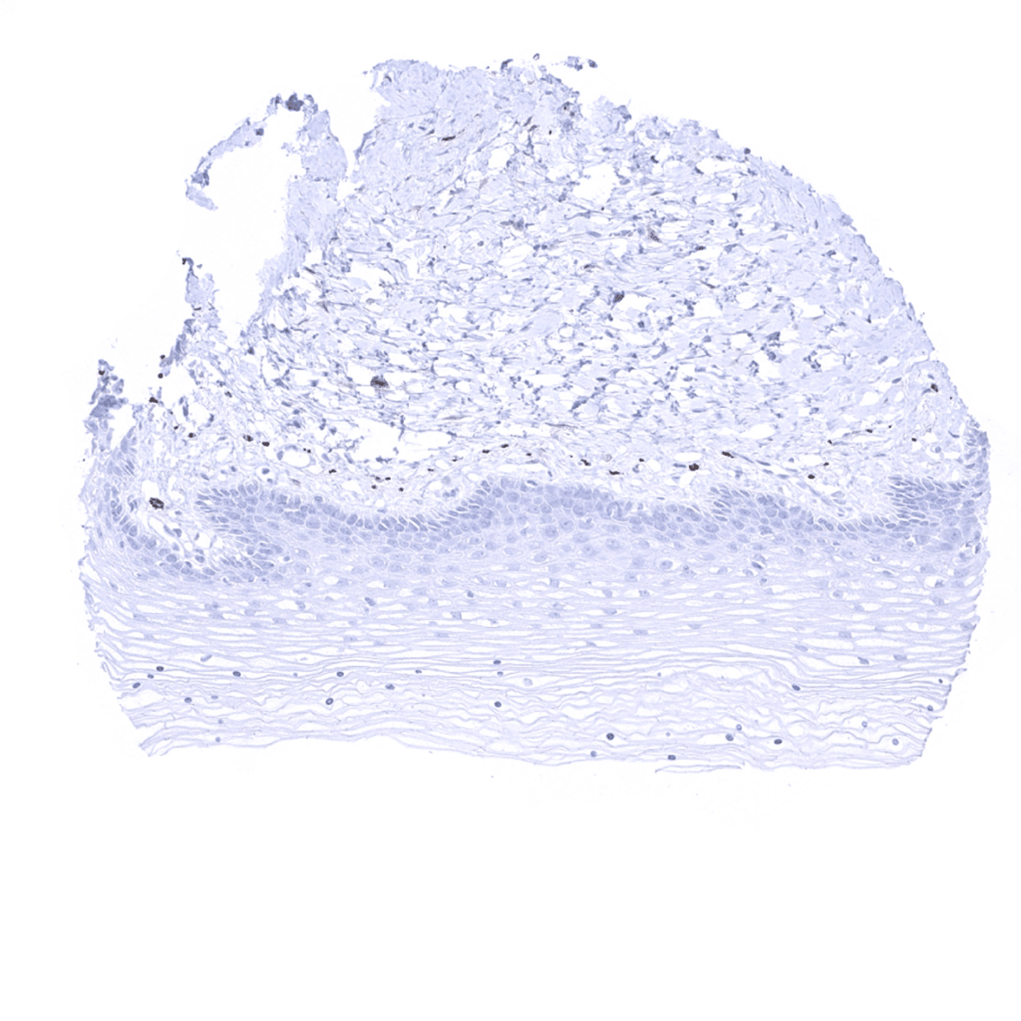
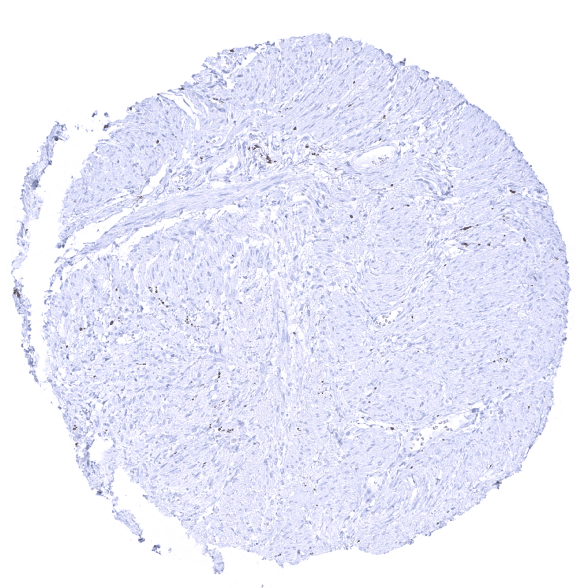
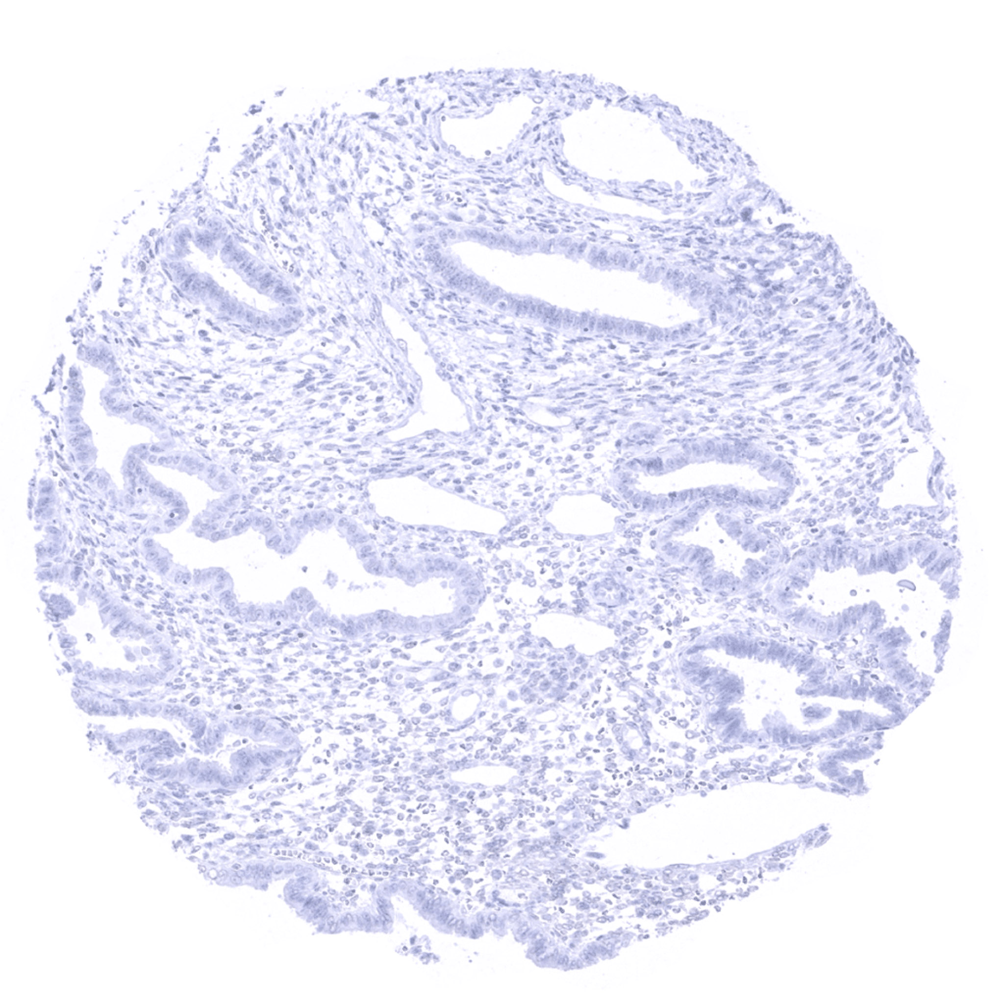
PGP9.5 antibody [MSVA-905R] HistoMAXTM


a
Go to Cancer Tissue Gallery
  
 Go back to product page
  

Go to HistoMAX page


Adrenal gland – A weak to moderate PGP9.5 positivity can occur in cortical cells adjacent to the medulla.

Adrenal gland – Strong PGP9.5 immunostaining in all medullary cells.

Aorta media – PGP9.5 staining is seen in some fibres of the media of the aorta.

Appendix, mucosa


Appendix, muscular wall

Bone marrow

Breast

Bronchus, mucosa – In the bronchus mucosa, PGP9.5 immunostaining occurs in a fraction of cells. A “mosaic pattern” displaying a mixture of positive and negative cells is typically seen.


Cerebellum (molecular layer, Purkinje cell layer, granule cell layer) – Strong PGP9.5 positivity in all neuronal cells and in axons. Glia cells are negative.

Cerebellum (white matter)

Cerebrum, grey matter – Strong PGP9.5 positivity in all neuronal cells and in axons. Glia cells are negative.

Cerebrum, white matter


Colon descendens, mucosa

Colon descendens, muscular wall – Strong PGP9.5 immunostaining of nerve fibres and ganglions in the muscular wall.

Duodenum, Brunner gland

Duodenum, mucosa


Epididymis – In the epididymis, PGP9.5 immunostaining is seen in a small fraction of cells in distal tubuli and collecting ducts. The staining intensity is variable and sometimes a “mosaic pattern” is seen.

Esophagus, squamous epithelium

Fallopian tube, mucosa – In the fallopian tube, a moderate PGP9.5 immunostaining is seen in scattered epithelial cells.

Fat


Gallbladder, epithelium

Heart muscle

Ileum, mucosa

Kidney, cortex – In the kidney, PGP9.5 immunostaining is seen in a fraction of cells in distal tubuli and collecting ducts. The staining intensity is variable and sometimes a “mosaic pattern” is seen.


Kidney, medulla – In the kidney, PGP9.5 immunostaining is seen in a fraction of cells in distal tubuli and collecting ducts. The staining intensity is variable and sometimes a “mosaic pattern” is seen.

Liver

Lung

Lymph node


Ovary, stroma – The ovarian stroma exhibits a moderate PGP9.5 immunostaining.

Pancreas – In the pancreas, a strong PGP9.5 immunostaining is seen in virtually all cells of islets of Langerhans. In addition small nerve fibres stain positive.

Parathyroid gland

Parotid gland – In the parotid gland, a strong PGP9.5 immunostaining is seen in small nerve fibres.


Pituitary gland, anterior lobe

Pituitary gland, posterior lobe

Placenta (amnion and chorion) – Strong PGP9.5 immunostaining of decidua cells. Cells of placental origin are PGP9.5 negative.

Pregnant uterus (decidua)


Placenta, early

Placenta, mature

Prostate

Rectum, mucosa


Seminal vesicle

Sinus paranasales

Skeletal muscle

Skin


Spleen

Stomach, antrum

Stomach, corpus

Testis – In the testis, a strong PGP9.5 immunostaining is seen in spermatogonia. PGP9.5 staining decreases sharply in spermatocytes where it is only faint. A moderate PGP9.5 staining is seen in Leydig cells while Sertoli cells remain PGP9.5 negative.


Thymus – Scattered PGP9.5 positive cells occur in the thymus.

Thyroid gland

Tonsil – Weak PGP9.5 immunostaining is seen in a fraction of lymphocytes in the tonsil.

Tonsil, surface epithelium – Staining of the epithelium surface represents ink (no true immunostaining).


Urinary bladder, muscular wall

Urinary bladder, urothelium

Uterus endocervix

Uterus ectocervix


Uterus myometrium

Uterus, endometrium (proliferation)

Uterus, endometrium, (secretion)


| TOP |