14-3-3 sigma antibody


14-3-3 sigma antibody detects 14-3-3 sigma protein at cytosol on mouse lung by immunohistochemical analysis.
Sample: Paraffin-embedded mouse lung.
14-3-3 sigma antibody (GTX100289) dilution: 1:500.
Antigen Retrieval: Trilogy™ (EDTA based, pH 8.0) buffer, 15min

14-3-3 sigma antibody [C1C3] detects 14-3-3 sigma protein at cytoplasm by immunofluorescent analysis.
Sample: A431 cells were fixed in ice-cold Methanol for 5 min.
Green: 14-3-3 sigma protein stained by 14-3-3 sigma antibody [C1C3] (GTX100289) diluted at 1:500.
Blue: Hoechst 33343 staining.

14-3-3 sigma antibody detects SFN protein by western blot analysis.
A. 30 μg Neuro2A whole cell lysate/extract
B. 30 μg GL261 whole cell lysate/extract
12% SDS-PAGE
14-3-3 sigma antibody (GTX100289) dilution: 1:3000
The HRP-conjugated anti-rabbit IgG antibody (GTX213110-01) was used to detect the primary antibody.

14-3-3 sigma antibody detects SFN protein by western blot analysis.
A. 30 μg 293T whole cell lysate/extract
B. 30 μg A431 whole cell lysate/extract
C. 30 μg HeLa whole cell lysate/extract
D. 30 μg HepG2 whole cell lysate/extract
12% SDS-PAGE
14-3-3 sigma antibody (GTX100289) dilution: 1:3000
The HRP-conjugated anti-rabbit IgG antibody (GTX213110-01) was used to detect the primary antibody.

Whole cell extract (30 μg) was separated by 12% SDS-PAGE, and the membrane was blotted with 14-3-3 sigma antibody (GTX100289) diluted at 1:3000. The HRP-conjugated anti-rabbit IgG antibody (GTX213110-01) was used to detect the primary antibody.

Immunohistochemical analysis of paraffin-embedded 59T xenograft, using 14-3-3 sigma(GTX100289) antibody at 1:100 dilution.
Antigen Retrieval: Trilogy™ (EDTA based, pH 8.0) buffer, 15min
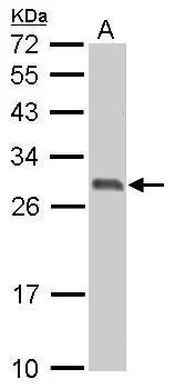
14-3-3 sigma antibody detects SFN protein by western blot analysis.
A. 30 μg PC-12 whole cell lysate/extract
12% SDS-PAGE
14-3-3 sigma antibody (GTX100289) dilution: 1:3000
The HRP-conjugated anti-rabbit IgG antibody (GTX213110-01) was used to detect the primary antibody.

Various whole cell extracts (30 μg) were separated by 12% SDS-PAGE, and the membrane was blotted with 14-3-3 sigma antibody (GTX100289) diluted at 1:3000. The HRP-conjugated anti-rabbit IgG antibody (GTX213110-01) was used to detect the primary antibody. Corresponding RNA expression data for the same cell lines are based on Human Protein Atlas program.
-
HostRabbit
-
ClonalityPolyclonal
-
IsotypeIgG
-
ApplicationsWB ICC/IF IHC-P
-
ReactivityHuman, Mouse, Rat