53BP1 antibody


Non-transfected (–) and transfected (+) HeLa whole cell extracts (50 μg) were separated by 5% SDS-PAGE, and the membrane was blotted with 53BP1 antibody (GTX70310) diluted at 1:500. The HRP-conjugated anti-rabbit IgG antibody (GTX213110-01) was used to detect the primary antibody.
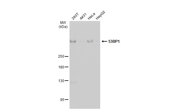
Various whole cell extracts (30 μg) were separated by 5% SDS-PAGE, and the membrane was blotted with 53BP1 antibody (GTX70310) diluted at 1:2000. The HRP-conjugated anti-rabbit IgG antibody (GTX213110-01) was used to detect the primary antibody.

53BP1 antibody detects 53BP1 protein at nucleus by immunohistochemical analysis.Sample: Paraffin-embedded human breast carcinoma.53BP1 stained by 53BP1 antibody (GTX70310) diluted at 1:500.Antigen Retrieval: Citrate buffer, pH 6.0, 15 min

53BP1 antibody detects 53BP1 protein at nucleus by immunofluorescent analysis.Sample: HeLa cells were fixed in ice-cold MeOH for 5 min.Green: 53BP1 stained by 53BP1 antibody (GTX70310) diluted at 1:500.Red: alpha Tubulin, a cytoskeleton marker, stained by alpha Tubulin antibody [GT114] (GTX628802) diluted at 1:500.Scale bar= 10 μm.

53BP1 antibody detects 53BP1 protein at nucleus by immunohistochemical analysis.Sample: Paraffin-embedded human breast carcinoma.53BP1 stained by 53BP1 antibody (GTX70310) diluted at 1:4000.Antigen Retrieval: Citrate buffer, pH 6.0, 15 min

53BP1 antibody detects 53BP1 protein at nucleus by immunohistochemical analysis.Sample: Paraffin-embedded human colon cancer.53BP1 stained by 53BP1 antibody (GTX70310) diluted at 1:4000.Antigen Retrieval: Citrate buffer, pH 6.0, 15 min

Various whole cell extracts (30 μg) were separated by 5% SDS-PAGE, and the membrane was blotted with 53BP1 antibody (GTX70310) diluted at 1:500. The HRP-conjugated anti-rabbit IgG antibody (GTX213110-01) was used to detect the primary antibody.

53BP1 antibody detects 53BP1 protein at nucleus by immunohistochemical analysis.Sample: Paraffin-embedded human breast carcinoma.53BP1 stained by 53BP1 antibody (GTX70310) diluted at 1:1000.Antigen Retrieval: Citrate buffer, pH 6.0, 15 min
-
HostRabbit
-
ClonalityPolyclonal
-
IsotypeIgG
-
ApplicationsWB ICC/IF IHC-P IHC
-
ReactivityHuman, Mouse