Arginase 1 antibody


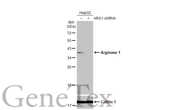
Non-transfected (–) and transfected (+) HepG2 whole cell extracts (30 μg) were separated by 12% SDS-PAGE, and the membrane was blotted with Arginase 1 antibody (GTX109242) diluted at 1:500. The HRP-conjugated anti-rabbit IgG antibody (GTX213110-01) was used to detect the primary antibody.

Non-transfected (–) and transfected (+) HepG2 whole cell extracts (30 μg) were separated by 10% SDS-PAGE, and the membrane was blotted with Arginase 1 antibody (GTX109242) diluted at 1:1000. The HRP-conjugated anti-rabbit IgG antibody (GTX213110-01) was used to detect the primary antibody.

Various tissue extracts (50 μg) were separated by 12% SDS-PAGE, and the membrane was blotted with Arginase 1 antibody (GTX109242) diluted at 1:10000. The HRP-conjugated anti-rabbit IgG antibody (GTX213110-01) was used to detect the primary antibody. Corresponding RNA expression data for the same cell lines are based on NCBI mouse tissue.

Various whole cell extracts (30 μg) were separated by 12% SDS-PAGE, and the membrane was blotted with Arginase 1 antibody (GTX109242) diluted at 1:1000. The HRP-conjugated anti-rabbit IgG antibody (GTX213110-01) was used to detect the primary antibody. Corresponding RNA expression data for the same cell lines are based on Human Protein Atlas program.
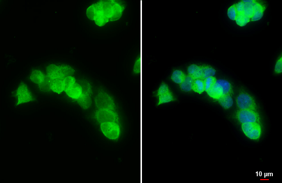
Arginase 1 antibody detects Arginase 1 protein at cytoplasm by immunofluorescent analysis.Sample: HepG2 cells were fixed in 4% paraformaldehyde at RT for 15 min.Green: Arginase 1 stained by Arginase 1 antibody (GTX109242) diluted at 1:500.Blue: Fluoroshield with DAPI (GTX30920).Scale bar= 10 μm.
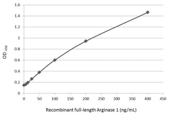
Sandwich ELISA detection of recombinant full-length Arginase 1 protein using GTX109242 as capture antibody at concentration of 5 μg/mL and GTX634218 as detection antibody at concentration of 1 μg/mL. Mouse IgG antibody (HRP) (GTX213111-01) was diluted at 1:10000 and used to detect the primary antibody.
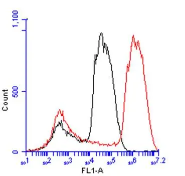
Arginase 1 antibody (GTX109242) detects ARG1 protein by flow cytometry analysis.
Sample: HepG2 cell.
Black: Unlabelled sample was used as a control.
Red: Arginase 1 antibody (GTX109242) dilution: 1:50.
Acquisition of 20,000 events were collected for FACS analysis.
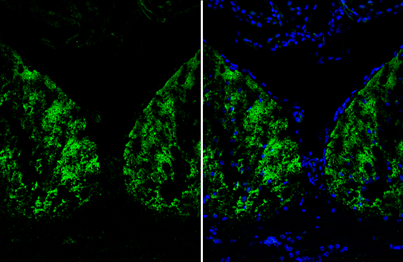
Arginase 1 antibody detects Arginase 1 protein by immunohistochemical analysis.Sample: Frozen-sectioned mouse hippocampus.Green: Arginase 1 stained by Arginase 1 antibody (GTX109242) diluted at 1:250.Blue: Fluoroshield with DAPI (GTX30920).
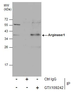
Immunoprecipitation of Arginase 1 protein from HepG2 whole cell extracts using 5 μg of Arginase 1 antibody (GTX109242).
Western blot analysis was performed using Arginase 1 antibody (GTX109242) diluted at 1:600.
EasyBlot anti-Rabbit IgG (GTX221666-01) was used as a secondary reagent.
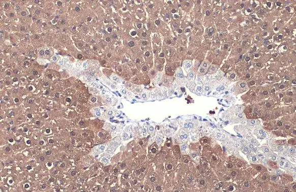
Arginase 1 antibody detects Arginase 1 protein at cytoplasm by immunohistochemical analysis.Sample: Paraffin-embedded mouse liver.Arginase 1 stained by Arginase 1 antibody (GTX109242) diluted at 1:500.Antigen Retrieval: Citrate buffer, pH 6.0, 15 min
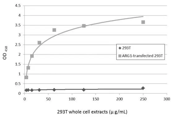
Sandwich ELISA detection of non-transfected and transfected 293T whole cell extracts using GTX634218 as capture antibody at concentration of 5 μg/mL and GTX109242 as detection antibody at concentration of 1 μg/mL. Rabbit IgG antibody (HRP) (GTX213110-01) was diluted at 1:10000 and used to detect the primary antibody.
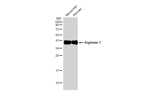
Various tissue extracts (50 μg) were separated by 12% SDS-PAGE, and the membrane was blotted with Arginase 1 antibody (GTX109242) diluted at 1:1000. The HRP-conjugated anti-rabbit IgG antibody (GTX213110-01) was used to detect the primary antibody.
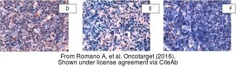
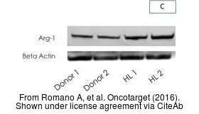
The data was published in the journal Oncotarget in 2016. PMID: 27637084
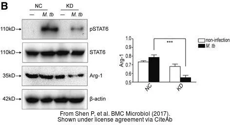
The data was published in the journal BMC Microbiol in 2017. PMID: 28835201
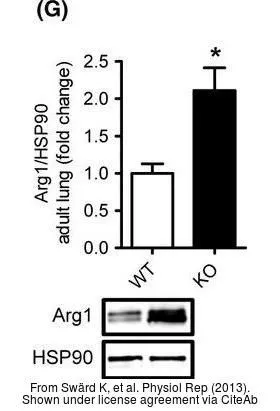
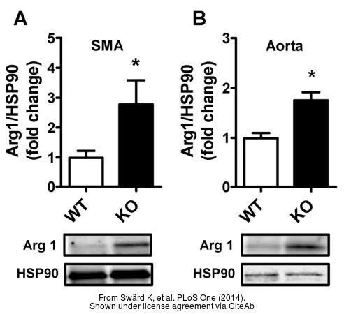
The data was published in the journal Physiol Rep in 2013. PMID: 24303100
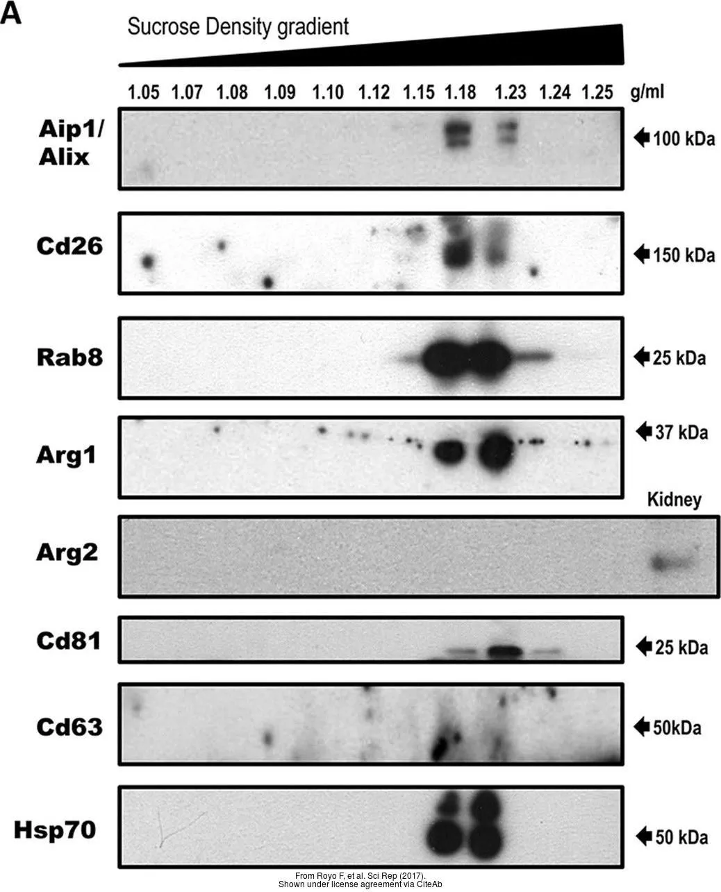
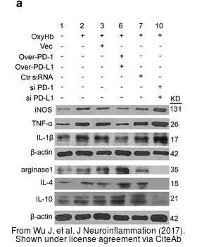
The data was published in the journal Sci Rep in 2017.PMID: 28211494
-
HostRabbit
-
ClonalityPolyclonal
-
IsotypeIgG
-
ApplicationsWB ICC/IF IHC-P IHC-Fr FCM IP ELISA Sandwich ELISA
-
ReactivityHuman, Mouse, Rat