Cofilin 1 antibody


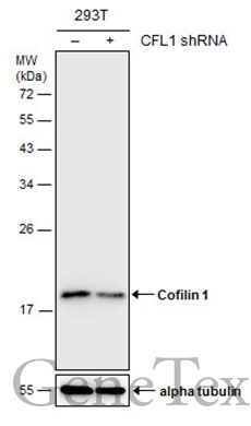
Non-transfected (–) and transfected (+) 293T whole cell extracts (30 μg) were separated by 12% SDS-PAGE, and the membrane was blotted with Cofilin 1 antibody (GTX102156) diluted at 1:1000. The HRP-conjugated anti-rabbit IgG antibody (GTX213110-01) was used to detect the primary antibody.
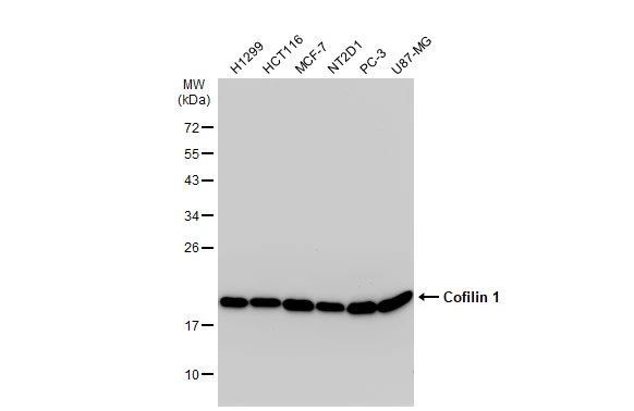
Various whole cell extracts (30 μg) were separated by 12% SDS-PAGE, and the membrane was blotted with Cofilin 1 antibody (GTX102156) diluted at 1:1000. The HRP-conjugated anti-rabbit IgG antibody (GTX213110-01) was used to detect the primary antibody.
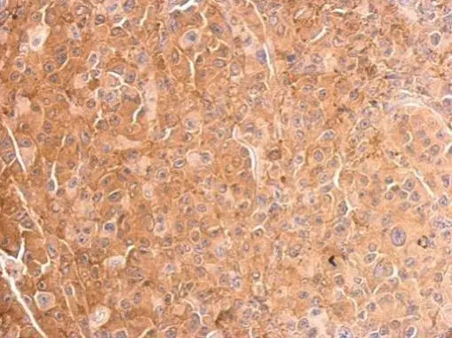
Cofilin 1 antibody detects CFL1 protein at cytosol on HBL435 xenograft by immunohistochemical analysis.
Sample: Paraffin-embedded HBL435 xenograft.
Cofilin 1 antibody (GTX102156) dilution: 1:500.
Antigen Retrieval: Trilogy™ (EDTA based, pH 8.0) buffer, 15min
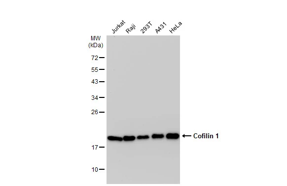
Various whole cell extracts (30 μg) were separated by 12% SDS-PAGE, and the membrane was blotted with Cofilin 1 antibody (GTX102156) diluted at 1:1000. The HRP-conjugated anti-rabbit IgG antibody (GTX213110-01) was used to detect the primary antibody.
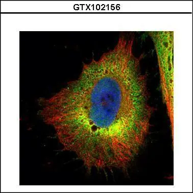
Confocal immunofluorescence analysis (Olympus FV10i) of paraformaldehyde-fixed HeLa, using Cofilin 1 (non-muscle)(GTX102156) antibody (Green) at 1:500 dilution. Alpha-tubulin filaments were labeled with GTX11304 (Red) at 1:2000.
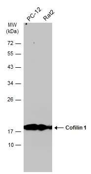
Various whole cell extracts (30 μg) were separated by 12% SDS-PAGE, and the membrane was blotted with Cofilin 1 antibody (GTX102156) diluted at 1:1000. The HRP-conjugated anti-rabbit IgG antibody (GTX213110-01) was used to detect the primary antibody.
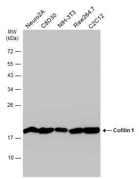
Various whole cell extracts (30 μg) were separated by 12% SDS-PAGE, and the membrane was blotted with Cofilin 1 antibody (GTX102156) diluted at 1:1000. The HRP-conjugated anti-rabbit IgG antibody (GTX213110-01) was used to detect the primary antibody.
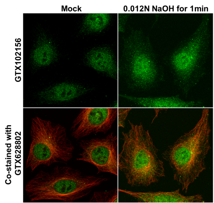
Cofilin 1 antibody detects Cofilin 1 protein at cytoplasm and nucleus by immunofluorescent analysis.
Samples: HeLa cells mock (left) and treated with 0.012 N NaOH/PBS for 1 min (right) were fixed in 4% paraformaldehyde at RT for 15 min.
Green: Cofilin 1 protein stained by Cofilin 1 antibody (GTX102156) diluted at 1:1000.
Red: alpha Tubulin, a cytoskeleton marker, stained by alpha Tubulin antibody [GT114] (GTX628802) diluted at 1:1000.
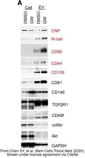
The data was published in the journal Stem Cells Transl Med in 2020.PMID: 31943851
-
HostRabbit
-
ClonalityPolyclonal
-
IsotypeIgG
-
ApplicationsWB ICC/IF IHC-P IHC-Fr
-
ReactivityHuman, Mouse, Rat