Collagen I antibody

IHC-P analysis of normal mouse kidney tissue using GTX20292 Collagen I antibody.
Antigen retrieval: not performed.
Dilution : 1:100
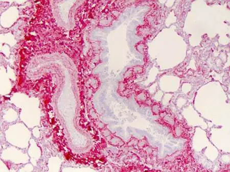
IHC-P analysis of human lung tissue using GTX20292 Collagen I antibody.
Strong staining was observed in the extracellular matrix of the lung. Epithelial cells were negative.
Dilution : 1:400
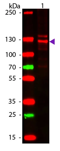
WB analysis of human Collagen Type I protein using GTX20292 Collagen I antibody.
Loading : 50 ng
Dilution : 1:1000
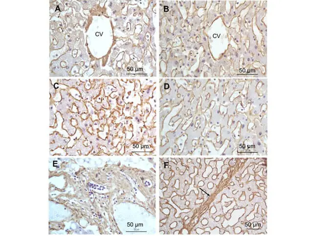
IHC-P analysis of liver tissue using GTX20292 Collagen I antibody.
Panel A : Central Vein (CV) fibrosis
Panel B : Non-fibrotic Central Vein (CV)
Panel C : Perisinusodial fibrosis
Panel D : Non-fibrotic area
Panel E : Protat tract fibrosis
Panel F : Septal fibrosis (arrow)
Antigen retrieval: not performed.
Dilution : 1:1250

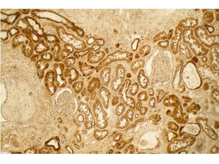
IHC-P analysis of normal kidney tissue section using GTX20292 Collagen I antibody.
Antigen retrieval : No antigen retrieval was performed.
Dilution : 1:100
Localization : Distal tubules in normal kidney tissue.
Note : the absence of staining of glomeruli.

Western blot of Human Collagen Type I. Load: 50 ng Human Collagen Type 1. Primary antibody: Collagen Type I antibody (GTX20292) at 1:1,000 overnight at 4ºC. Secondary antibody: DyLight™ 649 rabbit secondary antibody at 1:20,000 for 30 min at RT. Block for 30 min at RT. Predicted/Observed size: 139 & 130 kDa.

IHC-P analysis of human lung tissue section using GTX20292 Collagen I antibody.
Antigen retrieval : user optimized
Dilution : 1:400
Localization : Strong staining was observed in the extracellular matrix of the lung. Epithelial cells were negative.
Staining : antibody as precipitated red signal with a hematoxylin purple nuclear counterstain.


The data was published in the journal Front Pharmacol in 2016. PMID: 27199755
-
HostRabbit
-
ClonalityPolyclonal
-
IsotypeIgG
-
ApplicationsWB ICC/IF IHC-P FCM IP Dot ELISA Multiplexing
-
ReactivityHuman, Mouse, Rat, Bovine, Hamster, Pig