DDB1 antibody

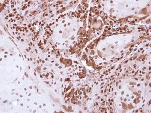
Immunohistochemical analysis of paraffin-embedded Cal27 Xenograft , using DDB1(GTX100130) antibody at 1:500 dilution.
Antigen Retrieval: Trilogy™ (EDTA based, pH 8.0) buffer, 15min
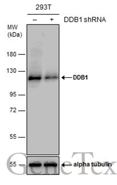
Non-transfected (–) and transfected (+) 293T whole cell extracts (30 μg) were separated by 7.5% SDS-PAGE, and the membrane was blotted with DDB1 antibody (GTX100130) diluted at 1:10000. The HRP-conjugated anti-rabbit IgG antibody (GTX213110-01) was used to detect the primary antibody.
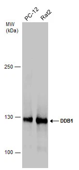
Various whole cell extracts (30 μg) were separated by 5% SDS-PAGE, and the membrane was blotted with DDB1 antibody (GTX100130) diluted at 1:1000. The HRP-conjugated anti-rabbit IgG antibody (GTX213110-01) was used to detect the primary antibody.
Sample (30 μg of whole cell lysate)
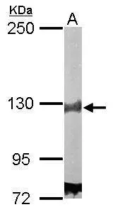
A:NIH-3T3
7.5% SDS PAGE
GTX100130 diluted at 1:1000
The HRP-conjugated anti-rabbit IgG antibody (GTX213110-01) was used to detect the primary antibody.
Sample (50 μg of whole cell lysate)
A: Mouse brain
5% SDS PAGE
GTX100130 diluted at 1:5000
The HRP-conjugated anti-rabbit IgG antibody (GTX213110-01) was used to detect the primary antibody.
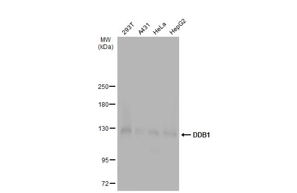
Various whole cell extracts (30 μg) were separated by 5% SDS-PAGE, and the membrane was blotted with DDB1 antibody (GTX100130) diluted at 1:1000. The HRP-conjugated anti-rabbit IgG antibody (GTX213110-01) was used to detect the primary antibody.
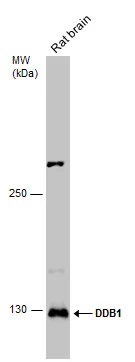
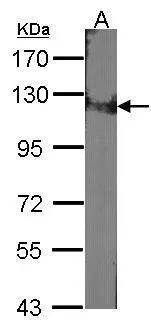
Rat tissue extract (50 μg) was separated by 5% SDS-PAGE, and the membrane was blotted with DDB1 antibody (GTX100130) diluted at 1:1000. The HRP-conjugated anti-rabbit IgG antibody (GTX213110-01) was used to detect the primary antibody.
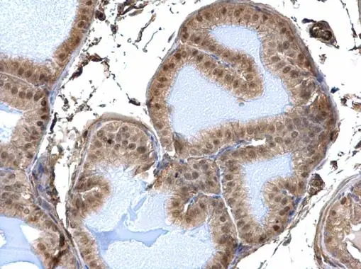
DDB1 antibody detects DDB1 protein at nucleus and cytosol on mouse prostate by immunohistochemical analysis.
Sample: Paraffin-embedded mouse prostate.
DDB1 antibody (GTX100130) dilution: 1:500.
Antigen Retrieval: Trilogy™ (EDTA based, pH 8.0) buffer, 15min
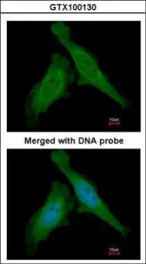
Immunofluorescence analysis of paraformaldehyde-fixed HeLa, using DDB1(GTX100130) antibody at 1:200 dilution.
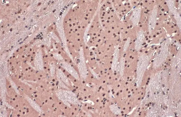
DDB1 antibody detects DDB1 protein at nucleus by immunohistochemical analysis.Sample: Paraffin-embedded mouse brain.DDB1 stained by DDB1 antibody (GTX100130) diluted at 1:500.Antigen Retrieval: Citrate buffer, pH 6.0, 15 min
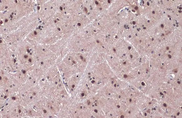
DDB1 antibody detects DDB1 protein at nucleus by immunohistochemical analysis.Sample: Paraffin-embedded rat brain.DDB1 stained by DDB1 antibody (GTX100130) diluted at 1:500.Antigen Retrieval: Citrate buffer, pH 6.0, 15 min
-
HostRabbit
-
ClonalityPolyclonal
-
IsotypeIgG
-
ApplicationsWB ICC/IF IHC-P IP
-
ReactivityHuman, Mouse, Rat