FIS1 antibody


Non-transfected (–) and transfected (+) HeLa whole cell extracts (30 μg) were separated by 15% SDS-PAGE, and the membrane was blotted with FIS1 antibody (GTX111010) diluted at 1:5000. The HRP-conjugated anti-rabbit IgG antibody (GTX213110-01) was used to detect the primary antibody.

FIS1 antibody detects FIS1 protein at mitochondria by immunofluorescent analysis.
Sample: HeLa cells were fixed in 2% paraformaldehyde/culture medium at 37oC for 30 min.
Green: FIS1 protein stained by FIS1 antibody (GTX111010) diluted at 1:2000.
Red: MitoTrackerR Red CMXRos, a mitochondria tracker.
Blue: Hoechst 33342 staining.
Scale bar = 10 μm.
Sample (50 μg of whole cell lysate)
A: mouse brain
15% SDS PAGE
GTX111010 diluted at 1:5000
The HRP-conjugated anti-rabbit IgG antibody (GTX213110-01) was used to detect the primary antibody.
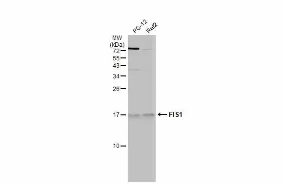
Various whole cell extracts (30 μg) were separated by 15% SDS-PAGE, and the membrane was blotted with FIS1 antibody (GTX111010) diluted at 1:500. The HRP-conjugated anti-rabbit IgG antibody (GTX213110-01) was used to detect the primary antibody.

FIS1 antibody detects FIS1 protein by western blot analysis. Mouse tissue extracts (50 μg) was separated by 15% SDS-PAGE, and the membrane was blotted with FIS1 antibody (GTX111010) diluted by 1:5000. The HRP-conjugated anti-rabbit IgG antibody (GTX213110-01) was used to detect the primary antibody.
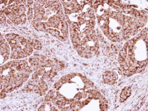
Immunohistochemical analysis of paraffin-embedded NCIN87 xenograft, using FIS1(GTX111010) antibody at 1:100 dilution.
Antigen Retrieval: Trilogy™ (EDTA based, pH 8.0) buffer, 15min

TTC11 antibody detects TTC11 protein at mitochondria on mouse prostate by immunohistochemical analysis.
Sample: Paraffin-embedded mouse prostate.
TTC11 antibody (GTX111010) dilution: 1:500.
Antigen Retrieval: Trilogy™ (EDTA based, pH 8.0) buffer, 15min

FIS1 antibody detects FIS1 protein at cytoplasm by immunohistochemical analysis.Sample: Paraffin-embedded mouse heart.FIS1 stained by FIS1 antibody (GTX111010) diluted at 1:500.Antigen Retrieval: Citrate buffer, pH 6.0, 15 min

Various whole cell extracts (30 μg) were separated by 15% SDS-PAGE, and the membrane was blotted with FIS1 antibody (GTX111010) diluted at 1:10000.

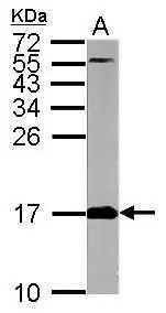
Sample (30 μg of whole cell lysate)
A: Raji
15% SDS PAGE
GTX111010 diluted at 1:10000
The HRP-conjugated anti-rabbit IgG antibody (GTX213110-01) was used to detect the primary antibody.
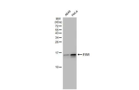
Various whole cell extracts (30 μg) were separated by 15% SDS-PAGE, and the membrane was blotted with FIS1 antibody (GTX111010) diluted at 1:10000. The HRP-conjugated anti-rabbit IgG antibody (GTX213110-01) was used to detect the primary antibody.


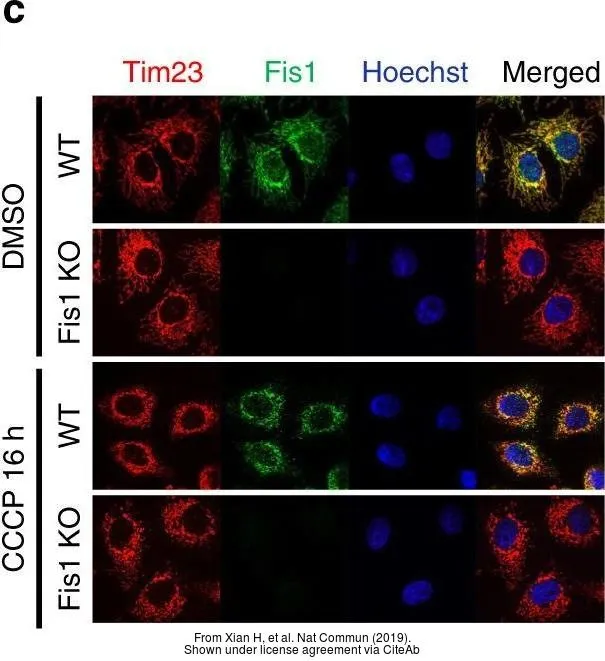

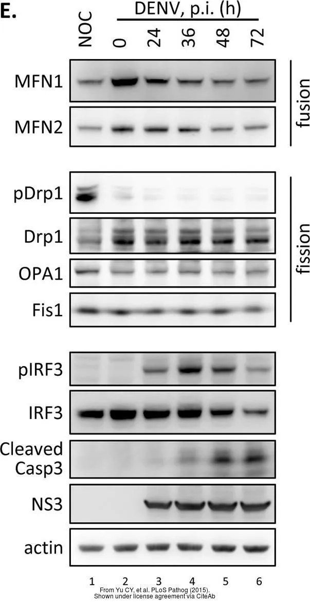
The data was published in the journal PLoS Pathog in 2015.PMID: 26717518
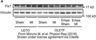
The data was published in the journal Physiol Rep in 2018.PMID: 29932506
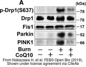
The data was published in the journal FEBS Open Bio in 2019.PMID: 30761259
-
HostRabbit
-
ClonalityPolyclonal
-
IsotypeIgG
-
ApplicationsWB ICC/IF IHC-P
-
ReactivityHuman, Mouse, Rat