Heme Oxygenase 1 antibody


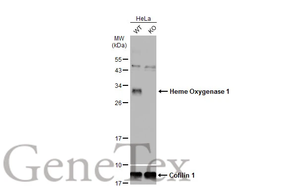
Wild-type (WT) and Heme Oxygenase 1 knockout (KO) HeLa cell extracts (30 μg) were separated by 12% SDS-PAGE, and the membrane was blotted with Heme Oxygenase 1 antibody (GTX101147) diluted at 1:500. The HRP-conjugated anti-rabbit IgG antibody (GTX213110-01) was used to detect the primary antibody, and the signal was developed with Trident ECL plus-Enhanced.
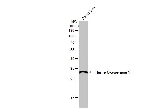
Rat tissue extract (50 μg) was separated by 12% SDS-PAGE, and the membrane was blotted with Heme Oxygenase 1 antibody (GTX101147) diluted at 1:1000. The HRP-conjugated anti-rabbit IgG antibody (GTX213110-01) was used to detect the primary antibody.
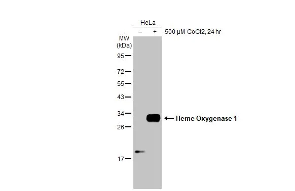
Untreated (–) and treated (+) HeLa whole cell extracts (30 μg) were separated by 12% SDS-PAGE, and the membrane was blotted with Heme Oxygenase 1 antibody (GTX101147) diluted at 1:5000. The HRP-conjugated anti-rabbit IgG antibody (GTX213110-01) was used to detect the primary antibody.
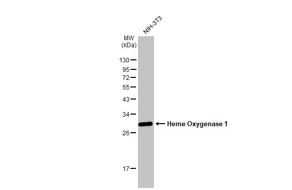
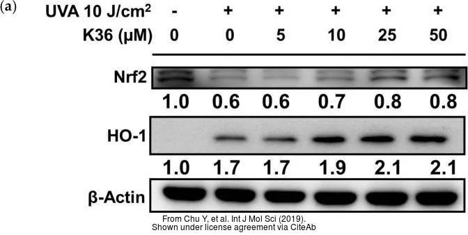
Whole cell extract (30 μg) was separated by 12% SDS-PAGE, and the membrane was blotted with Heme Oxygenase 1 antibody (GTX101147) diluted at 1:1000. The HRP-conjugated anti-rabbit IgG antibody (GTX213110-01) was used to detect the primary antibody.
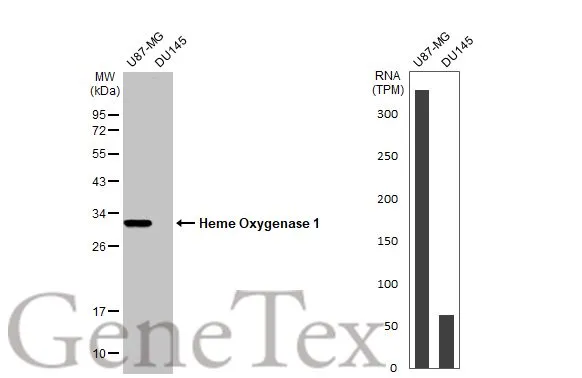
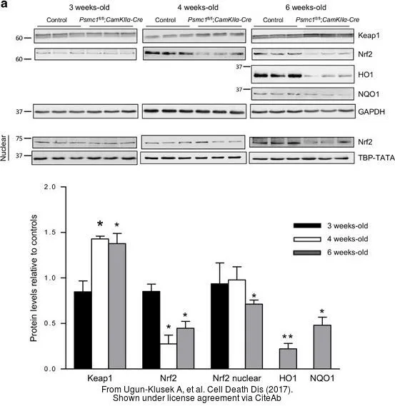
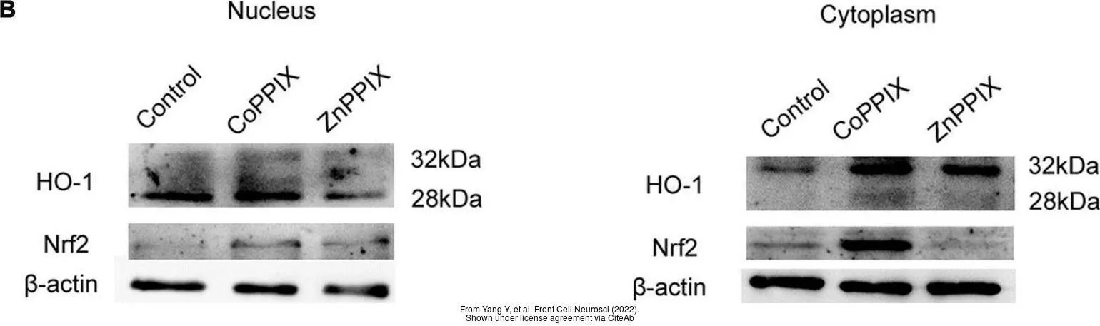
Various whole cell extracts (30 μg) were separated by 12% SDS-PAGE, and the membrane was blotted with Heme Oxygenase 1 antibody (GTX101147) diluted at 1:1000. The HRP-conjugated anti-rabbit IgG antibody (GTX213110-01) was used to detect the primary antibody. Corresponding RNA expression data for the same cell lines are based on Human Protein Atlas program.
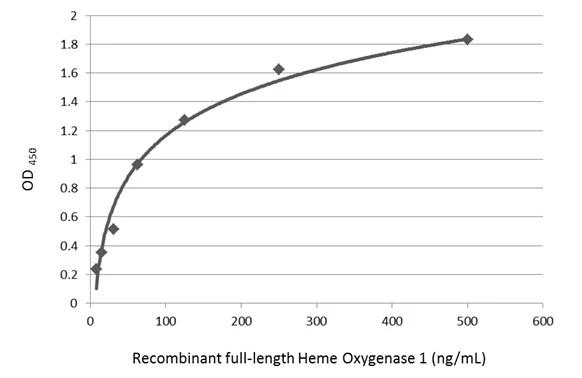
Sandwich ELISA detection of recombinant full-length Heme Oxygenase 1 protein using GTX633677 as capture antibody at concentration of 5 μg/mL and GTX101147 as detection antibody at concentration of 1 μg/mL. Rabbit IgG antibody (HRP) (GTX213110-01) was diluted at 1:10000 and used to detect the primary antibody.
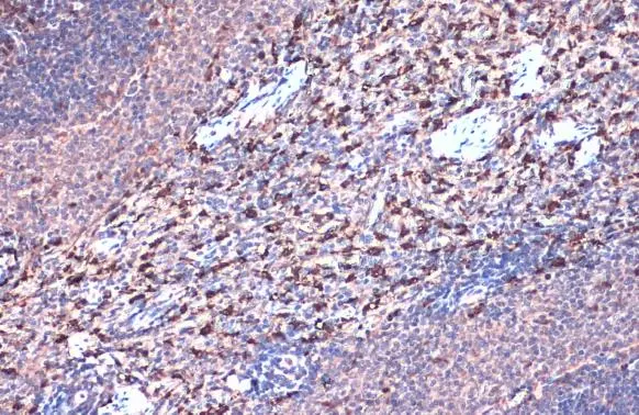
Heme Oxygenase 1 antibody detects Heme Oxygenase 1 protein at cytoplasm by immunohistochemical analysis.Sample: Paraffin-embedded mouse spleen.Heme Oxygenase 1 stained by Heme Oxygenase 1 antibody (GTX101147) diluted at 1:500.Antigen Retrieval: Citrate buffer, pH 6.0, 15 min
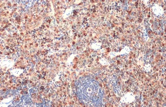
Heme Oxygenase 1 antibody detects Heme Oxygenase 1 protein at cytoplasm by immunohistochemical analysis.Sample: Paraffin-embedded rat spleen.Heme Oxygenase 1 stained by Heme Oxygenase 1 antibody (GTX101147) diluted at 1:500.Antigen Retrieval: Citrate buffer, pH 6.0, 15 min
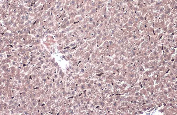
Heme Oxygenase 1 antibody detects Heme Oxygenase 1 protein at cytoplasm by immunohistochemical analysis.Sample: Paraffin-embedded mouse liver.Heme Oxygenase 1 stained by Heme Oxygenase 1 antibody (GTX101147) diluted at 1:500.Antigen Retrieval: Citrate buffer, pH 6.0, 15 min
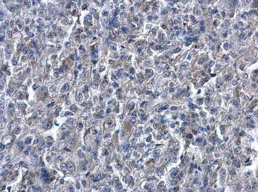
Heme Oxygenase 1 antibody detects Heme Oxygenase 1 protein at cytoplasm on human renal carcinoma by immunohistochemical analysis.
Sample: Paraffin-embedded human renal carcinoma .
Heme Oxygenase 1 antibody (GTX101147) diluted at 1:500.
Antigen Retrieval: Trilogy™ (EDTA based, pH 8.0) buffer, 15min
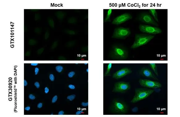
Heme Oxygenase 1 antibody detects Heme Oxygenase 1 protein at endoplasmic reticulum and nucleus by immunofluorescent analysis.
Sample: Mock and treated HeLa cells were fixed in 4% paraformaldehyde at RT for 15 min.
Green: Heme Oxygenase 1 stained by Heme Oxygenase 1 antibody (GTX101147) diluted at 1:500.
Blue: Fluoroshield with DAPI (GTX30920).
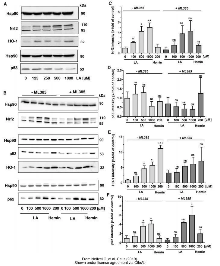
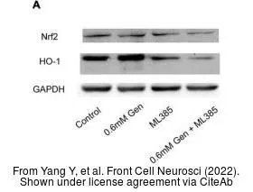
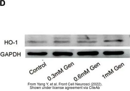
The data was published in the 2022 in Front Cell Neurosci. PMID: 35496911
-
HostRabbit
-
ClonalityPolyclonal
-
IsotypeIgG
-
ApplicationsWB ICC/IF IHC-P ELISA Sandwich ELISA
-
ReactivityHuman, Mouse, Rat, Monkey