LC3B antibody


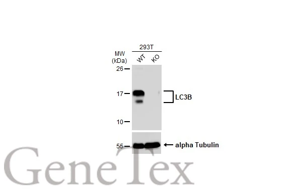
Wild-type (WT) and LC3B knockout (KO) 293T cell extracts (30 μg) were separated by 15% SDS-PAGE, and the membrane was blotted with LC3B antibody (GTX127375) diluted at 1:500. The HRP-conjugated anti-rabbit IgG antibody (GTX213110-01) was used to detect the primary antibody, and the signal was developed with Trident ECL plus-Enhanced.
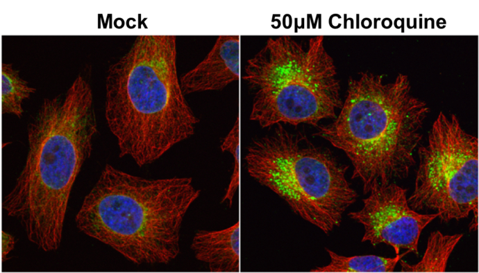
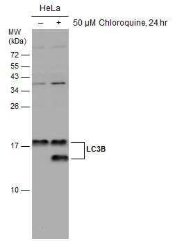
LC3B antibody detects LC3B protein at autophagosome by immunofluorescent analysis.
Samples: HeLa cells mock (left) and treated with 50μM Chloroquine for 24 hr (right) were fixed in 4% paraformaldehyde at RT for 15 min.
Green: LC3B protein stained by LC3B antibody (GTX127375) diluted at 1:2000.
Red: alpha Tubulin, a cytoskeleton marker, stained by alpha Tubulin antibody [GT114] (GTX628802) diluted at 1:1000.
Blue: Hoechst 33342 staining.
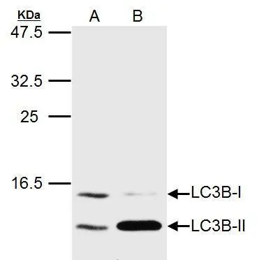
LC3B antibody detects MAP1LC3B protein by western blot analysis.
A. 20 μg Huh7 whole cell lysate/extract (untreated)
B. 20 μg Huh7 whole cell lysate/extract (3uM-Thapsigargin treatment for 12hr)
LC3B antibody (GTX127375) dilution: 1:1500
The HRP-conjugated anti-rabbit IgG antibody (GTX213110-01) was used to detect the primary antibody.
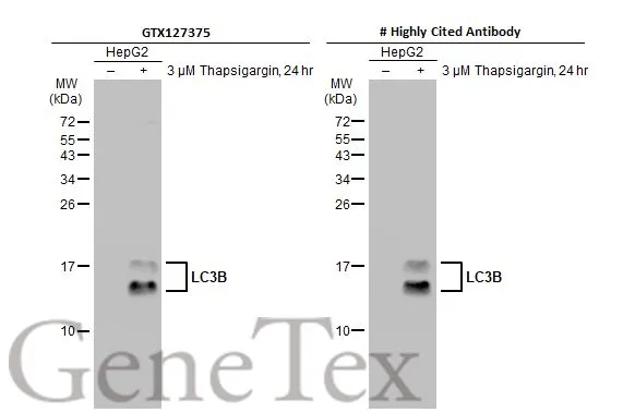
Untreated (–) and treated (+) HepG2 whole cell extracts (30 μg) were separated by 15% SDS-PAGE, and the membranes were blotted with LC3B antibody (GTX127375) diluted at 1:1000 and competitor's antibody (# Highly Cited Antibody ) diluted at 1:1000. The HRP-conjugated anti-rabbit IgG antibody (GTX213110-01) was used to detect the primary antibody.
*The competitor is not affiliated with GeneTex and does not endorse this product.
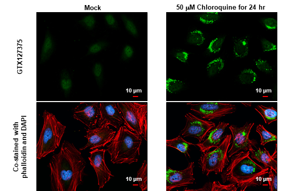
LC3B antibody detects LC3B protein at autophagosome by immunofluorescent analysis.Sample: Mock and treated HeLa cells were fixed in 4% paraformaldehyde at RT for 15 min.Green: LC3B stained by LC3B antibody (GTX127375) diluted at 1:500.Red: phalloidin, a cytoskeleton marker, diluted at 1:200.Blue: Fluoroshield with DAPI (GTX30920).Scale bar= 10 μm.
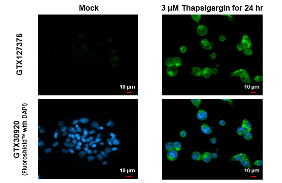
LC3B antibody detects LC3B protein at vesicle by immunofluorescent analysis.Sample: Mock and treated HepG2 cells were fixed in 4% paraformaldehyde at RT for 15 min.Green: LC3B stained by LC3B antibody (GTX127375) diluted at 1:500.Red: alpha Tubulin, a cytoskeleton marker, stained by alpha Tubulin antibody [GT114] (GTX628802) diluted at 1:1000.Blue: Fluoroshield with DAPI (GTX30920).
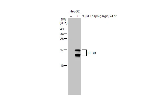
Untreated (–) and treated (+) HepG2 whole cell extract (30 μg) were separated by 15% SDS-PAGE, and the membrane was blotted with LC3B antibody (GTX127375) diluted at 1:500. The HRP-conjugated anti-rabbit IgG antibody (GTX213110-01) was used to detect the primary antibody.
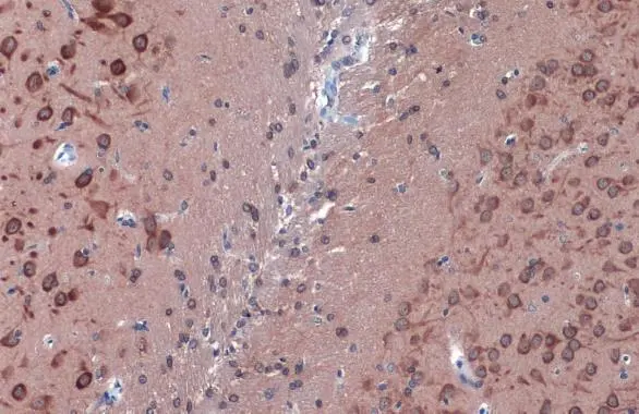
LC3B antibody detects LC3B protein at cytoplasm by immunohistochemical analysis.Sample: Paraffin-embedded rat brain.LC3B stained by LC3B antibody (GTX127375) diluted at 1:500.Antigen Retrieval: Citrate buffer, pH 6.0, 15 min
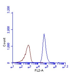
LC3B antibody (GTX127375) detects LC3B protein by flow cytometry analysis.
Sample: HeLa cell fixed in 4% paraformaldehyde at 4oC for 5 min.
Brown: Unlabelled sample was also used as a control.
Blue: LC3B antibody (GTX127375) dilution: 1:100.
Acquisition of >20,000 events were collected using Argon ion laser (488nm) and 525/30 bandpass filter.
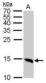
LC3B antibody detects MAP1LC3B protein by western blot analysis.
A. 50 μg Rat brain lysate/extract
15% SDS-PAGE
LC3B antibody (GTX127375) dilution: 1:1000
The HRP-conjugated anti-rabbit IgG antibody (GTX213110-01) was used to detect the primary antibody.
Untreated (–) and treated (+) HeLa whole cell extracts (30 μg) were separated by 15% SDS-PAGE, and the membrane was blotted with LC3B antibody (GTX127375) diluted at 1:2500. The HRP-conjugated anti-rabbit IgG antibody (GTX213110-01) was used to detect the primary antibody.
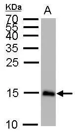
LC3B antibody detects MAP1LC3B protein by western blot analysis.
A. 50 μg mouse brain lysate/extract
15% SDS-PAGE
LC3B antibody (GTX127375) dilution: 1:1000
The HRP-conjugated anti-rabbit IgG antibody (GTX213110-01) was used to detect the primary antibody.
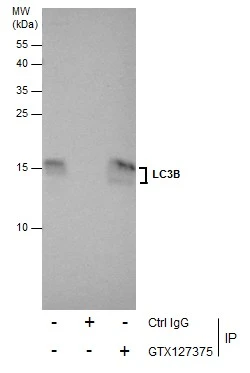
Immunoprecipitation of LC3B protein from U87-MG whole cell extracts using 5 μg of LC3B antibody (GTX127375).
Western blot analysis was performed using LC3B antibody (GTX127375).
EasyBlot anti-Rabbit IgG (GTX221666-01) was used as a secondary reagent.
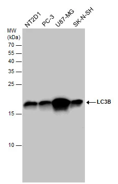
LC3B antibody detects LC3B protein by western blot analysis. Various whole cell extracts (30 μg) were separated by 15% SDS-PAGE, and the membrane was blotted with LC3B antibody (GTX127375) diluted at a dilution of 1:1000. The HRP-conjugated anti-rabbit IgG antibody (GTX213110-01) was used to detect the primary antibody.
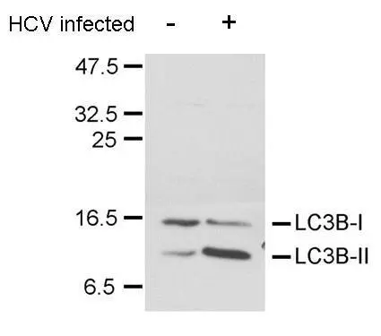
LC3B antibody detects LC3B protein in HCV-infected samples by western blot analysis.
A. 20 μg Huh7 whole cell lysate/extract (un-infected)
B. 20 μg Huh7 whole cell lysate/extract (HCV-infected)
LC3B antibody (GTX127375) dilution: 1:1500
The HRP-conjugated anti-rabbit IgG antibody (GTX213110-01) was used to detect the primary antibody.
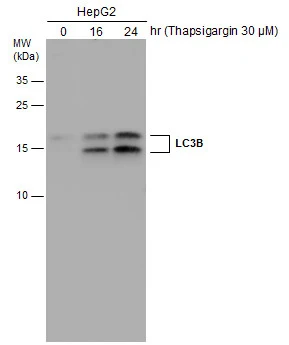
HepG2 cells were untreated or treated with 3 μM thapsigargin for 16 and 24 hrs. Whole cell extracts (30 μg) were separated by 15% SDS-PAGE, and the membrane was blotted with LC3B antibody (GTX127375) diluted at 1:1000. The HRP-conjugated anti-rabbit IgG antibody (GTX213110-01) was used to detect the primary antibody.
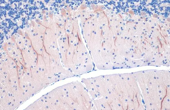
LC3B antibody detects LC3B protein at cytoplasm by immunohistochemical analysis.Sample: Paraffin-embedded mouse cerebellum.LC3B stained by LC3B antibody (GTX127375) diluted at 1:500.Antigen Retrieval: Citrate buffer, pH 6.0, 15 min
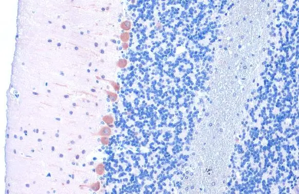
LC3B antibody detects LC3B protein at cytoplasm by immunohistochemical analysis.Sample: Paraffin-embedded rat cerebellum.LC3B stained by LC3B antibody (GTX127375) diluted at 1:500.Antigen Retrieval: Citrate buffer, pH 6.0, 15 min
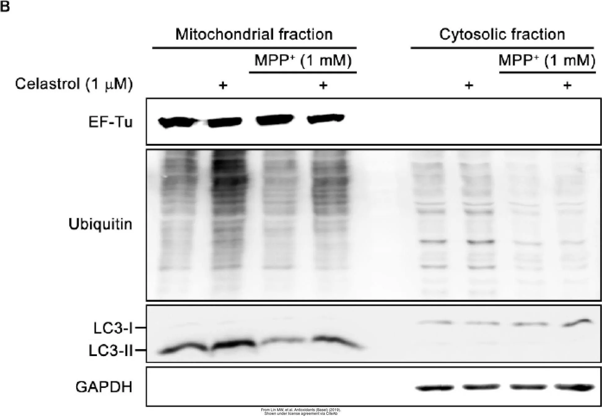
The data was published in the journal Antioxidants (Basel) in 2019.PMID: 31906147
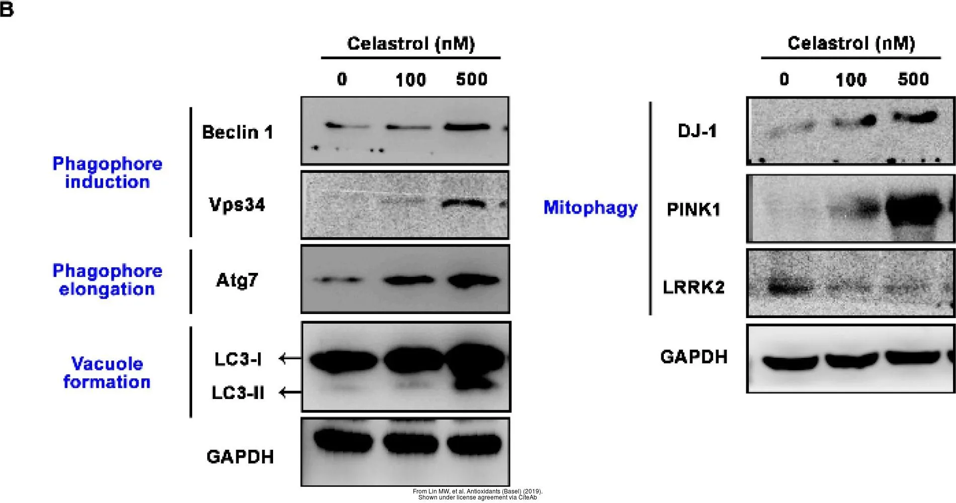
The data was published in the journal Antioxidants (Basel) in 2019.PMID: 31906147
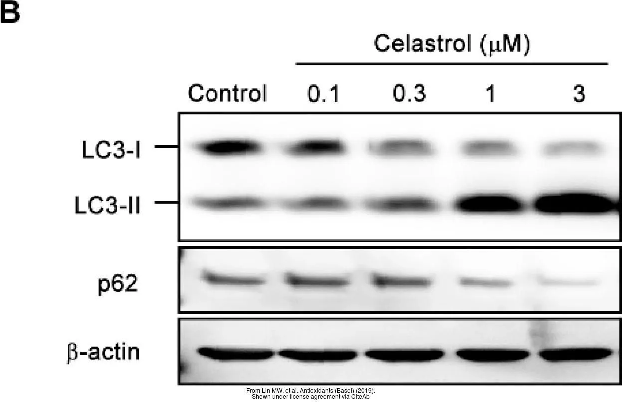
The data was published in the journal Antioxidants (Basel) in 2019.PMID: 31906147
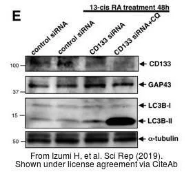
The data was published in the journal Sci Rep in 2019. PMID: 30783186

The data was published in the journal Sci Rep in 2019.PMID: 31341250
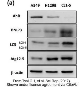
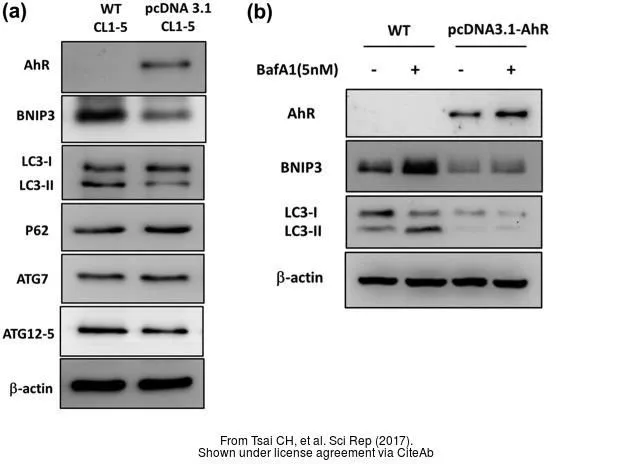
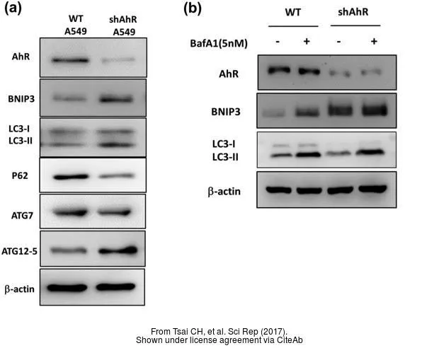
The data was published in the journal Sci Rep in 2017.PMID: 28195146
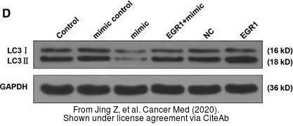
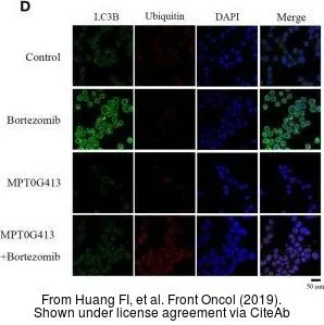
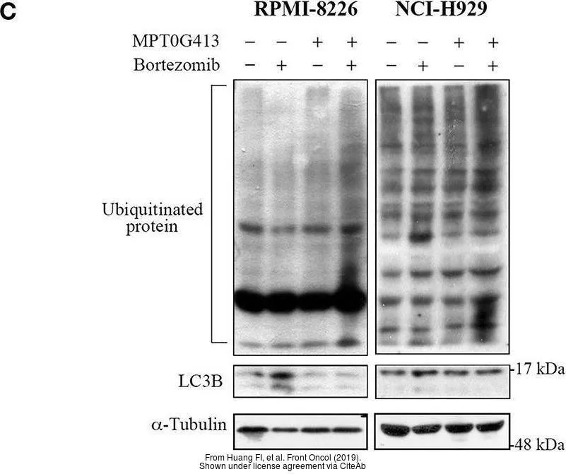
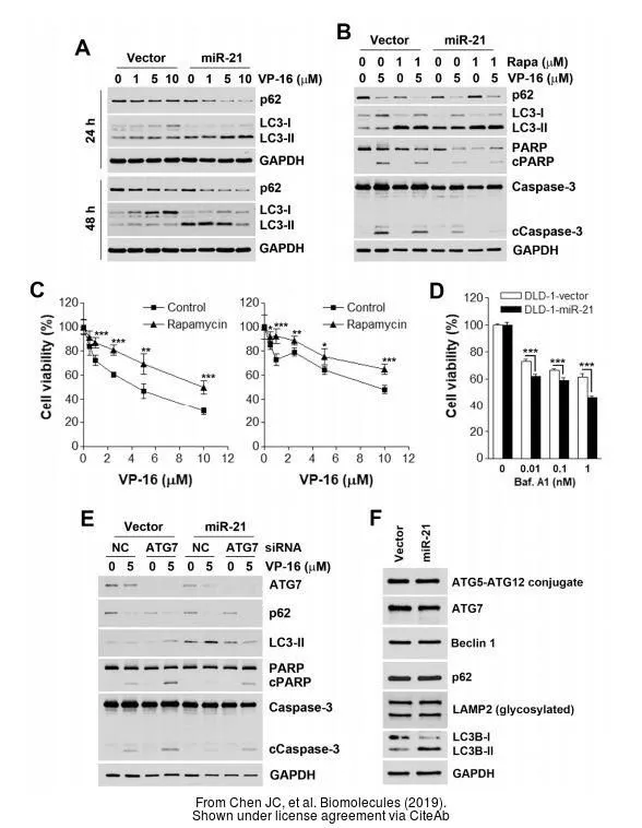
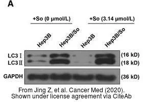
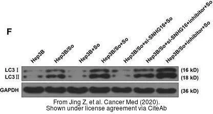
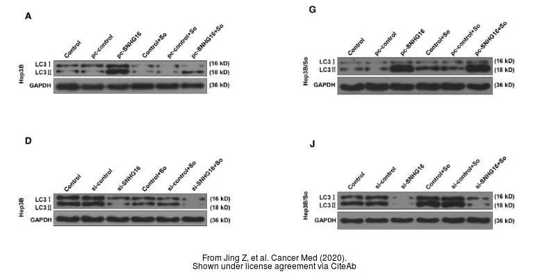
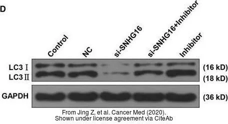
The data was published in the Cancer Med in 2020. PMID: 32324343
-
HostRabbit
-
ClonalityPolyclonal
-
IsotypeIgG
-
ApplicationsWB ICC/IF IHC-P IHC-Fr FCM IP EM
-
ReactivityHuman, Mouse, Rat, Guinea pig, Pig, Mosquito