N-Cadherin antibody


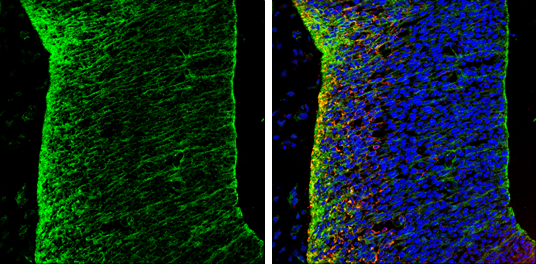
N-Cadherin antibody detects N-Cadherin protein expression by immunohistochemical analysis.
Sample: Frozen sectioned E13.5 Rat brain.
Green: N-Cadherin protein stained by N-Cadherin antibody (GTX127345) diluted at 1:250.
Red: beta Tubulin 3/ TUJ1, a mature neuron marker, stained by beta Tubulin 3/ TUJ1 antibody [GT11710] (GTX631836) diluted at 1:500.
Blue: Fluoroshield with DAPI (GTX30920).
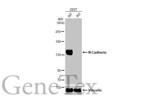
Wild-type (WT) and N-Cadherin knockout (KO) 293T cell extracts (30 μg) were separated by 5% SDS-PAGE, and the membrane was blotted with N-Cadherin antibody (GTX127345) diluted at 1:1000. The HRP-conjugated anti-rabbit IgG antibody (GTX213110-01) was used to detect the primary antibody.
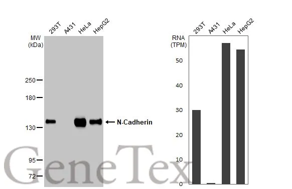
Various whole cell extracts (30 μg) were separated by 5% SDS-PAGE, and the membrane was blotted with N-Cadherin antibody (GTX127345) diluted at 1:1000. The HRP-conjugated anti-rabbit IgG antibody (GTX213110-01) was used to detect the primary antibody. Corresponding RNA expression data for the same cell lines are based on Human Protein Atlas program.
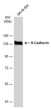
Whole cell extract (30 μg) was separated by 7.5% SDS-PAGE, and the membrane was blotted with N-Cadherin antibody (GTX127345) diluted at 1:1000. The HRP-conjugated anti-rabbit IgG antibody (GTX213110-01) was used to detect the primary antibody.
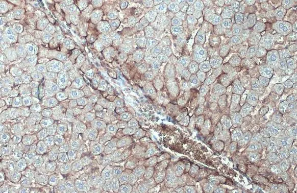
N-Cadherin antibody detects N-Cadherin protein at cell membrane by immunohistochemical analysis.Sample: Paraffin-embedded mouse liver.N-Cadherin stained by N-Cadherin antibody (GTX127345) diluted at 1:500.Antigen Retrieval: Citrate buffer, pH 6.0, 15 min
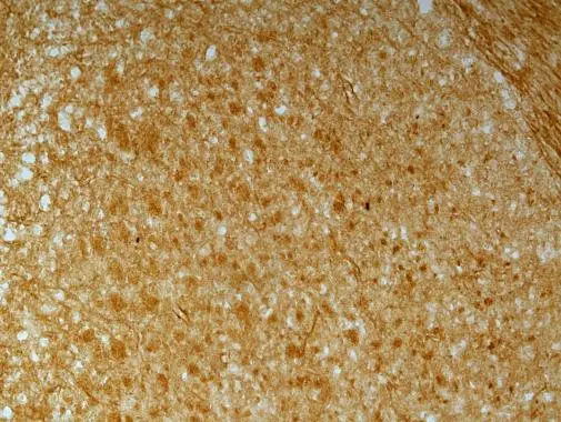
N-Cadherin antibody detects N-Cadherin protein on embryonic mouse brain by immunohistochemical analysis. Sample: Frozen section of embryonic mouse brain (mE18.5). N-Cadherin antibody (GTX127345) diluted at 1:500.
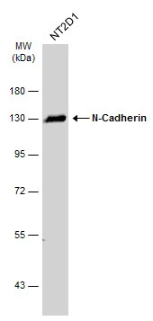
Whole cell extract (30 μg) was separated by 7.5% SDS-PAGE, and the membrane was blotted with N-Cadherin antibody (GTX127345) diluted at 1:1000. The HRP-conjugated anti-rabbit IgG antibody (GTX213110-01) was used to detect the primary antibody.
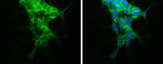
N-Cadherin antibody detects N-Cadherin protein at cell membrane by immunofluorescent analysis.
Sample: SH-SY5Y cells were fixed in 4% paraformaldehyde at RT for 15 min.
Green: N-Cadherin protein stained by N-Cadherin antibody (GTX127345) diluted at 1:500.
Blue: Hoechst 33342 staining.
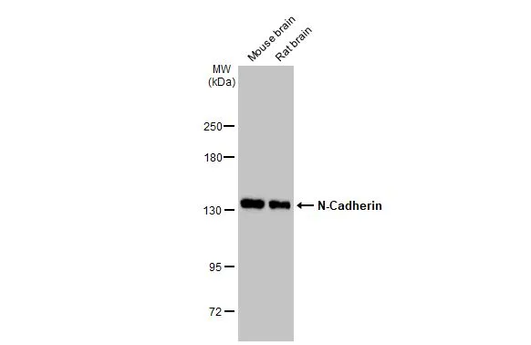
Various tissue extracts (50 μg) were separated by 5% SDS-PAGE, and the membrane was blotted with N-Cadherin antibody (GTX127345) diluted at 1:5000. The HRP-conjugated anti-rabbit IgG antibody (GTX213110-01) was used to detect the primary antibody.
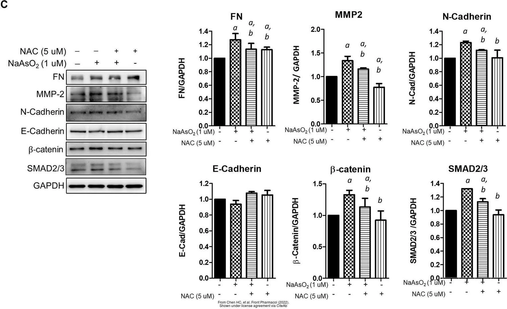
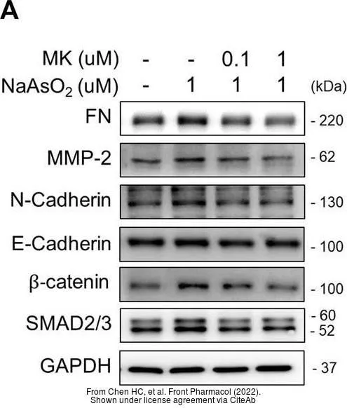
The data was published in the 2022 in Front Pharmacol. PMID: 35517780
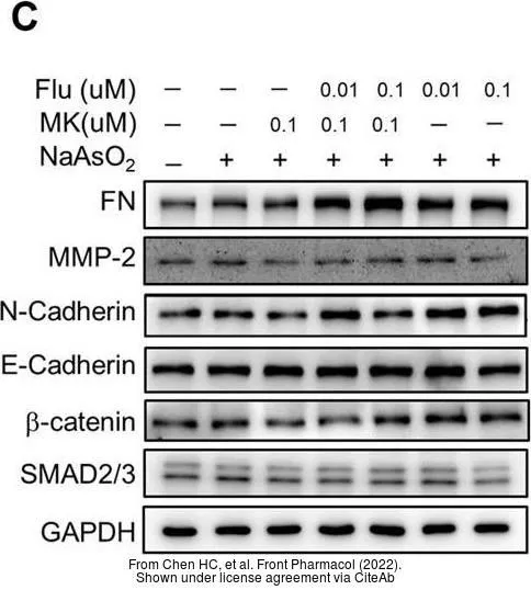
The data was published in the 2022 in Front Pharmacol. PMID: 35517780
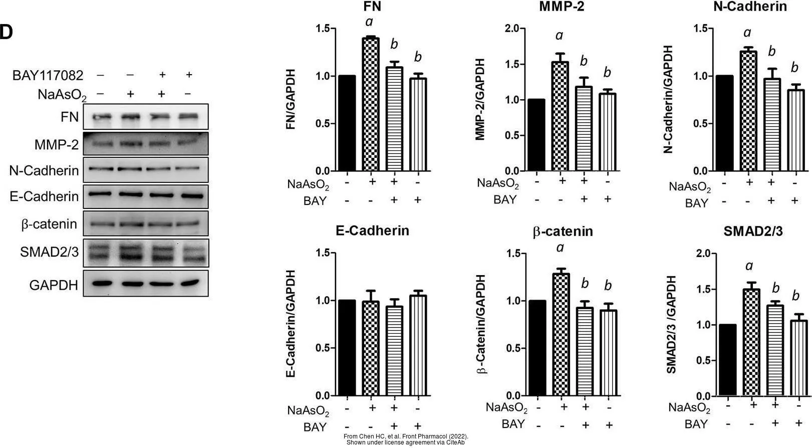
The data was published in the 2022 in Front Pharmacol. PMID: 35517780
-
HostRabbit
-
ClonalityPolyclonal
-
IsotypeIgG
-
ApplicationsWB ICC/IF IHC-P IHC-Fr
-
ReactivityHuman, Mouse, Rat, Monkey