NFkB p65 antibody


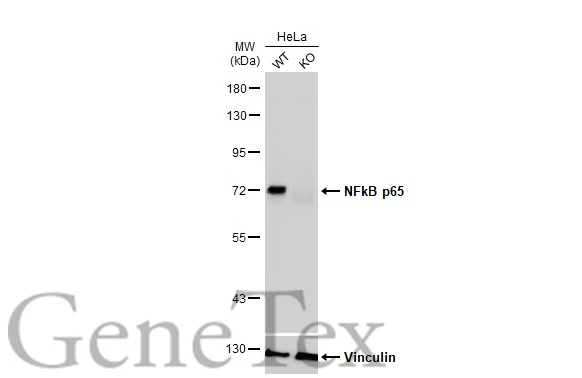
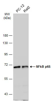
Wild-type (WT) and NFkB p65 knockout (KO) HeLa cell extracts (30 μg) were separated by 7.5% SDS-PAGE, and the membrane was blotted with NFkB p65 antibody (GTX107678) diluted at 1:1000. The HRP-conjugated anti-rabbit IgG antibody (GTX213110-01) was used to detect the primary antibody.
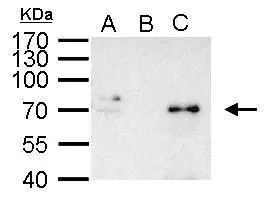
NFkB p65 antibody immunoprecipitates NFkB p65 protein in IP experiments. IP Sample: 1000 μg HeLa whole cell lysate/extract A. 40 μg HeLa whole cell lysate/extract B. Control with 2.5 μg of preimmune rabbit IgG C. Immunoprecipitation of NFkB p65 protein by 2.5 μg of NFkB p65 antibody (GTX107678) 10% SDS-PAGE The immunoprecipitated NFkB p65 protein was detected by NFkB p65 antibody (GTX107678) diluted at 1:1000. EasyBlot anti-rabbit IgG (GTX221666-01) was used as a secondary reagent.
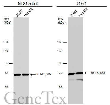
Various whole cell extracts (30 μg) were separated by 7.5% SDS-PAGE, and the membranes were blotted with NFkB p65 antibody (GTX107678) diluted at 1:1000 and competitor's antibody (#4764) diluted at 1:1000. The HRP-conjugated anti-rabbit IgG antibody (GTX213110-01) was used to detect the primary antibody.
*The competitor is not affiliated with GeneTex and does not endorse this product.
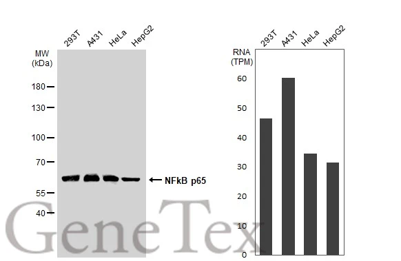
Various whole cell extracts (30 μg) were separated by 10% SDS-PAGE, and the membrane was blotted with NFkB p65 antibody (GTX107678) diluted at 1:1000. The HRP-conjugated anti-rabbit IgG antibody (GTX213110-01) was used to detect the primary antibody. Corresponding RNA expression data for the same cell lines are based on Human Protein Atlas program.
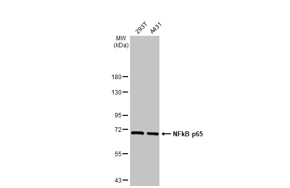
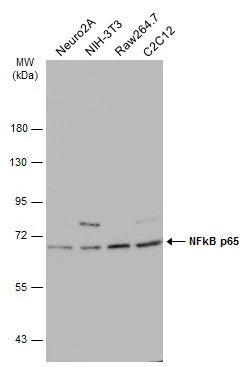
Various whole cell extracts (30 μg) were separated by 7.5% SDS-PAGE, and the membrane was blotted with NFkB p65 antibody (GTX107678) diluted at 1:1000. The HRP-conjugated anti-rabbit IgG antibody (GTX213110-01) was used to detect the primary antibody.
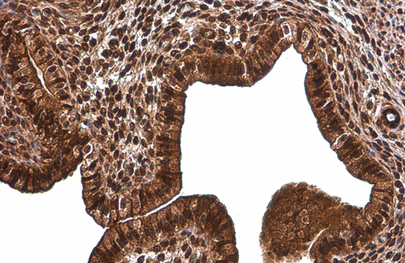
NFkB p65 antibody detects NFkB p65 protein at cytoplasm and nucleus by immunohistochemical analysis.Sample: Paraffin-embedded rat lung.NFkB p65 stained by NFkB p65 antibody (GTX107678) diluted at 1:500.Antigen Retrieval: Citrate buffer, pH 6.0, 15 min
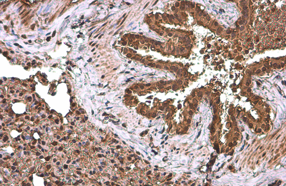
NFkB p65 antibody detects NFkB p65 protein at cytoplasm and nucleus by immunohistochemical analysis.Sample: Paraffin-embedded mouse cervix.NFkB p65 stained by NFkB p65 antibody (GTX107678) diluted at 1:500.Antigen Retrieval: Citrate buffer, pH 6.0, 15 min
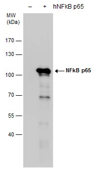
NFkB p65 antibody detects NFkB p65 protein by western blot analysis. Non-transfected (-) and NFkB p65 -transfected (+, including GFP-tag) 293T whole cell extracts (15 μg) were separated by 7.5% SDS-PAGE, and the membrane was blotted with NFkB p65 antibody (GTX107678) diluted by 1:5000. The HRP-conjugated anti-rabbit IgG antibody (GTX213110-01) was used to detect the primary antibody.
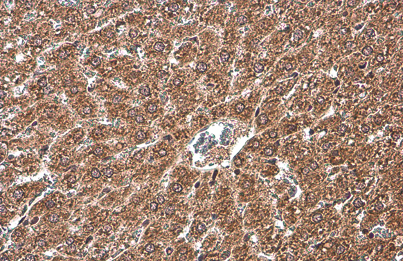
NFkB p65 antibody detects NFkB p65 protein at cytoplasm by immunohistochemical analysis.Sample: Paraffin-embedded rat liver.NFkB p65 stained by NFkB p65 antibody (GTX107678) diluted at 1:500.Antigen Retrieval: Citrate buffer, pH 6.0, 15 min
Various whole cell extracts (30 μg) were separated by 7.5% SDS-PAGE, and the membrane was blotted with NFkB p65 antibody (GTX107678) diluted at 1:1000. The HRP-conjugated anti-rabbit IgG antibody (GTX213110-01) was used to detect the primary antibody.
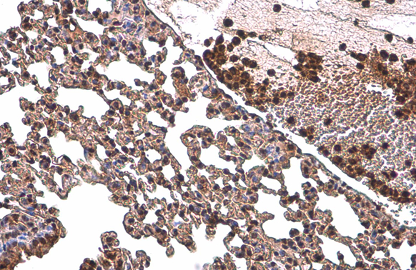
NFkB p65 antibody detects NFkB p65 protein at cytoplasm and nucleus by immunohistochemical analysis.Sample: Paraffin-embedded mouse lung.NFkB p65 stained by NFkB p65 antibody (GTX107678) diluted at 1:500.Antigen Retrieval: Citrate buffer, pH 6.0, 15 min
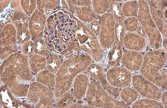
NFkB p65 antibody detects NFkB p65 protein at cytoplasm by immunohistochemical analysis.Sample: Paraffin-embedded mouse kidney.NFkB p65 stained by NFkB p65 antibody (GTX107678) diluted at 1:500.Antigen Retrieval: Citrate buffer, pH 6.0, 15 min
Various whole cell extracts (30 μg) were separated by 7.5% SDS-PAGE, and the membrane was blotted with NFkB p65 antibody (GTX107678) diluted at 1:1000. The HRP-conjugated anti-rabbit IgG antibody (GTX213110-01) was used to detect the primary antibody.
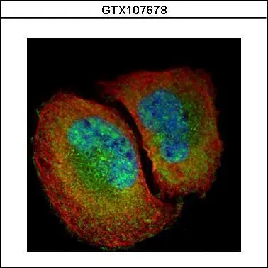
Confocal immunofluorescence analysis (Olympus FV10i) of paraformaldehyde-fixed U2OS, using NFkB p65(GTX107678) antibody (Green) at 1:500 dilution. Alpha-tubulin filaments were labeled with GTX11304 (Red) at 1:2000.
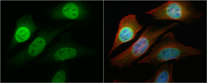
NFkB p65 antibody detects NFkB p65 protein at cytoplasm and nucleus by immunofluorescent analysis.
Sample: HeLa cells were fixed in 4% paraformaldehyde at RT for 15 min.
Green: NFkB p65 protein stained by NFkB p65 antibody (GTX107678) diluted at 1:1000.
Red: phalloidin, a cytoskeleton marker, stained by phalloidin (invitrogen, A12380) diluted at 1:200.
Blue: Hoechst 33342 staining.
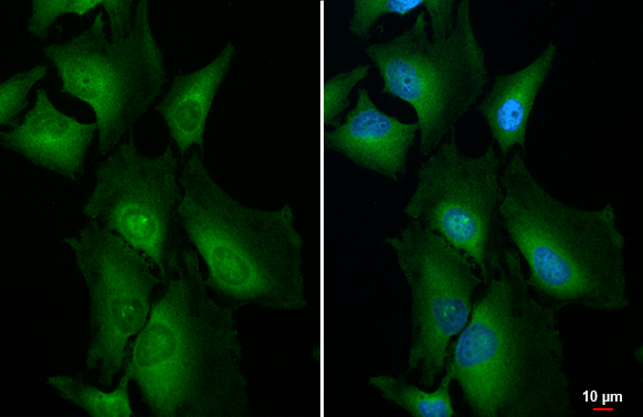
NFkB p65 antibody detects NFkB p65 protein at cytoplasm, nucleus and endoplasmic reticulum by immunofluorescent analysis.Sample: HeLa cells were fixed in 4% paraformaldehyde at RT for 15 min.Green: NFkB p65 stained by NFkB p65 antibody (GTX107678) diluted at 1:500.Blue: Fluoroshield with DAPI (GTX30920).
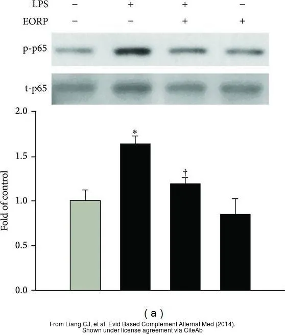
The data was published in the journal Evid Based Complement Alternat Med in 2014. PMID: 24723958
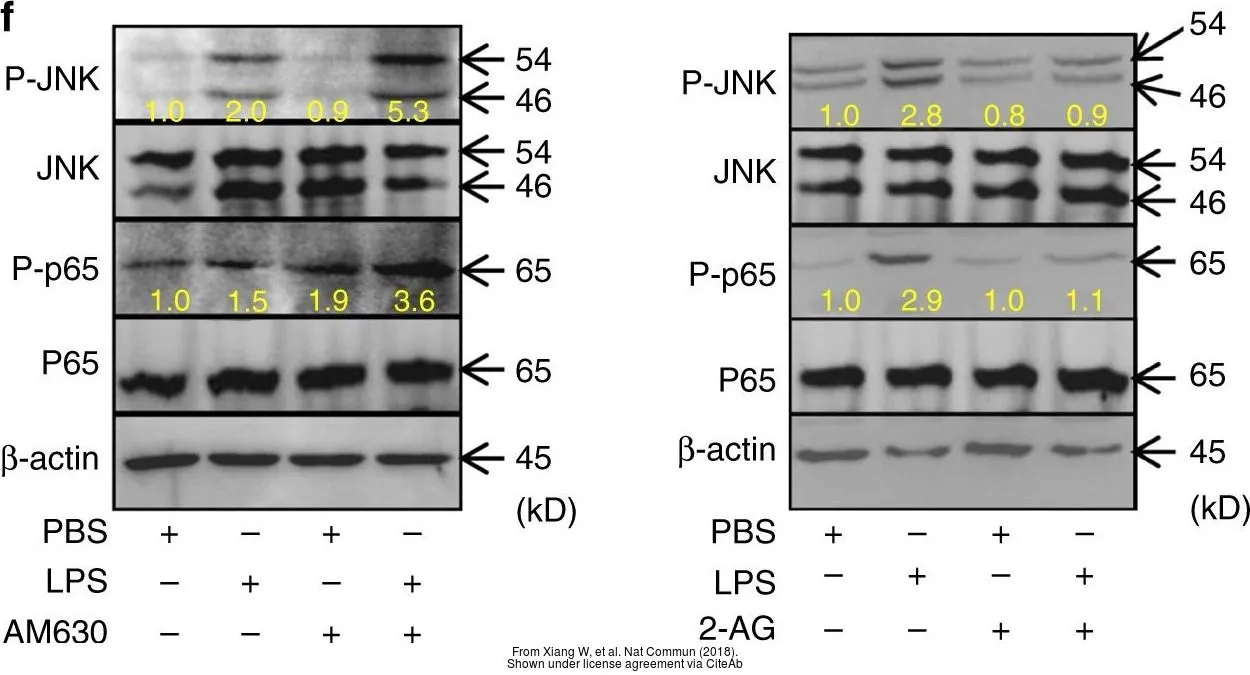
The data was published in the journal Nat Commun in 2018.PMID: 29968710
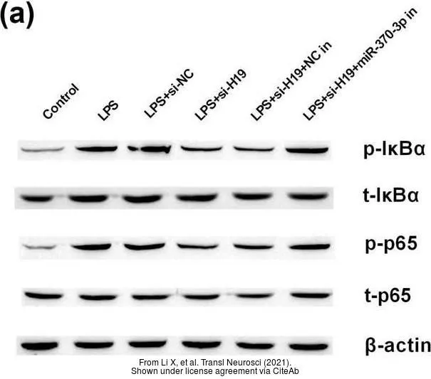
The data was published in the 2021 in Transl Neurosci. PMID: 33708438
-
HostRabbit
-
ClonalityPolyclonal
-
IsotypeIgG
-
ApplicationsWB ICC/IF IHC-P IP EMSA
-
ReactivityHuman, Mouse, Rat