OCT6 antibody
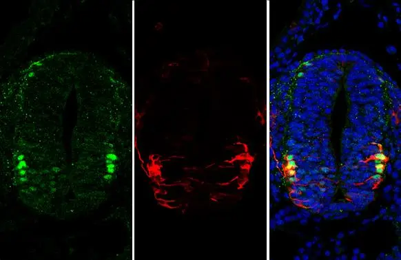
OCT6 antibody detects OCT6 protein at nucleus by immunohistochemical analysis.Sample: Paraffin-embedded mouse E10.5 embryo.Green: OCT6 stained by OCT6 antibody (GTX134063) diluted at 1:250.Red: beta Tubulin 3/ Tuj1, a cytoskeleton marker, stained by beta Tubulin 3/ Tuj1 antibody [GT11710] (GTX631836) diluted at 1:500.Blue: Fluoroshield with DAPI (GTX30920).Antigen Retrieval: Citrate buffer, pH 6.0, 15 min
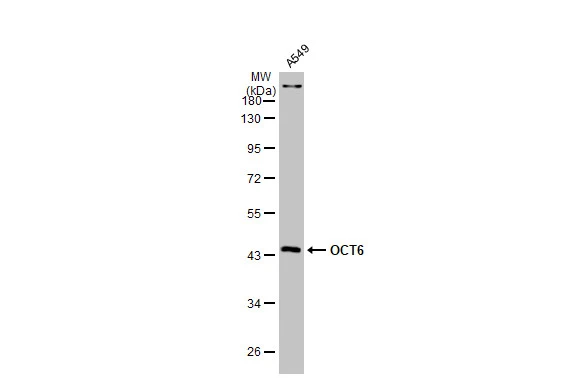
Whole cell extract (30 μg) was separated by 10% SDS-PAGE, and the membrane was blotted with OCT6 antibody (GTX134063) diluted at 1:1000. The HRP-conjugated anti-rabbit IgG antibody (GTX213110-01) was used to detect the primary antibody.

Mouse tissue extract (50 μg) was separated by 10% SDS-PAGE, and the membrane was blotted with OCT6 antibody (GTX134063) diluted at 1:500. The HRP-conjugated anti-rabbit IgG antibody (GTX213110-01) was used to detect the primary antibody, and the signal was developed with Trident ECL plus-Enhanced.

OCT6 antibody detects OCT6 protein at nucleus by immunofluorescent analysis.Sample: A549 cells were fixed in 4% paraformaldehyde at RT for 15 min.Green: OCT6 stained by OCT6 antibody (GTX134063) diluted at 1:500.Red: phalloidin, a cytoskeleton marker, diluted at 1:100.Scale bar= 10 μm.
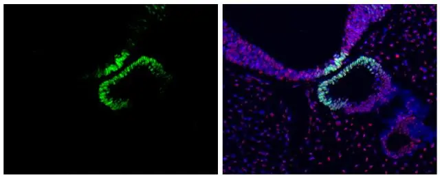
OCT6 antibody detects OCT6 protein at nucleus by immunohistochemical analysis.Sample: Paraffin-embedded mouse E10.5 embryo.Green: OCT6 stained by OCT6 antibody (GTX134063) diluted at 1:500.Red: SOX2, a nucleus marker, stained by SOX2 antibody [GT1352] (GTX627405) diluted at 1:250.Blue: Fluoroshield with DAPI (GTX30920).Antigen Retrieval: Citrate buffer, pH 6.0, 15 min

OCT6 antibody detects OCT6 protein by immunohistochemical analysis.Sample: Frozen-sectioned mouse cerebral cortex.Green: OCT6 stained by OCT6 antibody (GTX134063) diluted at 1:250.Blue: Fluoroshield with DAPI (GTX30920).

OCT6 antibody detects OCT6 protein at nucleus by immunofluorescent analysis.
Sample: DIV9 rat hippocampal neuron and glia cells were fixed in 4% paraformaldehyde at RT for 15 min.
Green: 44840 stained by OCT6 antibody (GTX134063) diluted at 1:250.
Red: Tau, a cytoskeleton marker, stained by Tau antibody [GT287] (GTX634809) diluted at 1:500.
-
HostRabbit
-
ClonalityPolyclonal
-
IsotypeIgG
-
ApplicationsWB ICC/IF IHC-P IHC-Fr
-
ReactivityHuman, Mouse, Rat