PI3 kinase p150 antibody


Non-transfected (–) and transfected (+) 293T whole cell extracts (30 μg) were separated by 5% SDS-PAGE, and the membrane was blotted with PI3-Kinase p150 antibody (GTX132466) diluted at 1:1000.
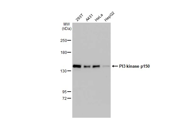
Various whole cell extracts (30 μg) were separated by 5% SDS-PAGE, and the membrane was blotted with PI3 kinase p150 antibody (GTX132466) diluted at 1:1000. The HRP-conjugated anti-rabbit IgG antibody (GTX213110-01) was used to detect the primary antibody.

Immunoprecipitation of PI3-Kinase p150 protein from HeLa whole cell extracts using 5 μg of PI3-Kinase p150 antibody (GTX132466).
Western blot analysis was performed using PI3-Kinase p150 antibody (GTX132466) diluted at 1:500.
EasyBlot anti-Rabbit IgG (GTX221666-01) was used as a secondary reagent.

Mouse tissue extract (50 μg) was separated by 5% SDS-PAGE, and the membrane was blotted with PI3-Kinase p150 antibody (GTX132466) diluted at 1:1000.

Various whole cell extracts (30 μg) were separated by 5% SDS-PAGE, and the membrane was blotted with PI3-Kinase p150 antibody (GTX132466) diluted at 1:1000.

Various whole cell extracts (30 μg) were separated by 5% SDS-PAGE, and the membrane was blotted with PI3-Kinase p150 antibody (GTX132466) diluted at 1:1000.

PI3-Kinase p150 antibody detects PI3-Kinase p150 protein at cytoplasm in mouse duodenum by immunohistochemical analysis.
Sample: Paraffin-embedded mouse duodenum.
PI3-Kinase p150 antibody (GTX132466) diluted at 1:500.
Antigen Retrieval: Citrate buffer, pH 6.0, 15 min
-
HostRabbit
-
ClonalityPolyclonal
-
IsotypeIgG
-
ApplicationsWB IHC-P IP
-
ReactivityHuman, Mouse