RAB7A antibody


*The competitor is not affiliated with GeneTex and does not endorse this product.
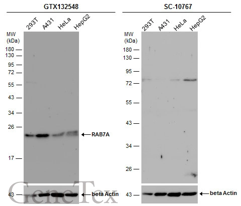
Various whole cell extracts (30 μg) were separated by 12% SDS-PAGE, and the membrane was blotted with RAB7A antibody (GTX132548) diluted at 1:1000 and competitor's antibody (SC-10767) diluted by 1:200.
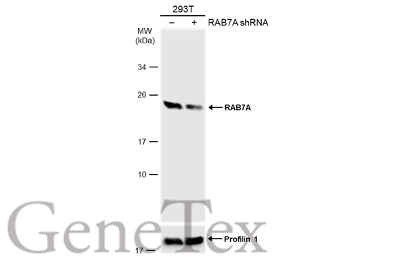
Non-transfected (–) and transfected (+) 293T whole cell extracts (50 μg) were separated by 12% SDS-PAGE, and the membrane was blotted with RAB7A antibody (GTX132548) diluted at 1:1000. The HRP-conjugated anti-rabbit IgG antibody (GTX213110-01) was used to detect the primary antibody, and the signal was developed with Trident ECL plus-Enhanced.
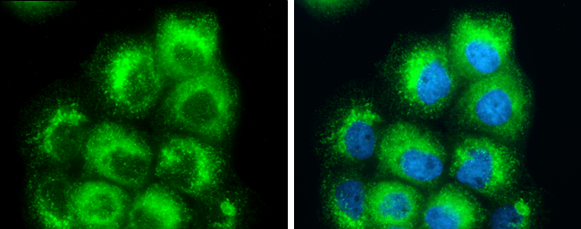
Rab7A antibody detects Rab7A protein at cytoplasm by immunofluorescent analysis.
Sample: A431 cells were fixed in 4% paraformaldehyde at RT for 15 min.
Green: Rab7A protein stained by Rab7A antibody (GTX132548) diluted at 1:500.
Blue: Hoechst 33342 staining.
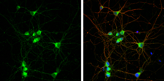
RAB7A antibody detects RAB7A protein by immunofluorescent analysis.Sample: DIV9 rat E18 primary cortical neuron cells were fixed in 4% paraformaldehyde at RT for 15 min.Green: RAB7A stained by RAB7A antibody (GTX132548) diluted at 1:500.Red: beta Tubulin 3/ Tuj1, stained by beta Tubulin 3/ Tuj1 antibody [GT1338] (GTX631831) diluted at 1:500.Blue: Fluoroshield with DAPI (GTX30920).
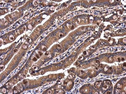
Rab7 antibody detects Rab7 protein at cytoplasm in rat intestine by immunohistochemical analysis.
Sample: Paraffin-embedded rat intestine.
Rab7 antibody (GTX132548) diluted at 1:500.
Antigen Retrieval: Citrate buffer, pH 6.0, 15 min
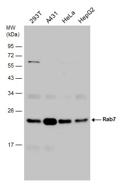
Various whole cell extracts (30 μg) were separated by 12% SDS-PAGE, and the membrane was blotted with Rab7 antibody (GTX132548) diluted at 1:1000.
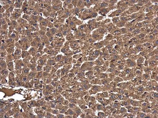
Rab7 antibody detects Rab7 protein at cytoplasm in mouse liver by immunohistochemical analysis.
Sample: Paraffin-embedded mouse liver.
Rab7 antibody (GTX132548) diluted at 1:500.
Antigen Retrieval: Citrate buffer, pH 6.0, 15 min
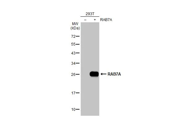
Non-transfected (–) and transfected (+) 293T whole cell extracts (30 μg) were separated by 12% SDS-PAGE, and the membrane was blotted with RAB7A antibody (GTX132548) diluted at 1:5000. The HRP-conjugated anti-rabbit IgG antibody (GTX213110-01) was used to detect the primary antibody.
-
HostRabbit
-
ClonalityPolyclonal
-
IsotypeIgG
-
ApplicationsWB ICC/IF IHC-P
-
ReactivityHuman, Mouse, Rat