RIF1 antibody

Cat. No. GTX48737
Cat. No.  GTX48737
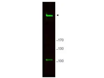
Anti-RIF1 antibody used in Western Blot (WB). GTX48737
GTX48737 WB Image

WB analysis of MEF whole cell lysate using GTX48737 RIF1 antibody.
Loading : 25 μg
Dilution : 1 μg/mL

1 / 2
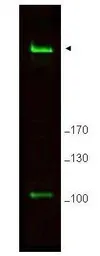
Anti-RIF1 antibody used in Western Blot (WB). GTX48737
GTX48737 WB Image

Western blot using GeneTex's Affinity Purified anti-Rif1 antibody shows detection of a band ~265 kDa corresponding to mouse Rif1 (arrowhead). Specific reactivity with this band is blocked when the antibody is pre-incubated with the immunizing peptide (data not shown). Approximately 25 μg of MEF whole cell lysate was separated by SDS-PAGE and transferred onto nitrocellulose. After blocking the membrane was probed with the primary antibody diluted to 1.0 ug/ml for 2 h at room temperature followed by washes and reaction with a 1:10,000 dilution of IRDye800 conjugated goat anti-Rabbit IgG [H&L] MX for 45 min at room temperature. IRDye800 fluorescence image was captured using the OdysseyR Infrared Imaging System developed by LI-COR. IRDye is a trademark of LI-COR, Inc. Other detection systems will yield similar results.

2 / 2
Anti-RIF1 antibody used in Western Blot (WB). GTX48737
  • Host
    Rabbit
  • Clonality
    Polyclonal
  • Isotype
    IgG
  • Applications
    WB ELISA
  • Reactivity
    Human, Mouse
See all RIF1 products