ROCK2 (phospho Ser1366) antibody


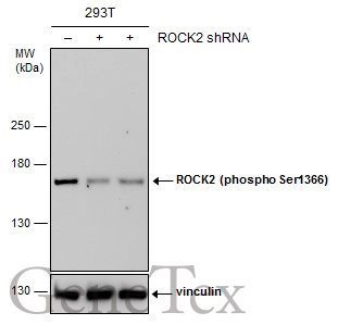
Non-transfected (–) and transfected (+) 293T whole cell extracts (30 μg) were separated by 5% SDS-PAGE, and the membrane was blotted with ROCK2 (phospho Ser1366) antibody (GTX122651) diluted at 1:500. The HRP-conjugated anti-rabbit IgG antibody (GTX213110-01) was used to detect the primary antibody.
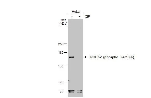
Untreated (–) and treated (+) HeLa whole cell extracts (30 μg) were separated by 5% SDS-PAGE, and the membrane was blotted with ROCK2 (phospho Ser1366) antibody (GTX122651) diluted at 1:1000. The HRP-conjugated anti-rabbit IgG antibody (GTX213110-01) was used to detect the primary antibody, and the signal was developed with Trident femto Western HRP Substrate.
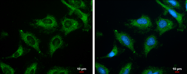
ROCK2 (phospho Ser1366) antibody detects ROCK2 (phospho Ser1366) protein at cytoplasm by immunofluorescent analysis.
Sample: HeLa cells were fixed in ice-cold MeOH for 5 min.
Green: ROCK2 (phospho Ser1366) protein stained by ROCK2 (phospho Ser1366) antibody (GTX122651) diluted at 1:500.
Blue: Hoechst 33342 staining.
Scale bar = 10 μm.
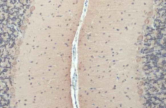
ROCK2 (phospho Ser1366) antibody detects ROCK2 (phospho Ser1366) protein at cell membrane and cytoplasm by immunohistochemical analysis.Sample: Paraffin-embedded rat cerebellum.ROCK2 (phospho Ser1366) stained by ROCK2 (phospho Ser1366) antibody (GTX122651) diluted at 1:500.Antigen Retrieval: Citrate buffer, pH 6.0, 15 min
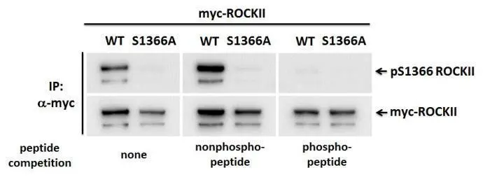
ROCKII (phospho Ser1366) antibody detects the phosphorylation of ROCKII at Ser 1366 residue in IP experiments.
HEK293T cells were transfected with wild type (WT) or phospho-mutant (S1366A) ROCKII expression constructs.
Overexpressed Myc-ROCKII proteins were immunoprecipitated with anti-myc antibody and probed with anti- ROCKII (phospho Ser1366) antibody (GTX122651, 1:1000 dilution) with or without peptide (0.2 mg/ml) competition as indicated.
The HRP-conjugated anti-rabbit IgG antibody (GTX213110-01) was used to detect the primary antibody.
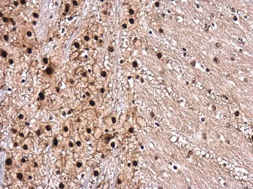
ROCK2 (phospho Ser1366) antibody detects ROCK2 (phospho Ser1366) protein at cytoplasm and nucleus in mouse brain by immunohistochemical analysis.
Sample: Paraffin-embedded mouse brain.
ROCK2 (phospho Ser1366) antibody (GTX122651) diluted at 1:500.
Antigen Retrieval: Citrate buffer, pH 6.0, 15 min
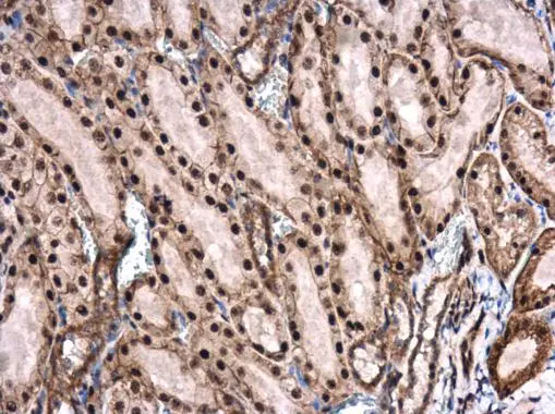
ROCK2 (phospho Ser1366) antibody detects ROCK2 (phospho Ser1366) protein at cytoplasm and nucleus in rat kidney by immunohistochemical analysis.
Sample: Paraffin-embedded rat kidney.
ROCK2 (phospho Ser1366) antibody (GTX122651) diluted at 1:500.
Antigen Retrieval: Citrate buffer, pH 6.0, 15 min
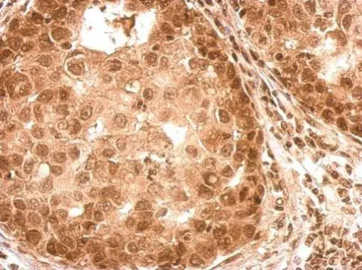
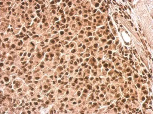
ROCK2 (phospho Ser1366) antibody detects ROCK2 protein at weak nucleus and cytosol on human breast tissue by immunohistochemical analysis.
Sample: Paraffin-embedded human breast tissue.
ROCK2 (phospho Ser1366) antibody (GTX122651) dilution: 1:500.
Antigen Retrieval: Trilogy™ (EDTA based, pH 8.0) buffer, 15min
ROCK2 (phospho Ser1366) antibody detects ROCK2 protein at nucleus and cytosol on HBL435 xenograft by immunohistochemical analysis.
Sample: Paraffin-embedded HBL435 xenograft.
ROCK2 (phospho Ser1366) antibody (GTX122651) dilution: 1:500.
Antigen Retrieval: Trilogy™ (EDTA based, pH 8.0) buffer, 15min
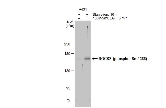
Untreated (–) and treated (+) A431 whole cell extracts (30 μg) were separated by 5% SDS-PAGE, and the membrane was blotted with ROCK2 (phospho Ser1366) antibody (GTX122651) diluted at 1:1000. The HRP-conjugated anti-rabbit IgG antibody (GTX213110-01) was used to detect the primary antibody, and the signal was developed with Trident ECL plus-Enhanced.
-
HostRabbit
-
ClonalityPolyclonal
-
IsotypeIgG
-
ApplicationsWB ICC/IF IHC-P IHC-Fr Immunoassay
-
ReactivityHuman, Mouse, Rat