Goat Anti-Rabbit IgG antibody (DyLight488)
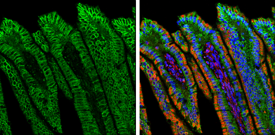
Double-labeled immunofluorescence photomicrographs of paraffin-embedded sections of mouse colon.
Green: E-Cadherin antibody (GTX100443) diluted at 1:500. The signal was developed using goat anti-rabbit IgG antibody (Dylight488) (GTX213110-04).
Red: alpha Tubulin antibody [GT114] (GTX628802) diluted at 1:500. The signal was developed using goat anti-mouse IgG antibody (Dylight594) (GTX213111-05).
Blue: Fluoroshield with DAPI (GTX30920).
Antigen Retrieval: Citrate buffer, pH 6.0, 15 min
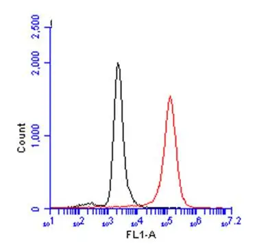
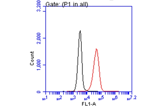
CD81 antibody (GTX101766) detects CD81 protein by flow cytometry analysis.
Sample: THP-1 cell.
Black: Unlabelled sample was used as a control.
Red: CD81 antibody (GTX101766) dilution: 1:50.
The Rabbit IgG antibody (DyLight488) (GTX213110-04) was used to detect the primary antibody.
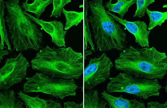
alpha Tubulin antibody detects alpha Tubulin protein at cytoskeleton by immunofluorescent analysis.Sample: HeLa cells were fixed in 4% paraformaldehyde at RT for 15 min.Green: alpha Tubulin stained by alpha Tubulin antibody (GTX112141) diluted at 1:500.The signal was developed using Goat Anti-Rabbit IgG antibody (DyLight488) (GTX213110-04) diluted at 1:2000.Blue: Fluoroshield with DAPI (GTX30920).
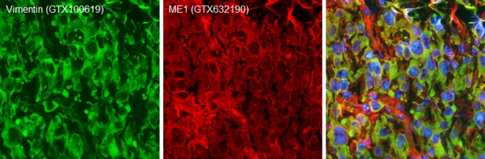
Double-labeled immunofluorescence photomicrographs of frozen sections of mouse brain.
Green: Vimentin antibody (GTX100619) diluted at 1:200. The signal was developed using goat anti-rabbit IgG antibody (Dylight488) (GTX213110-04).
Red: ME1 antibody [GT15611] (GTX632190) diluted at 1:200. The signal was developed using goat anti-mouse IgG antibody (Dylight594) (GTX213111-05).
Blue: Nuclear staining with Hoechst 33342.
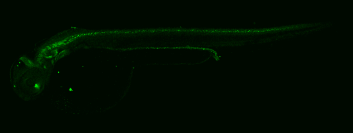
Whole mount immunohistochemical analysis of paraformaldehyde-fixed 2 day-post-fertilization zebrafish embryo using Pax2a antibody (GTX128127) detected by anti-rabbit IgG antibody (Dylight488) (GTX213110-04). GTX128127 diluted at 1:100 and incubated overnight at 4ºC. GTX213110-04 diluted at 1:500 and incubated 3 hours at room temperature.
Glypican 1 antibody [N3C3] (GTX104557) detects Glypican 1 protein by flow cytometry analysis.
Sample: A431 cell.
Black: Unlabelled sample was used as a control.
Red: Glypican 1 antibody [N3C3] (GTX104557) dilution: 1:50.
Acquisition of 20,000 events were collected using the Rabbit IgG (DyLight488) (GTX113110-04) secondary antibody for FACS analysis.
-
HostGoat
-
ClonalityPolyclonal
-
IsotypeIgG
-
ApplicationsWB ICC/IF IHC-P IHC-Fr IHC-Wm FACS
-
ReactivityRabbit