TOMM20 antibody


Mouse tissue extract (50 μg) was separated by 15% SDS-PAGE, and the membranes were blotted with TOMM20 antibody (GTX133756) diluted at 1:1000 and competitor's antibody (Competitor A) diluted at 1:500. The HRP-conjugated anti-rabbit IgG antibody (GTX213110-01) was used to detect the primary antibody.
*The competitor is not affiliated with GeneTex and does not endorse this product.

HepG2 and mitochondria extracts (30 μg) were separated by SDS-PAGE, and the membrane was blotted with TOMM20 antibody (GTX133756) diluted at 1:1000. The HRP-conjugated anti-rabbit IgG antibody (GTX213110-01) was used to detect the primary antibody.
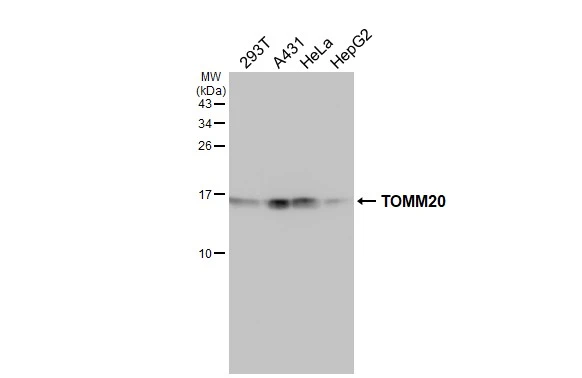
Various whole cell extracts (30 μg) were separated by 15% SDS-PAGE, and the membrane was blotted with TOMM20 antibody (GTX133756) diluted at 1:1000. The HRP-conjugated anti-rabbit IgG antibody (GTX213110-01) was used to detect the primary antibody.

TOMM20 antibody detects TOMM20 protein at mitochondria by immunofluorescent analysis.Sample: HeLa cells were fixed in 4% paraformaldehyde at RT for 15 min.Green: TOMM20 stained by TOMM20 antibody (GTX133756) diluted at 1:500.Red: alpha Tubulin, a cytoskeleton marker, stained by alpha Tubulin antibody [GT114] (GTX628802) diluted at 1:1000.Blue: Fluoroshield with DAPI (GTX30920).

Rat tissue extract (50 μg) was separated by 15% SDS-PAGE, and the membrane was blotted with TOMM20 antibody (GTX133756) diluted at 1:1000. The HRP-conjugated anti-rabbit IgG antibody (GTX213110-01) was used to detect the primary antibody.

TOMM20 antibody detects TOMM20 protein at mitochondria by immunohistochemical analysis.Sample: Paraffin-embedded rat kidney.TOMM20 stained by TOMM20 antibody (GTX133756) diluted at 1:500.
Antigen Retrieval: Citrate buffer, pH 6.0, 15 min

Rat tissue extract (50 μg) was separated by 15% SDS-PAGE, and the membrane was blotted with TOMM20 antibody (GTX133756) diluted at 1:1000. The HRP-conjugated anti-rabbit IgG antibody (GTX213110-01) was used to detect the primary antibody.

TOMM20 antibody detects TOMM20 protein at mitochondria by immunohistochemical analysis.Sample: Paraffin-embedded mouse kidney.TOMM20 stained by TOMM20 antibody (GTX133756) diluted at 1:500.Antigen Retrieval: Citrate buffer, pH 6.0, 15 min

TOMM20 antibody detects TOMM20 protein at mitochondria by immunohistochemical analysis.Sample: Paraffin-embedded mouse duodenum.TOMM20 stained by TOMM20 antibody (GTX133756) diluted at 1:500.
Antigen Retrieval: Citrate buffer, pH 6.0, 15 min

*The competitor is not affiliated with GeneTex and does not endorse this product.
Various whole cell extracts (30 μg) were separated by 15% SDS-PAGE, and the membranes were blotted with TOMM20 antibody (GTX133756) diluted at 1:1000 and competitor's antibody (sc-11415) diluted at 1:1000. The HRP-conjugated anti-rabbit IgG antibody (GTX213110-01) was used to detect the primary antibody.

Mouse tissue extract (50 μg) was separated by 15% SDS-PAGE, and the membrane was blotted with TOMM20 antibody (GTX133756) diluted at 1:1000. The HRP-conjugated anti-rabbit IgG antibody (GTX213110-01) was used to detect the primary antibody.

TOMM20 antibody detects TOMM20 protein at cytoplasm by immunohistochemical analysis.Sample: Paraffin-embedded mouse kidney.TOMM20 stained by TOMM20 antibody (GTX133756) diluted at 1:500.Antigen Retrieval: Citrate buffer, pH 6.0, 15 min

TOMM20 antibody detects TOMM20 protein at cytoplasm by immunohistochemical analysis.Sample: Paraffin-embedded rat kidney.TOMM20 stained by TOMM20 antibody (GTX133756) diluted at 1:500.Antigen Retrieval: Citrate buffer, pH 6.0, 15 min

TOMM20 antibody detects TOMM20 protein at mitochondria by immunofluorescent analysis.Sample: HeLa cells were fixed in 4% paraformaldehyde at RT for 15 min.Green: TOMM20 stained by TOMM20 antibody (GTX133756) diluted at 1:500.Red: alpha Tubulin, a cytoskeleton marker, stained by alpha Tubulin antibody [GT114] (GTX628802) diluted at 1:1000.Blue: Fluoroshield with DAPI (GTX30920).Scale bar= 10μm.

TOMM20 antibody detects TOMM20 protein at mitochondria by immunohistochemical analysis.Sample: Paraffin-embedded mouse kidney.TOMM20 stained by TOMM20 antibody (GTX133756) diluted at 1:615.Antigen Retrieval: Citrate buffer, pH 6.0, 15 min
-
HostRabbit
-
ClonalityPolyclonal
-
IsotypeIgG
-
ApplicationsWB ICC/IF IHC-P
-
ReactivityHuman, Mouse, Rat