Tau antibody

Cat. No. GTX130462
Cat. No.  GTX130462
Anti-Tau antibody used in Western Blot (WB). GTX130462
GTX130462 WB Image

Non-transfected (–) and transfected (+) SK-N-SH whole cell extracts (30 μg) were separated by 10% SDS-PAGE, and the membrane was blotted with Tau antibody (GTX130462) diluted at 1:500. The HRP-conjugated anti-rabbit IgG antibody (GTX213110-01) was used to detect the primary antibody, and the signal was developed with Trident ECL plus-Enhanced.

1 / 11
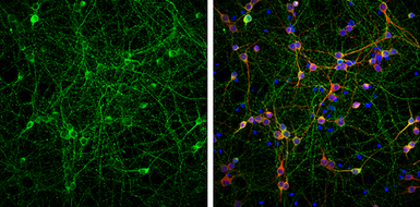
Anti-Tau antibody used in Immunocytochemistry/ Immunofluorescence (ICC/IF). GTX130462
GTX130462 ICC/IF Image

Tau antibody detects Tau protein at axon by immunofluorescent analysis.
Sample: DIV9 rat E18 primary cortical neurons were fixed in 4% paraformaldehyde at RT for 15 min.
Green: Tau protein stained by Tau antibody (GTX130462) diluted at 1:500.
Red: MAP2, stained by MAP2 antibody [HM-2] (GTX11267) diluted at 1:1000.
Blue: Fluoroshield with DAPI (GTX30920).

2 / 11
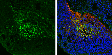
Anti-Tau antibody used in IHC (Frozen sections) (IHC-Fr). GTX130462
GTX130462 IHC-Fr Image

Tau antibody detects Tau protein expression by immunohistochemical analysis.
Sample: Frozen sectioned E13.5 Rat brain.
Green: Tau protein stained by Tau antibody (GTX130462) diluted at 1:250.
Red: beta Tubulin 3/ TUJ1, a mature neuron marker, stained by beta Tubulin 3/ TUJ1 antibody [GT11710] (GTX631836) diluted at 1:500.
Blue: Fluoroshield with DAPI (GTX30920).

3 / 11
Anti-Tau antibody used in IHC (Paraffin sections) (IHC-P). GTX130462
GTX130462 IHC-P Image

Tau antibody detects Tau protein at cytoplasm by immunohistochemical analysis.
Sample: Paraffin-embedded mouse brain.
Green: Tau stained by Tau antibody (GTX130462) diluted at 1:250.
Red: alpha Tubulin, a cytoskeleton marker, stained by alpha Tubulin antibody [GT114] (GTX628802) diluted at 1:1000.
Blue: Fluoroshield with DAPI (GTX30920).
Antigen Retrieval: Citrate buffer, pH 6.0, 15 min

4 / 11
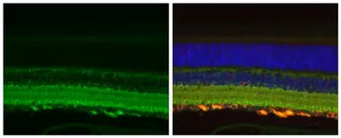
Anti-Tau antibody used in IHC (Paraffin sections) (IHC-P). GTX130462
GTX130462 IHC-P Image

Tau antibody detects Tau protein at cytoplasm by immunohistochemical analysis.
Sample: Paraffin-embedded mouse eye.
Green: Tau stained by Tau antibody (GTX130462) diluted at 1:500.
Red: beta Tubulin 3/ Tuj1, a cytoskeleton marker, stained by beta Tubulin 3/ Tuj1 antibody [GT11710] (GTX631836) diluted at 1:500.
Blue: Fluoroshield with DAPI (GTX30920).
Antigen Retrieval: Citrate buffer, pH 6.0, 15 min

5 / 11
Anti-Tau antibody used in Western Blot (WB). GTX130462
GTX130462 WB Image

Rat tissue extract (50 μg) was separated by 7.5% SDS-PAGE, and the membrane was blotted with Tau antibody (GTX130462) diluted at 1:100000. The HRP-conjugated anti-rabbit IgG antibody (GTX213110-01) was used to detect the primary antibody.

6 / 11
Anti-Tau antibody used in Western Blot (WB). GTX130462
GTX130462 WB Image

Whole cell extract (30 μg) was separated by 7.5% SDS-PAGE, and the membrane was blotted with Tau antibody (GTX130462) diluted at 1:1000. The HRP-conjugated anti-rabbit IgG antibody (GTX213110-01) was used to detect the primary antibody.

7 / 11
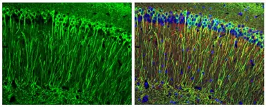
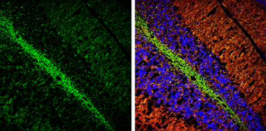
Anti-Tau antibody used in IHC (Frozen sections) (IHC-Fr). GTX130462
GTX130462 IHC-Fr Image

Tau antibody detects Tau protein expression by immunohistochemical analysis.
Sample: Frozen sectioned adult mouse cerebellum.
Green: Tau protein stained by Tau antibody (GTX130462) diluted at 1:250.
Red: beta Tubulin 3/ TUJ1, a stained by beta Tubulin 3/ TUJ1 antibody [GT11710] (GTX631836) diluted at 1:500.
Blue: Fluoroshield with DAPI (GTX30920).

8 / 11
Anti-Tau antibody used in Western Blot (WB). GTX130462
GTX130462 WB Image

Tau antibody detects Tau protein by western blot analysis. Mouse tissue extracts (50 μg) was separated by 7.5 % SDS-PAGE, and the membrane was blotted with Tau antibody (GTX130462) at a dilution of 1:10000.

9 / 11
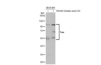
Anti-Tau antibody used in Western Blot (WB). GTX130462
GTX130462 WB Image

Untreated (–) and treated (+) SK-N-SH whole cell extract (30 μg) were separated by 10% SDS-PAGE, and the membrane was blotted with Tau antibody (GTX130462) diluted at 1:1000. The HRP-conjugated anti-rabbit IgG antibody (GTX213110-01) was used to detect the primary antibody, and the signal was developed with Trident ECL plus-Enhanced.

10 / 11
Anti-Tau antibody used in Western Blot (WB). GTX130462
GTX130462 WB Image

Mouse tissue extract (50 μg) was separated by 7.5% SDS-PAGE, and the membranes were blotted with Tau antibody (GTX130462) diluted at 1:4000 and competitor's antibody (#4019) diluted at 1:2000. The HRP-conjugated anti-rabbit IgG antibody (GTX213110-01) was used to detect the primary antibody.

*The competitor is not affiliated with GeneTex and does not endorse this product.

11 / 11
Anti-Tau antibody used in Western Blot (WB). GTX130462
Anti-Tau antibody used in Immunocytochemistry/ Immunofluorescence (ICC/IF). GTX130462
Anti-Tau antibody used in IHC (Frozen sections) (IHC-Fr). GTX130462
Anti-Tau antibody used in IHC (Paraffin sections) (IHC-P). GTX130462
Anti-Tau antibody used in Western Blot (WB). GTX130462
Anti-Tau antibody used in IHC (Frozen sections) (IHC-Fr). GTX130462
Anti-Tau antibody used in Western Blot (WB). GTX130462
  • Host
    Rabbit
  • Clonality
    Polyclonal
  • Isotype
    IgG
  • Applications
    WB ICC/IF IHC-P IHC-Fr IHC
  • Reactivity
    Human, Mouse, Rat
Summary
Tau antibody detects Tau protein, a phosphorylated microtubule-associated protein with a predicted molecular weight of 79 kDa. Tau protein has six isoforms in humans generated from a single gene. Expression changes of these Tau isoforms are controlled during neuronal development and in various pathological conditions. Dysfunction of Tau is associated with neurodegenerative diseases including Alzheimer’s disease and frontotemporal dementia.
See all Tau products