VCP antibody


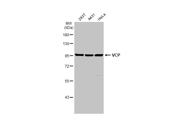
Various whole cell extracts (30 μg) were separated by 7.5% SDS-PAGE, and the membrane was blotted with VCP antibody (GTX101089) diluted at 1:500. The HRP-conjugated anti-rabbit IgG antibody (GTX213110-01) was used to detect the primary antibody.

VCP antibody detects VCP protein by western blot analysis.
A. 50 μg Rat brain lysate/extract
7.5% SDS-PAGE
VCP antibody (GTX101089) dilution: 1:10000
The HRP-conjugated anti-rabbit IgG antibody (GTX213110-01) was used to detect the primary antibody.

Sample (50 μg of whole cell lysate)
A: Mouse brain
7.5% SDS PAGE
GTX101089 diluted at 1:10000
The HRP-conjugated anti-rabbit IgG antibody (GTX213110-01) was used to detect the primary antibody.

VCP antibody detects VCP Protein expression by immunohistochemical analysis.
Sample: Frozen-sectioned adult mouse cerebellum.
Green: VCP stained by VCP antibody (GTX101089) diluted at 1:250.
Red: NF-H, stained by NF-H antibody [GT114] (GTX634289) diluted at 1:500.
Blue: Fluoroshield with DAPI (GTX30920).
Antigen Retrieval: Citrate buffer, pH 6.0, 10 min

Immunohistochemical analysis of paraffin-embedded human breast cancer, using VCP(GTX101089) antibody at 1:500 dilution.
Antigen Retrieval: Trilogy™ (EDTA based, pH 8.0) buffer, 15min

Immunoprecipitation of VCP protein from HeLa whole cell extracts using 5 μg of VCP antibody (GTX101089).
Western blot analysis was performed using VCP antibody (GTX101089) diluted at 1:500.
EasyBlot anti-Rabbit IgG (GTX221666-01) was used as a secondary reagent.

VCP antibody detects Vcp protein on whole-mount zebrafish embryos by immunohistochemical analysis.
Sample: Paraformaldehyde-fixed zebrafish embryos.
VCP antibody (GTX101089) dilution: 1:200.

Zebrafish tissue extract (30 μg) was separated by 7.5% SDS-PAGE, and the membrane was blotted with VCP antibody (GTX101089) diluted at 1:1000.

-
HostRabbit
-
ClonalityPolyclonal
-
IsotypeIgG
-
ApplicationsWB IHC-P IHC-Fr IHC-Wm IP
-
ReactivityHuman, Mouse, Rat, Zebrafish