VRL1 antibody

Cat. No. GTX16610
Cat. No.  GTX16610
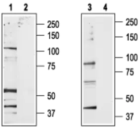
Anti-VRL1 antibody used in Western Blot (WB). GTX16610
GTX16610 WB Image

WB analysis of rat brain membrane (lanes 1 and 2) and RBL (lanes 3 and 4) lysates using GTX16610 VRL1 antibody preincubated with or without immunogen peptide.
Dilution : 1:200

1 / 3
Anti-VRL1 antibody used in IHC (Frozen sections) (IHC-Fr). GTX16610
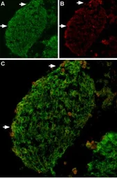
GTX16610 IHC-Fr Image

IHC-Fr analysis of mouse DRG tissue using GTX16610 VRL1 antibody.
Panel A : TRPV2 (green) appears in patches along the perimeter of the DRG (arrows).
Panel B : Neurons containing neurofilament 200 (red) are scattered in the DRG, also in patches (arrows).
Panel C : Merge of the two panels shows that the spatial distribution of neurofilament 200 and TRPV2 expression overlaps. However, DRGs showing robust expression of neurofilament 200 do not contain TRPV2.

2 / 3
Anti-VRL1 antibody used in Immunoprecipitation (IP). GTX16610
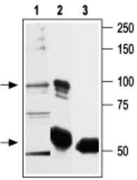
GTX16610 IP Image

IP analysis of RBL cell lysate using GTX16610 VRL1 antibody. The upper arrow indicates TRPV2 while the lower arrow indicates the IgG heavy chain. WB was performed with the GTX16610 antibody.
IP reaction : 7.5 μg
Lane 1 : RBL lysate
Lane 2 : Lysate immunoprecipitated with Anti-TRPV2   Antibody
Lane 3 : Lysate immunoprecipitated with pre-immune rabbit serum

3 / 3
Anti-VRL1 antibody used in Western Blot (WB). GTX16610
Anti-VRL1 antibody used in IHC (Frozen sections) (IHC-Fr). GTX16610
Anti-VRL1 antibody used in Immunoprecipitation (IP). GTX16610
  • Host
    Rabbit
  • Clonality
    Polyclonal
  • Isotype
    IgG
  • Applications
    WB IHC-Fr IP
  • Reactivity
    Mouse, Rat
See all VRL1 products