ZO-1 antibody


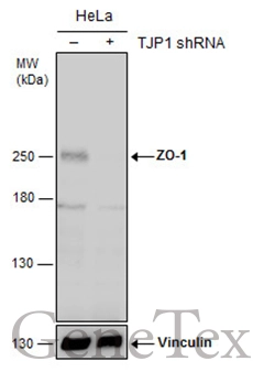
Non-transfected (–) and transfected (+) HeLa whole cell extracts (30 μg) were separated by 5% SDS-PAGE, and the membrane was blotted with ZO-1 antibody (GTX108592) diluted at 1:500. The HRP-conjugated anti-rabbit IgG antibody (GTX213110-01) was used to detect the primary antibody.
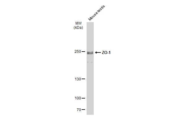
Mouse tissue extract (50 μg) was separated by 5% SDS-PAGE, and the membrane was blotted with ZO-1 antibody (GTX108592) diluted at 1:500. The HRP-conjugated anti-rabbit IgG antibody (GTX213110-01) was used to detect the primary antibody.
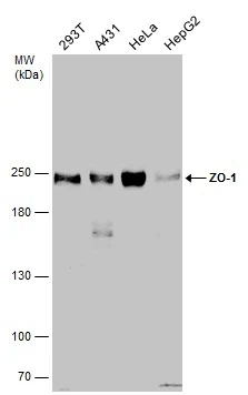
Various whole cell extracts (30 μg) were separated by 5% SDS-PAGE, and the membrane was blotted with ZO-1 antibody (GTX108592) diluted at 1:1000.
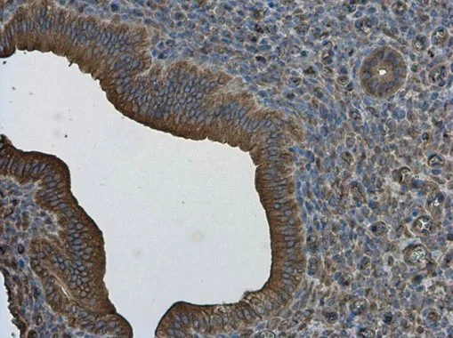
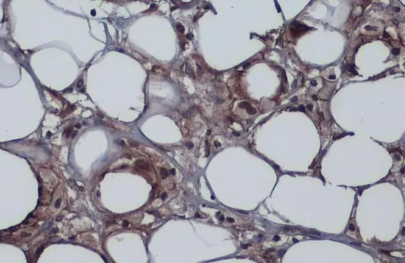
ZO-1 antibody detects ZO-1 protein at cell membrane and cytoplasm in mouse cervix by immunohistochemical analysis.
Sample: Paraffin-embedded mouse cervix.
ZO-1 antibody (GTX108592) diluted at 1:500.
Antigen Retrieval: Citrate buffer, pH 6.0, 15 min
ZO-1 antibody detects ZO-1 protein at cell membrane by immunohistochemical analysis.Sample: Paraffin-embedded human breast carcinoma.ZO-1 stained by ZO-1 antibody (GTX108592) diluted at 1:500.Antigen Retrieval: Citrate buffer, pH 6.0, 15 min
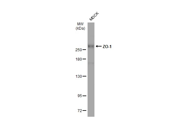
Whole cell extract (30 μg) was separated by 5% SDS-PAGE, and the membrane was blotted with ZO-1 antibody (GTX108592) diluted at 1:1000. The HRP-conjugated anti-rabbit IgG antibody (GTX213110-01) was used to detect the primary antibody.
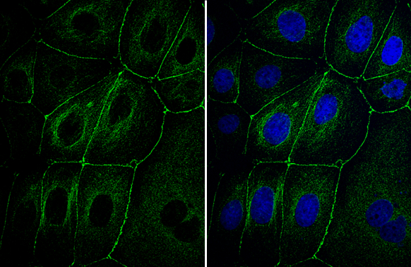
ZO-1 antibody detects ZO-1 protein at cell membrane and cytoplasm by immunofluorescent analysis.Sample: MDCK cells were fixed in 4% paraformaldehyde at RT for 15 min.Green: ZO-1 stained by ZO-1 antibody (GTX108592) diluted at 1:1000.
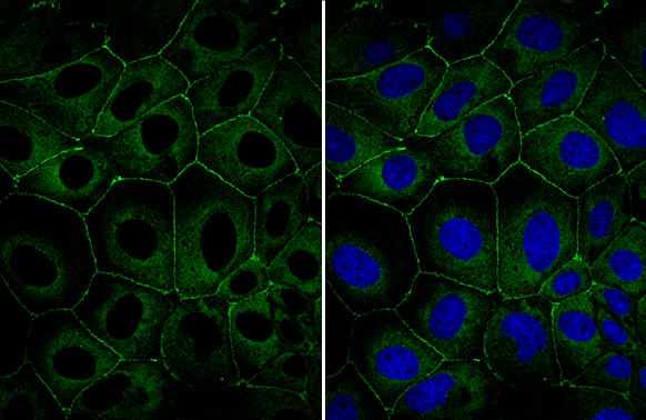
ZO-1 antibody detects ZO-1 protein at cell membrane and cytoplasm by immunofluorescent analysis.Sample: MDCK cells were fixed in 4% paraformaldehyde at RT for 15 min.Green: ZO-1 stained by ZO-1 antibody (GTX108592) diluted at 1:1000.
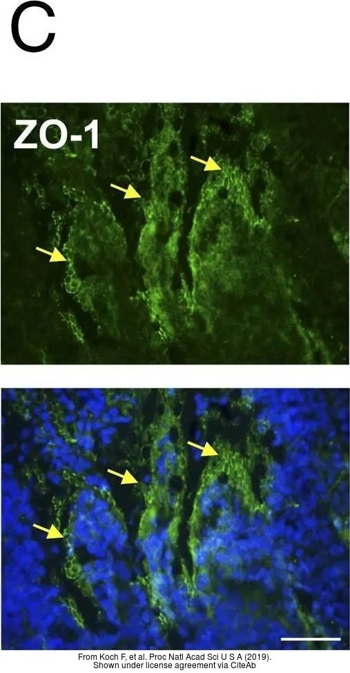
The data was published in the journal Proc Natl Acad Sci U S A in 2019. PMID: 31064871
-
HostRabbit
-
ClonalityPolyclonal
-
IsotypeIgG
-
ApplicationsWB ICC/IF IHC-P IHC-Fr
-
ReactivityHuman, Mouse, Bovine, Dog, Pig, Monkey