cIAP1 antibody


Non-transfected (–) and transfected (+) 293T whole cell extracts (50 μg) were separated by 7.5% SDS-PAGE, and the membrane was blotted with cIAP1 antibody (GTX110087) diluted at 1:500. The HRP-conjugated anti-rabbit IgG antibody (GTX213110-01) was used to detect the primary antibody.

Sample (50 μg of whole cell lysate)
A: Rat muscle
7.5% SDS PAGE
GTX110087 diluted at 1:1000
The HRP-conjugated anti-rabbit IgG antibody (GTX213110-01) was used to detect the primary antibody.

cIAP1 antibody detects cIAP1 protein at cytoplasm on mouse kidney by immunohistochemical analysis.
Sample: Paraffin-embedded mouse kidney.
cIAP1 antibody (GTX110087) diluted at 1:500.
Antigen Retrieval: Trilogy™ (EDTA based, pH 8.0) buffer, 15min

Whole cell extract (30 μg) was separated by 7.5% SDS-PAGE, and the membrane was blotted with cIAP1 antibody (GTX110087) diluted at 1:1000. The HRP-conjugated anti-rabbit IgG antibody (GTX213110-01) was used to detect the primary antibody.

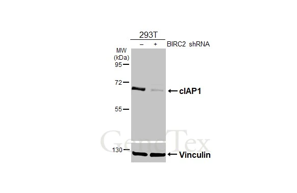
Non-transfected (–) and transfected (+) 293T whole cell extracts (30 μg) were separated by 7.5% SDS-PAGE, and the membrane was blotted with cIAP1 antibody (GTX110087) diluted at 1:1000. The HRP-conjugated anti-rabbit IgG antibody (GTX213110-01) was used to detect the primary antibody.

*The competitor is not affiliated with GeneTex and does not endorse this product.
Whole cell extract (30 μg) was separated by 7.5% SDS-PAGE, and the membrane was blotted with cIAP1 antibody (GTX110087) diluted at 1:1000 and competitor's antibody (SC-7943) diluted by 1:200. The HRP-conjugated anti-rabbit IgG antibody (GTX213110-01) was used to detect the primary antibody.

cIAP1 antibody detects cIAP1 protein at cytoplasm by immunohistochemical analysis.Sample: Paraffin-embedded mouse liver.cIAP1 stained by cIAP1 antibody (GTX110087) diluted at 1:500.Antigen Retrieval: Citrate buffer, pH 6.0, 15 min
-
HostRabbit
-
ClonalityPolyclonal
-
IsotypeIgG
-
ApplicationsWB IHC-P
-
ReactivityHuman, Mouse, Rat