Country / Location Selection
Search suggestions
CD5 antibody [MSVA-005R] HistoMAX
Go to HistoMAX page
Colon- Colorectal adenocarcinoma showing a weak to moderate membraneous CD5 immunostaining in about 50% of tumor cells.
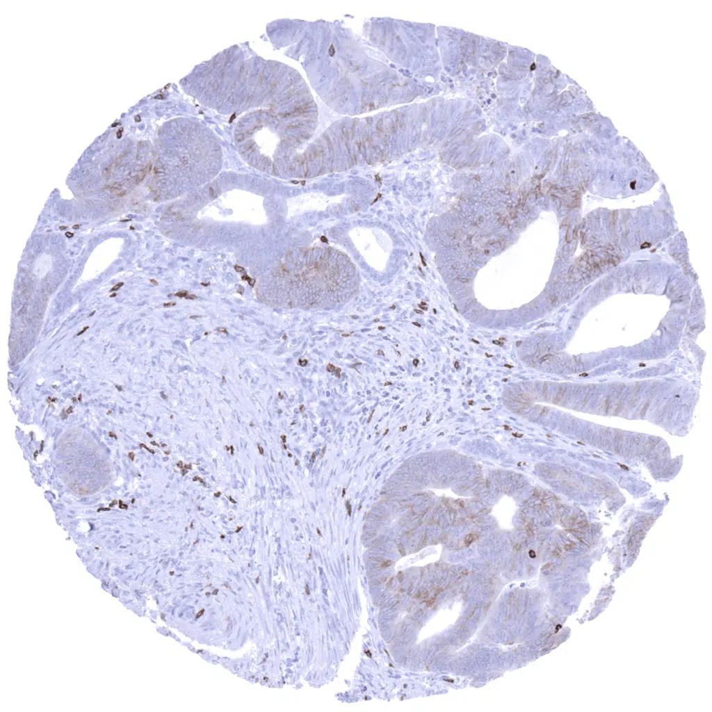
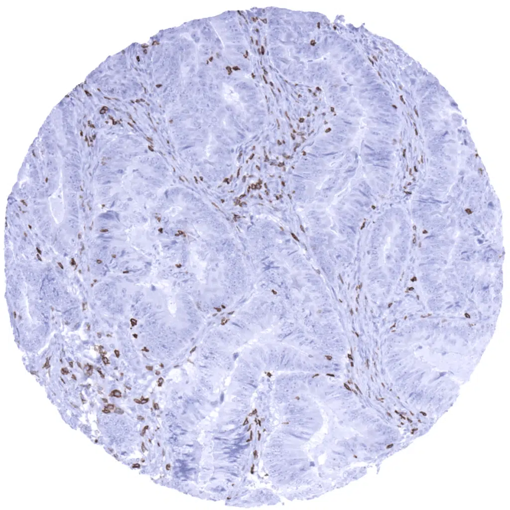
Colon- Colorectal adenocarcinoma with a large number of CD5 positive T-lymphocytes in the stroma.
Colon- Colorectal adenocarcinoma with numerous CD5 positive T-lymphocytes in the stroma.
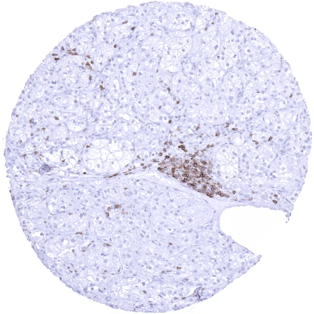
Kidney- Clear cell renal cell carcinoma with focal infiltrate of CD5 positive T-lymphocytes.
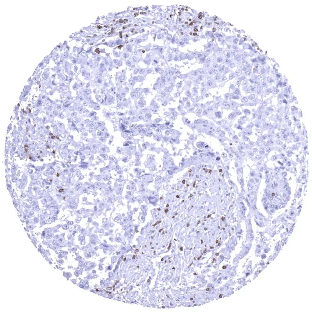
Kidney- Papillary renal cell carcinoma with CD5 positive T-lymphocytes in the stroma.
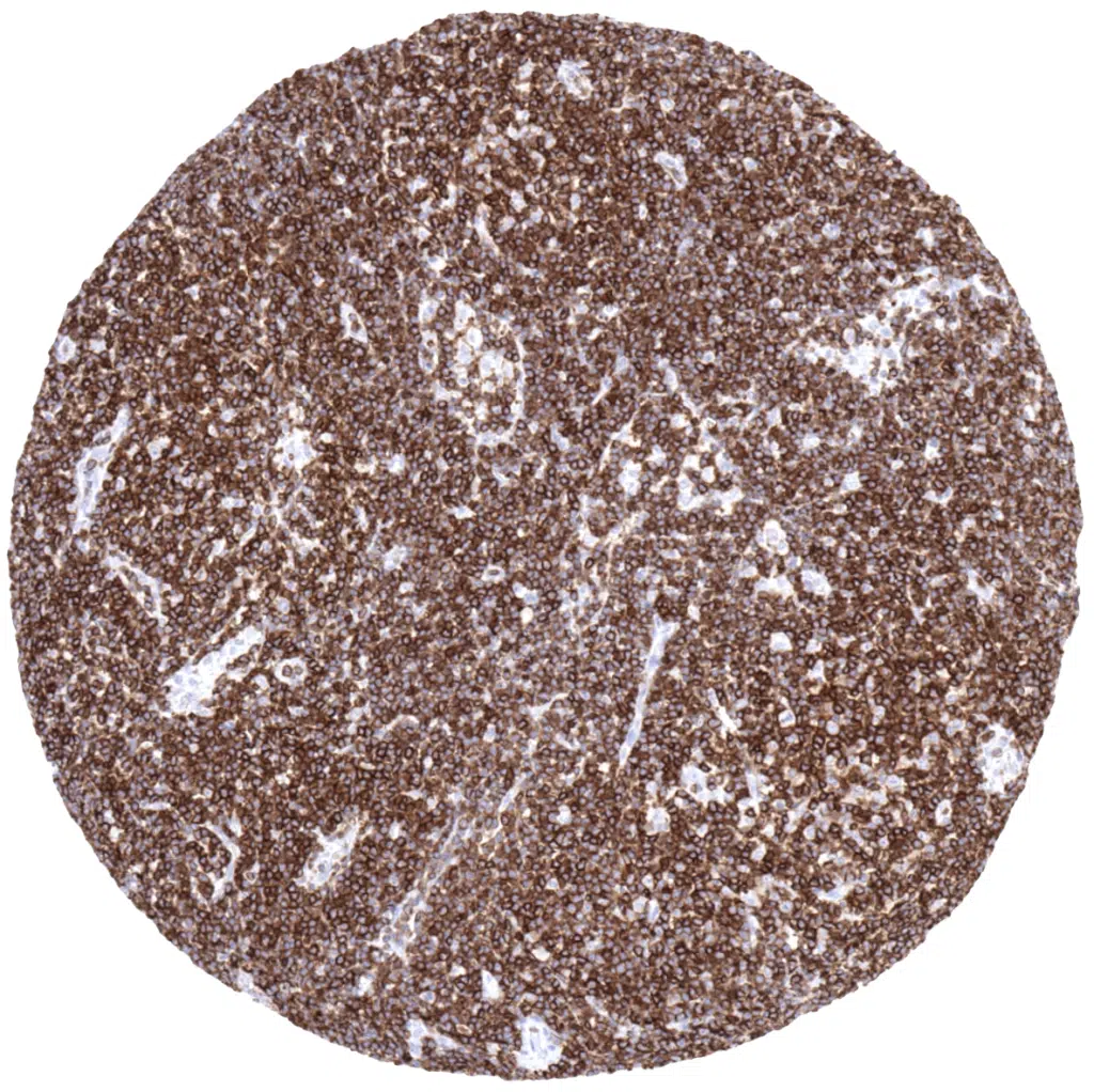
Lymph node- B-CLL with strong CD5 positivity of all tumor cells.
Lymph node- CD5 negative peripheral T-cell lymphoma
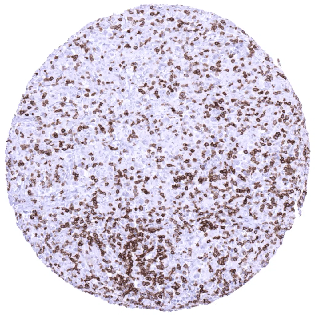
Lymph node- Diffuse large B-cell lymphoma containing few interspersed CD5 positive T-lymphocytes.
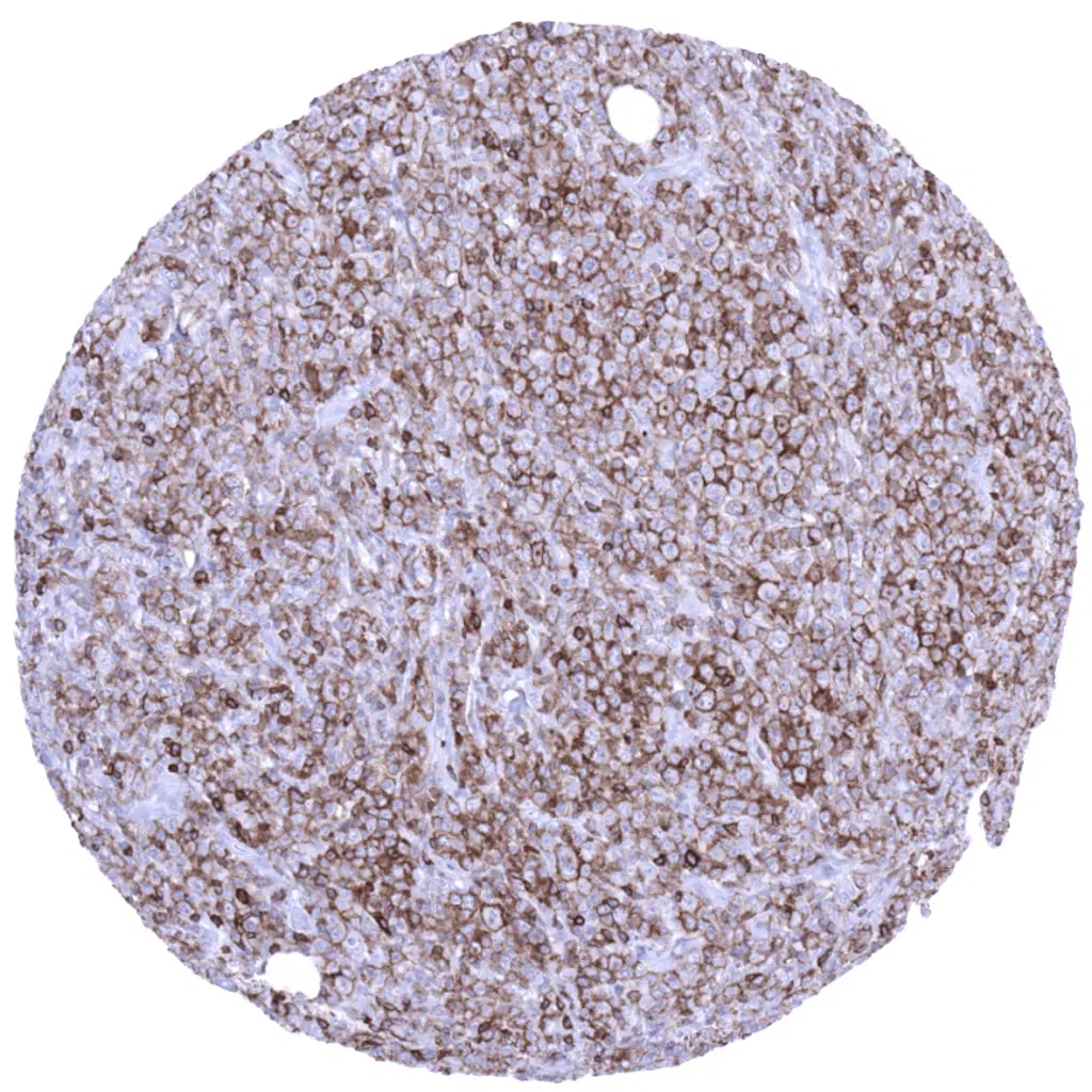
Lymph node- Diffuse large B-cell lymphoma showing moderate to strong CD5 positivity of all tumor cells.
Lymph node- Diffuse large B-cell lymphoma with interspersed CD5 positive T-lymphocytes.
Lymph node- Hodgkin's lymphoma containing a moderate number of CD5 positive T-lymphocytes.
Lymph node- Hodgkin's lymphoma containing numerous CD5 positive T-lymphocytes.
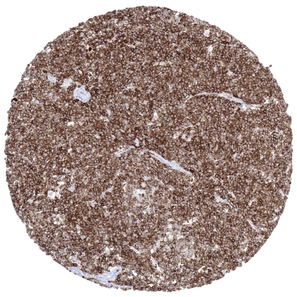
Lymph node- Mantle cell lymphoma showing strong CD5 positivity of all tumor cells.
Testis- Seminoma containing numerous CD5 positive T-lymphocytes.
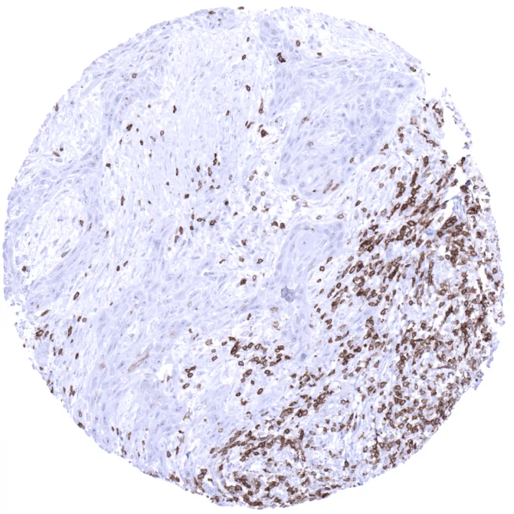
Uterine cervix- Squamous cell carcinoma containing numerous CD5 positive T-lymphocytes in the stroma.
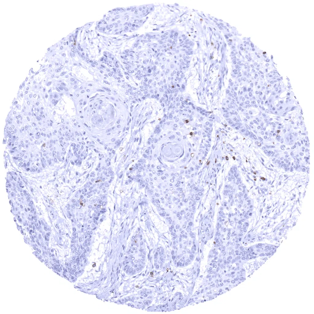
Uterine cervix- Squamous cell carcinoma with few CD5 positive T-lymphocytes.
Uterine cervix- Squamous cell carcinoma with only few CD5 positive T-lymphocytes.
Vagina- Squamous cell carcinoma exhibiting a dense infitrate of CD5 positive T-lymphocytes at the invasive tumor margin.
| TOP |