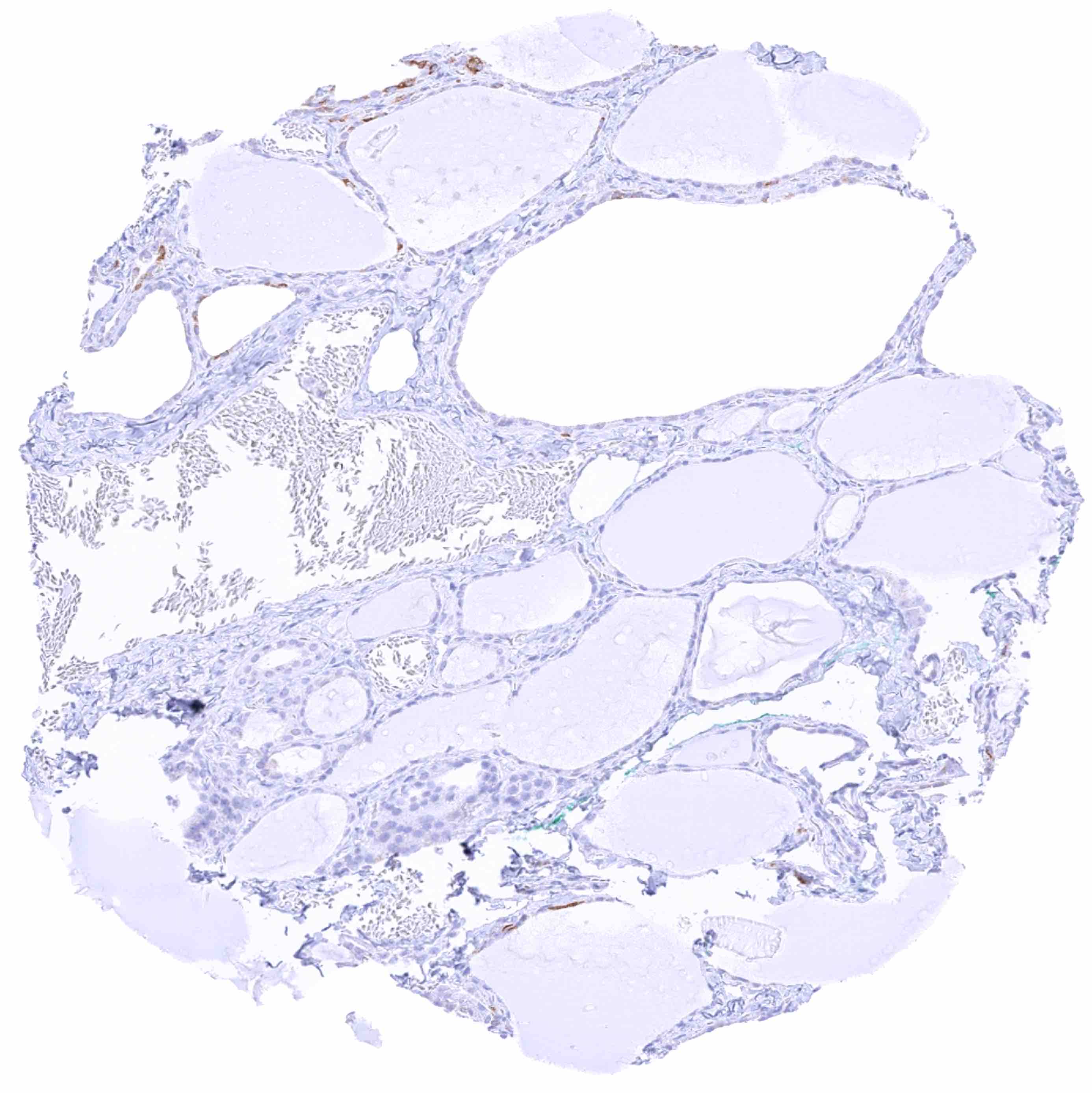
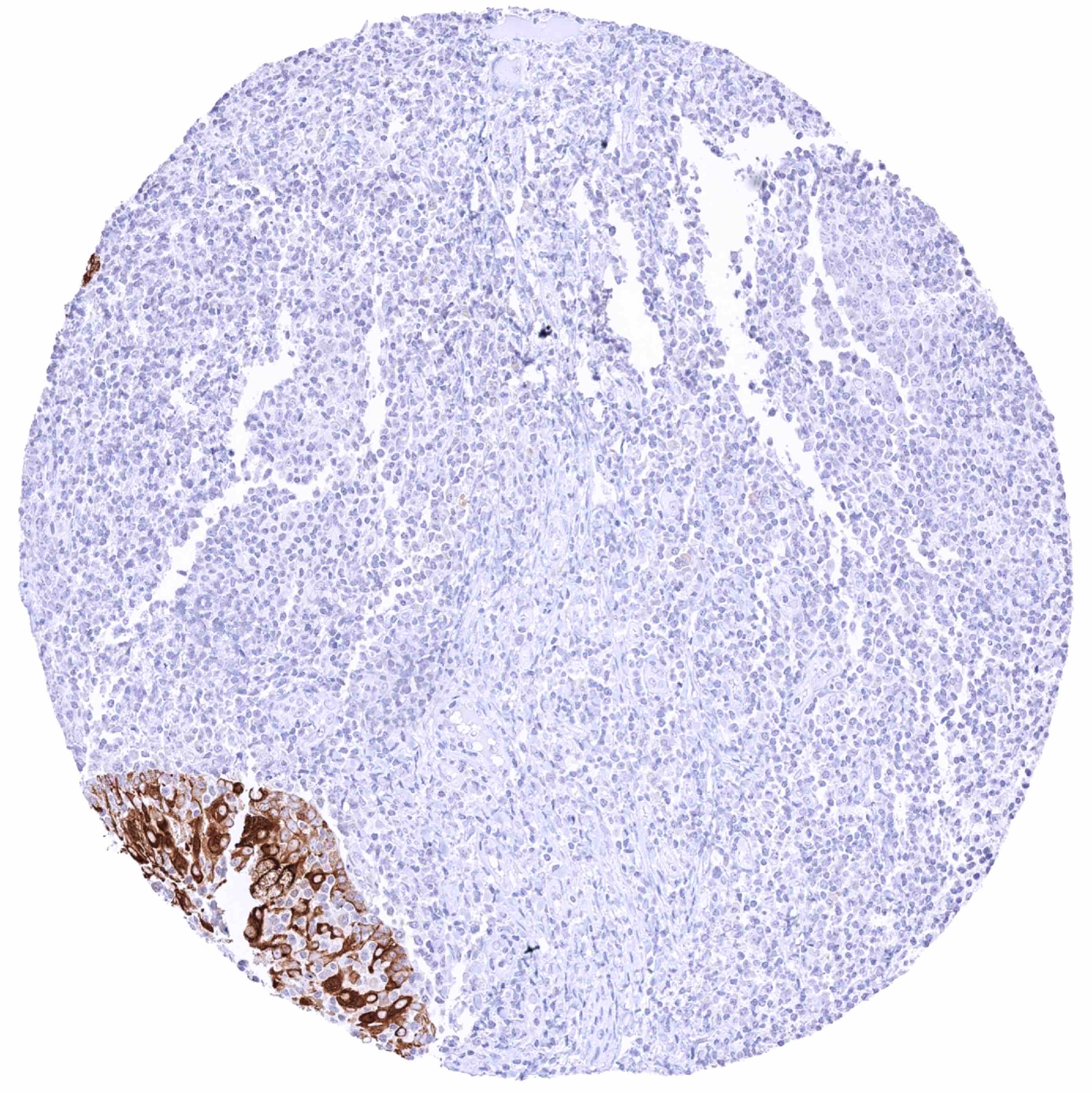
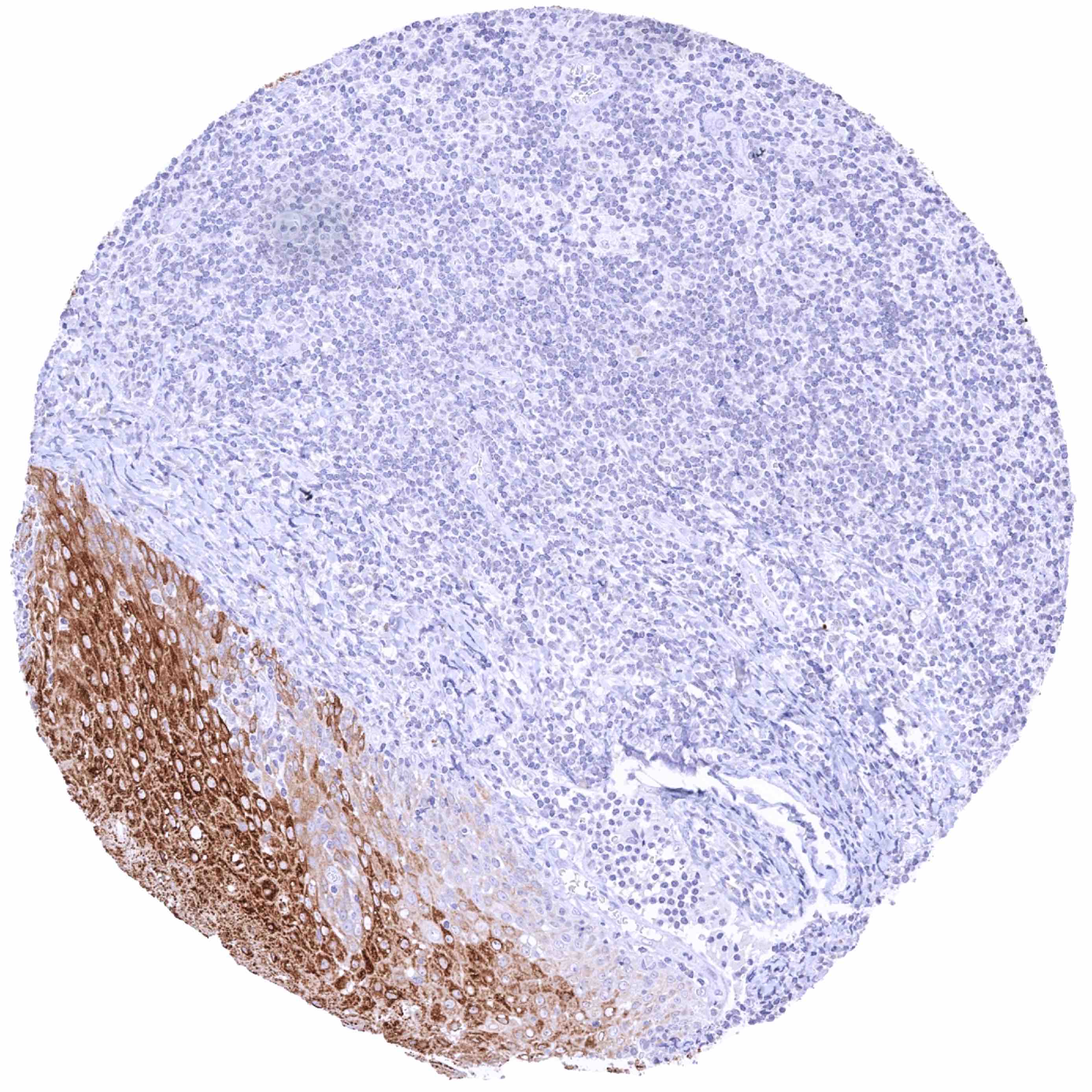
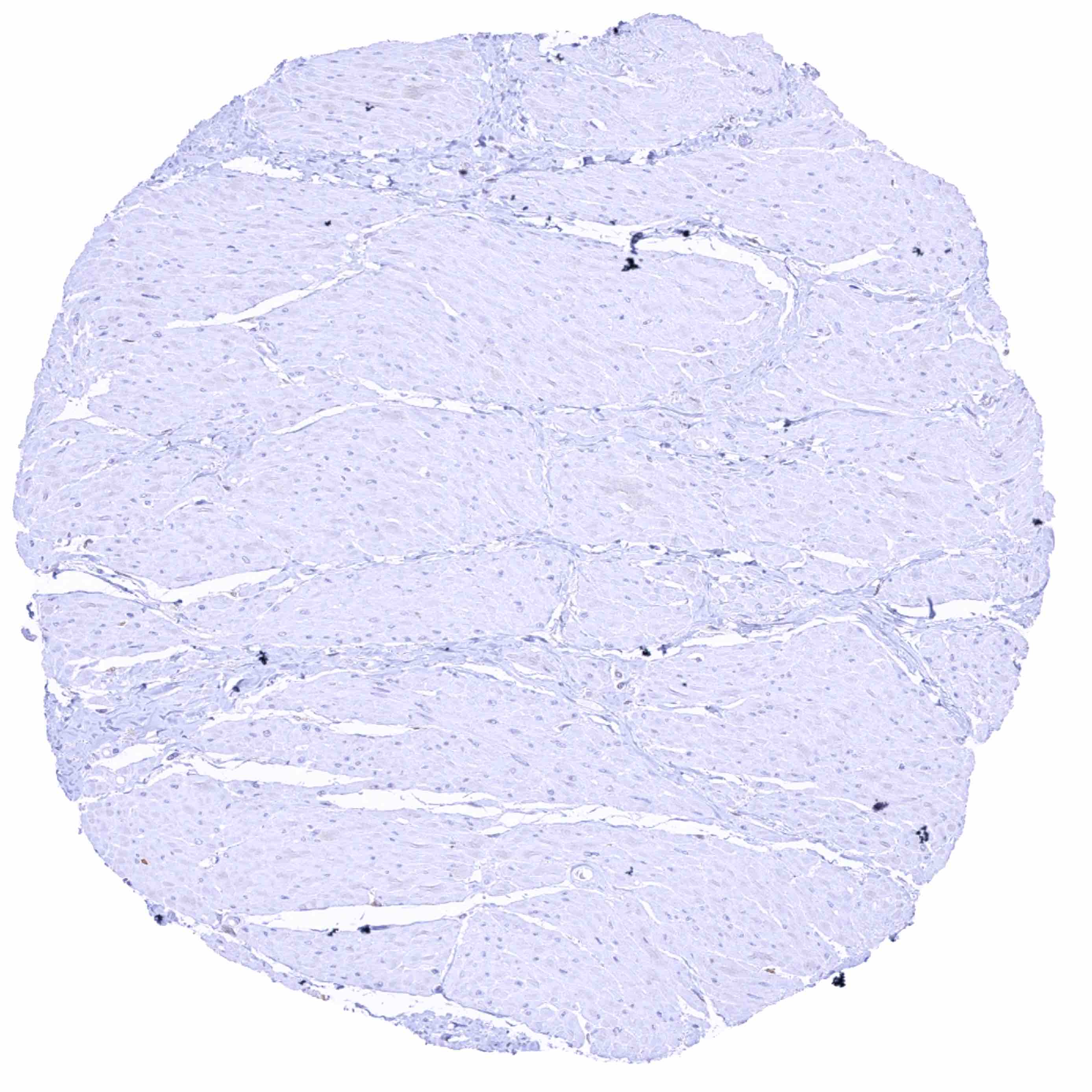
Normal Tissue Gallery- GTX640557

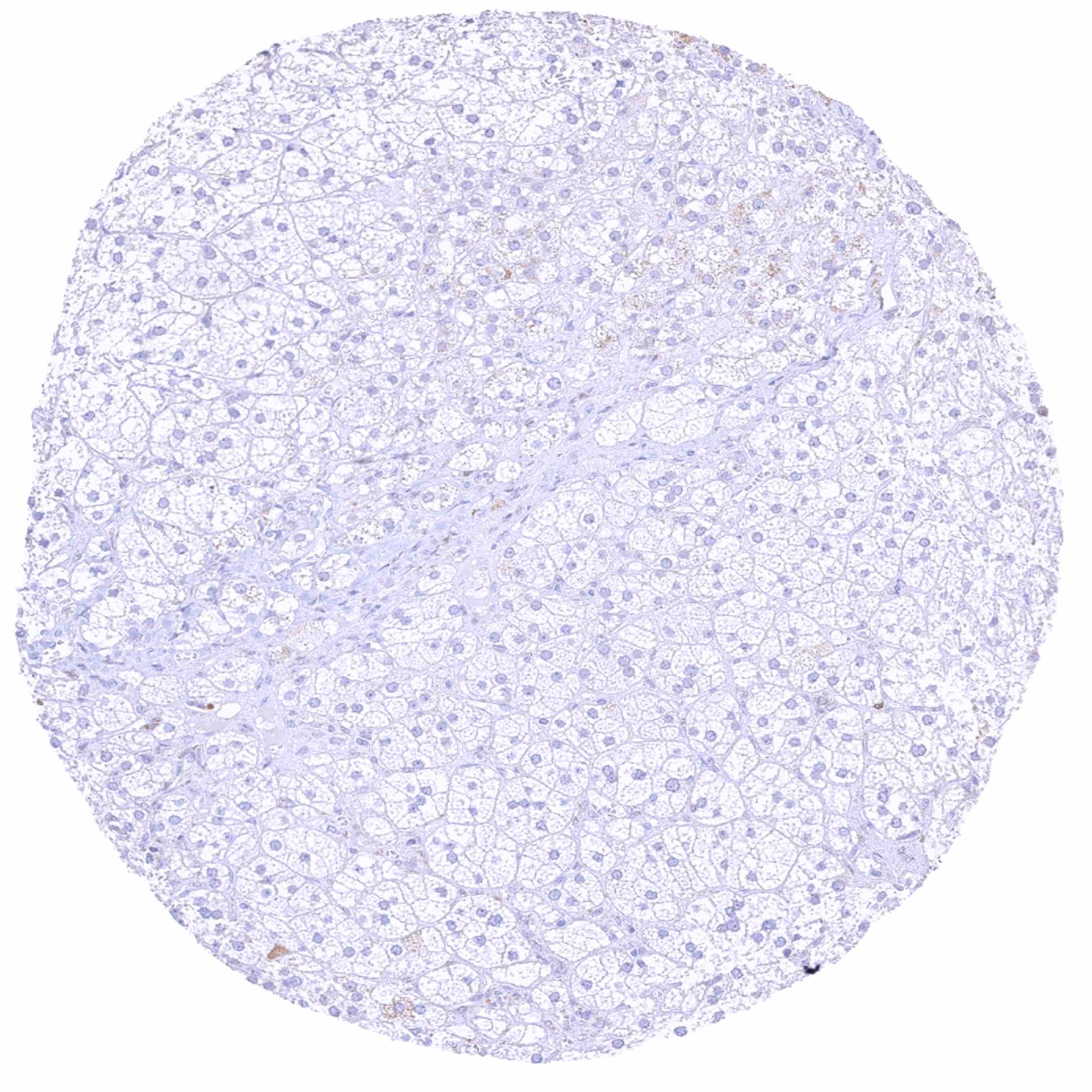
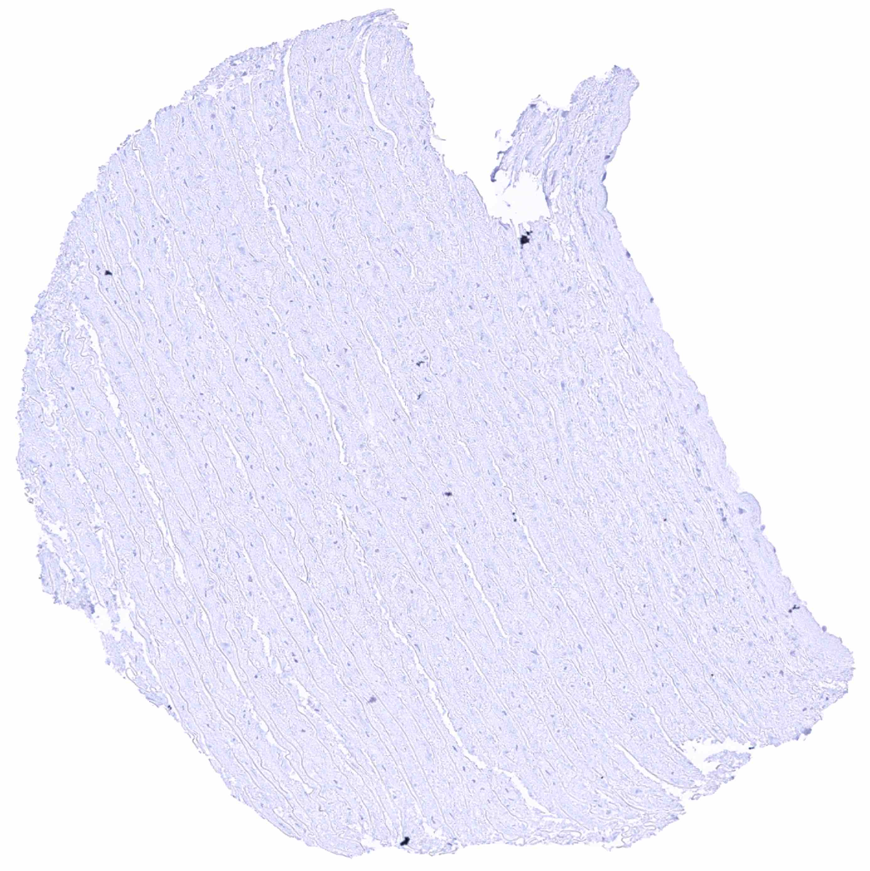
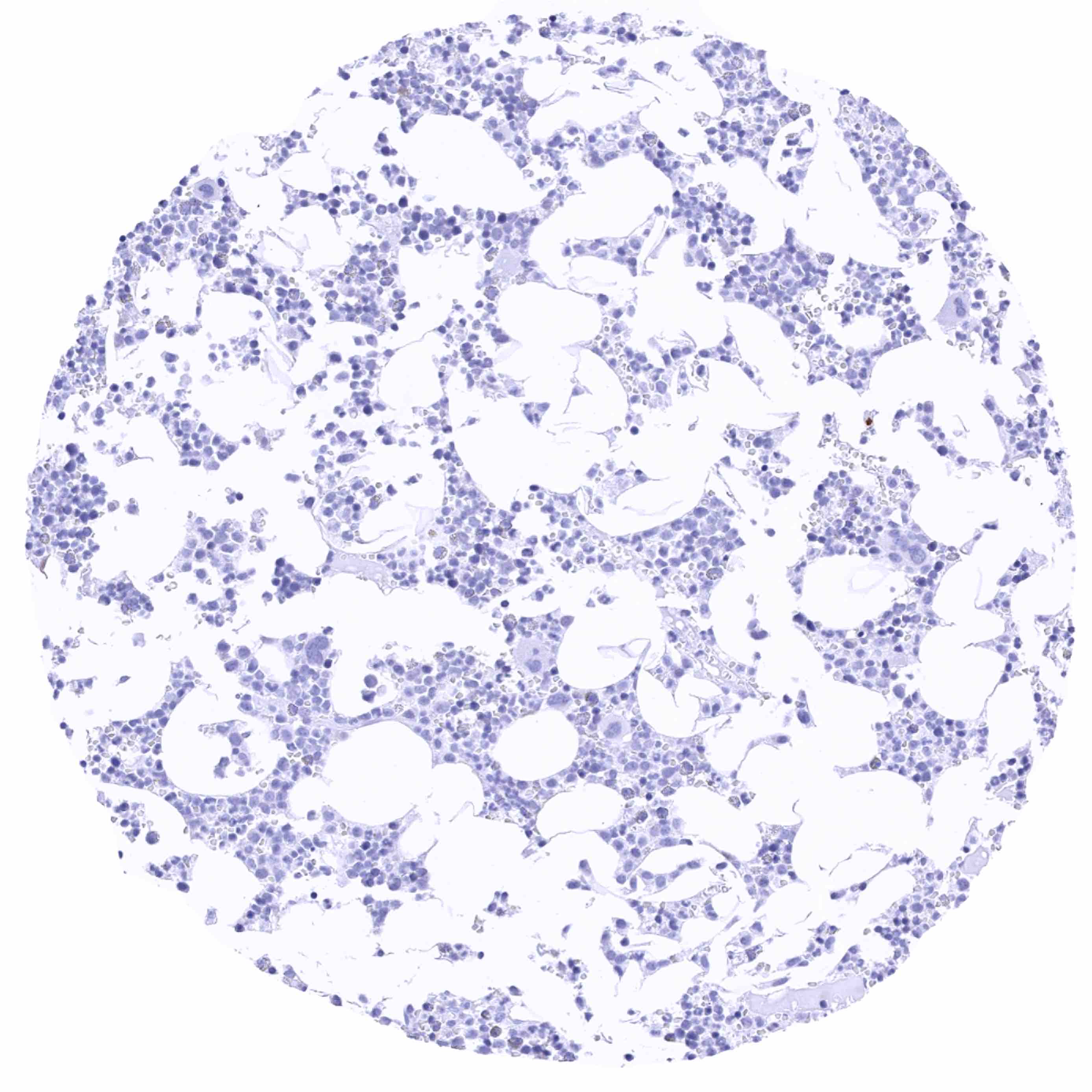
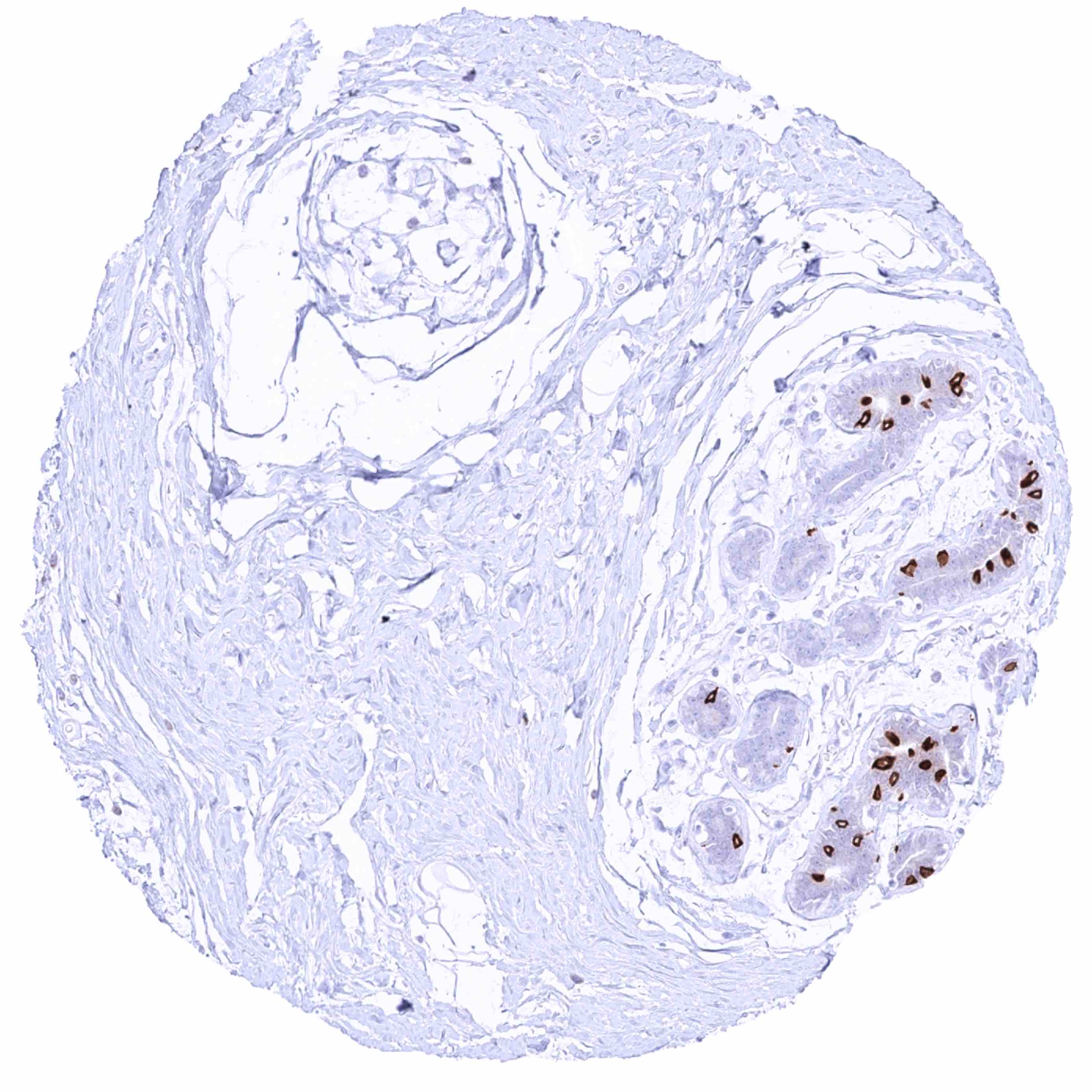
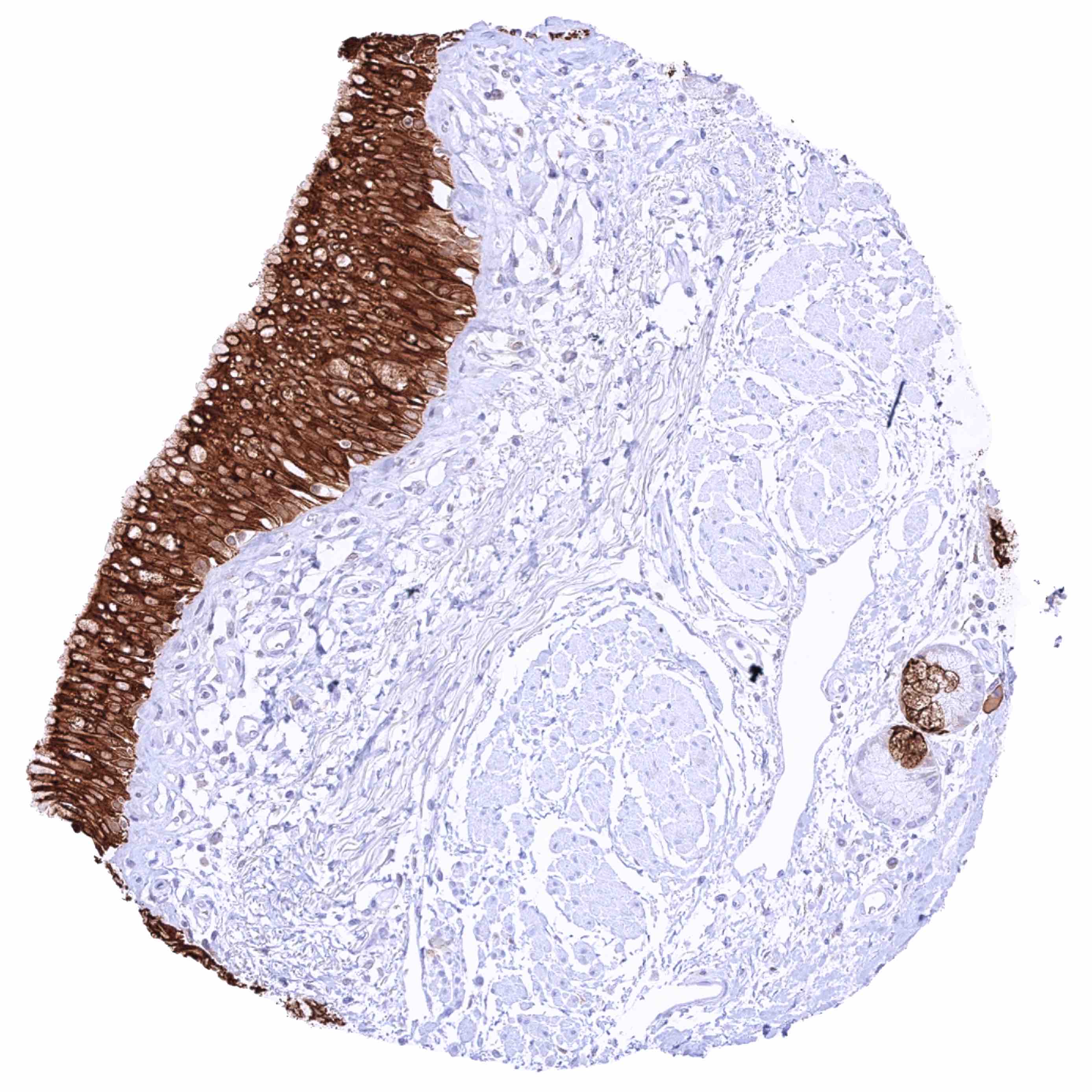
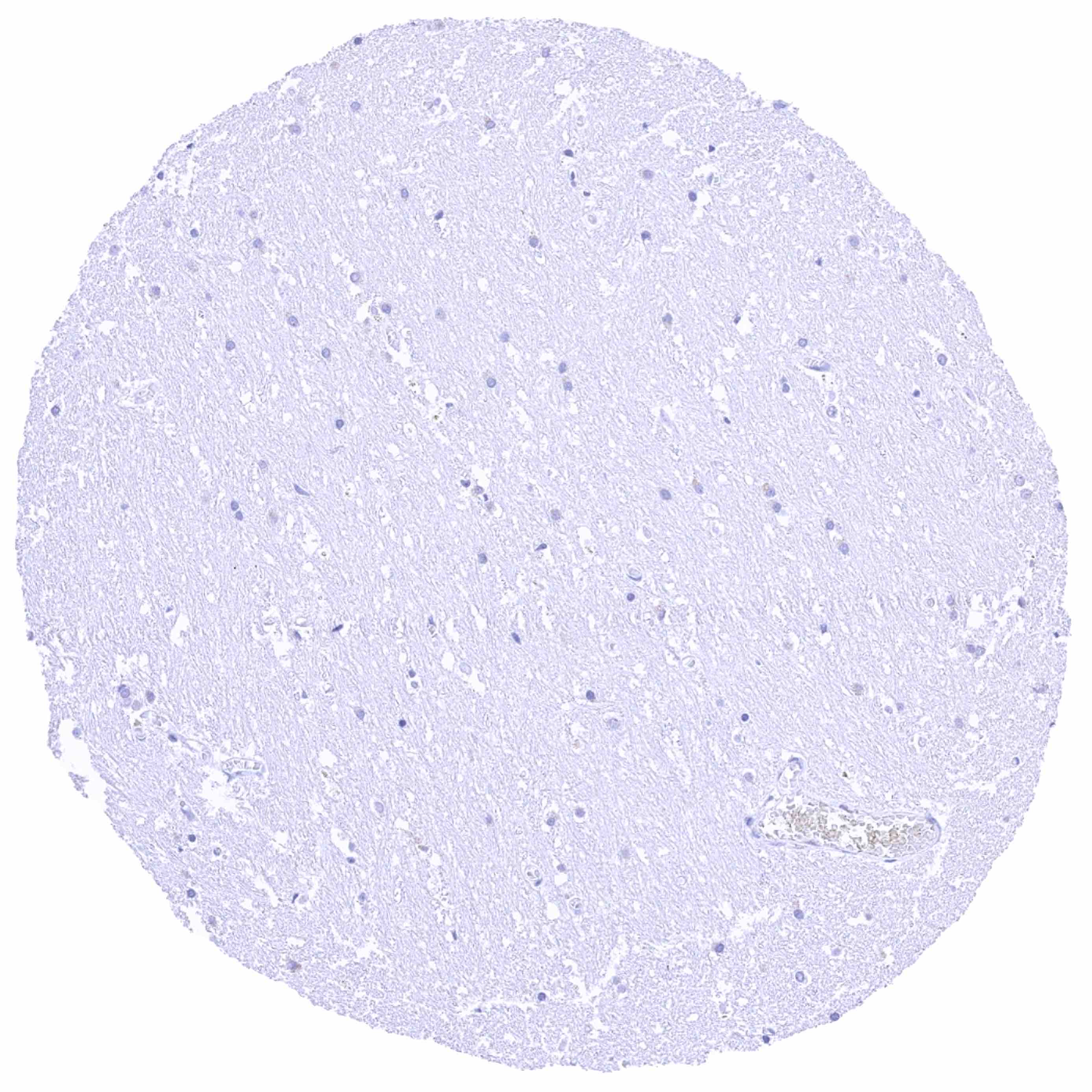
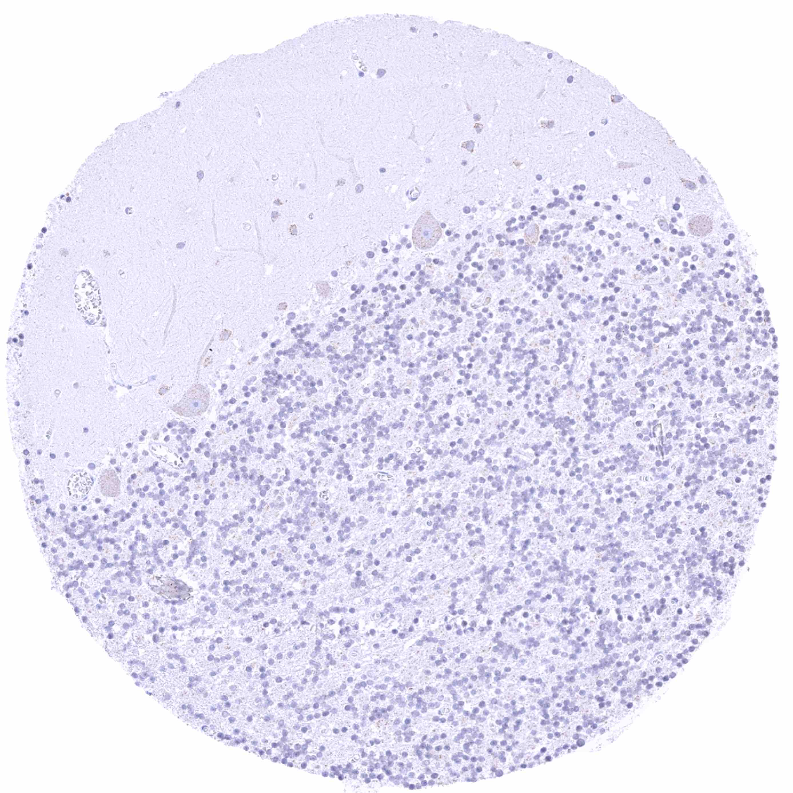
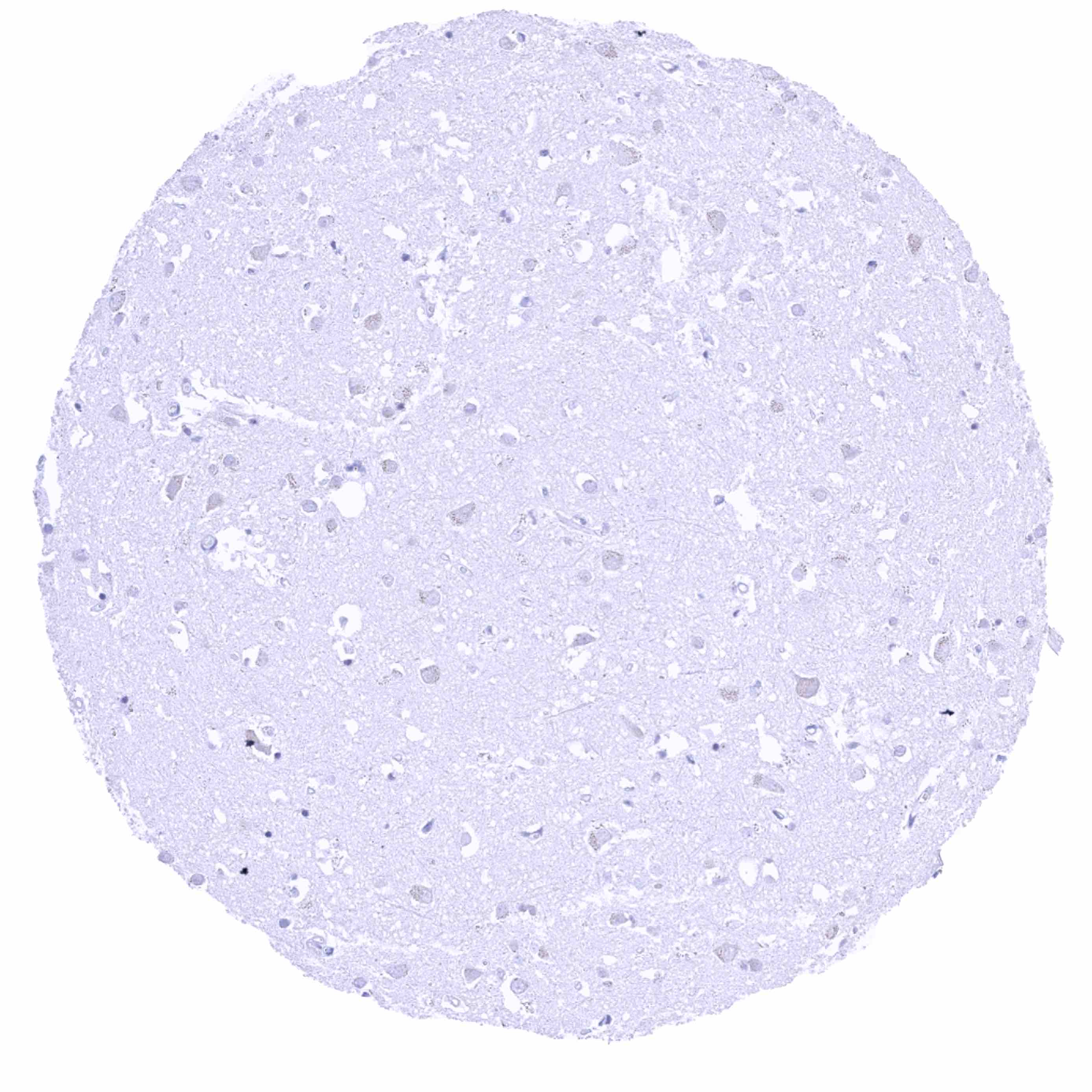
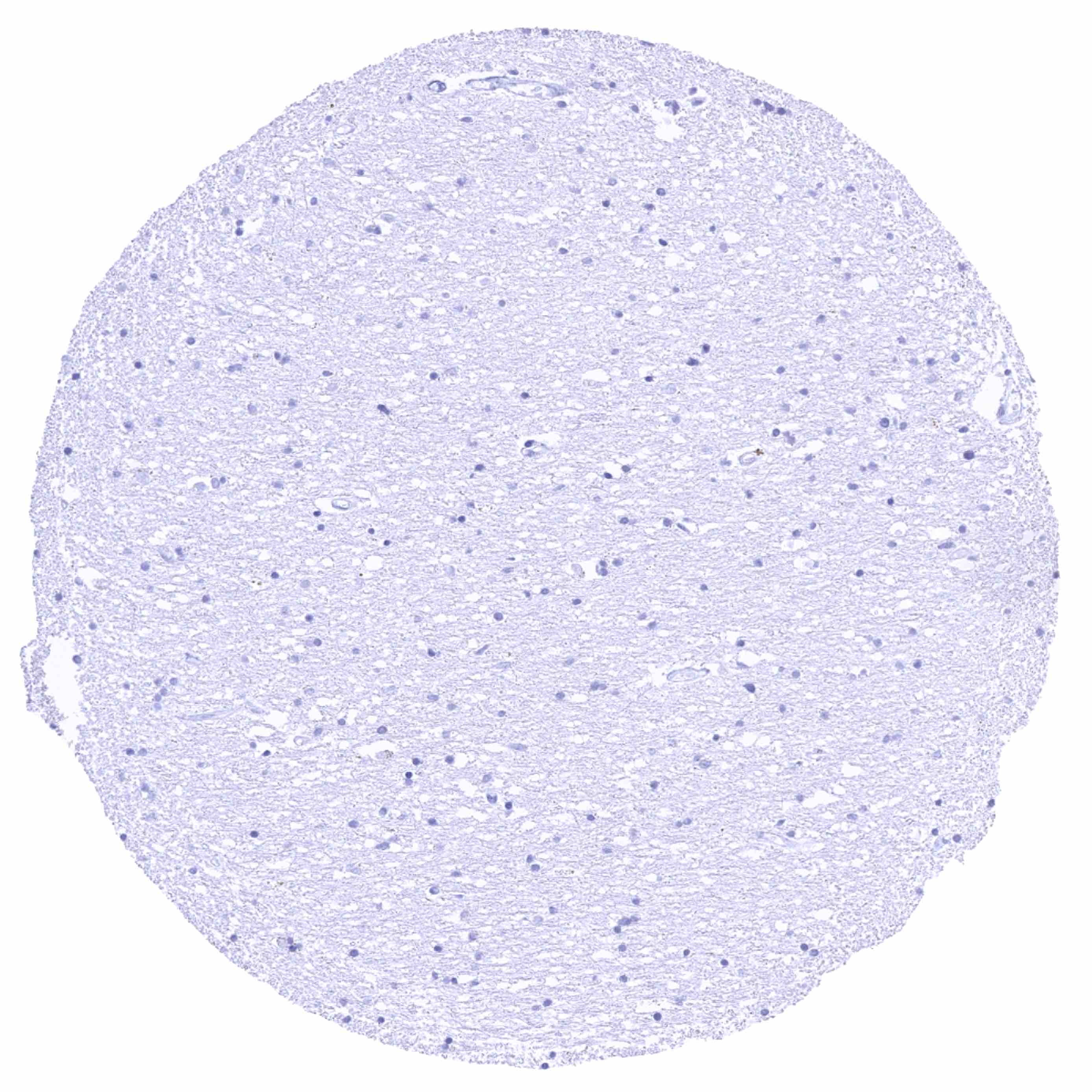
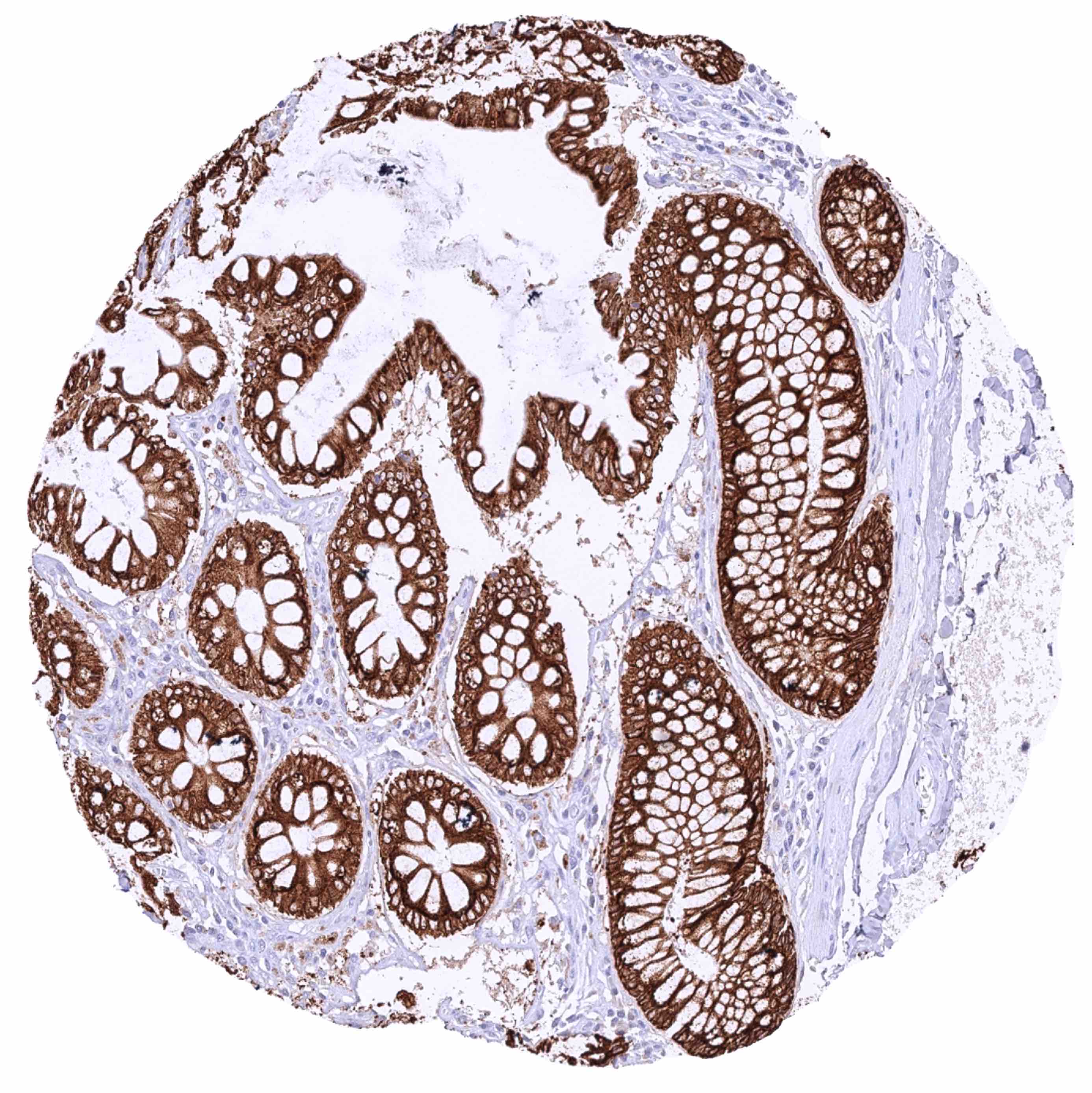
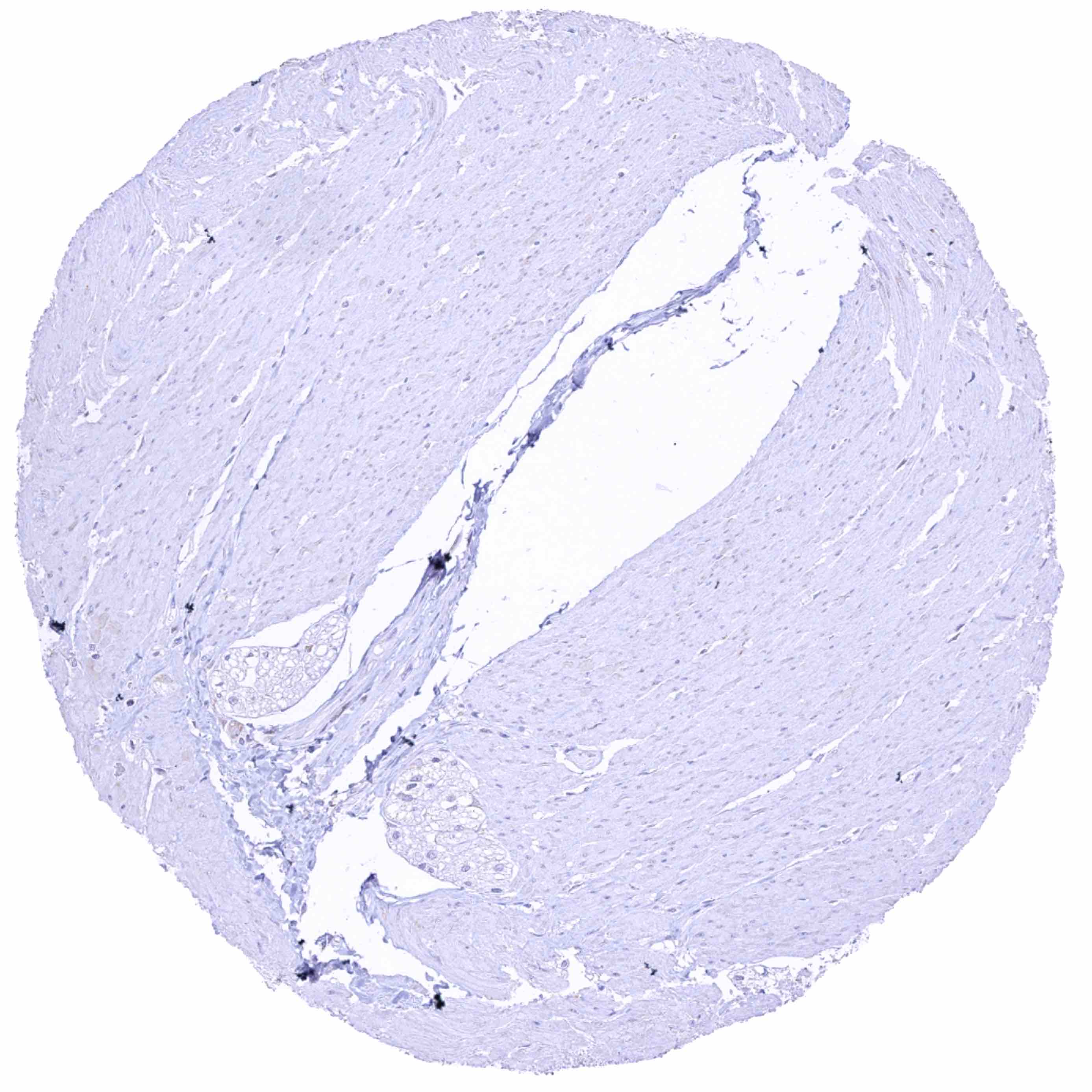
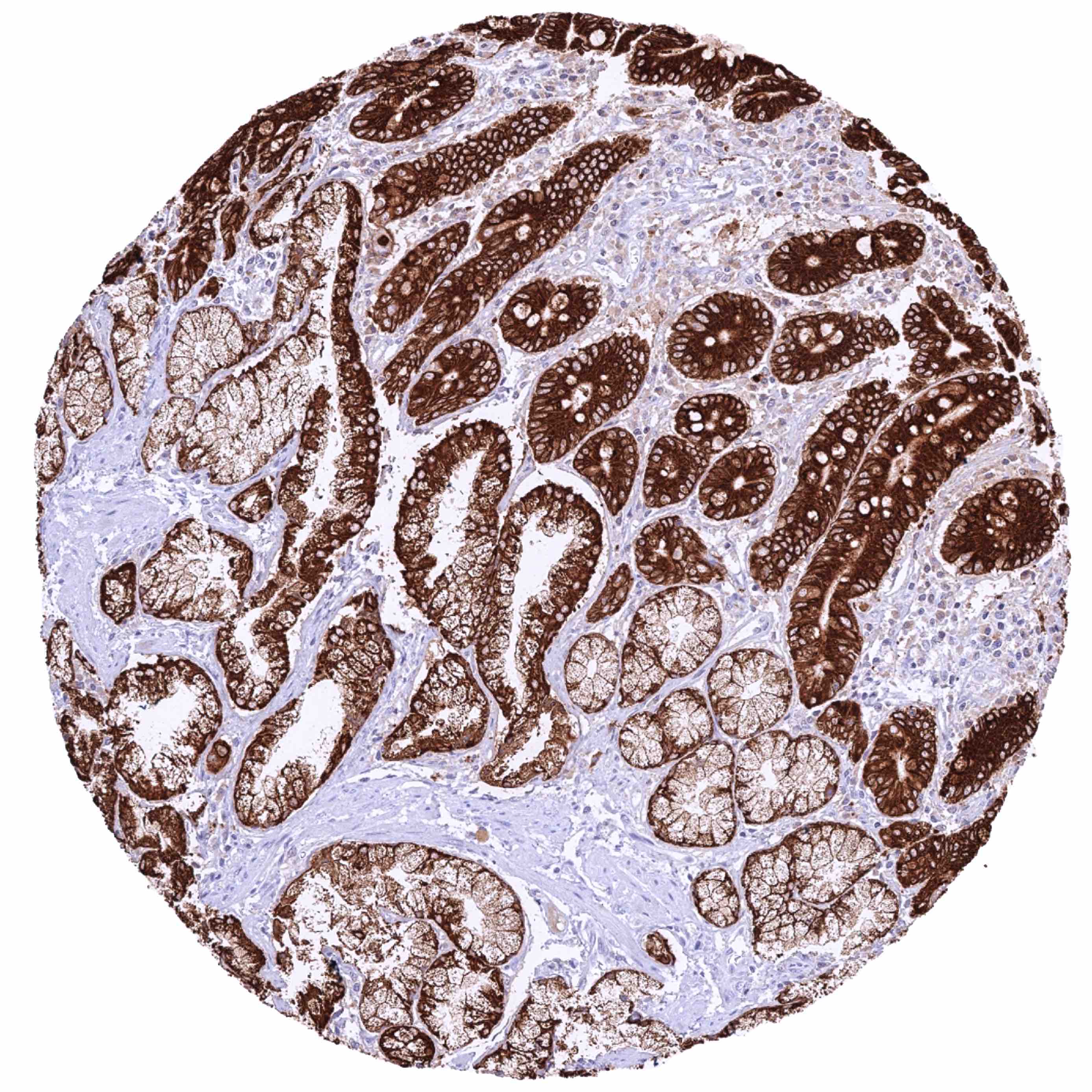
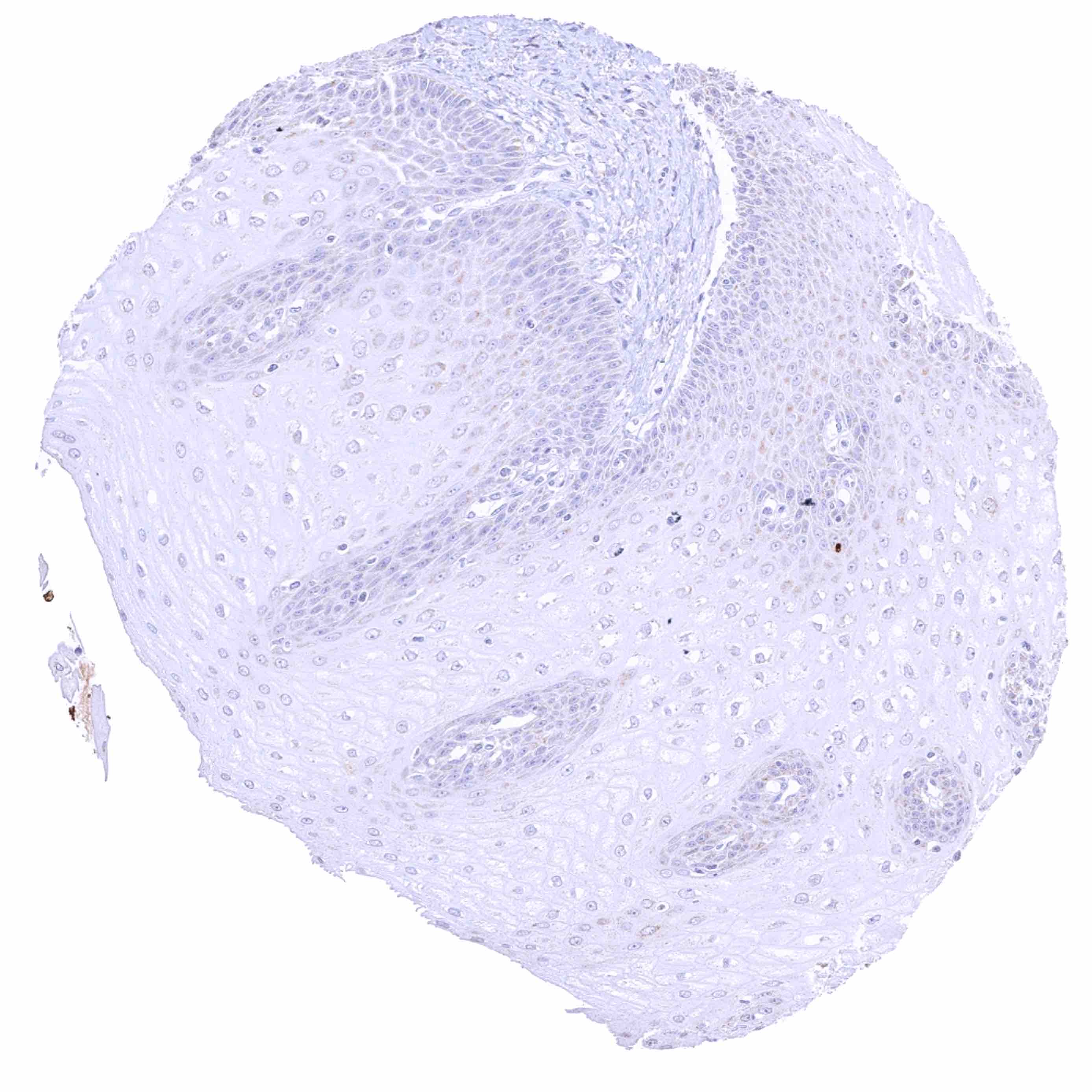
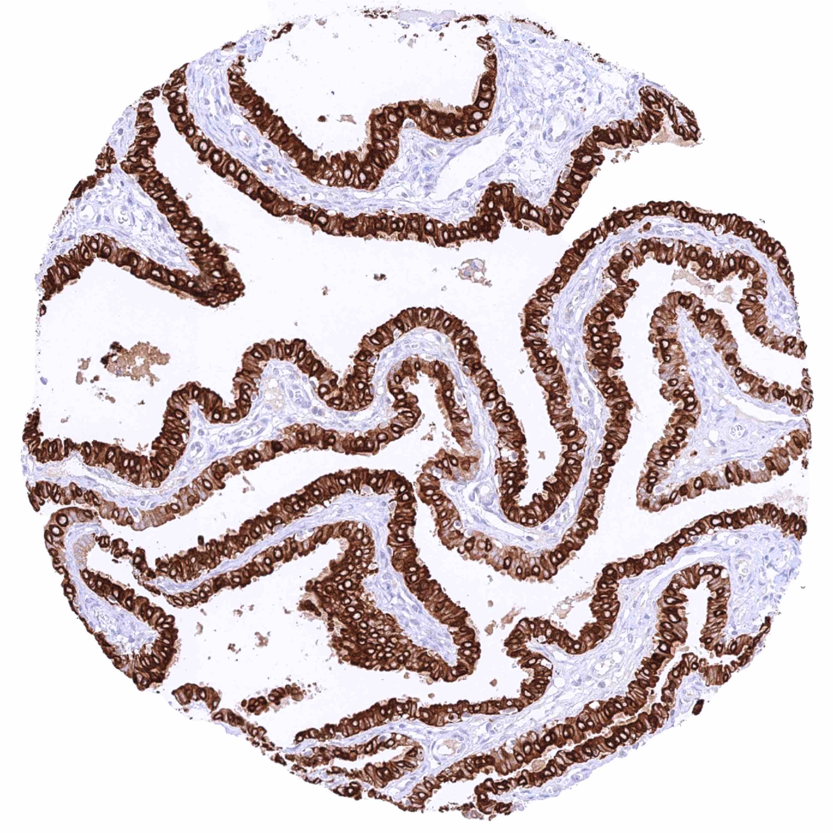
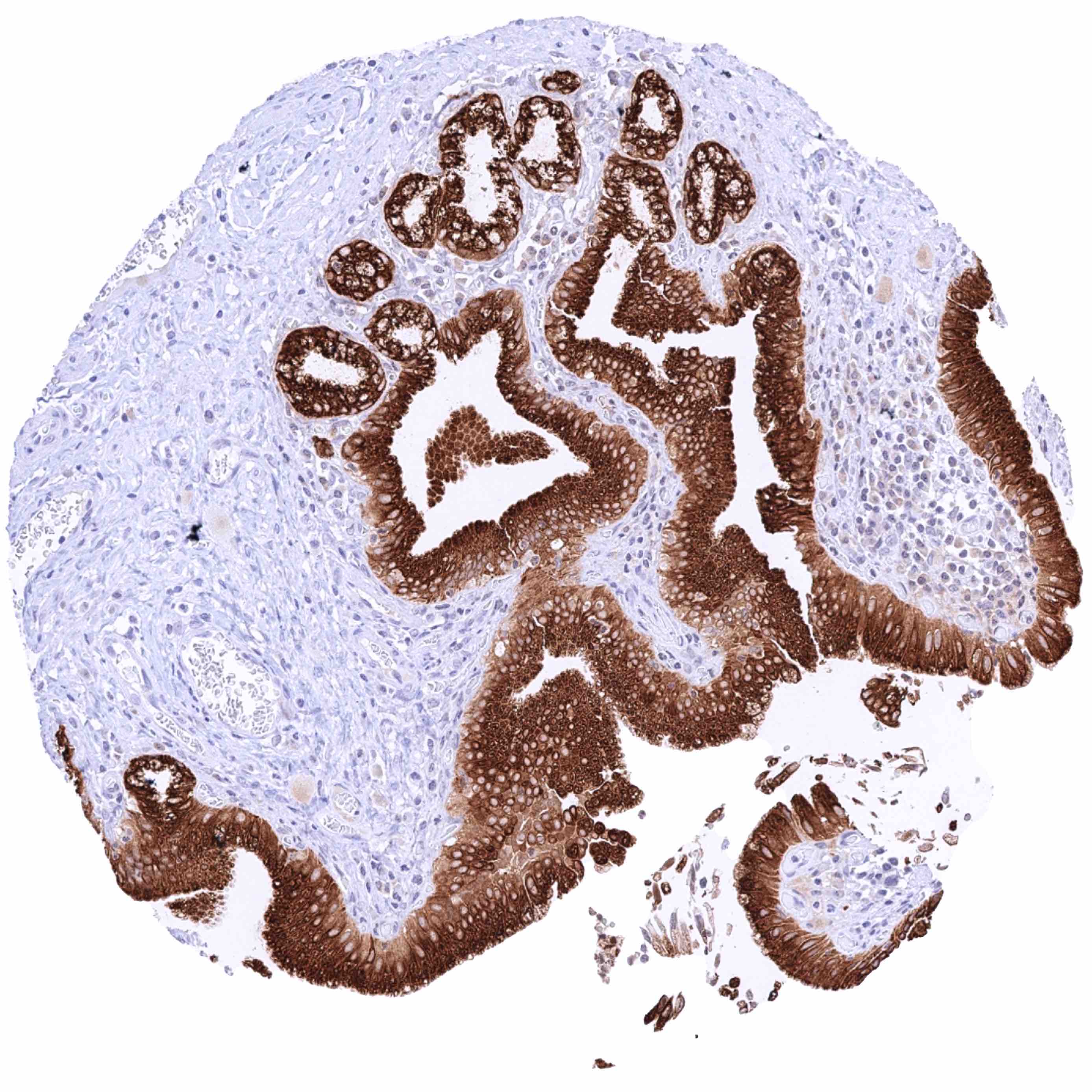
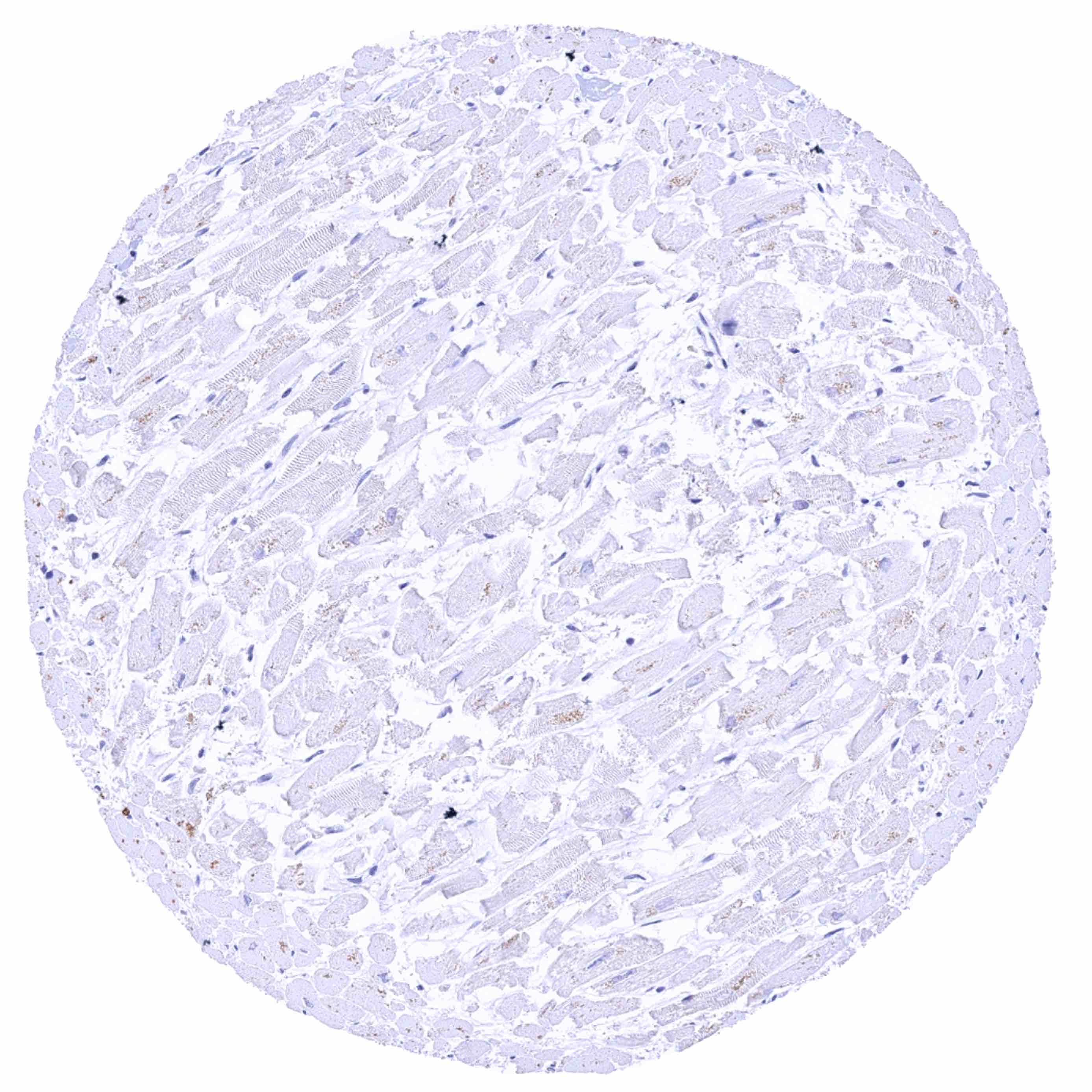
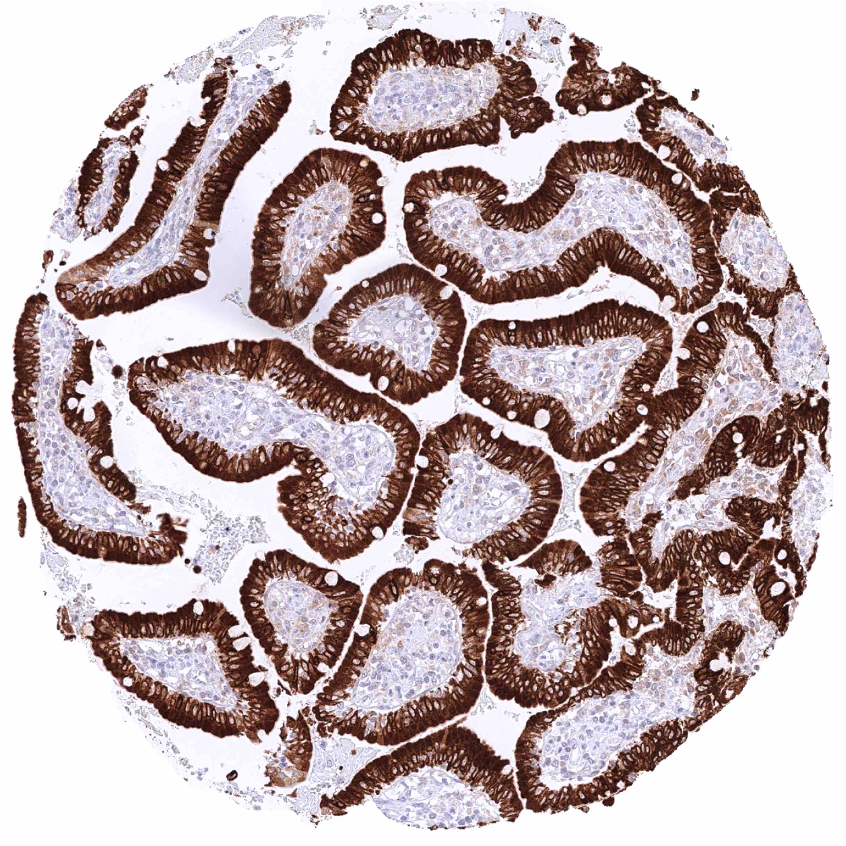
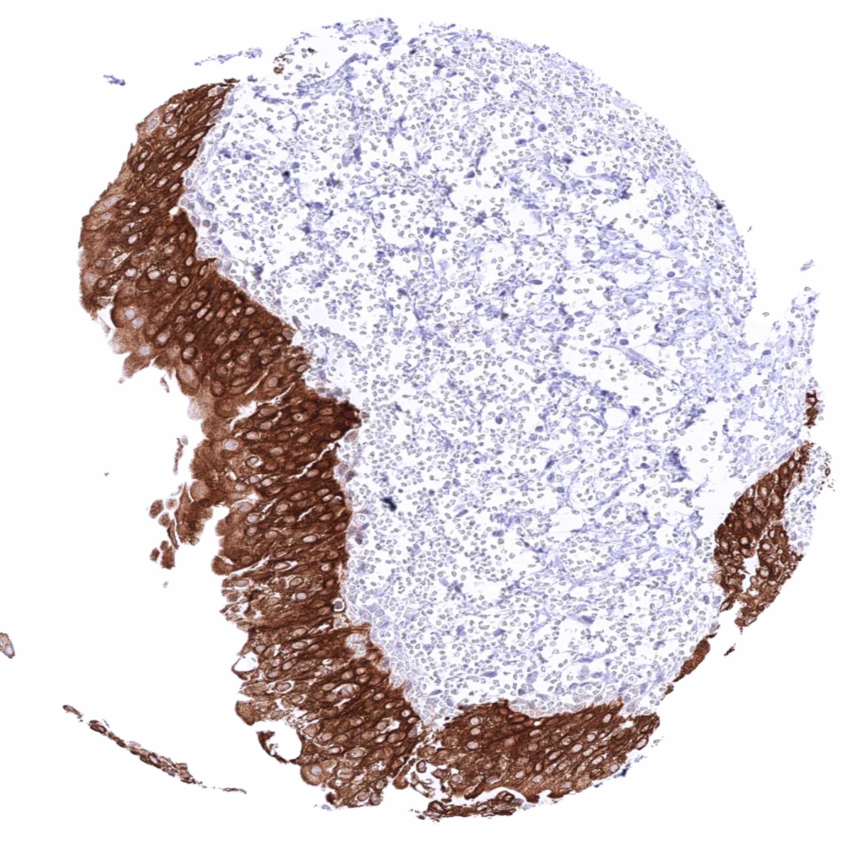
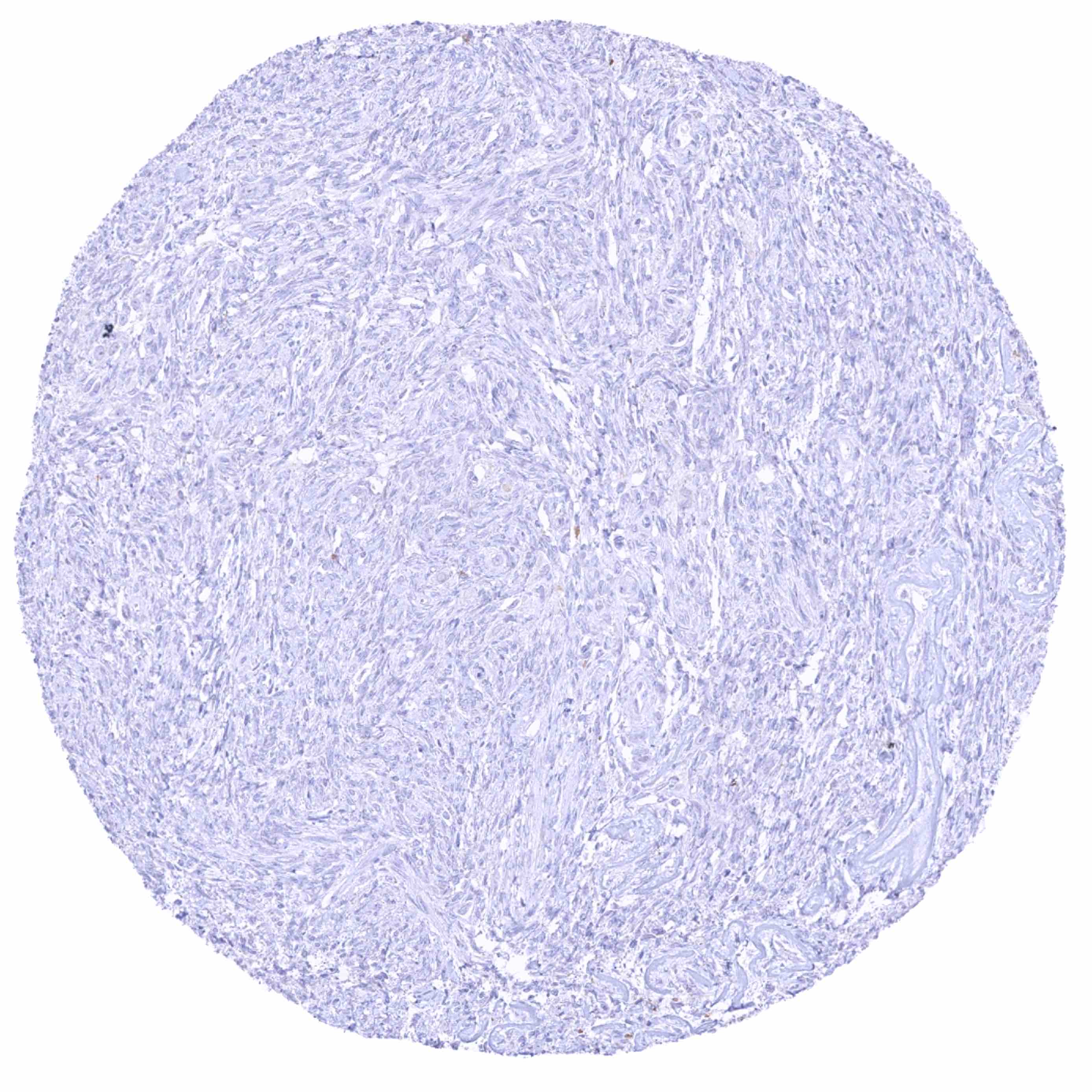
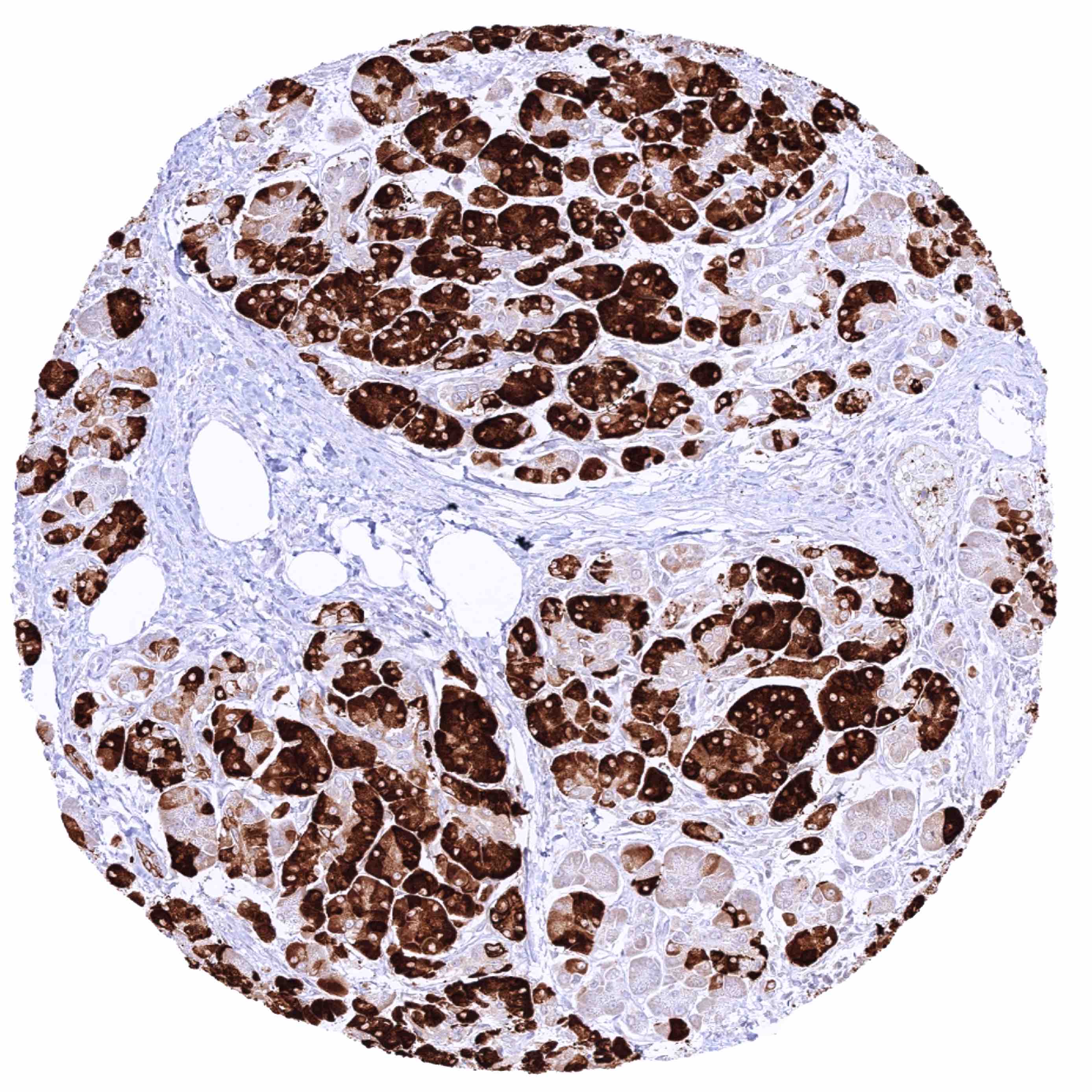
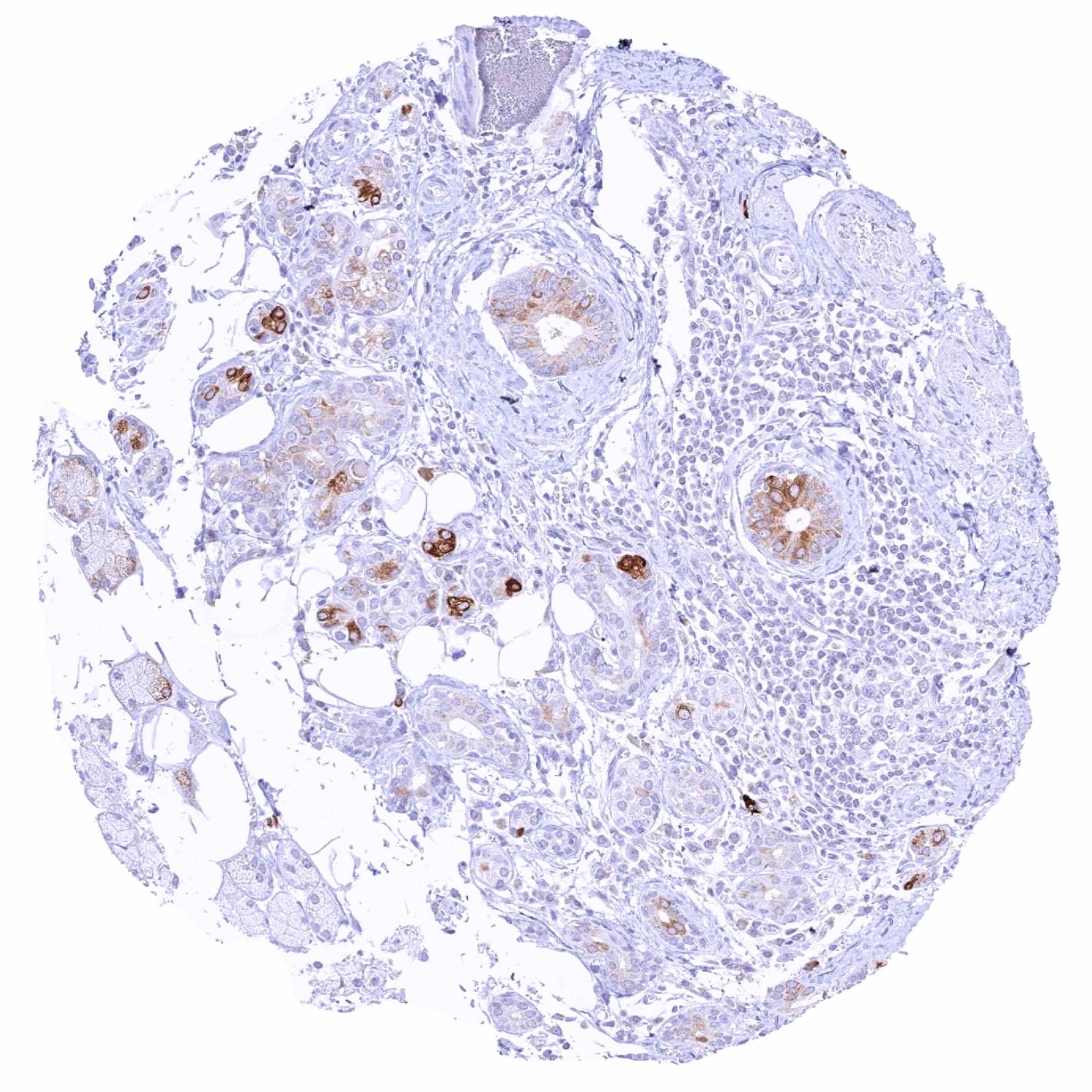
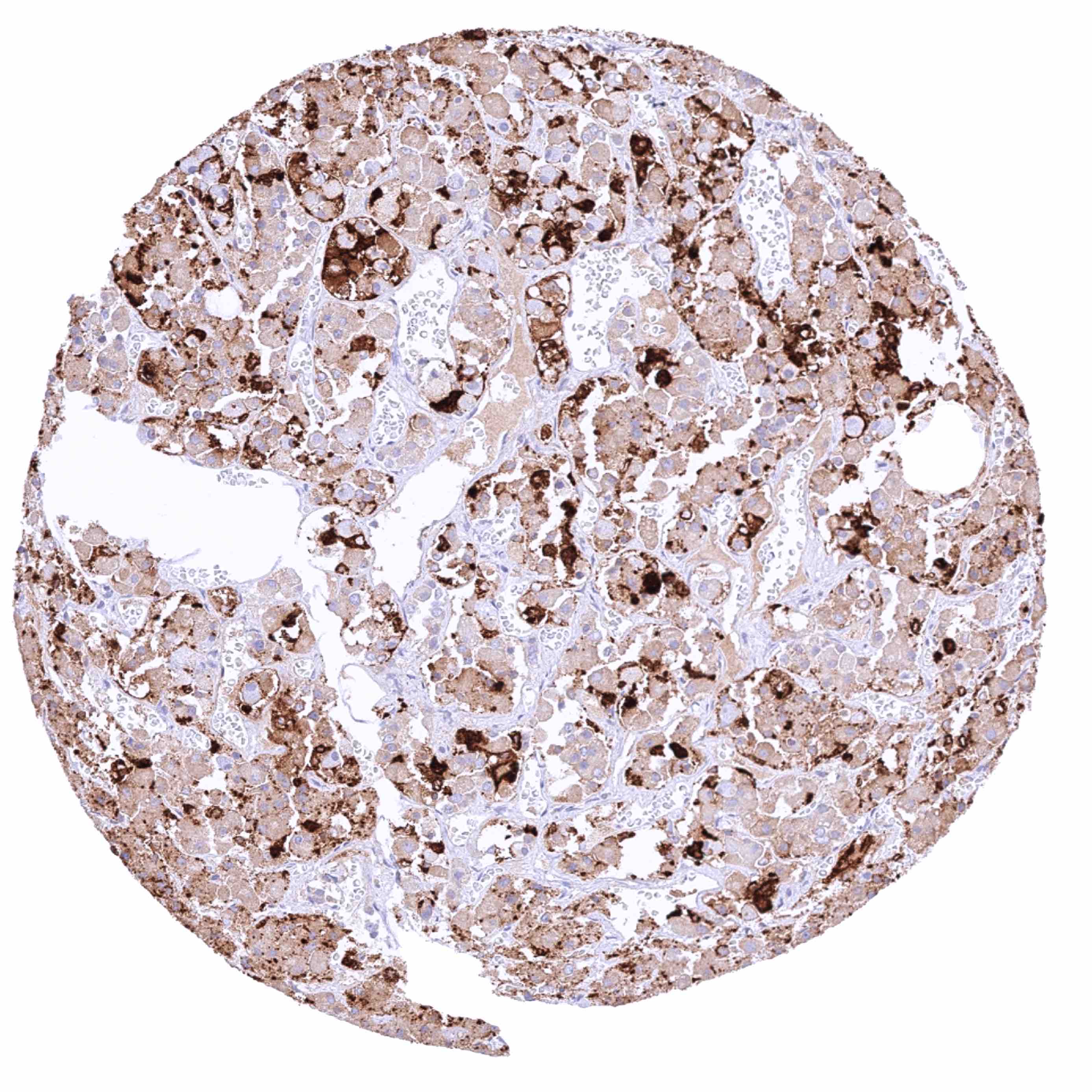
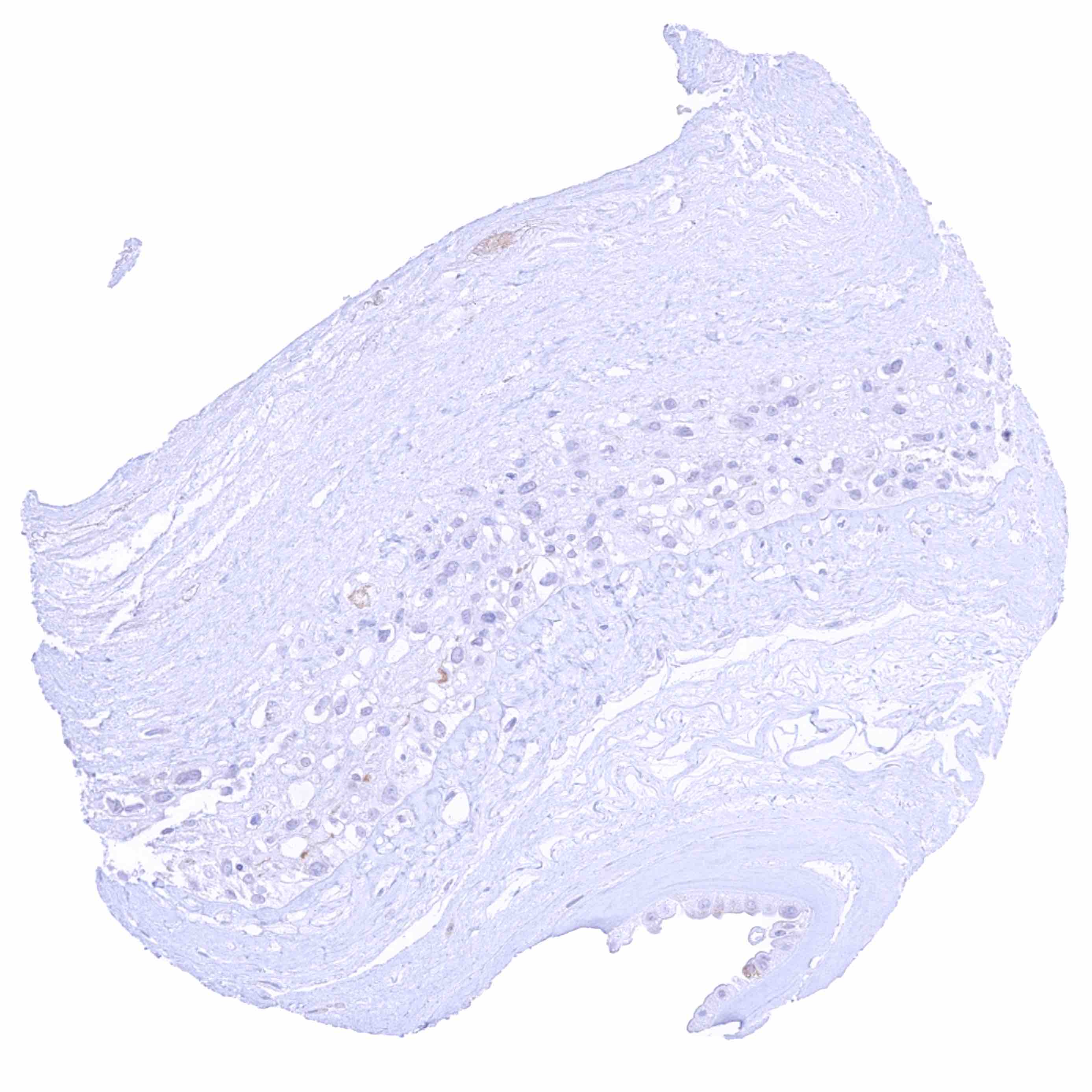
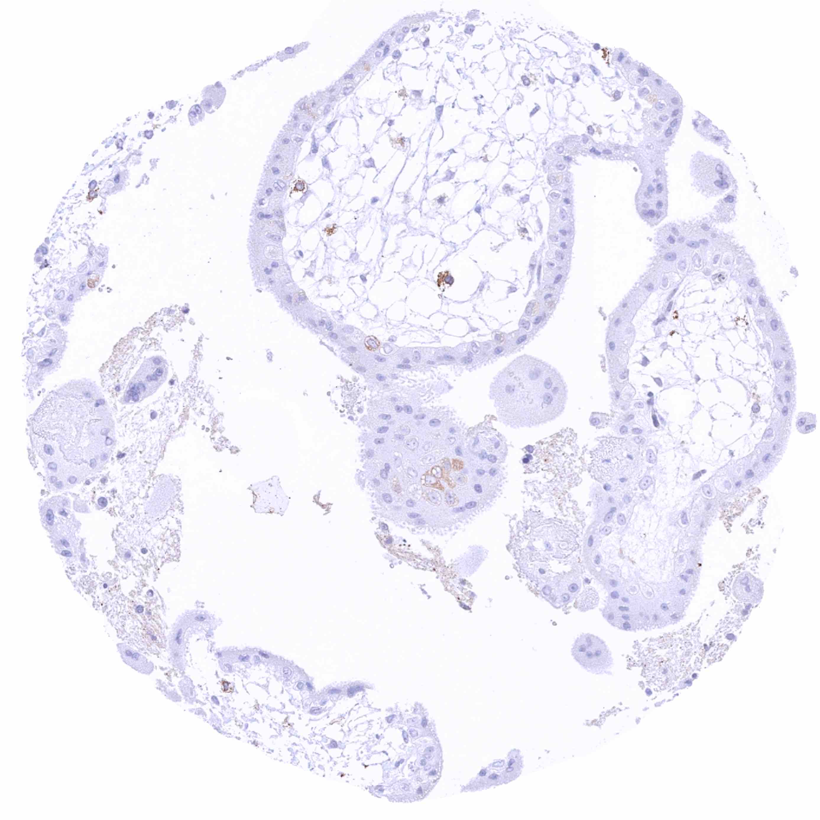
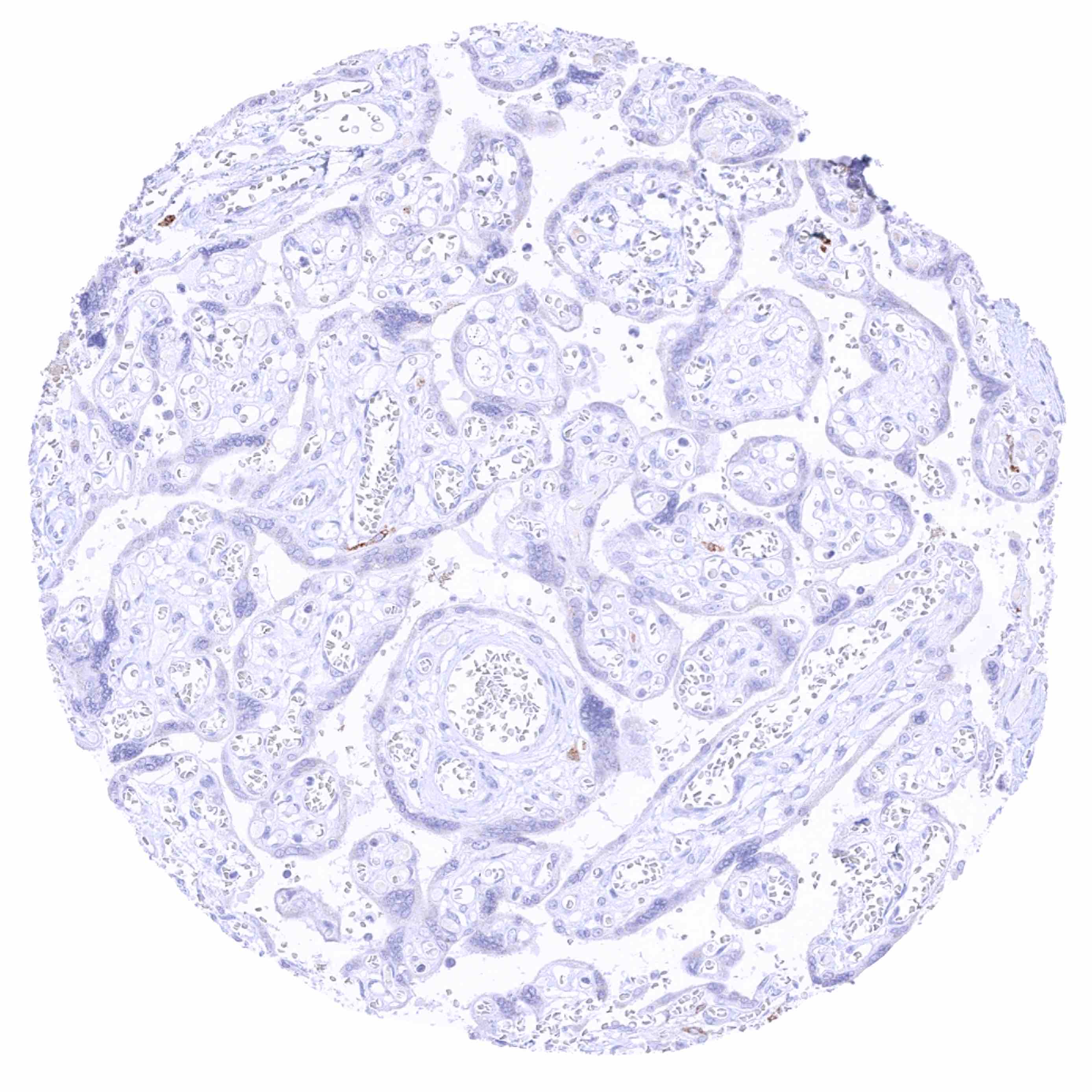
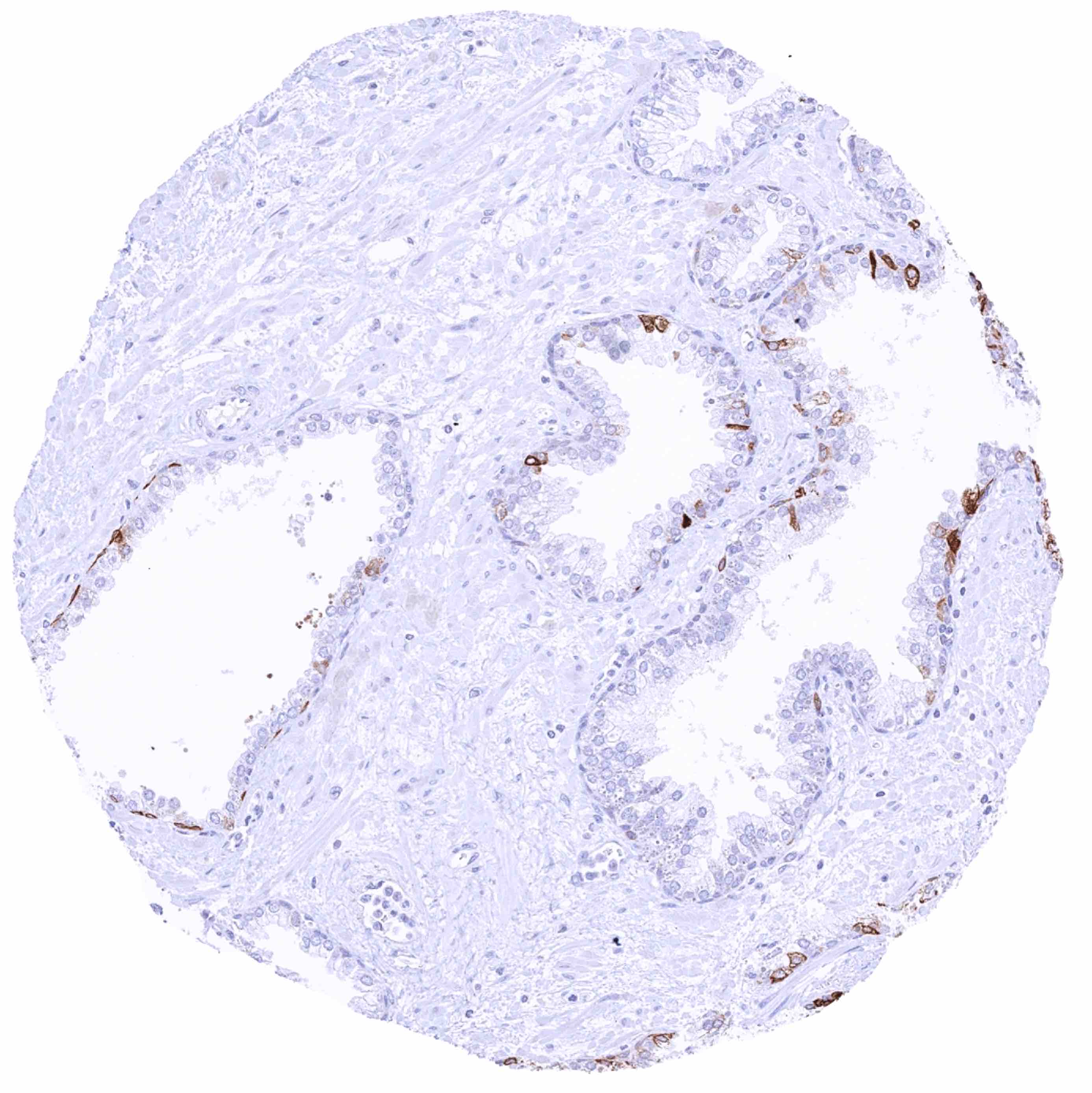
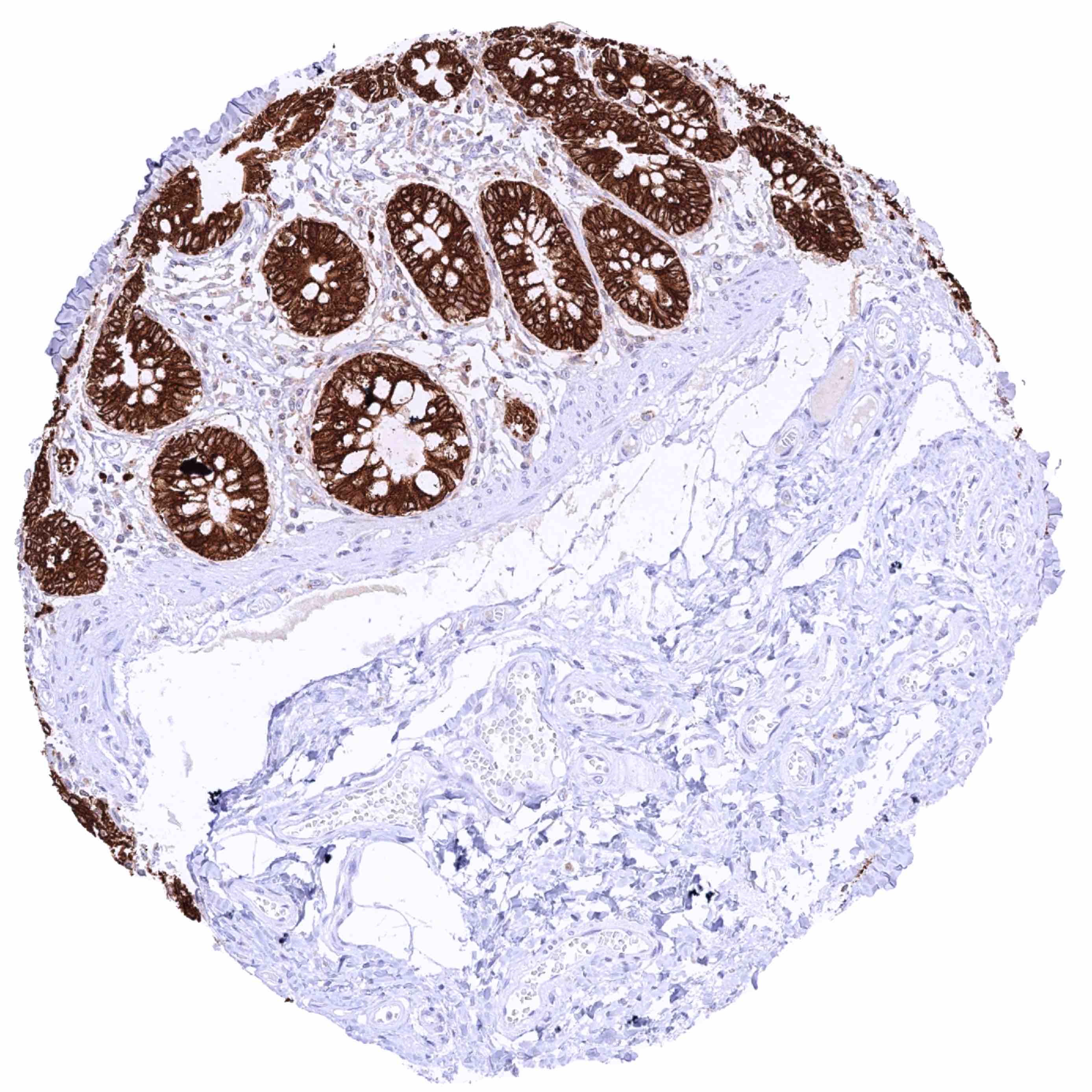
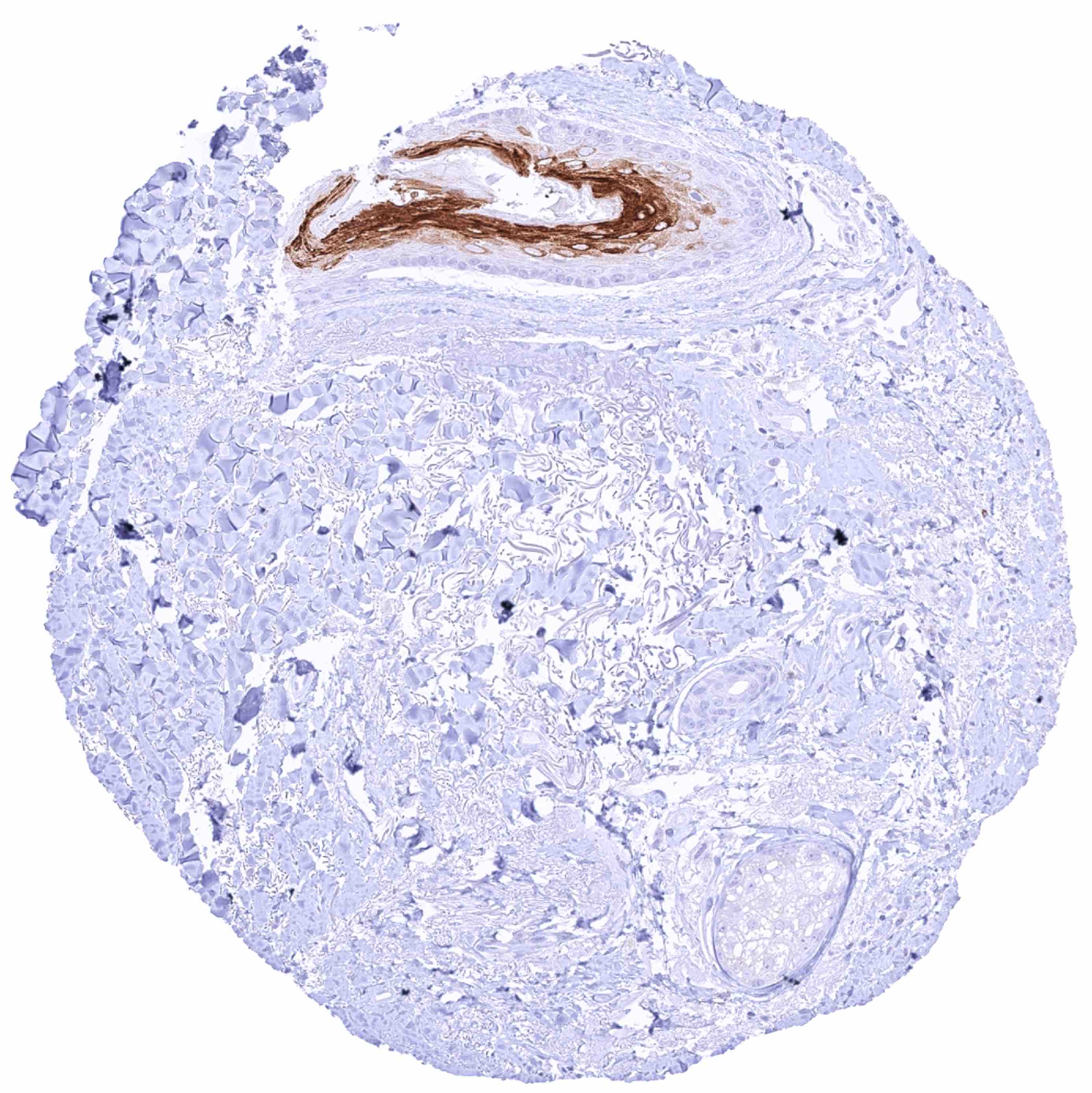
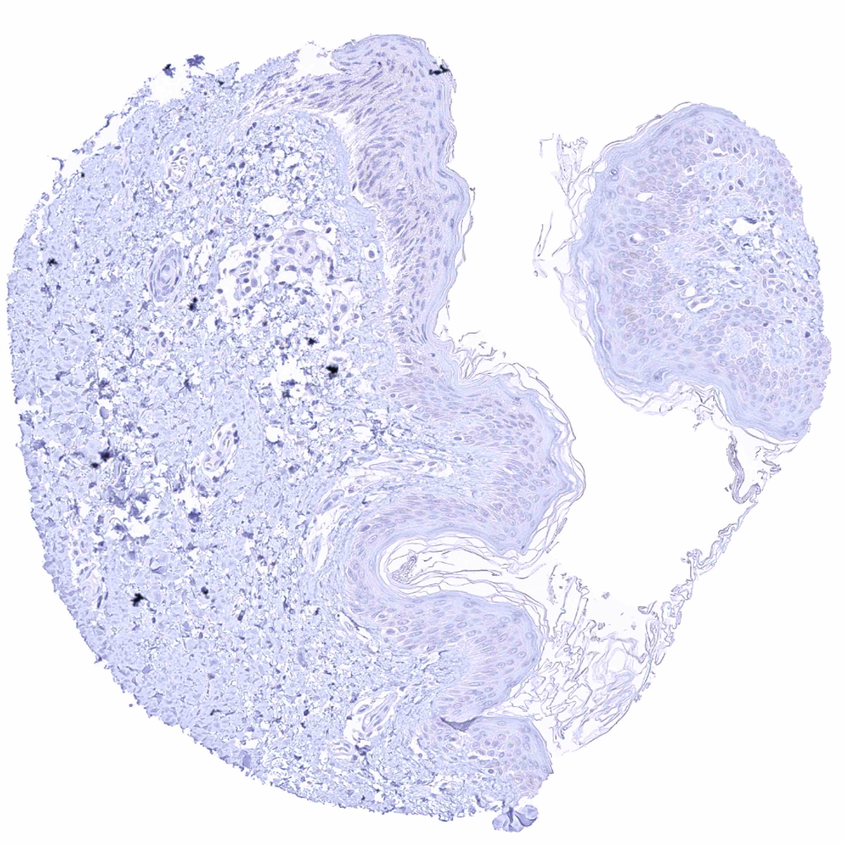
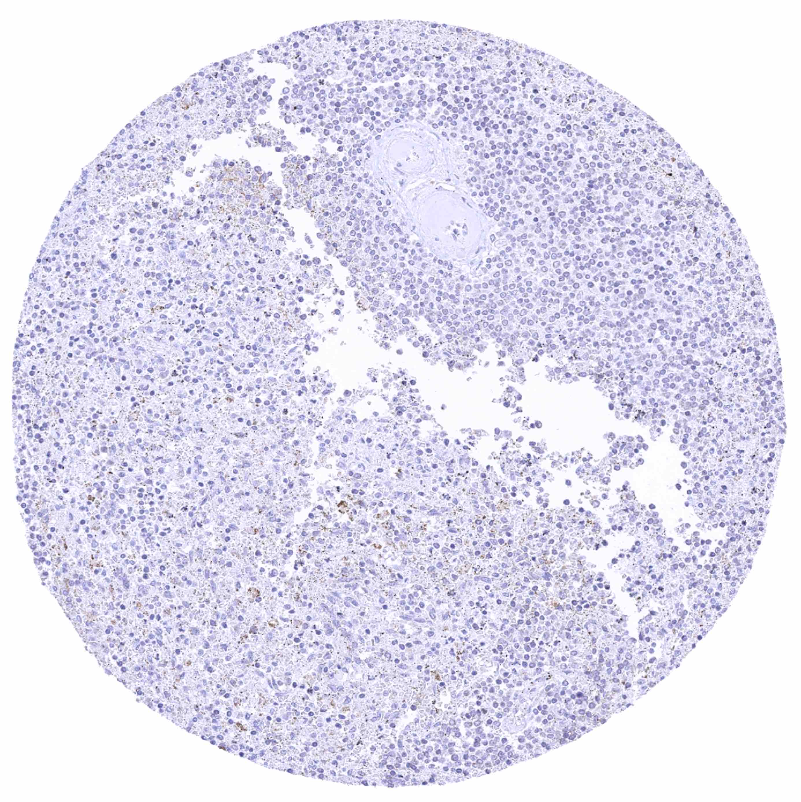
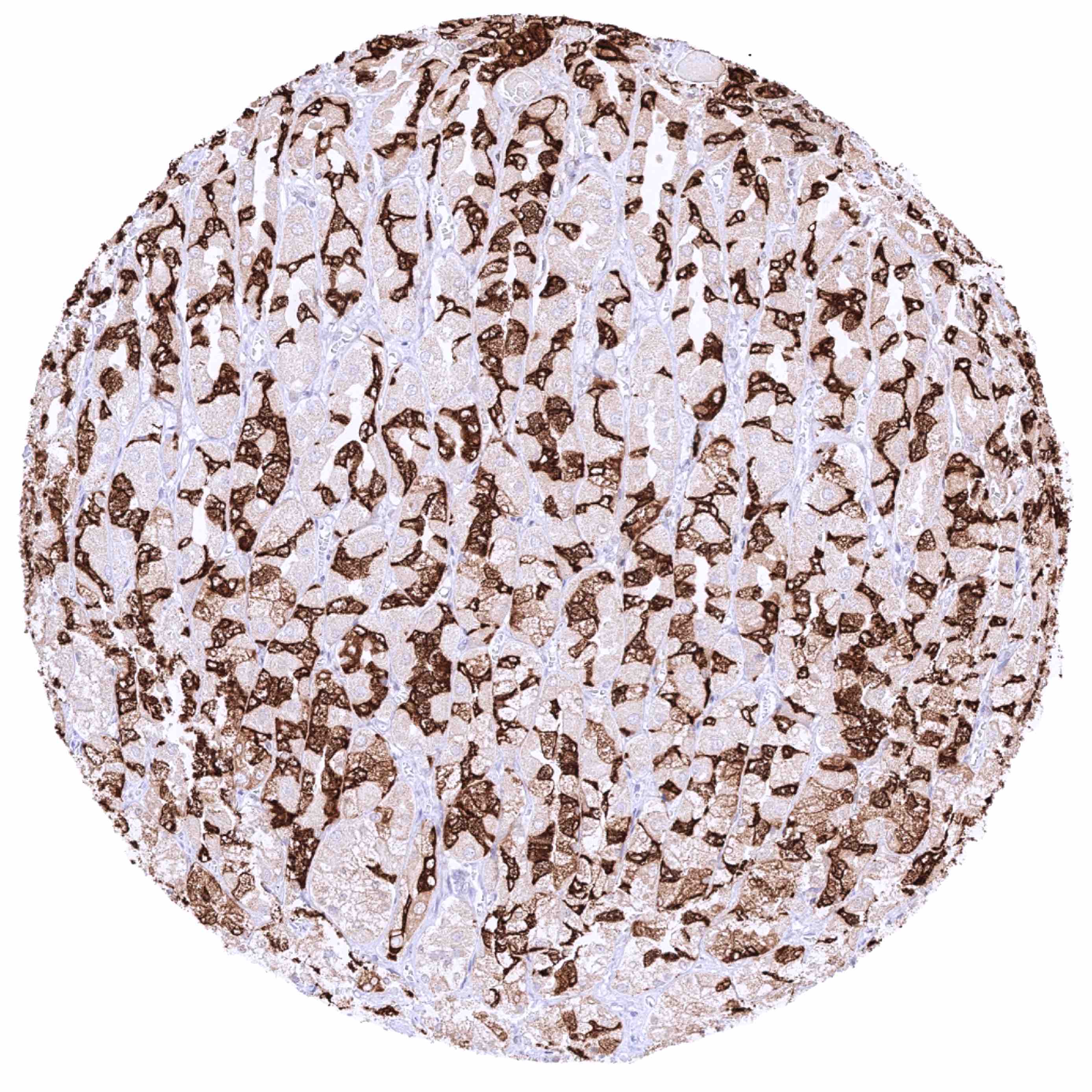
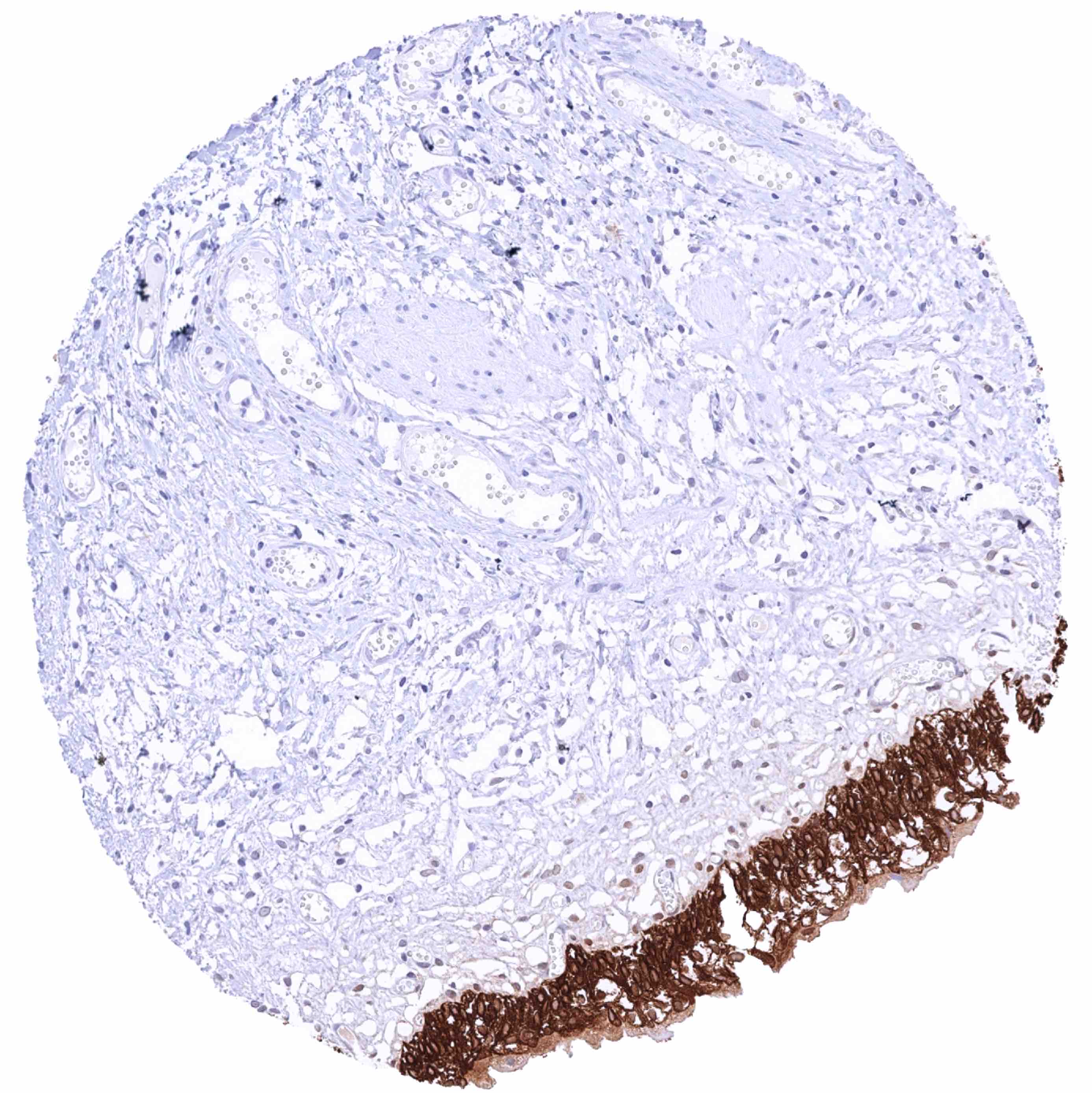
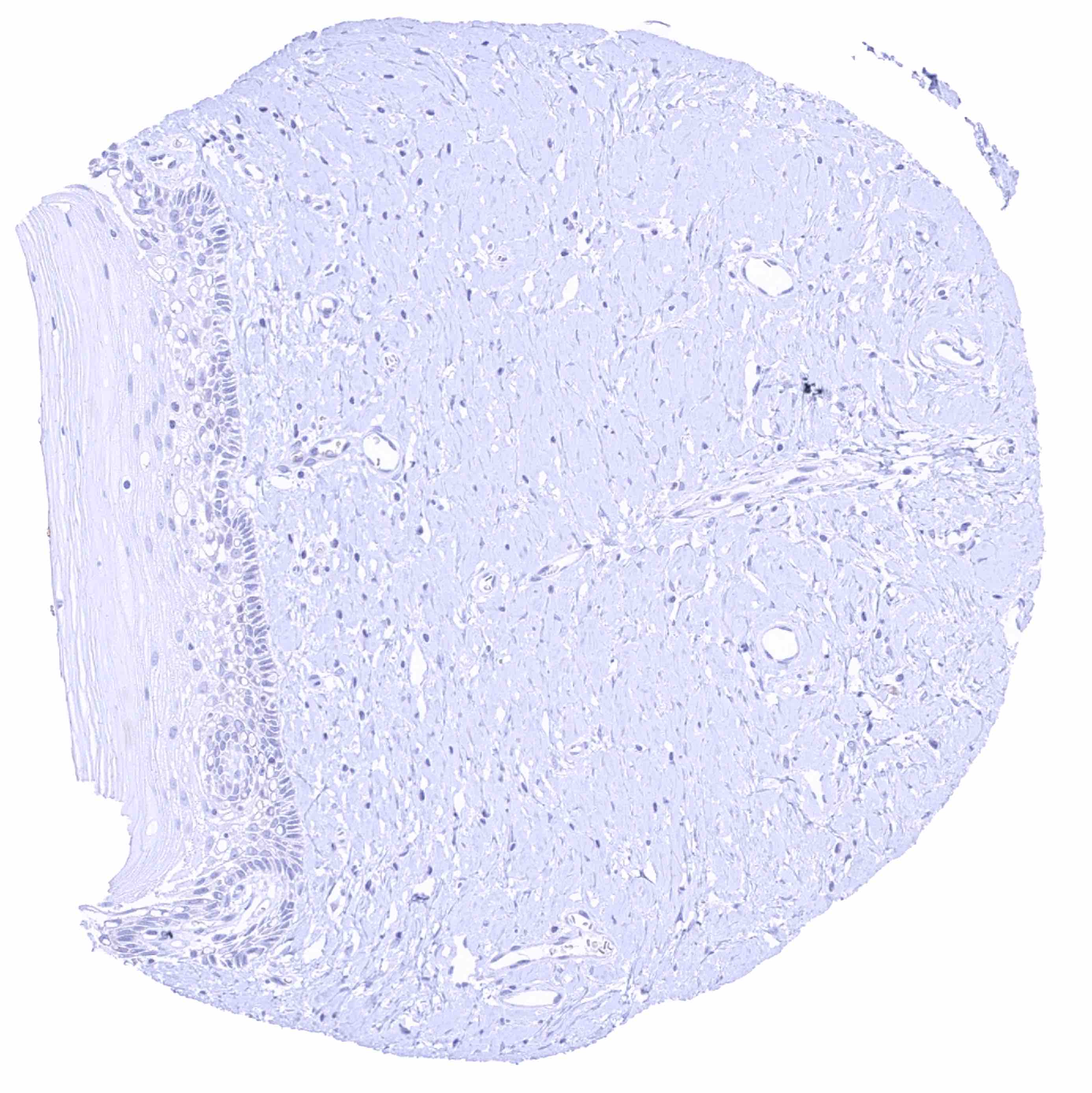
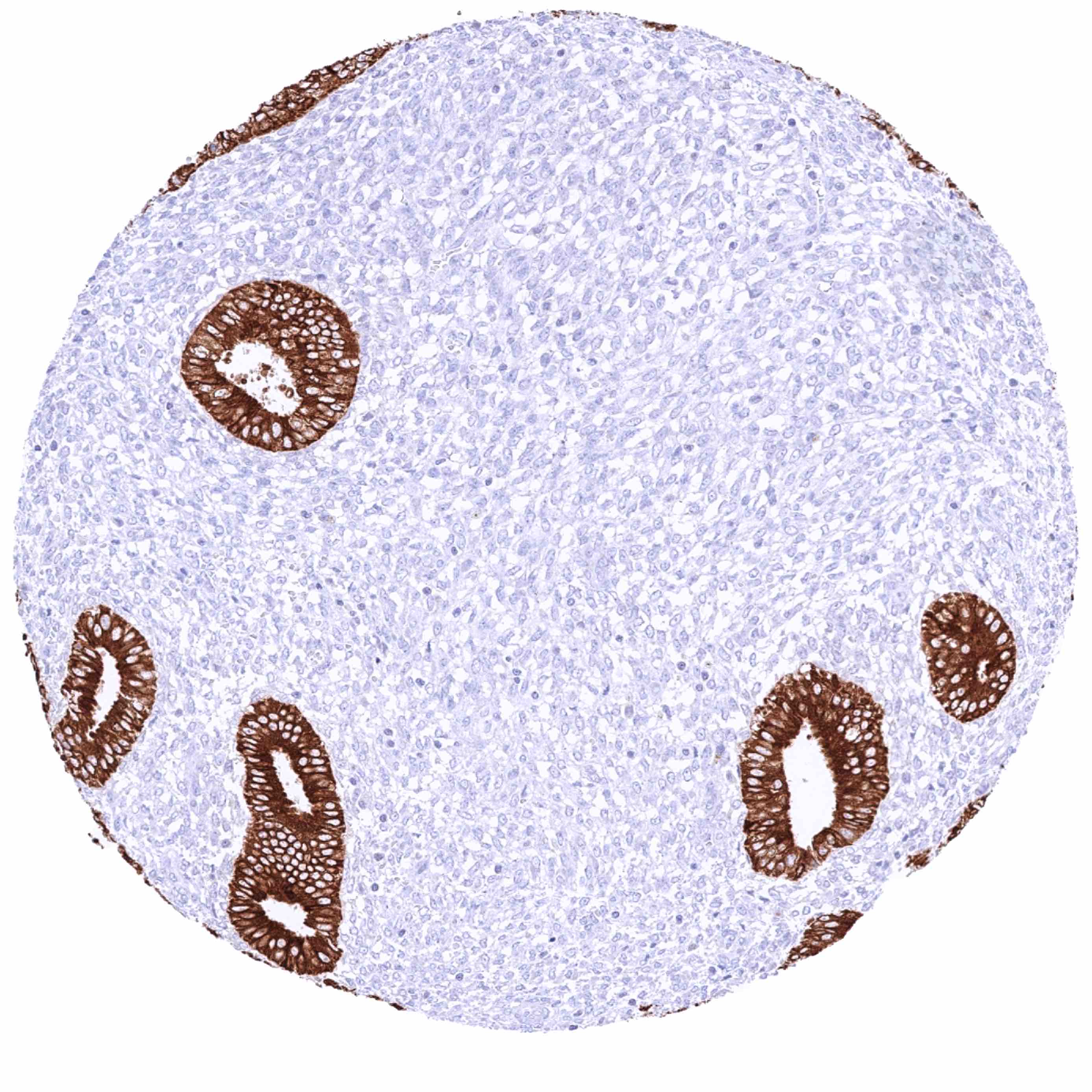
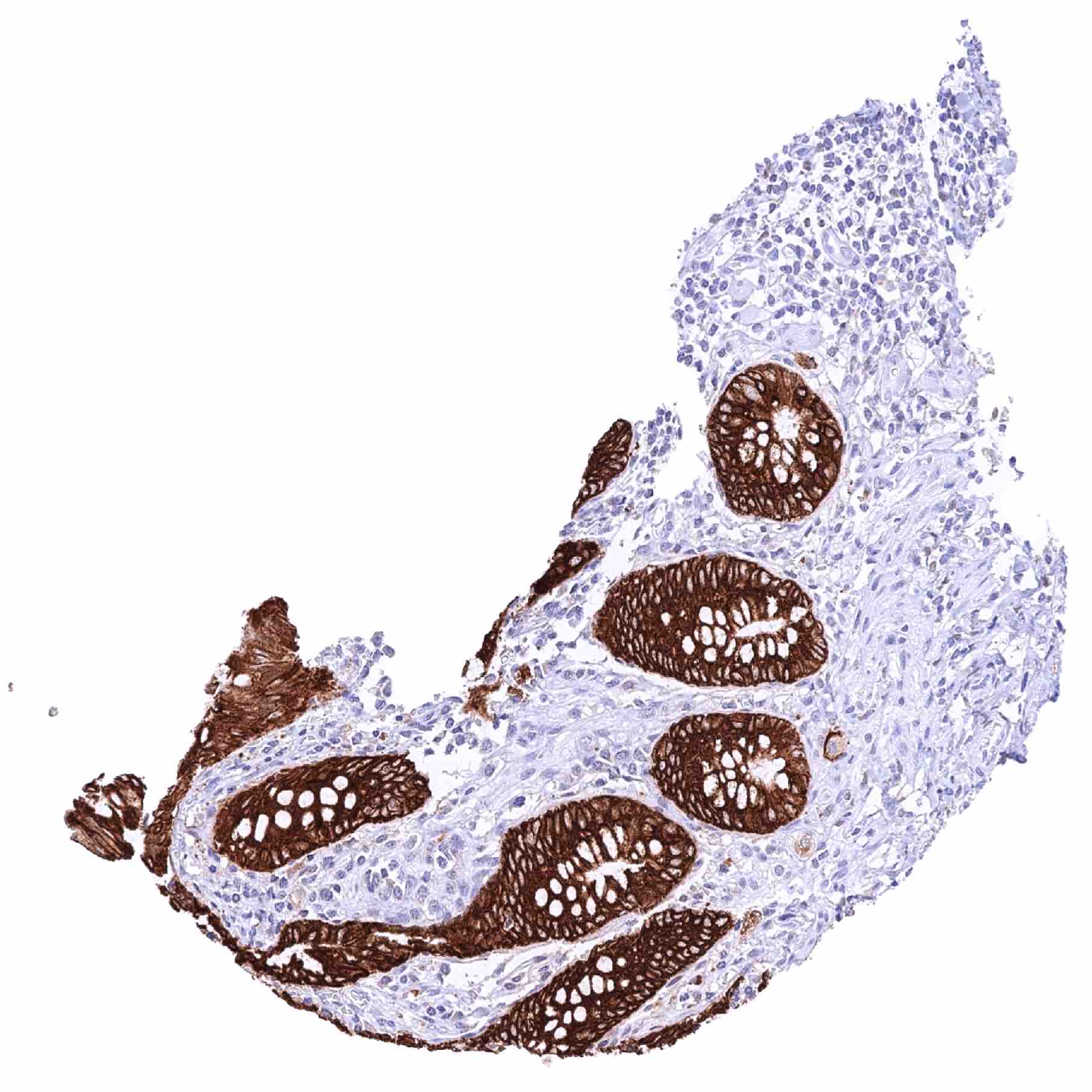
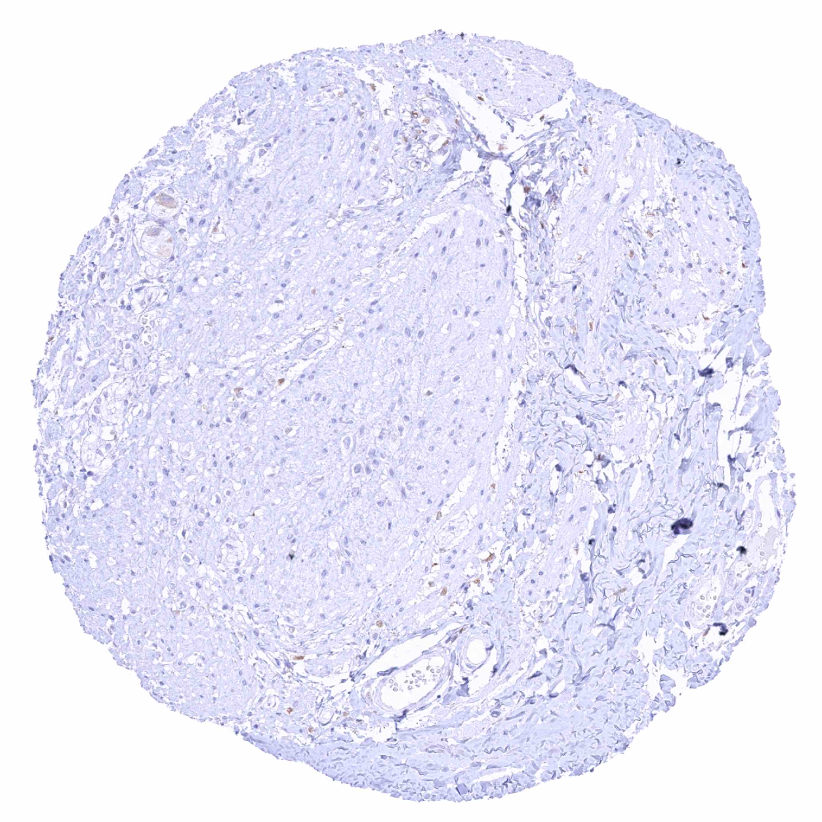
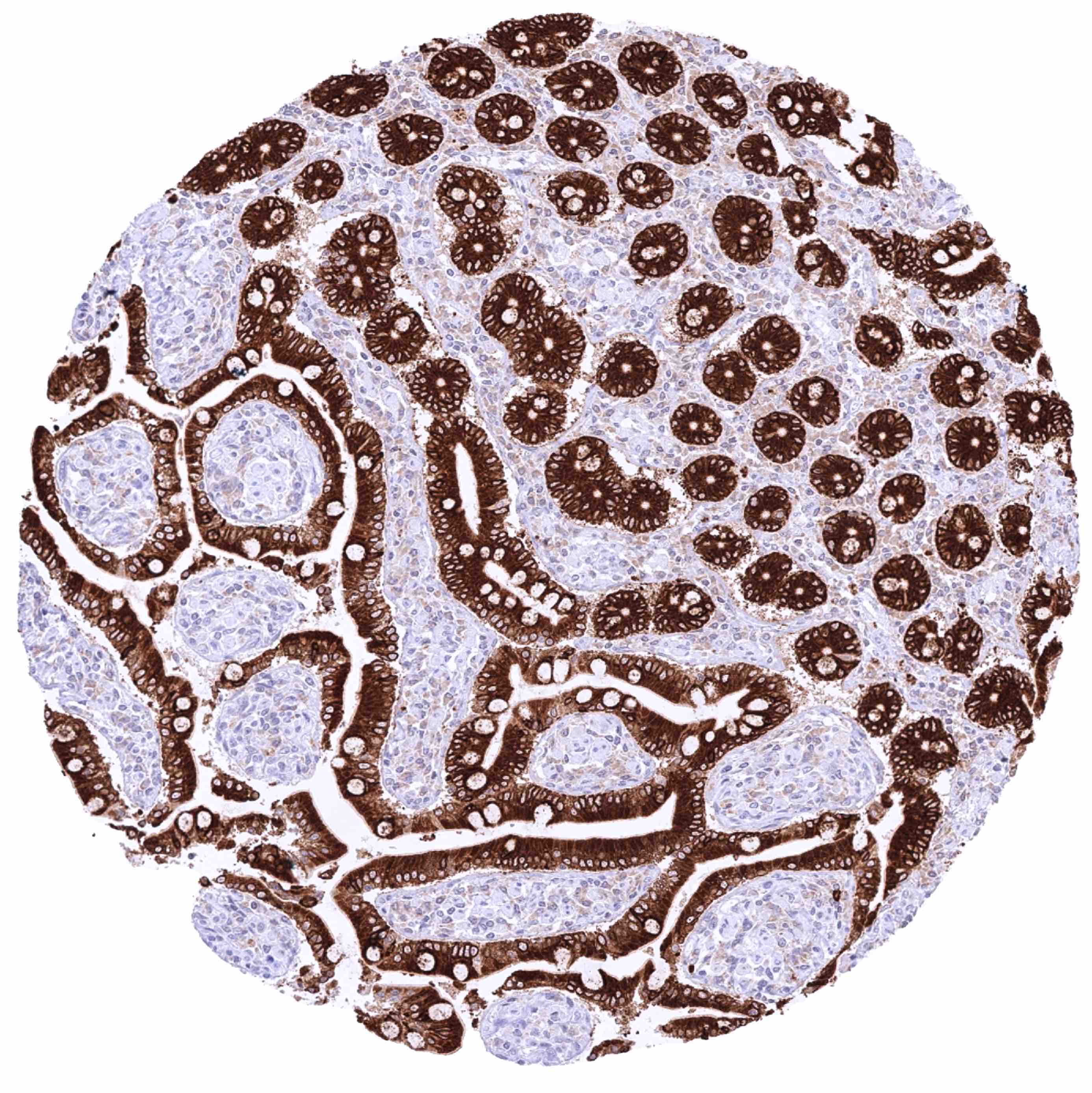
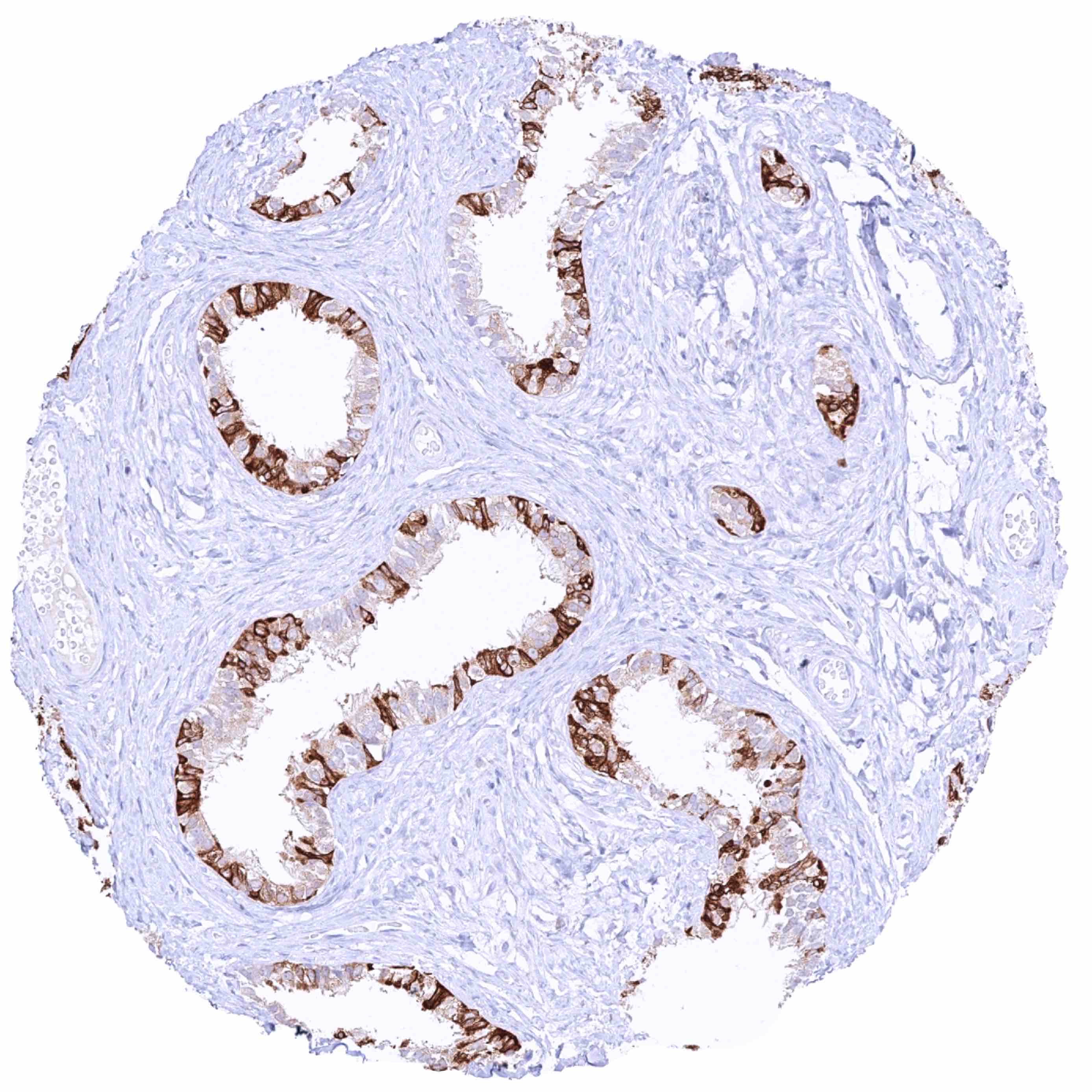
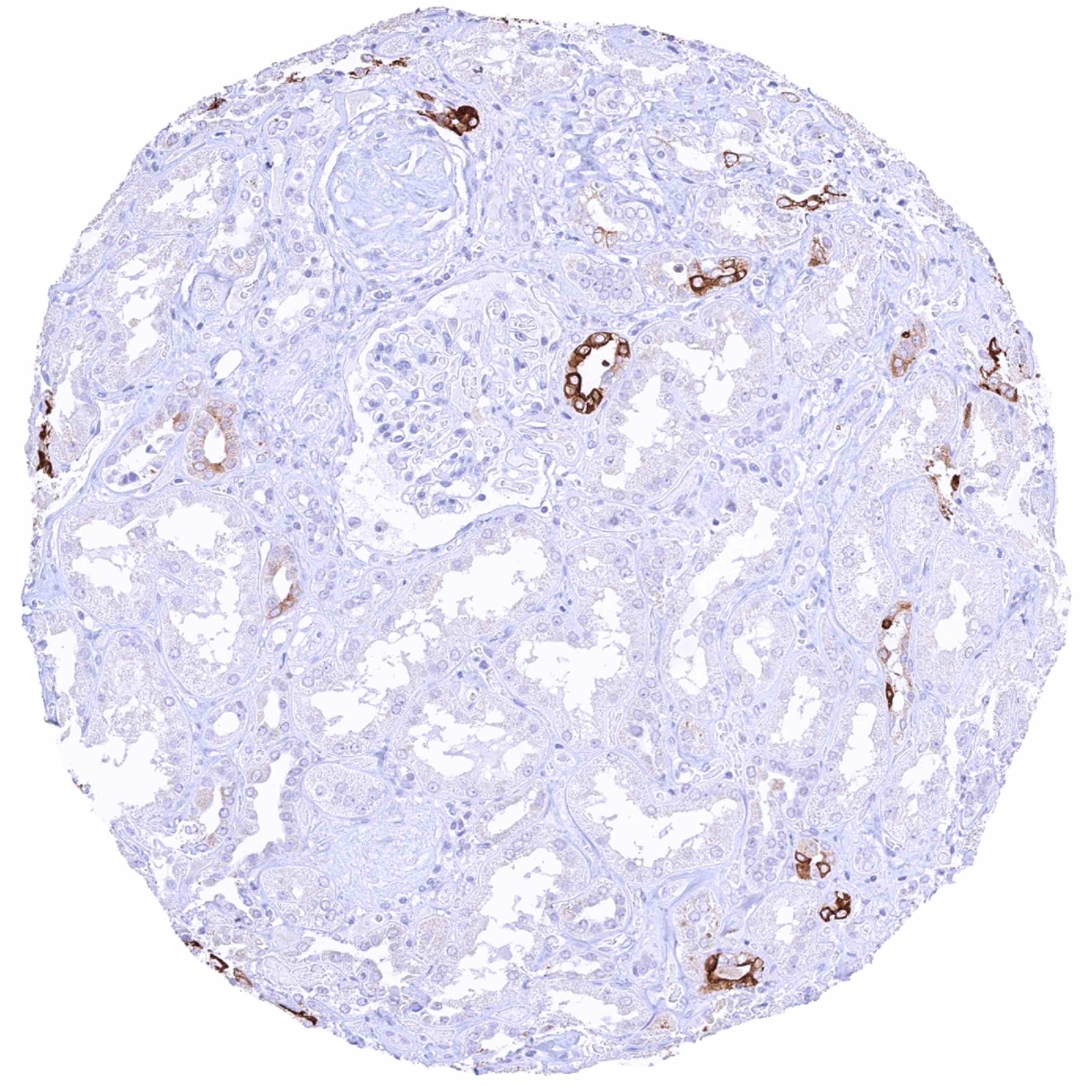
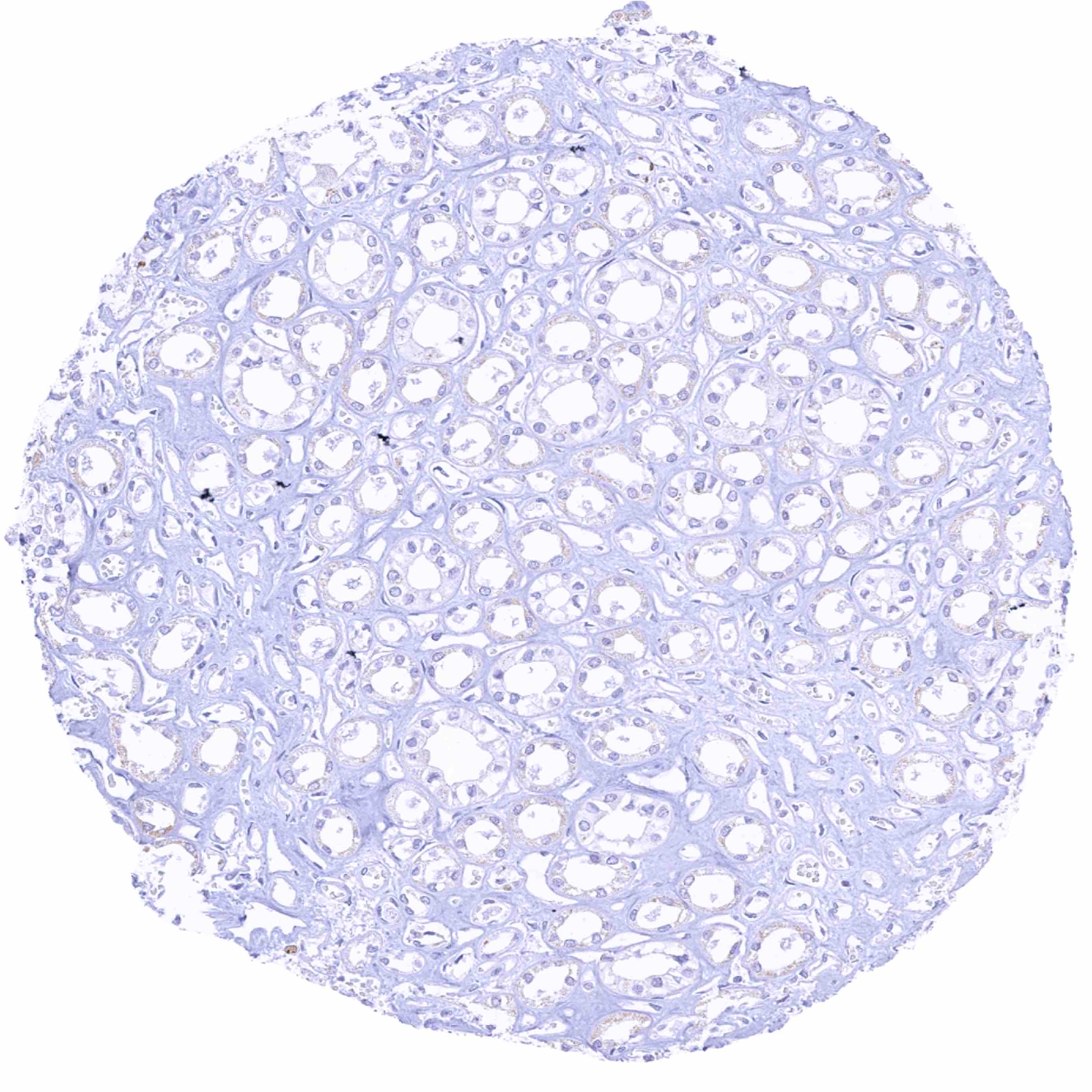
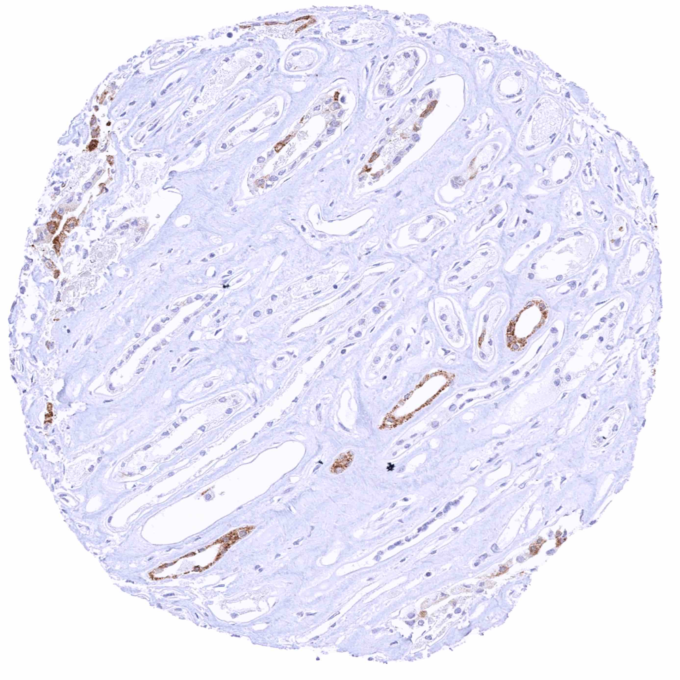
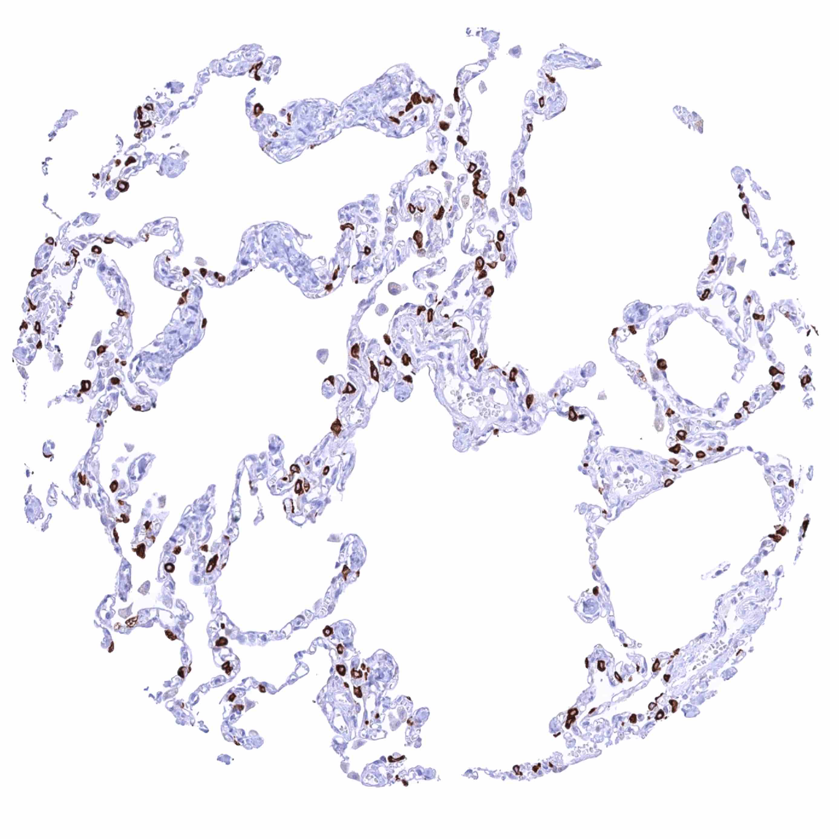
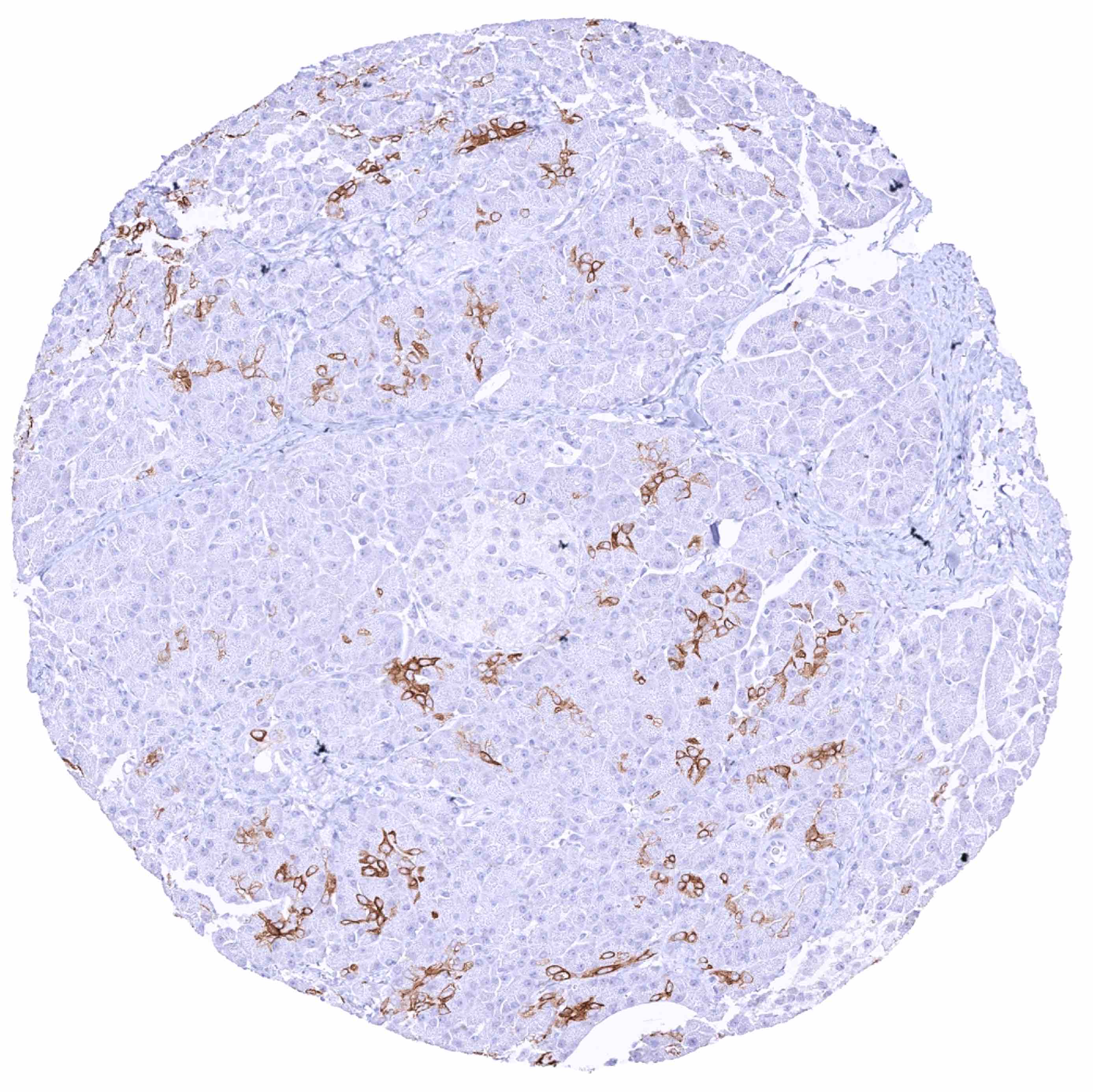
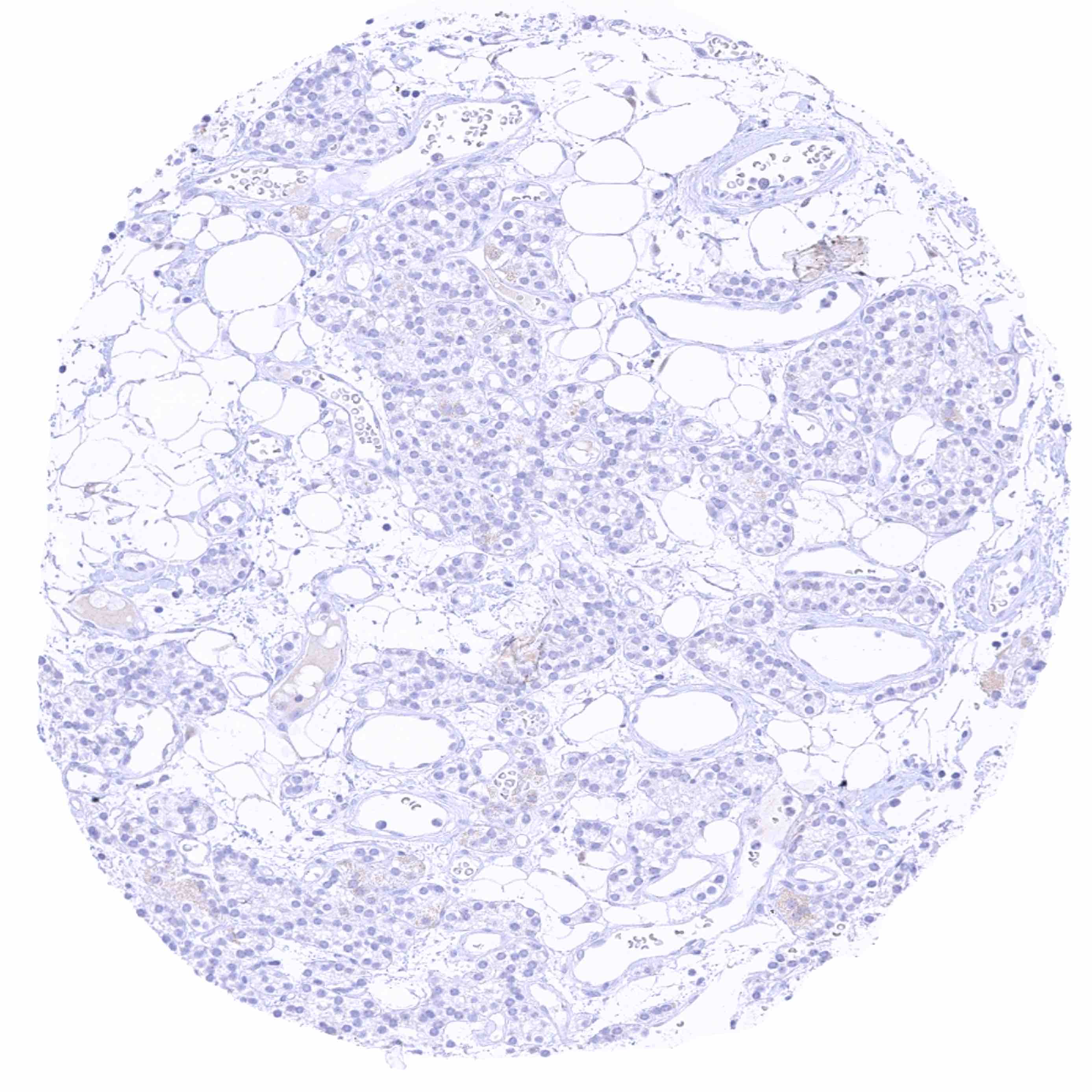
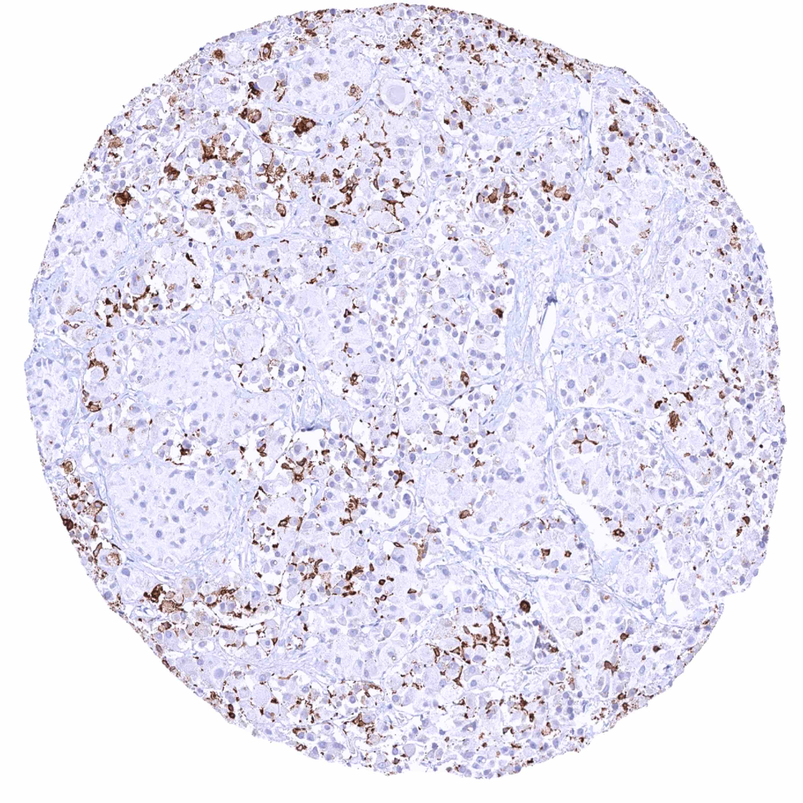
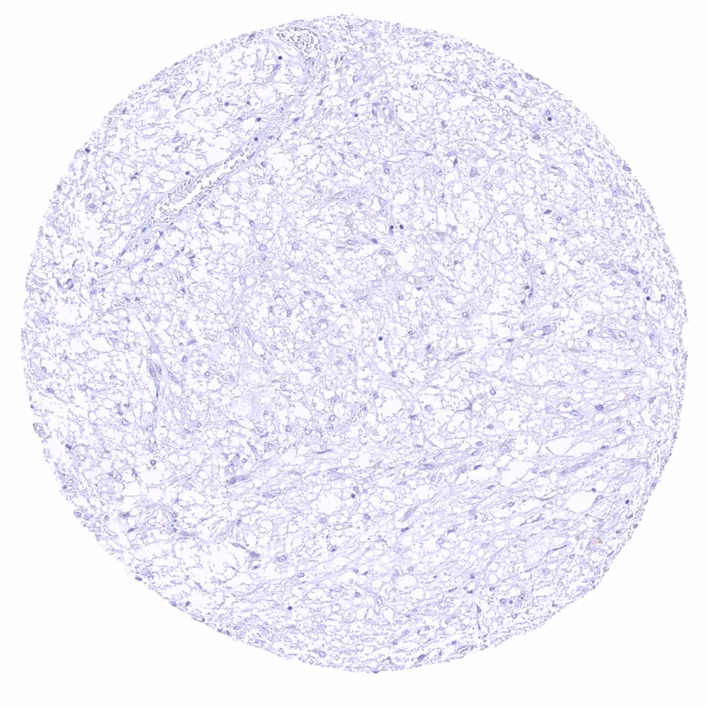
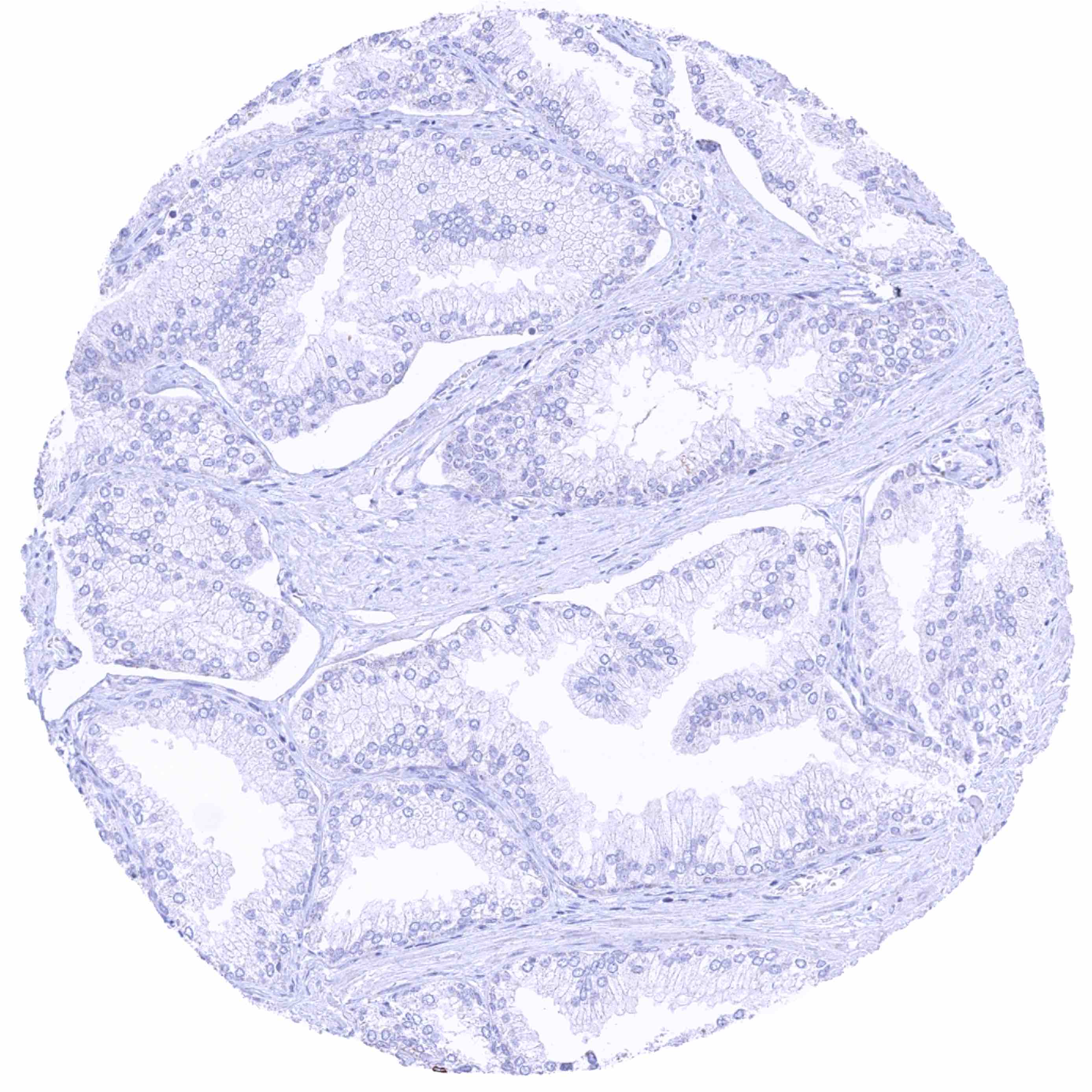
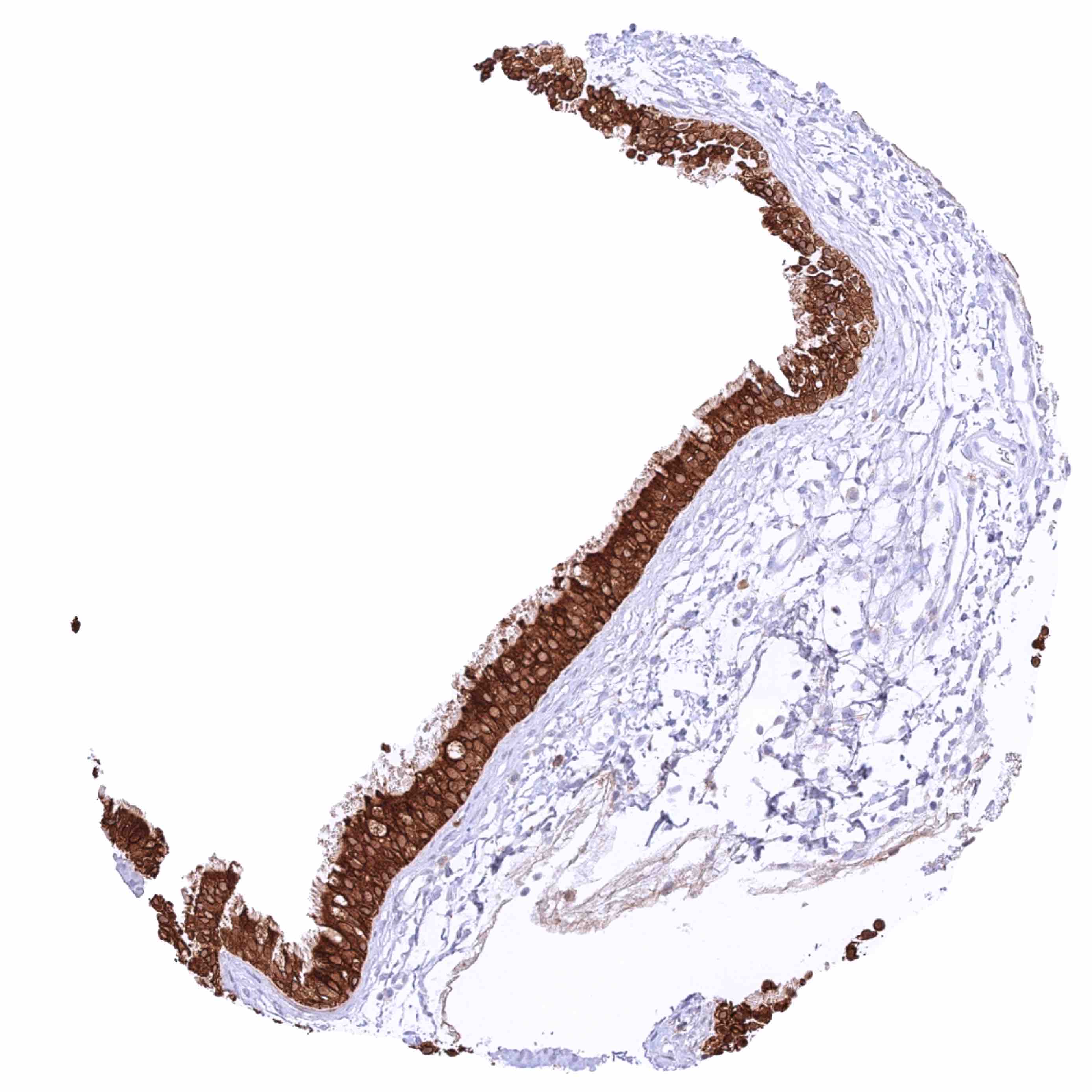
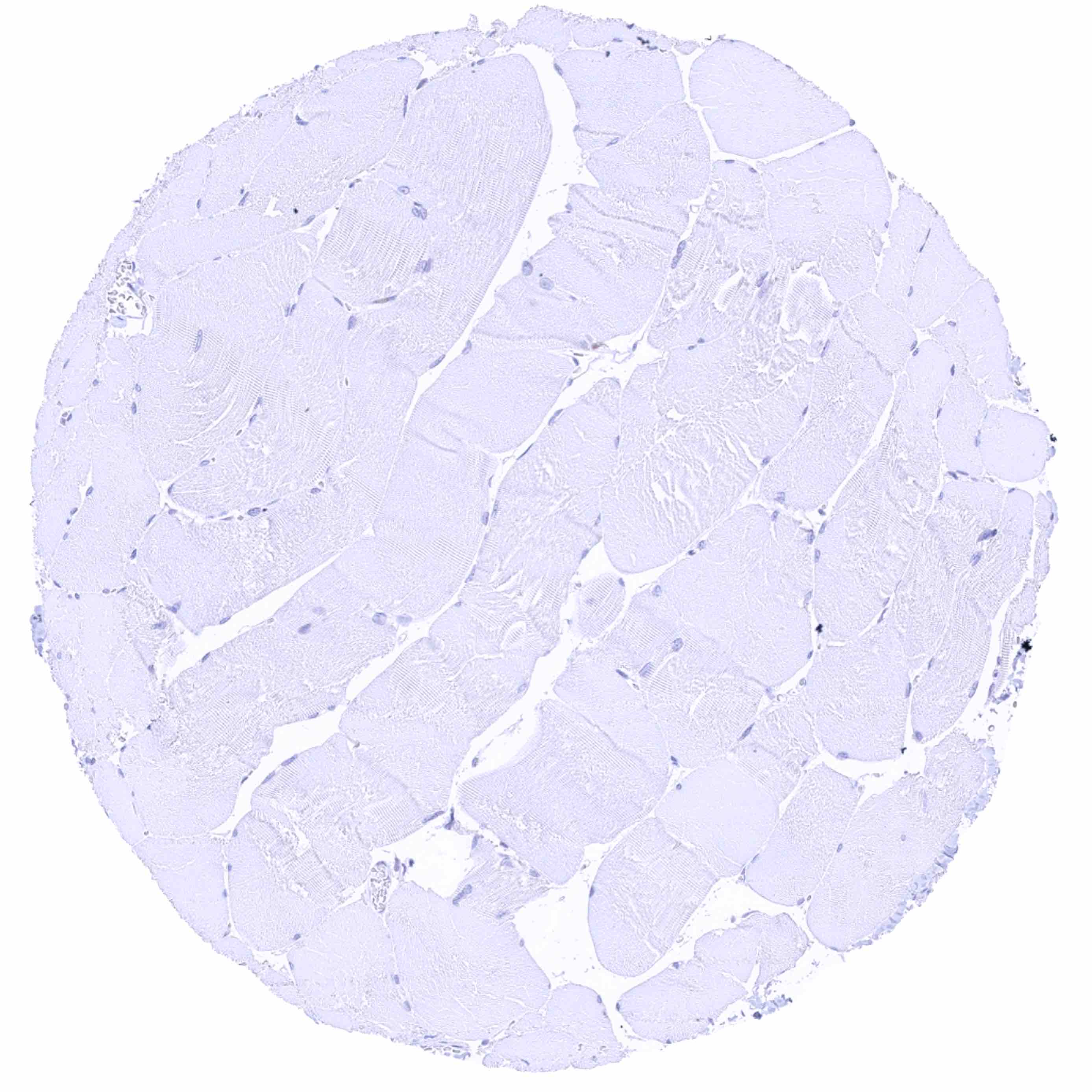
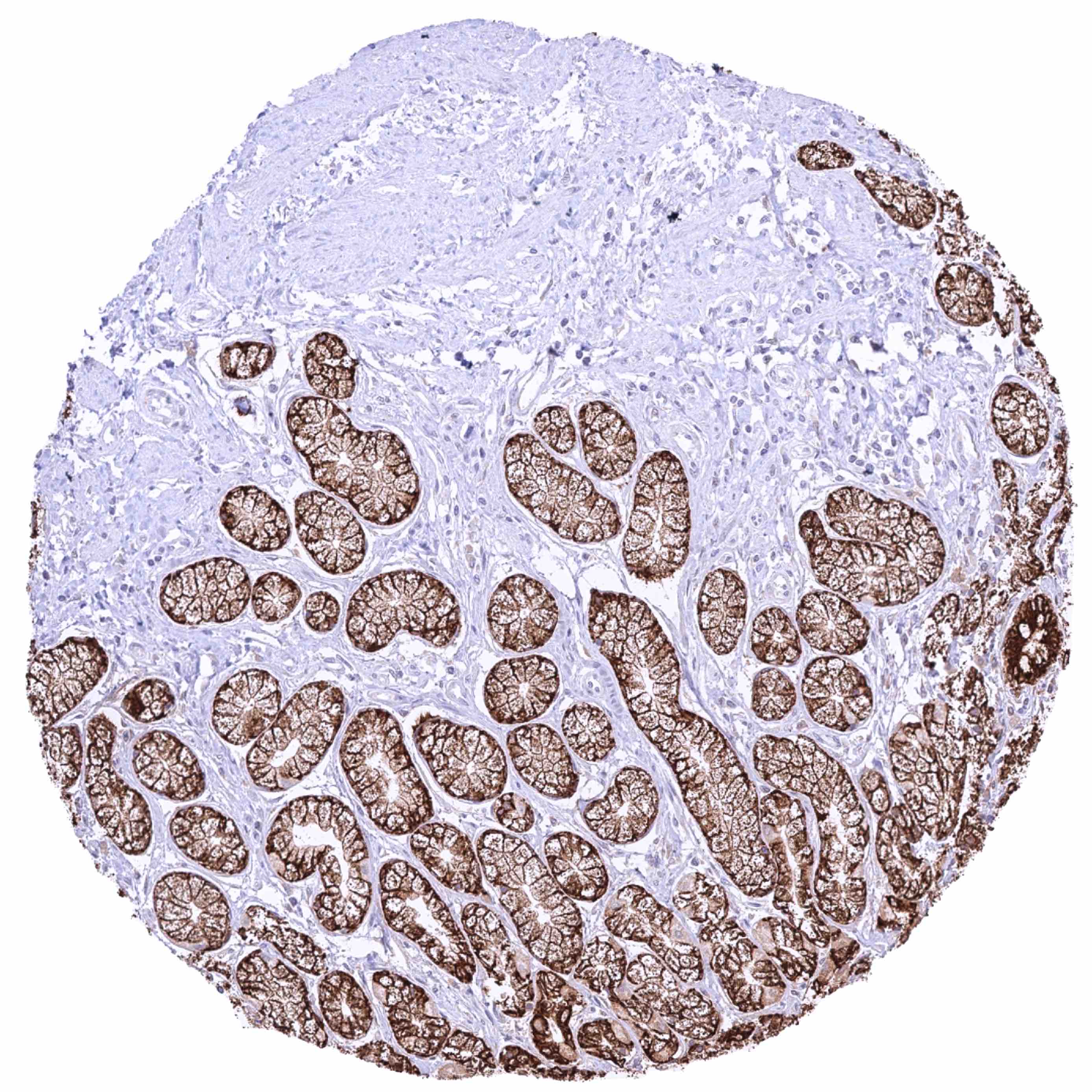
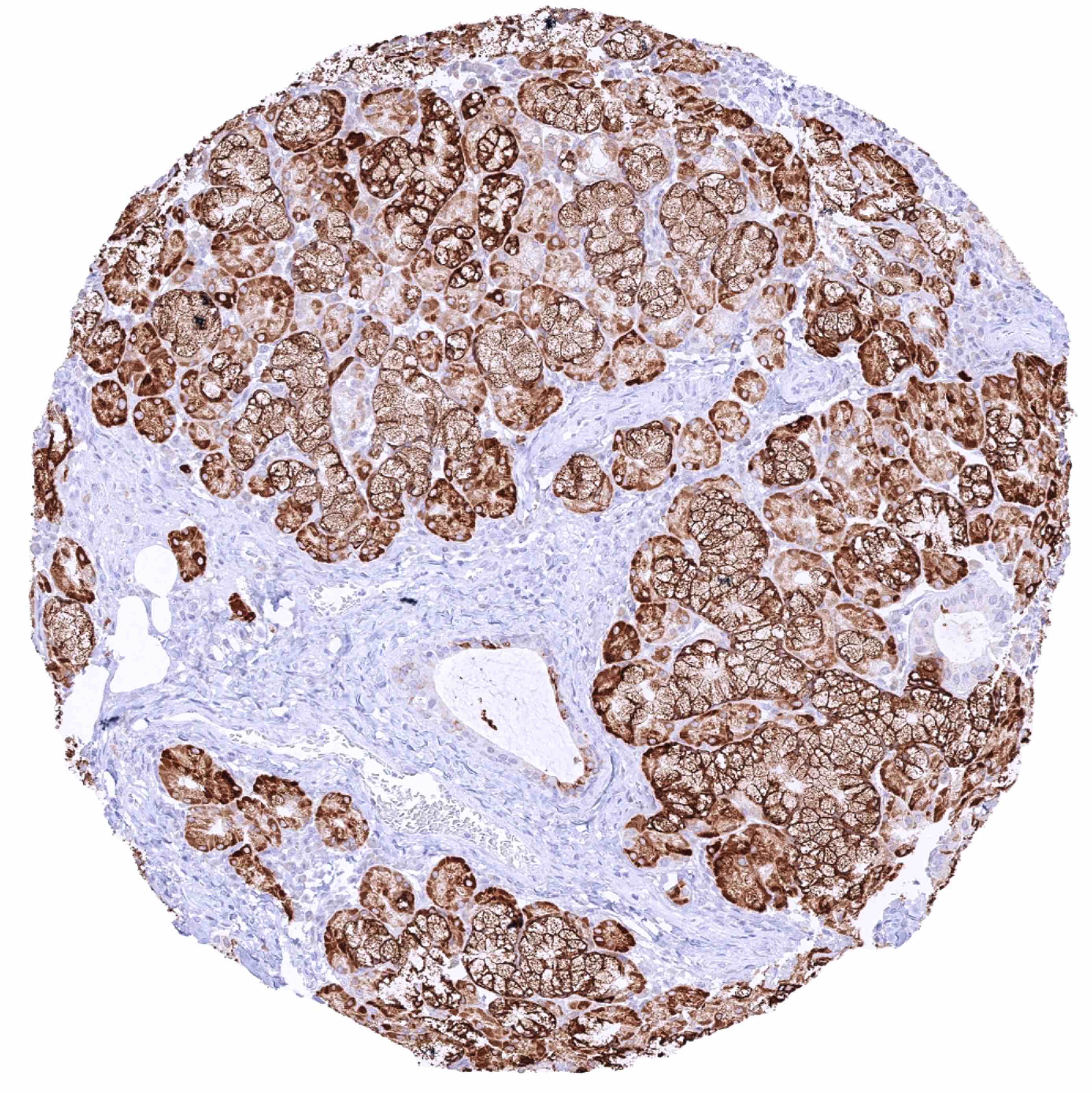
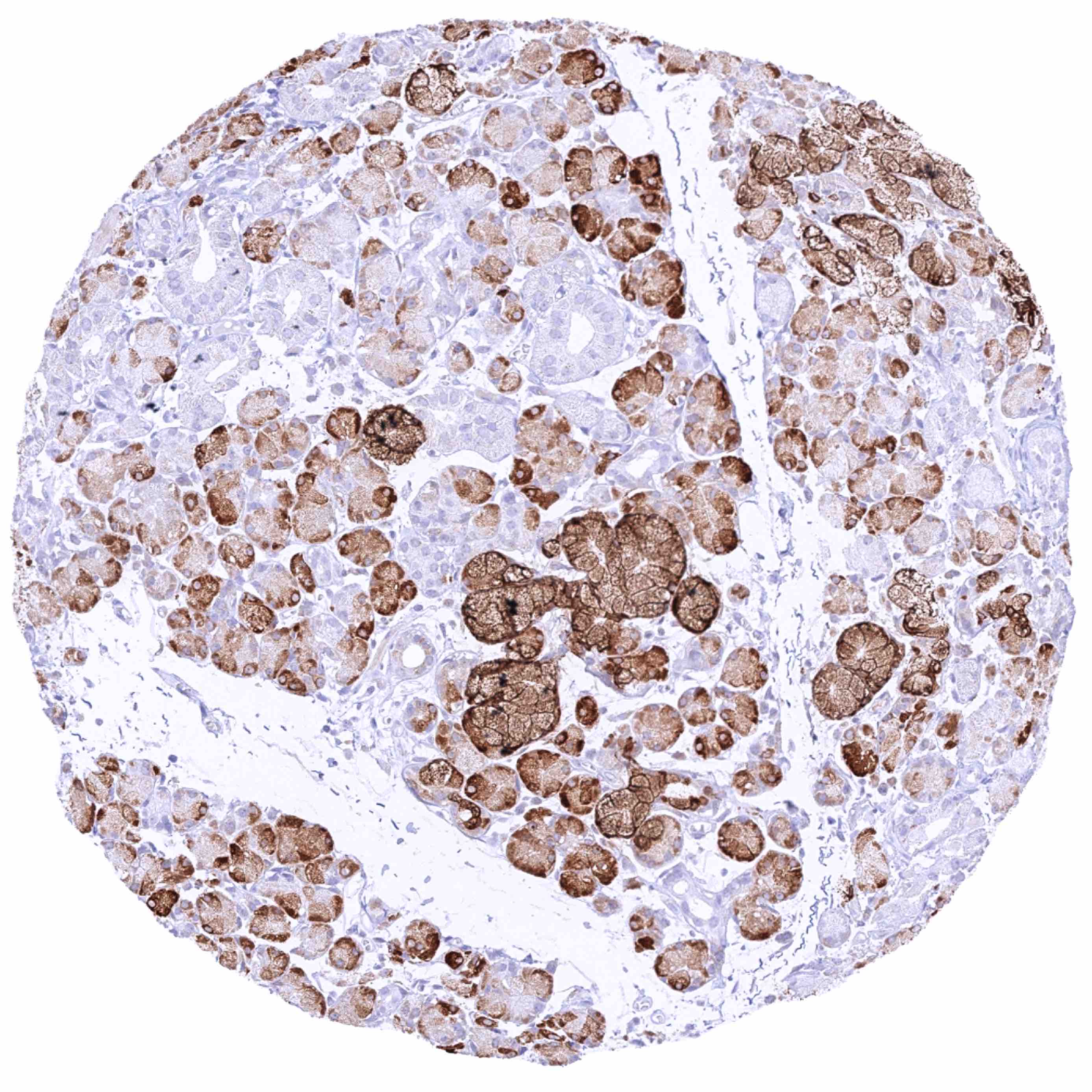
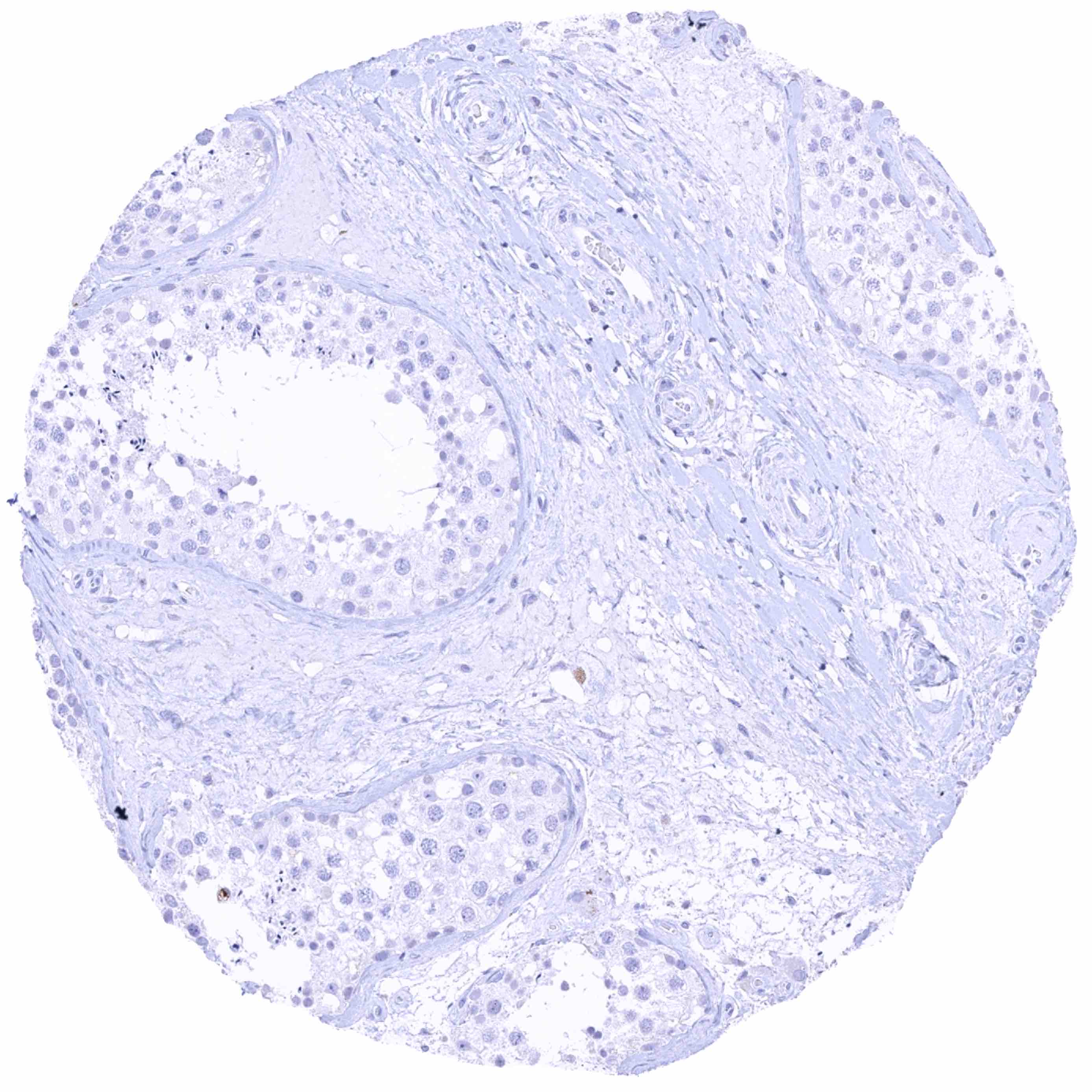
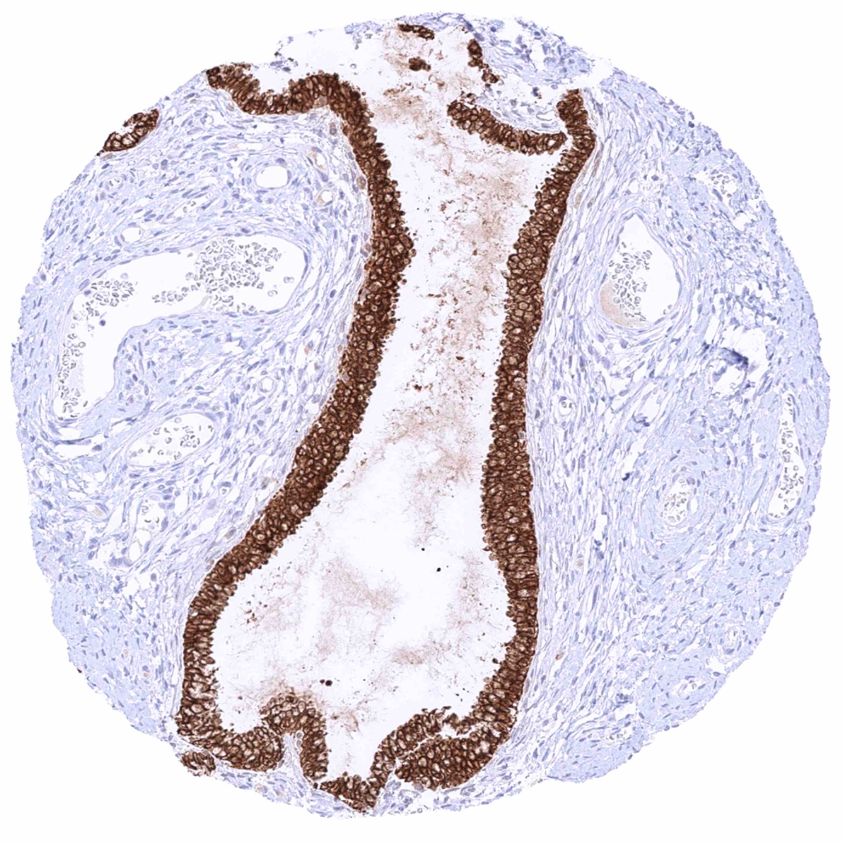
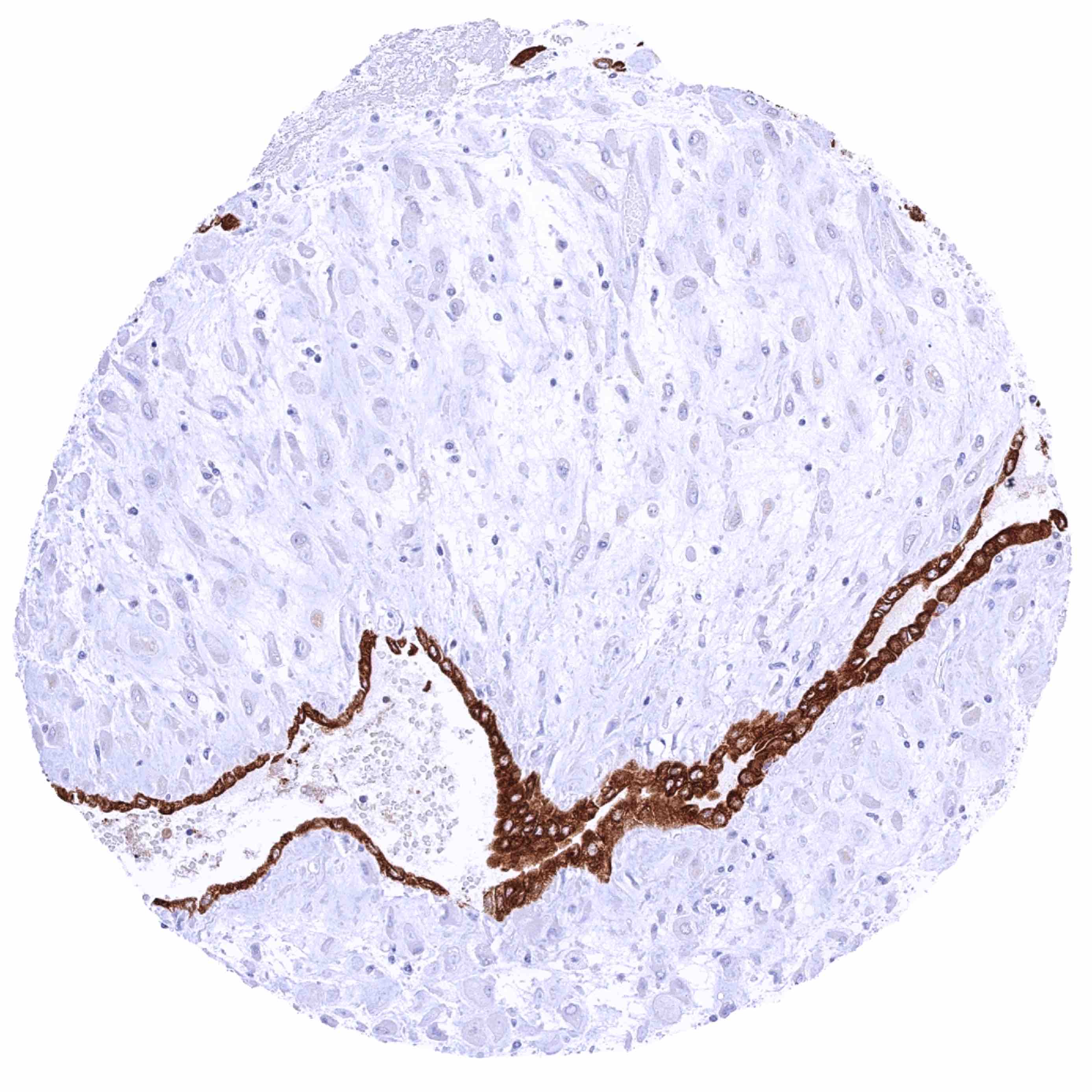
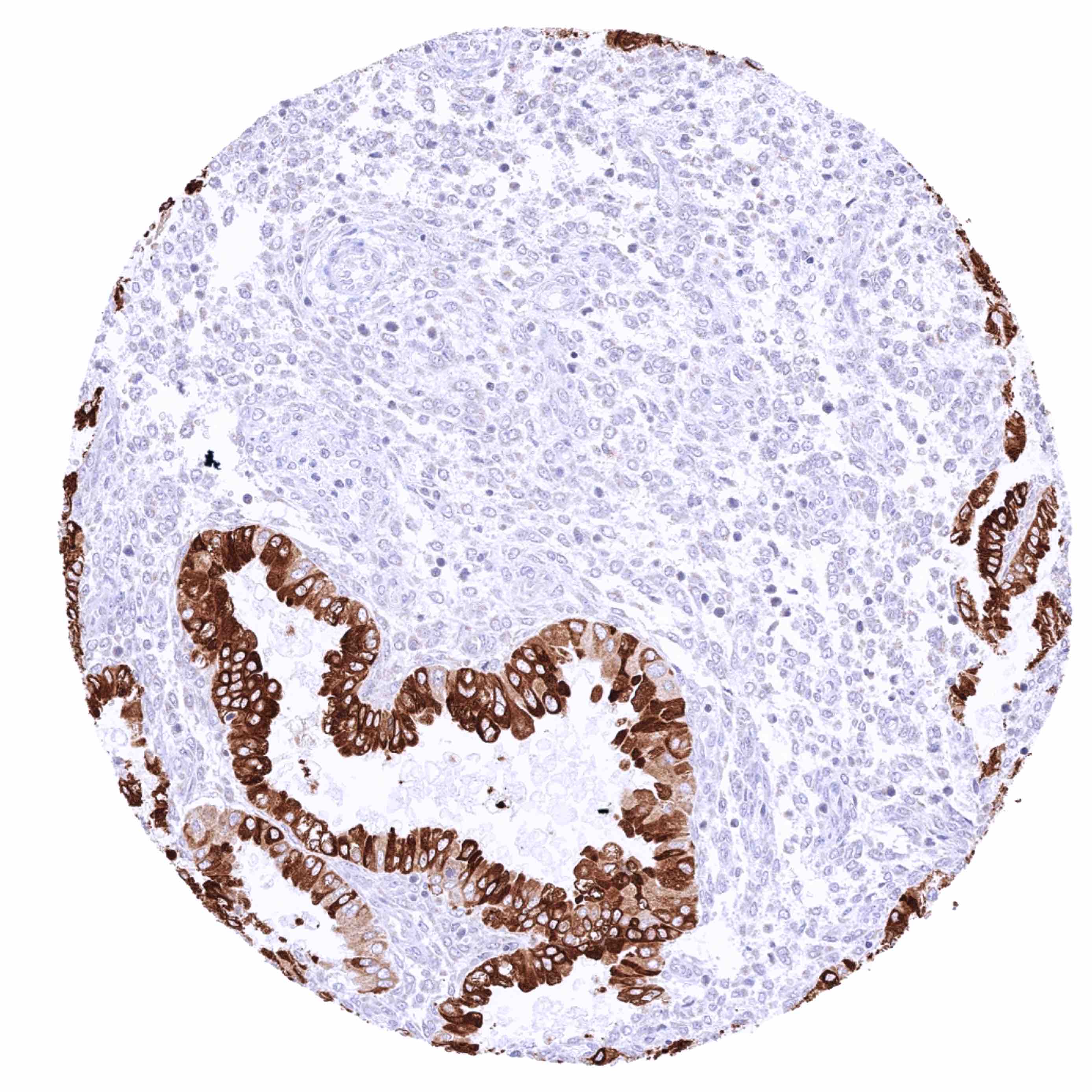
Anterior Gradient 2 antibody [HMV325] HistoMAXTM


Go to Cancer Tissue Gallery
  
 Go back to product page
  

Go to HistoMAX page


Adrenal gland

Aorta, endothelium

Appendix, mucosa – Strong, predominantly cytoplasmic AGR2 staining of all epithelial cells

Appendix, muscular wall


Bone marrow

Breast – Strong, predominantly cytoplasmic AGR2 staining of a subset of luminal cells

Bronchus, mucosa – Strong, predominantly cytoplasmic AGR2 staining of all epithelial cells

Cerebellum (white matter)


Cerebellum, cortex (molecular layer, Purkinje cell layer, granule cell layer)

Cerebrum, grey matter

Cerebrum, white matter

Colon descendens, mucosa – Strong, predominantly cytoplasmic AGR2 staining of all epithelial cells


Colon descendens, muscular wall

Duodenum, Brunner gland – Moderate to strong, predominantly cytoplasmic AGR2 staining of Brunner gland cells. Strong AGR2 staining of duodenal epithelial cells

Duodenum, mucosa – Strong, predominantly cytoplasmic AGR2 staining of all epithelial cells

Epididymis (Cauda) – Strong, predominantly cytoplasmic AGR2 staining of a subset of epithelial cells


Esophagus, squamous epithelium

Fallopian tube, mucosa

Fat

Gallbladder, epithelium – Strong, predominantly cytoplasmic AGR2 staining of all epithelial cells


Heart muscle

Ileum, mucosa – Strong, predominantly cytoplasmic AGR2 staining of all epithelial cells

Kidney, cortex – Moderate to strong predominantly cytoplasmic AGR2 staining of a small subset of tubuli-collecting ducts

Kidney, medulla – Lack of AGR2 staining in this sample


Kidney, medulla – Moderate to strong predominantly cytoplasmic AGR2 staining of a small subset of tubuli-collecting ducts

Kidney, pelvis, urothelium – Strong, predominantly cytoplasmic AGR2 staining of all epithelial cells

Liver

Lung – Strong, predominantly cytoplasmic AGR2 staining of a large subset of pneumocytes


Lymph node

Ovary, corpus luteum

Ovary, stroma

Pancreas – In this sample, a strong, predominantly cytoplasmic AGR2 staining is seen in numerous individual or groups of acinar cells


Pancreas – Moderate to strong, predominantly cytoplasmic AGR2staining of intercalated duct cells. Islet cells are AGR2 negative

Parathyroid gland

Parotid gland

Pituitary gland, anterior lobe – Predominantly cytoplasmic AGR2 staining of variable intensity in a large subset of epithelial cells


Pituitary gland, anterior lobe – Predominantly cytoplasmic AGR2 staining of variable intensity in a subset of epithelial cells

Pituitary gland, posterior lobe

Placenta (amnion and chorion)

Placenta, early


Placenta, mature

Prostate – Complete absence of AGR2 staining in this sample

Prostate – Strong, predominantly cytoplasmic AGR2 staining of all acinar and basal cells in this sample

Prostate – Strong, predominantly cytoplasmic AGR2 staining of few acinar and basal cells


Rectum, mucosa – Strong, predominantly cytoplasmic AGR2 staining of all epithelial cells

Seminal vesicle

Sinus paranasales – Strong, predominantly cytoplasmic AGR2 staining of all epithelial cells

Skeletal muscle


Skin, hairfollicel and sebaceous glands – Strong cytoplasmic AGR2 staining of the inner (Huxley) layers of a hair follicle

Skin

Spleen

Stomach, antrum – Strong, predominantly cytoplasmic AGR2 staining of all epithelial cells


Stomach, corpus – Strong, predominantly cytoplasmic AGR2 staining of all epithelial cells except parietal cells which only show a faint staining

Sublingual gland – Strong, predominantly cytoplasmic AGR2 staining of subset of mucinous glandular cells. Weak to moderate AGR2 staining of a subset of excretory duct cells

Submandibular gland – Moderate to strong, predominantly cytoplasmic AGR2 staining of subset of glandular cells (especially mucinous)

Testis


Thymus – Strong cytoplasmic AGR2 staining of a fraction of (squamous) epithelial cells associated with a corpuscle of Hassall's

Thyroid gland

Tonsil – Strong cytoplasmic AGR2 staining of a subset of squamous epithelial cells of tonsil crypts

Tonsil, surface epithelium


Urinary bladder, muscular wall

Urinary bladder, urothelium – Strong, predominantly cytoplasmic AGR2 staining of all epithelial cells. Staining intensity is lowest in umbrella cells

Uterus, ectocervix

Uterus, endocervix – Strong, predominantly cytoplasmic AGR2 staining of all epithelial cells


Uterus, endometrium (pregnancy)

Uterus, endometrium (proliferation) – Strong, predominantly cytoplasmic AGR2 staining of all epithelial cells

Uterus, endometrium (secretion) – Strong, predominantly cytoplasmic AGR2 staining of virtually all epithelial cells

Uterus, myometrium


| TOP |