Normal Tissue Gallery- GTX639928

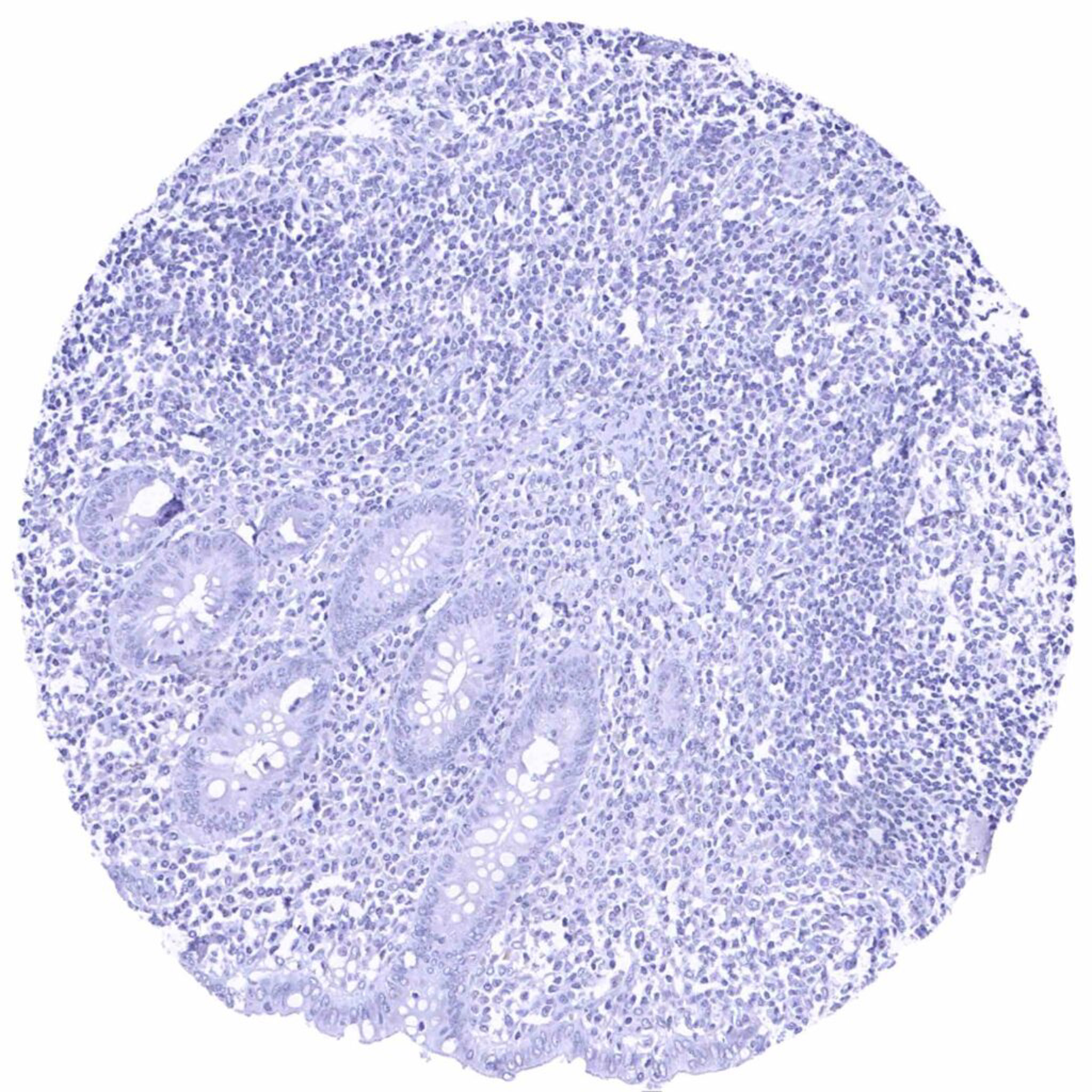
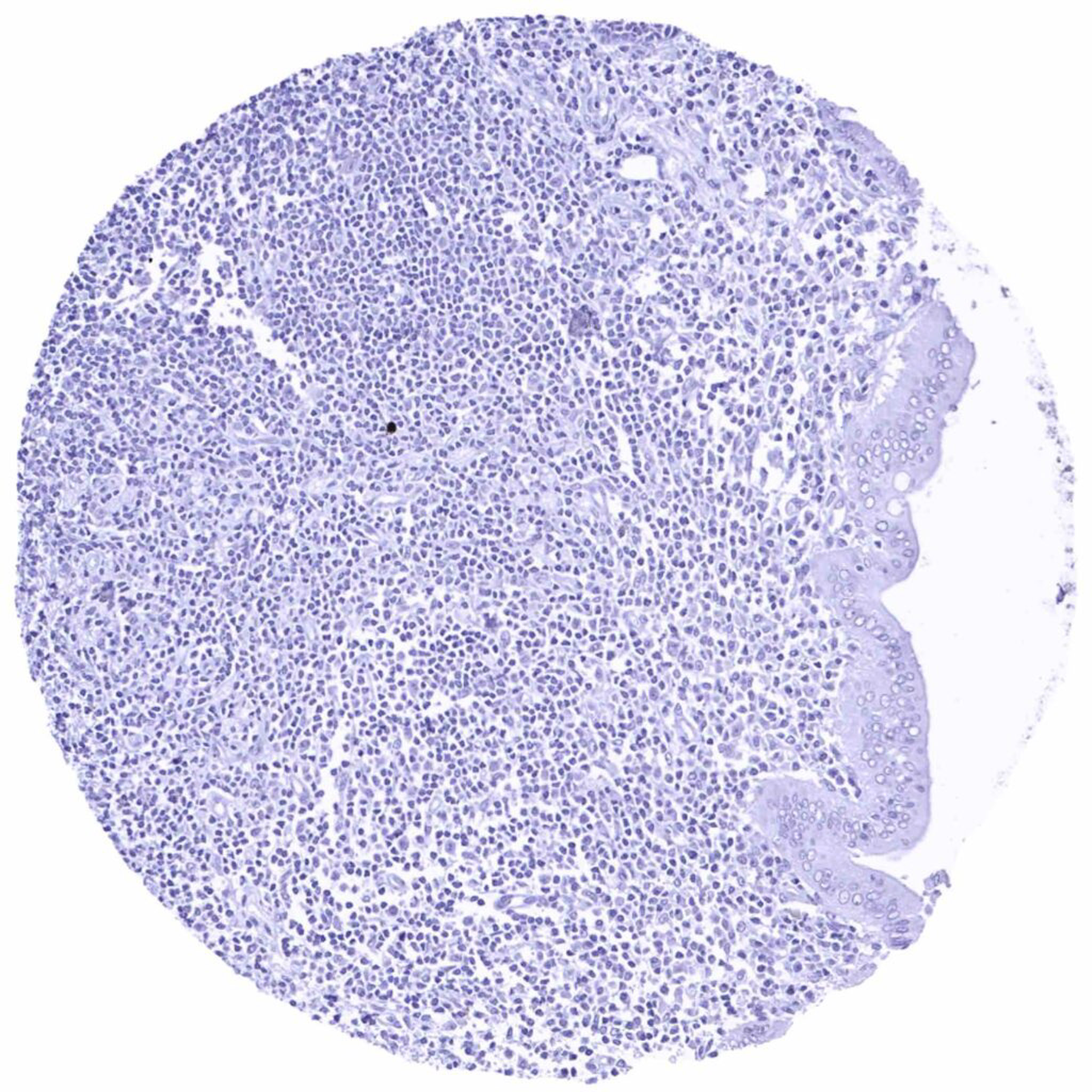
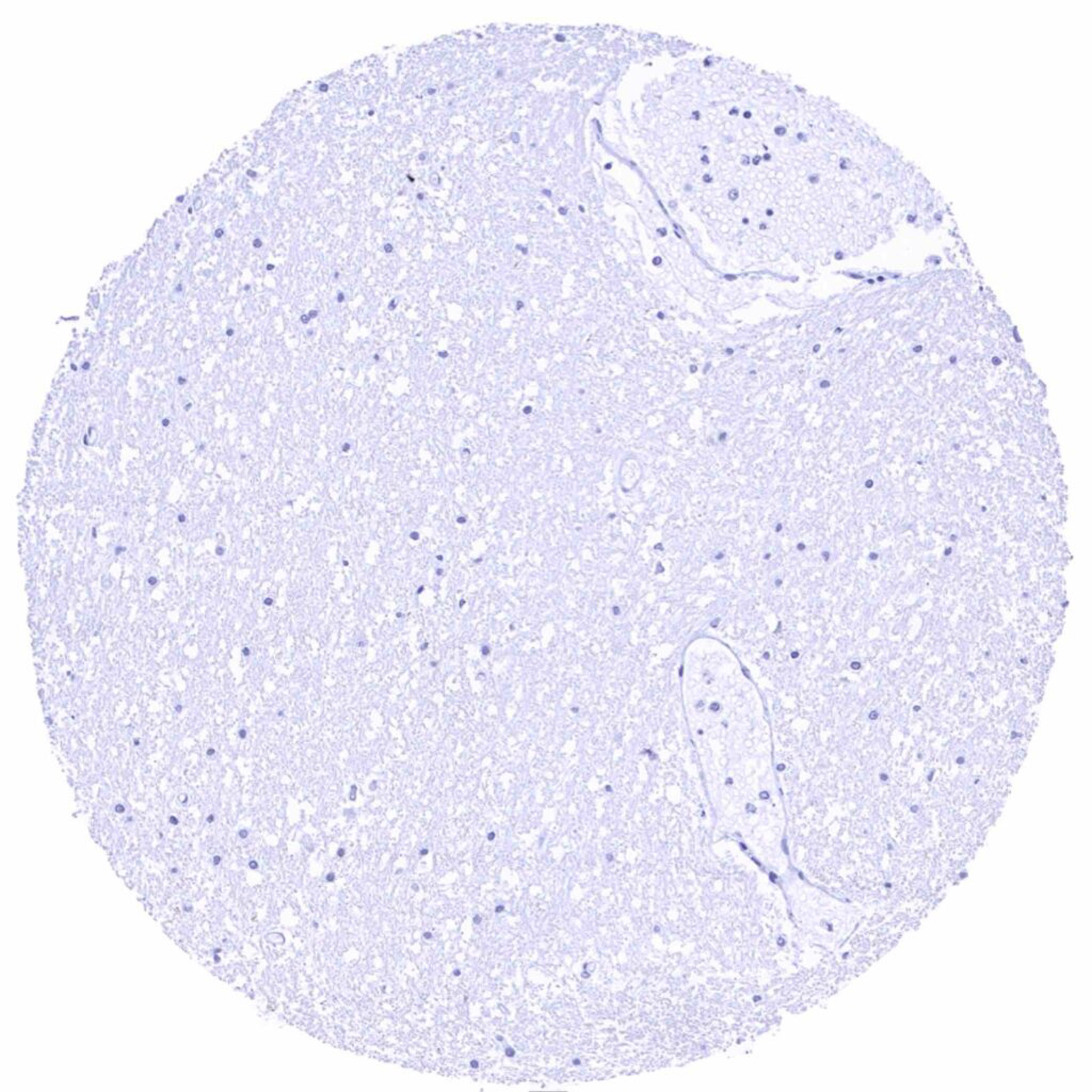
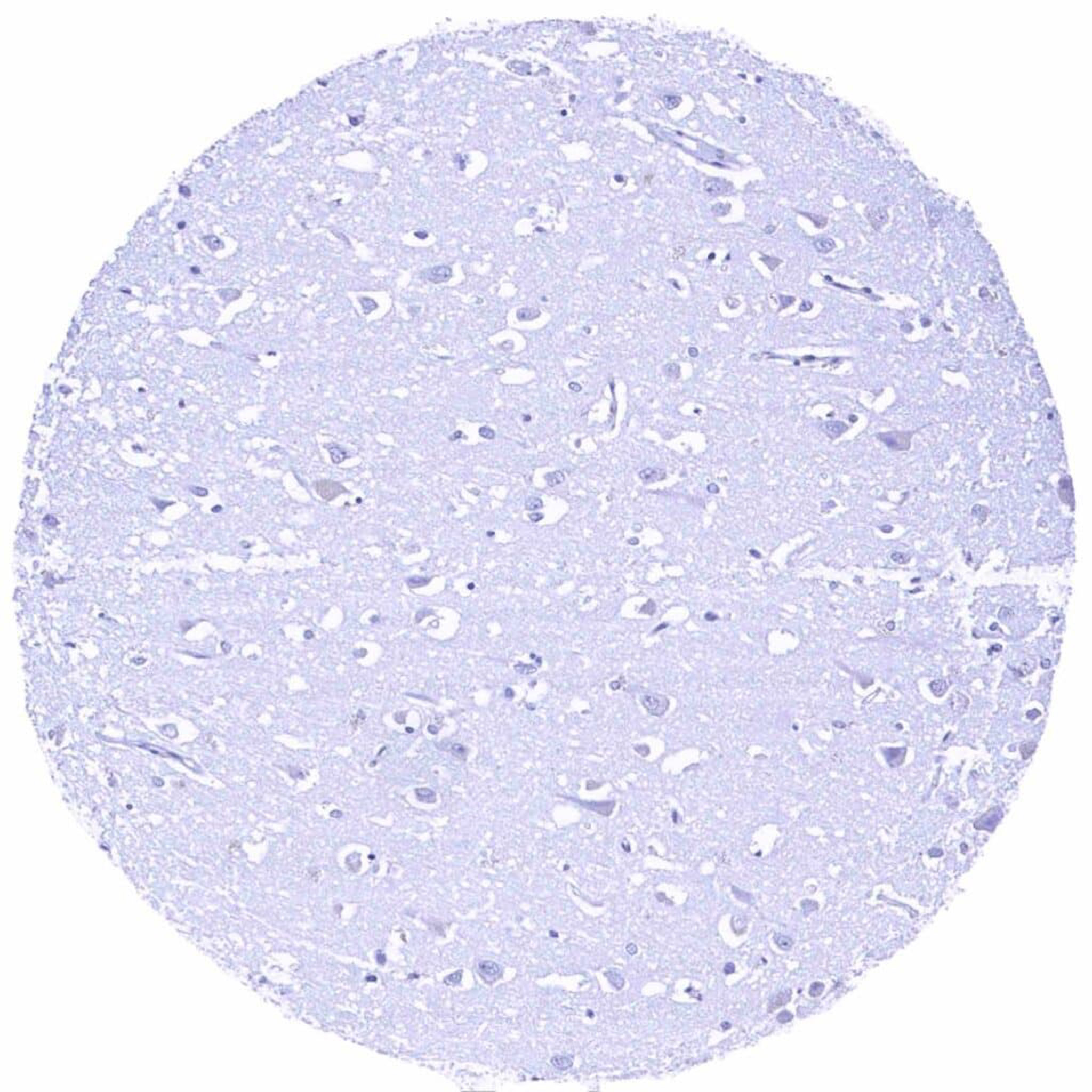
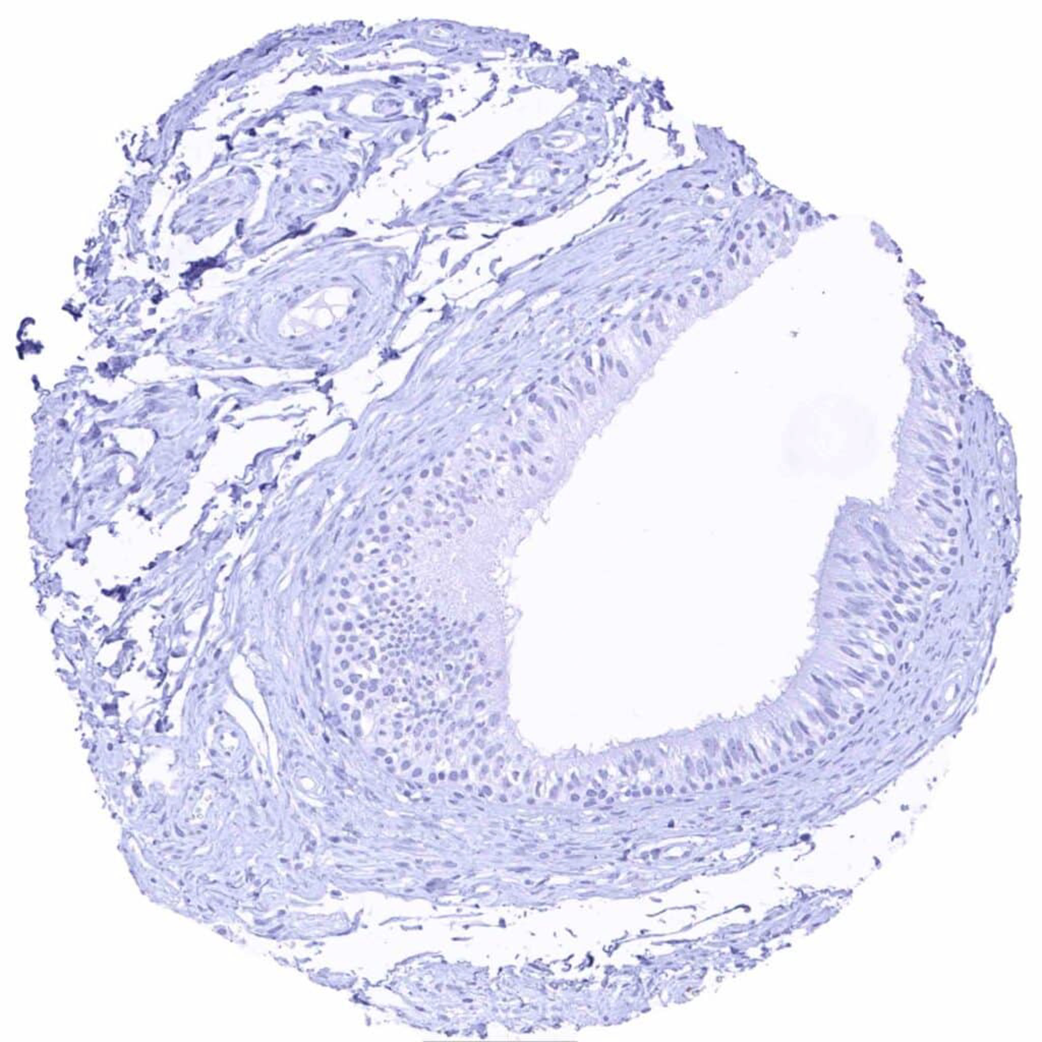
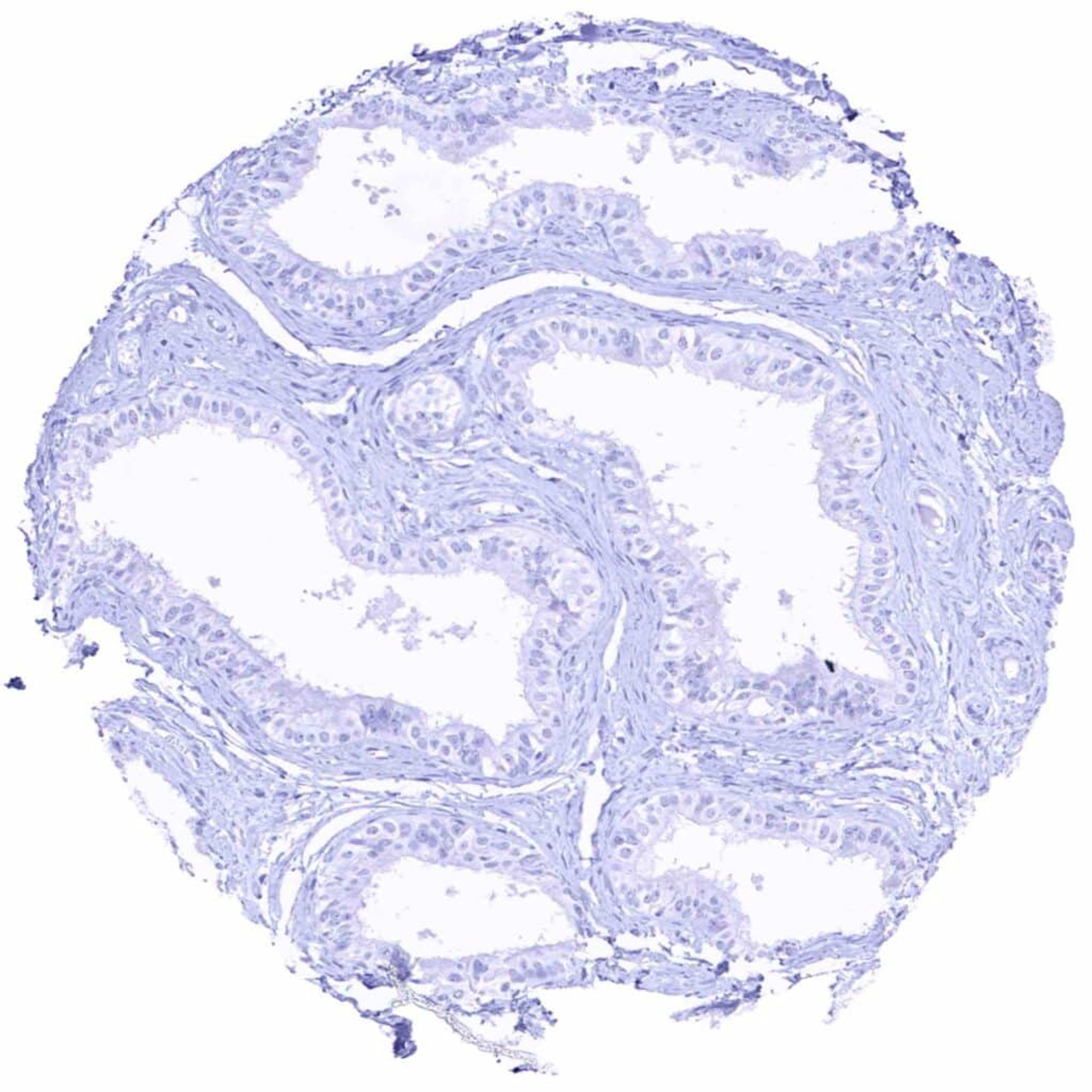
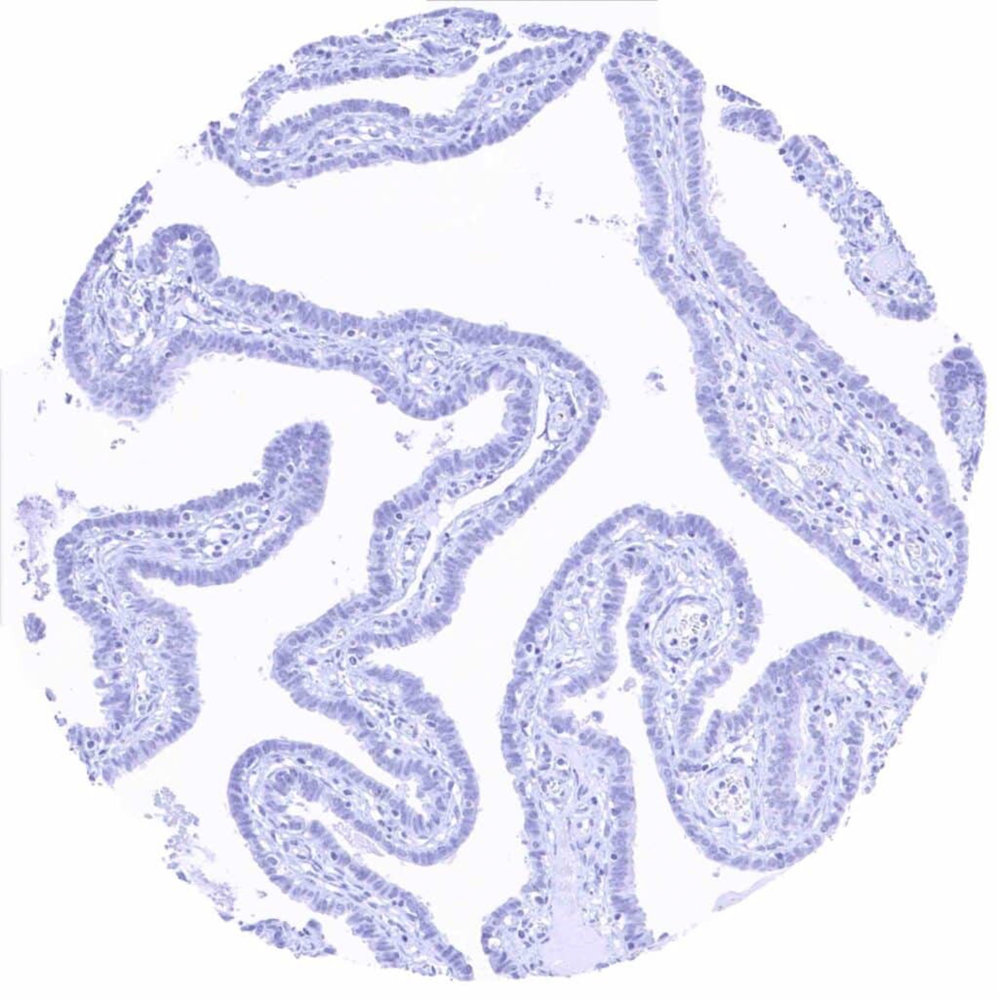
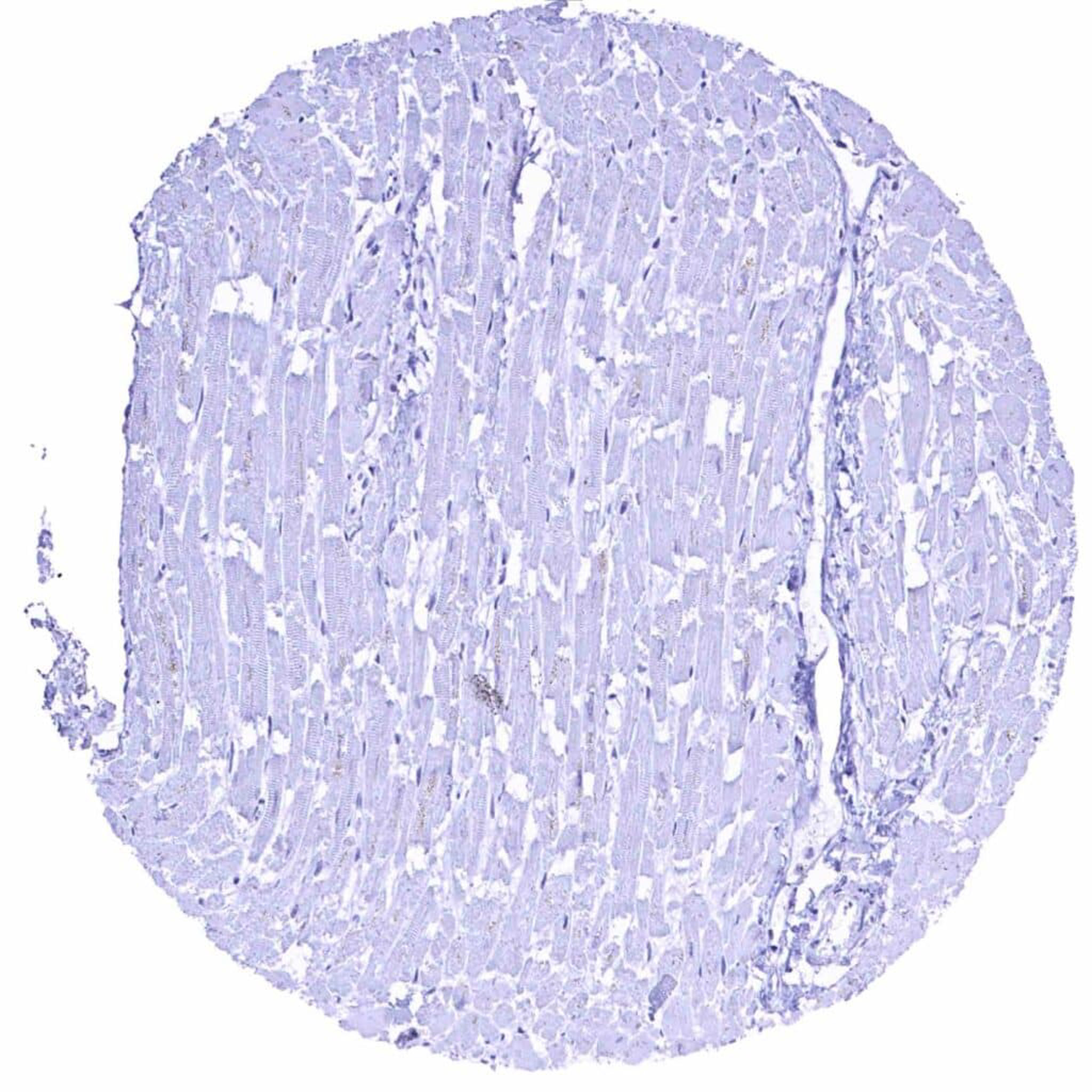
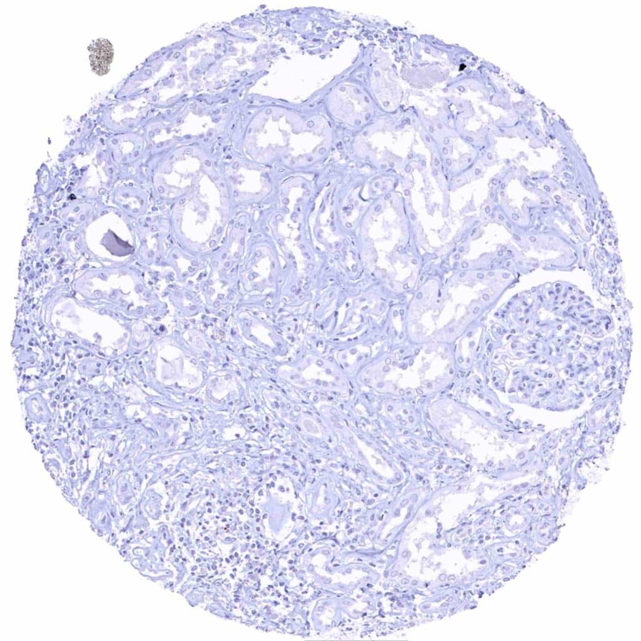
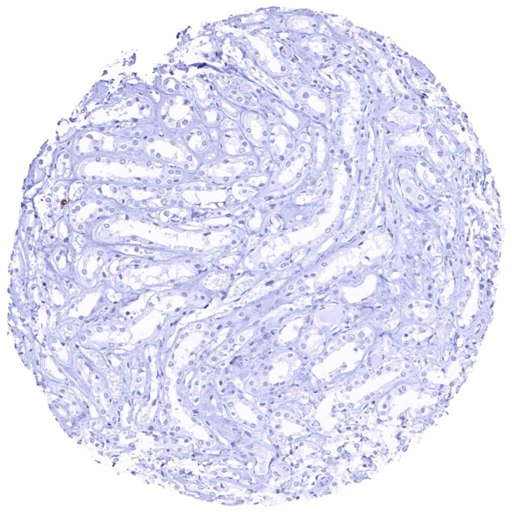
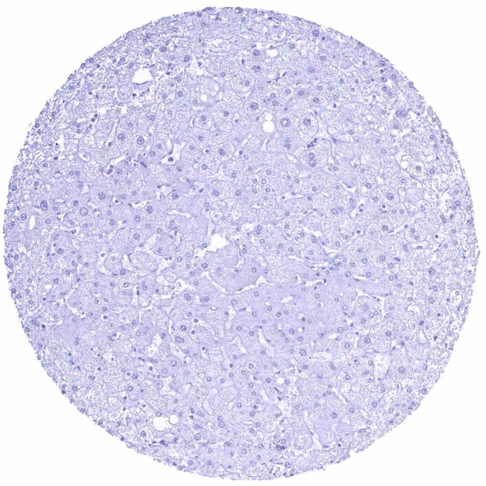
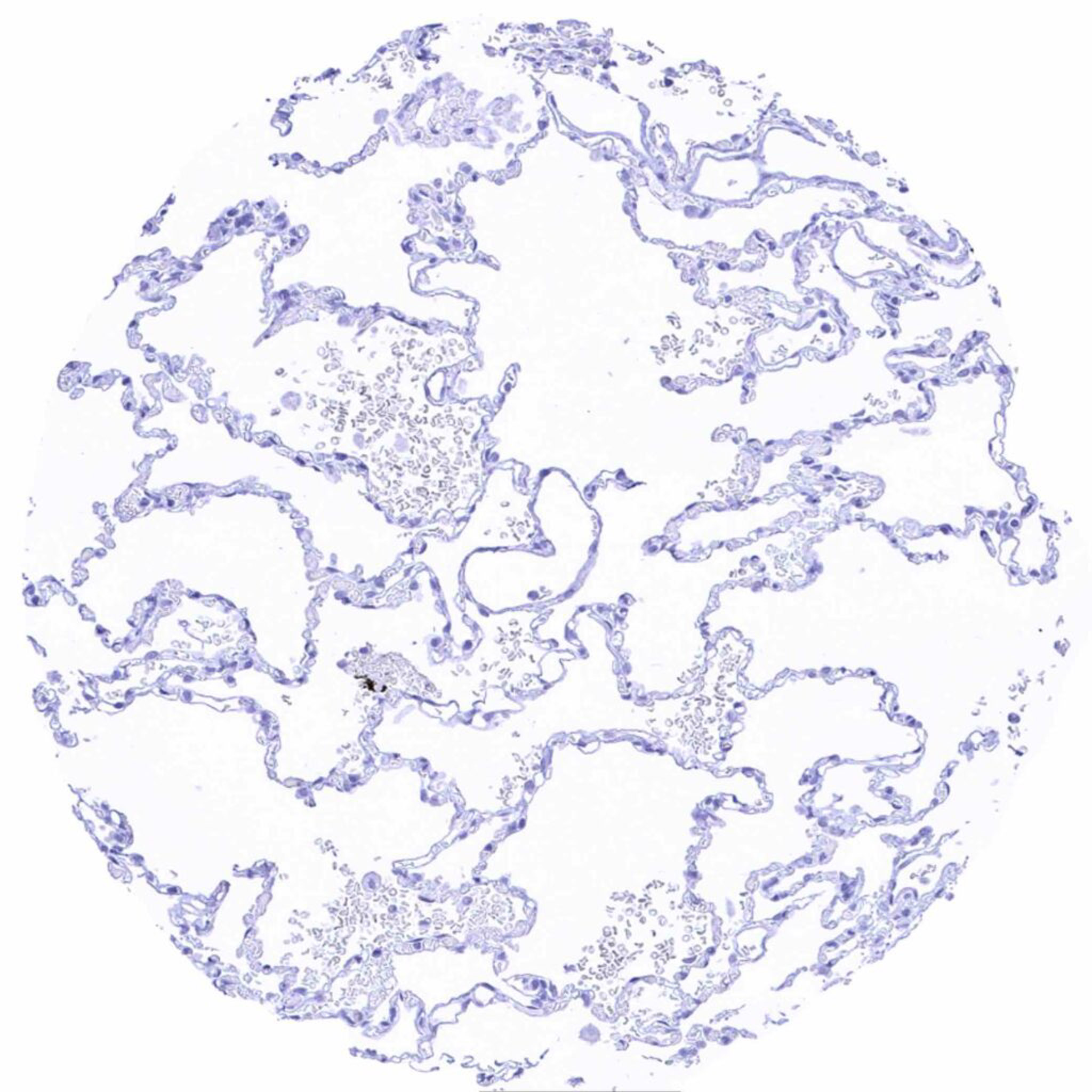
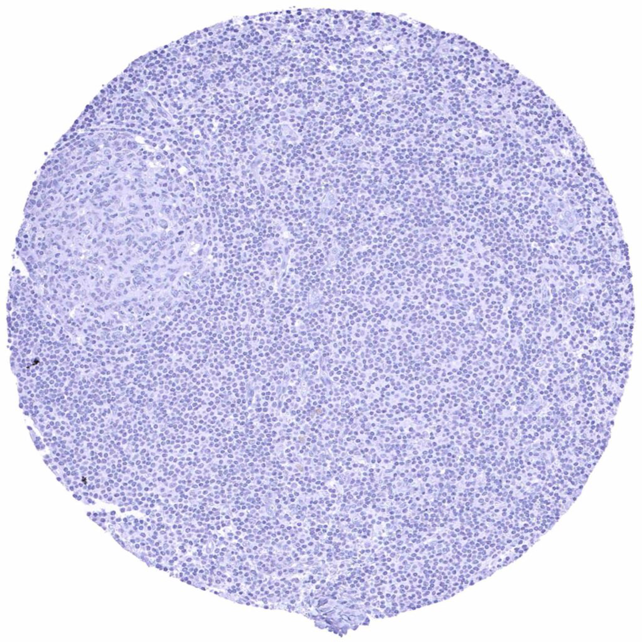
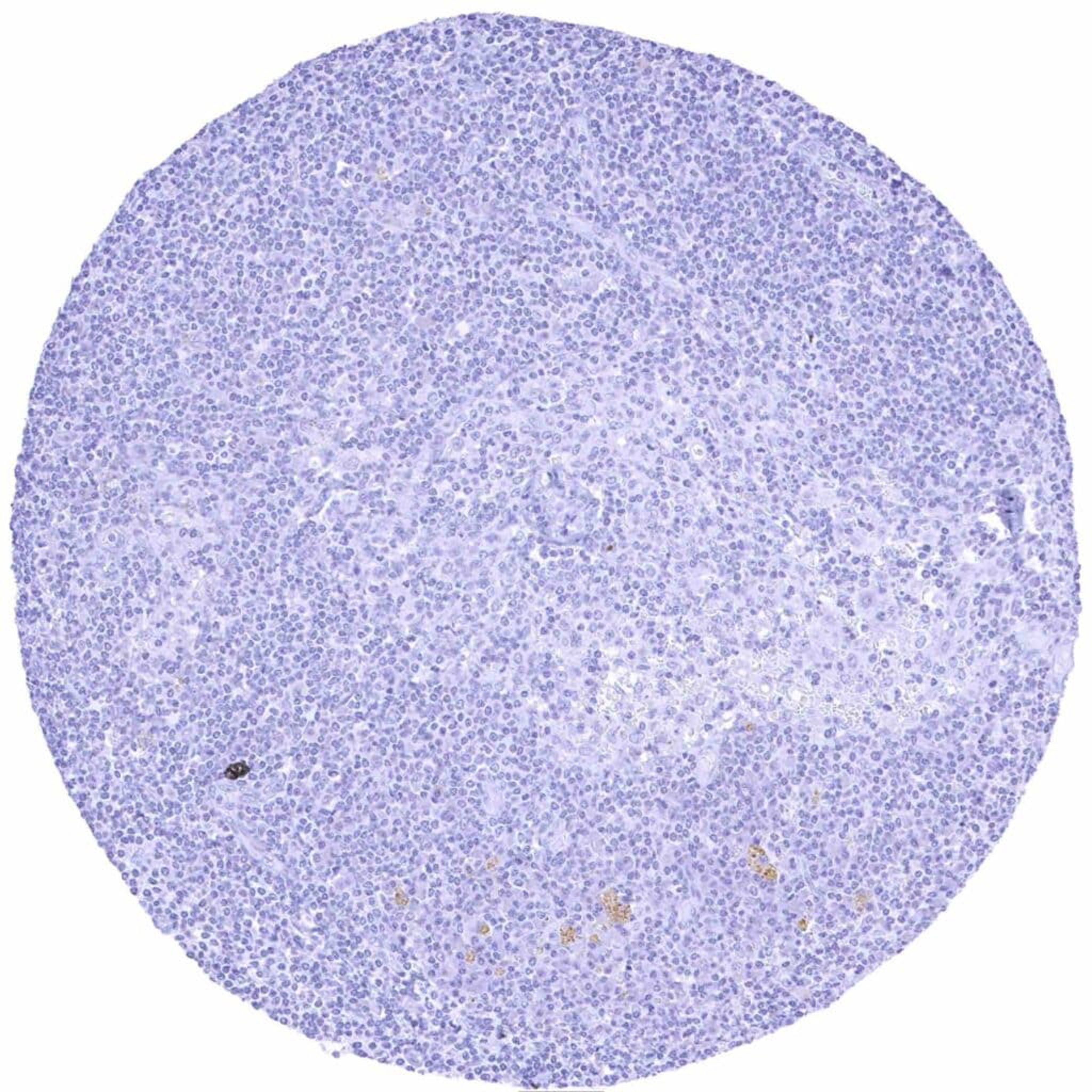
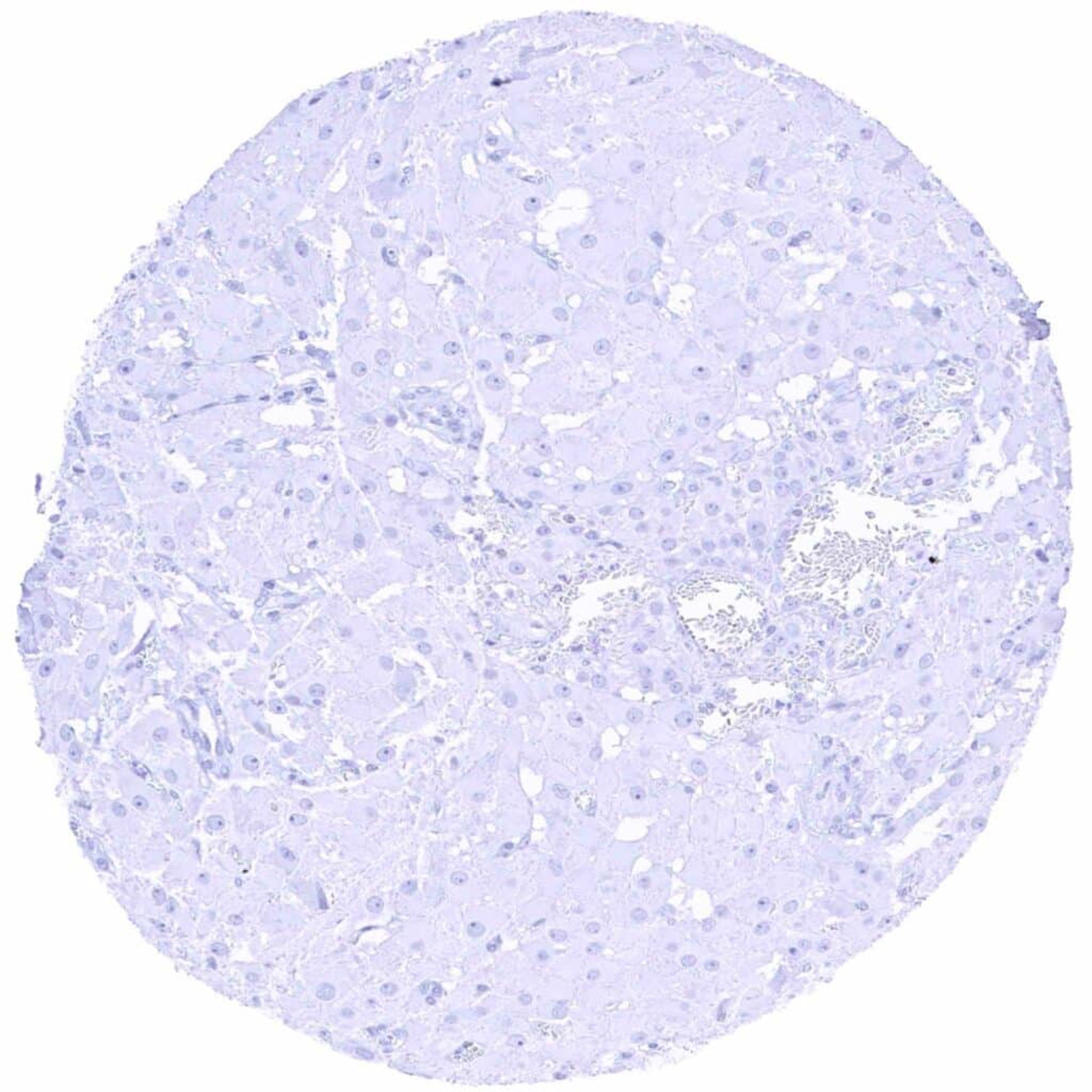
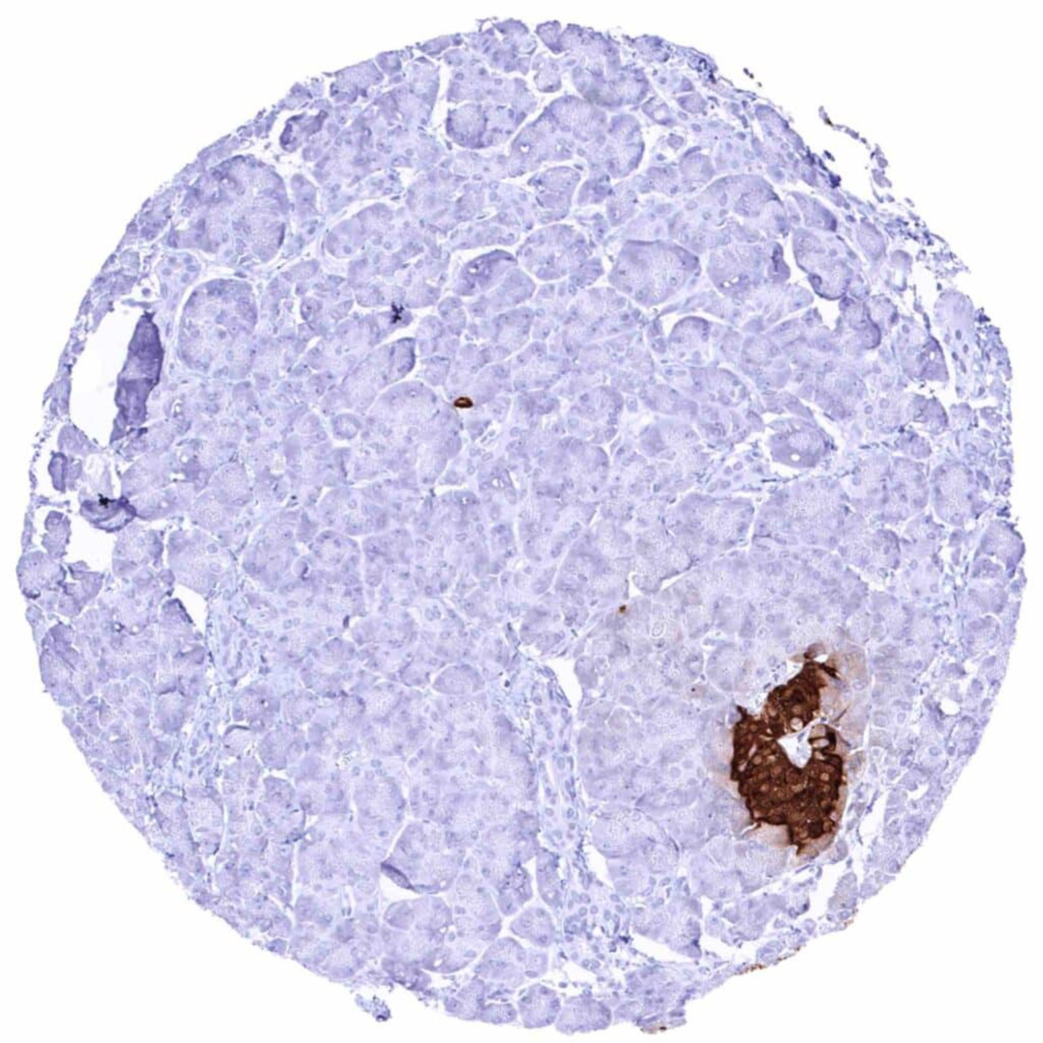
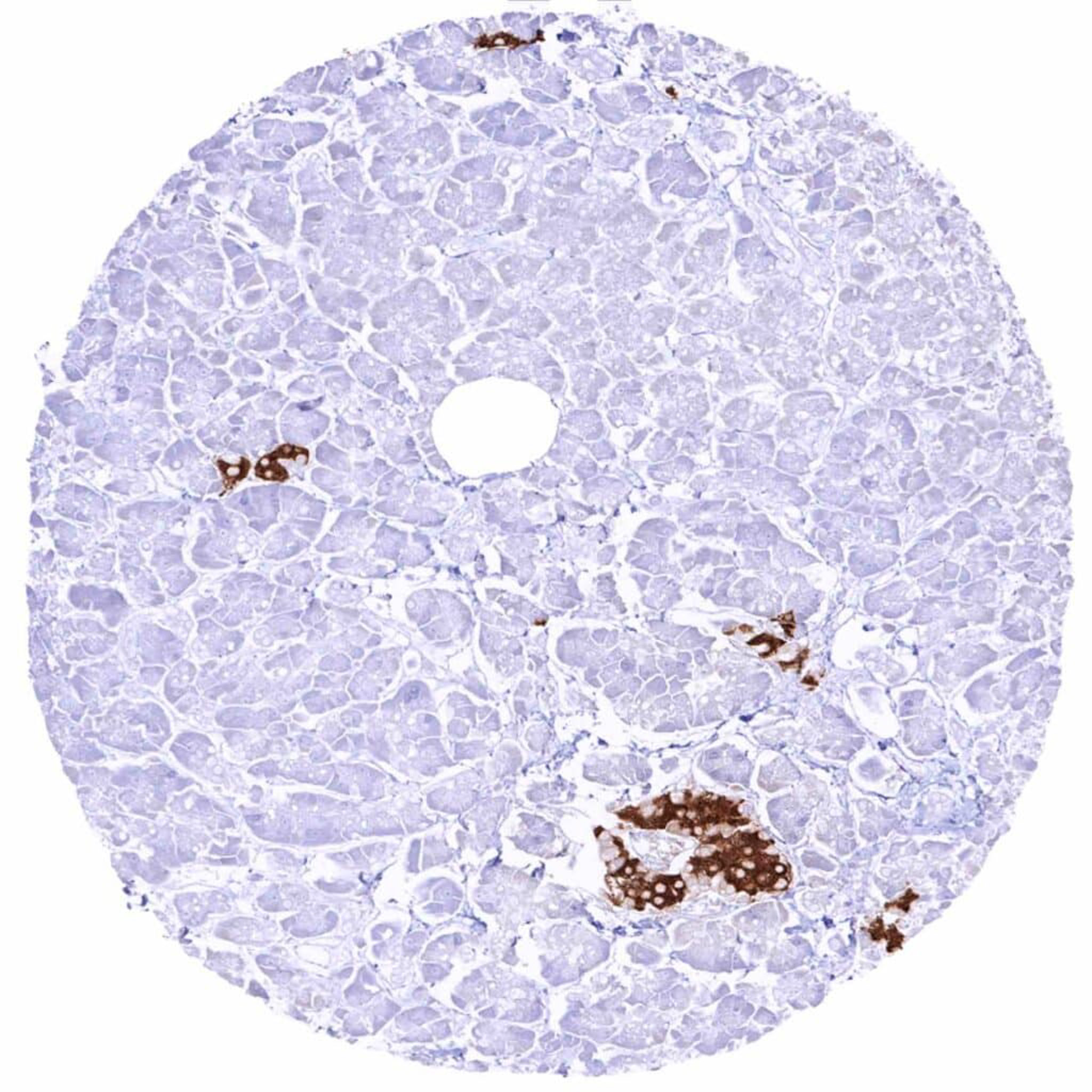
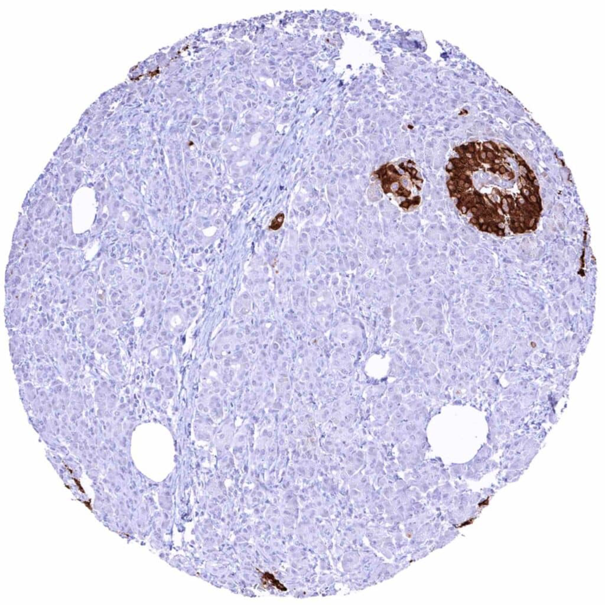
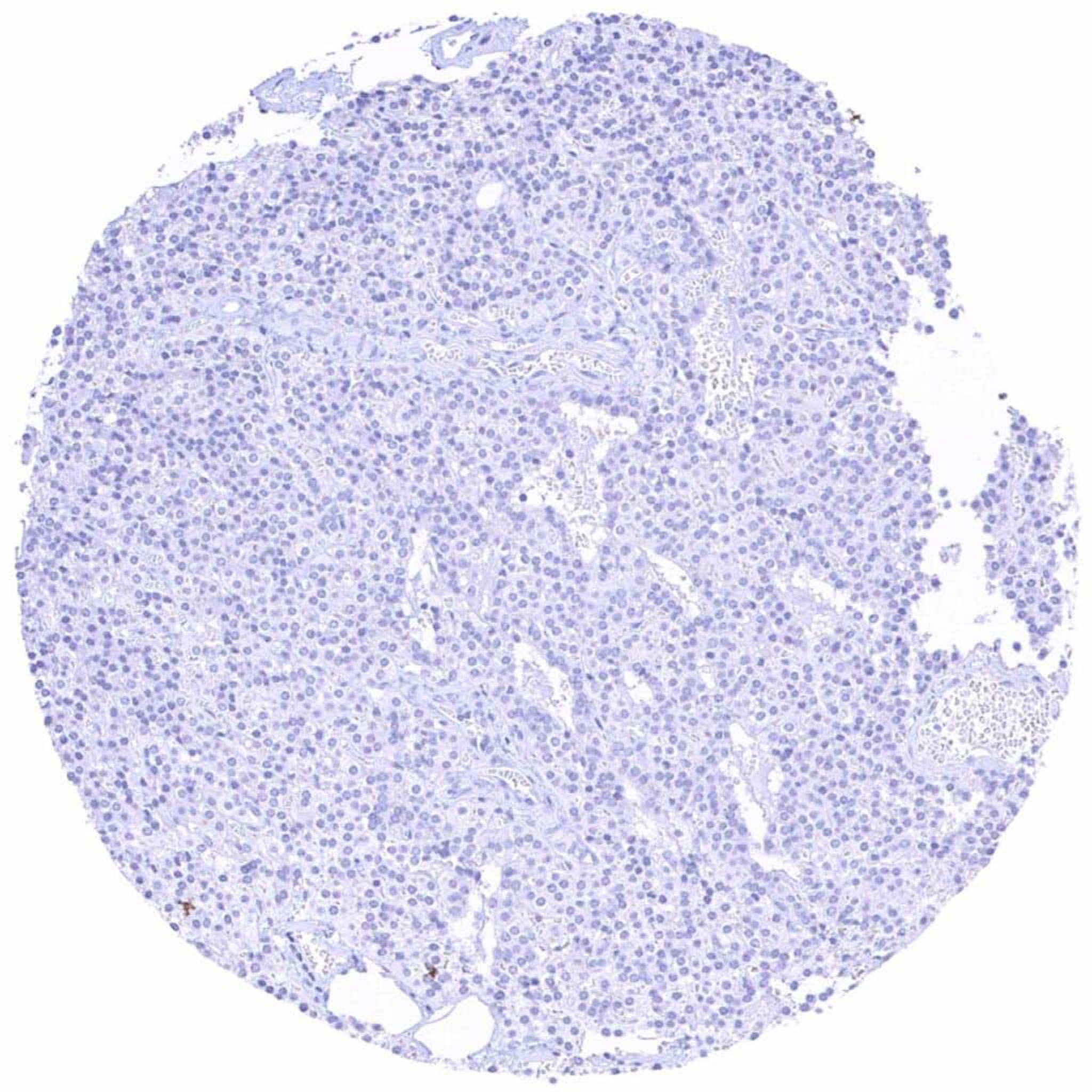
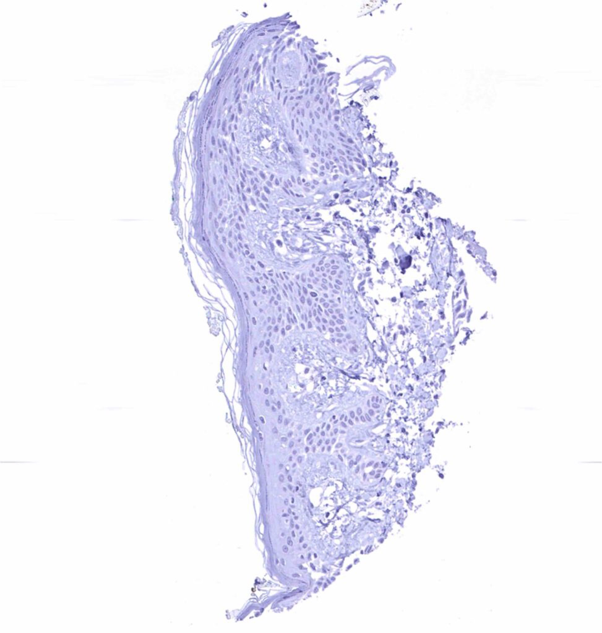
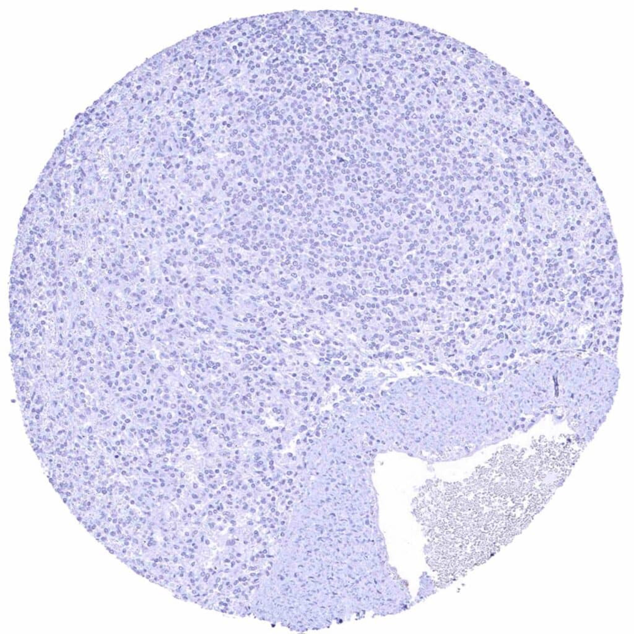
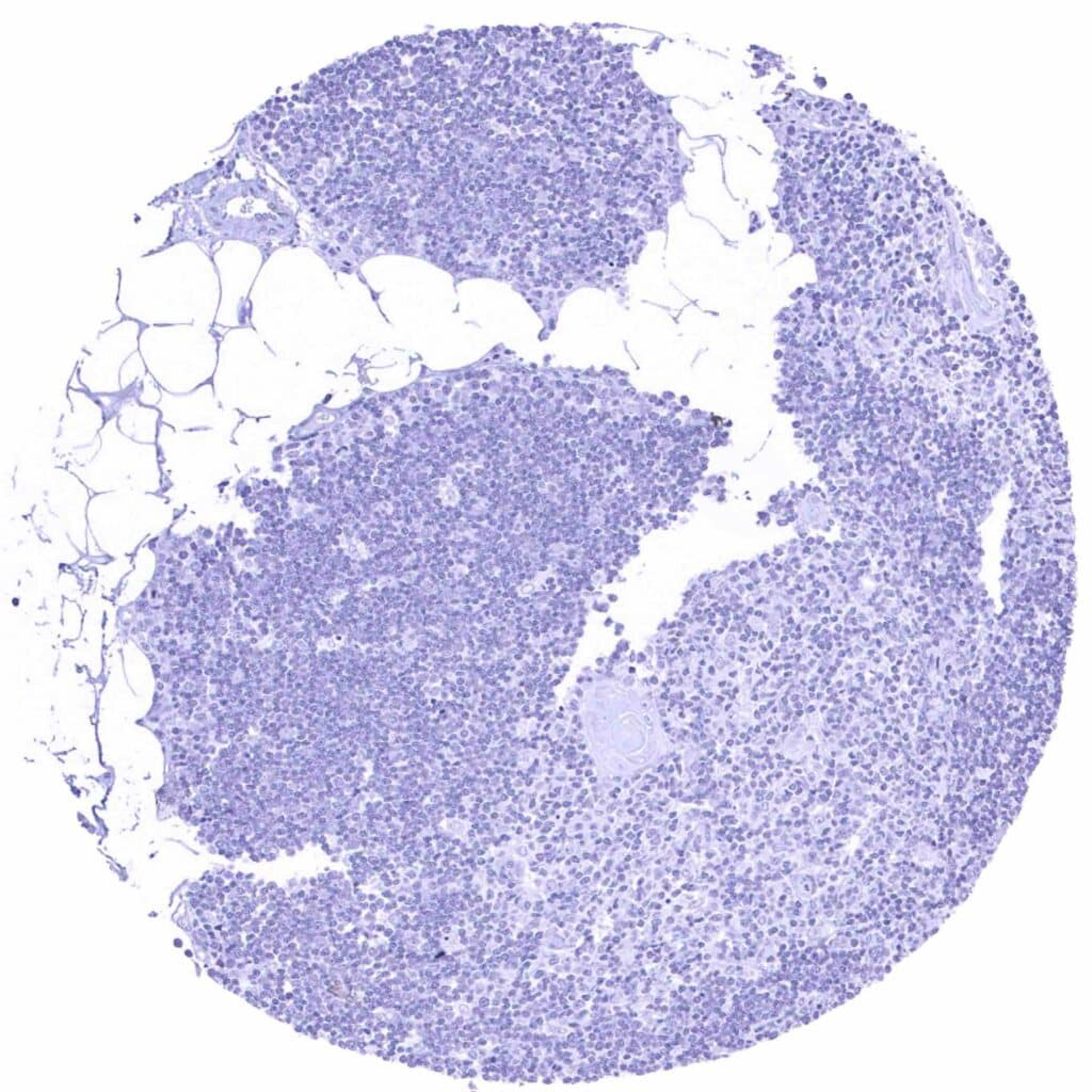
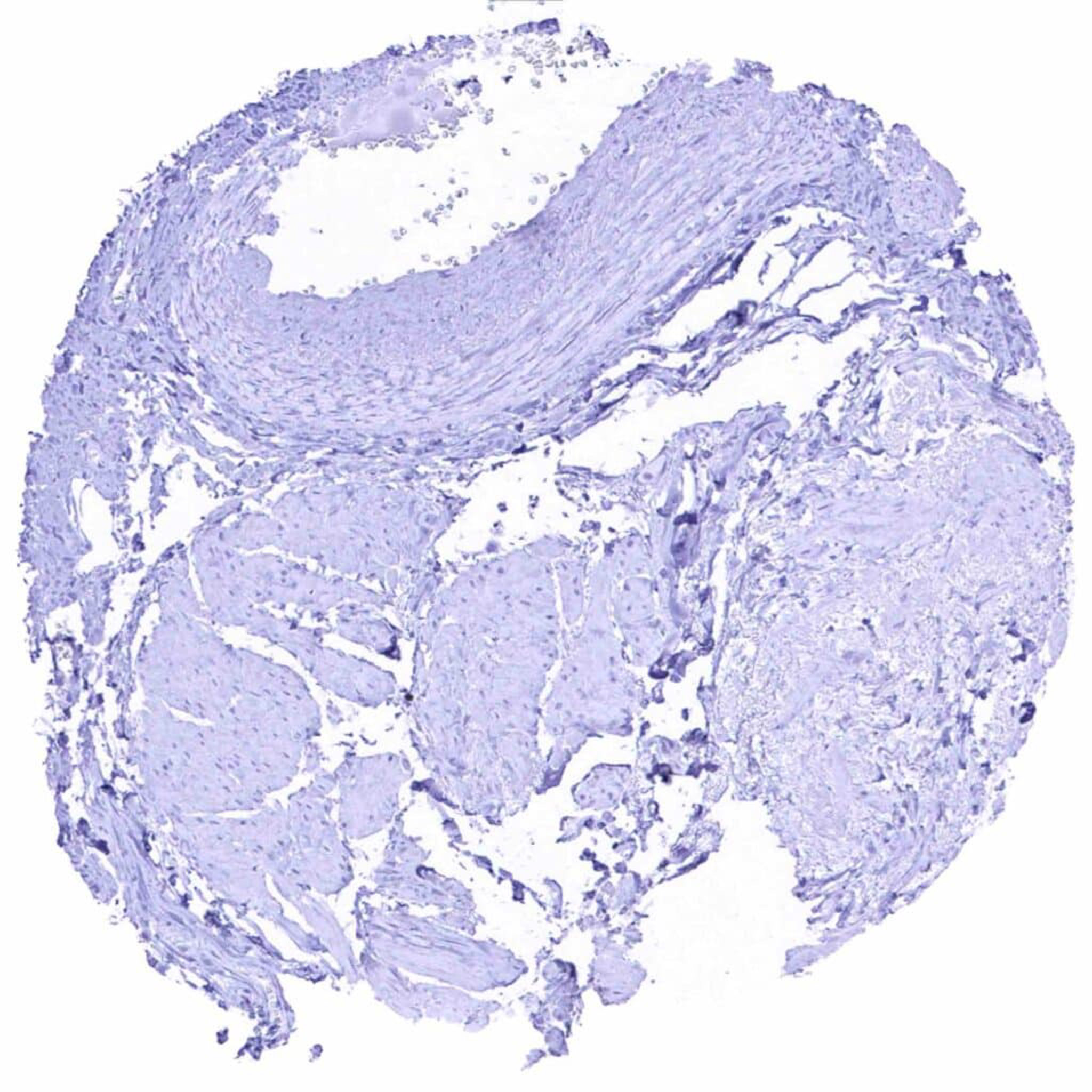
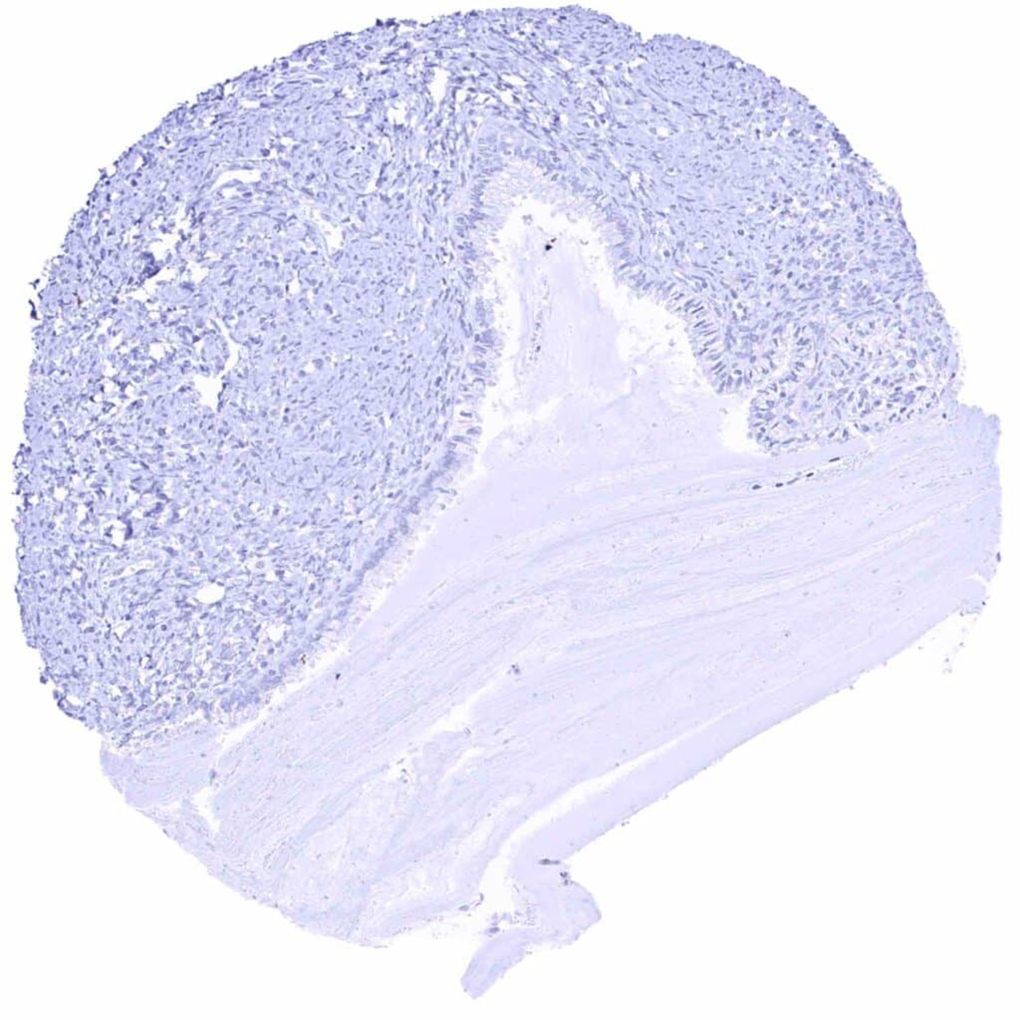
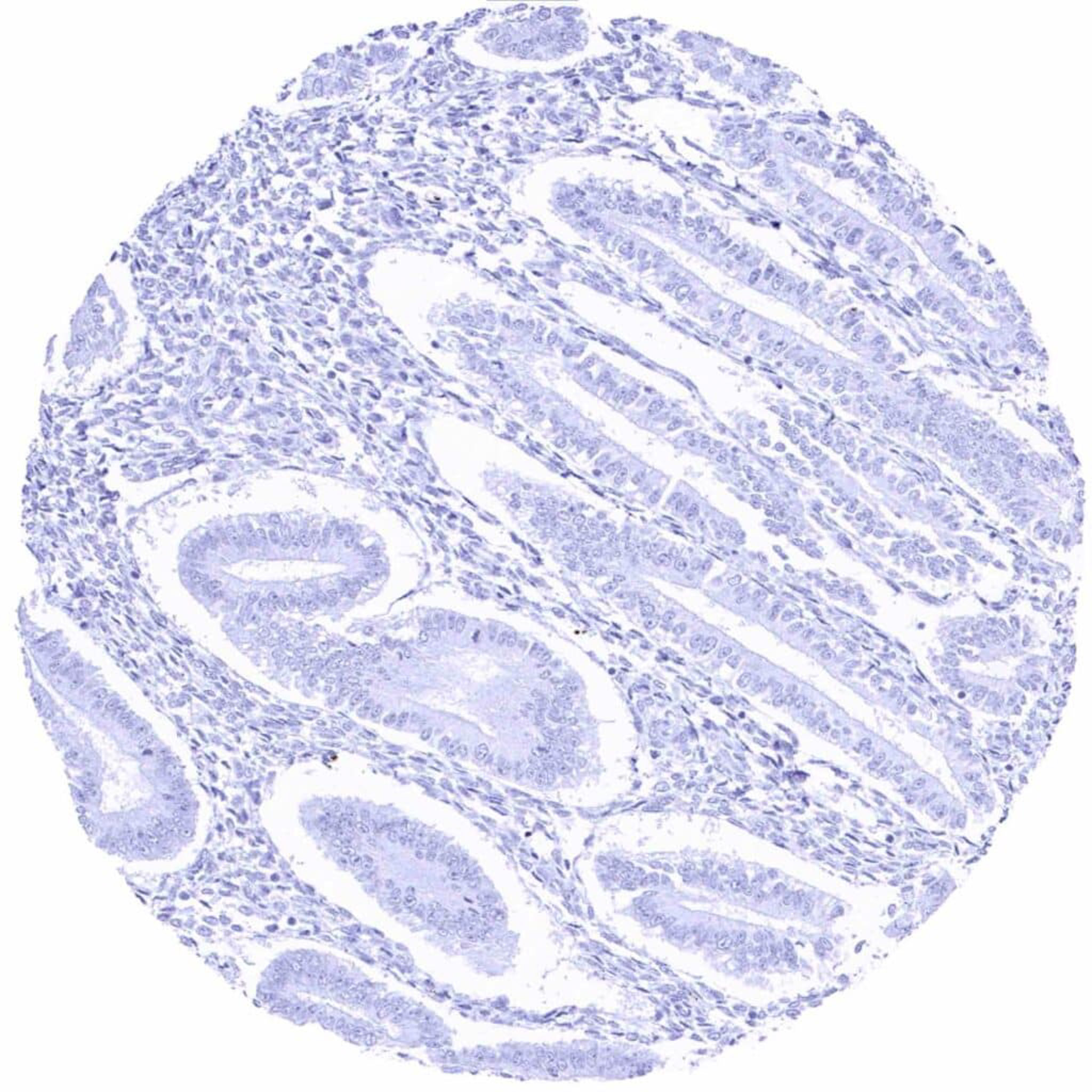
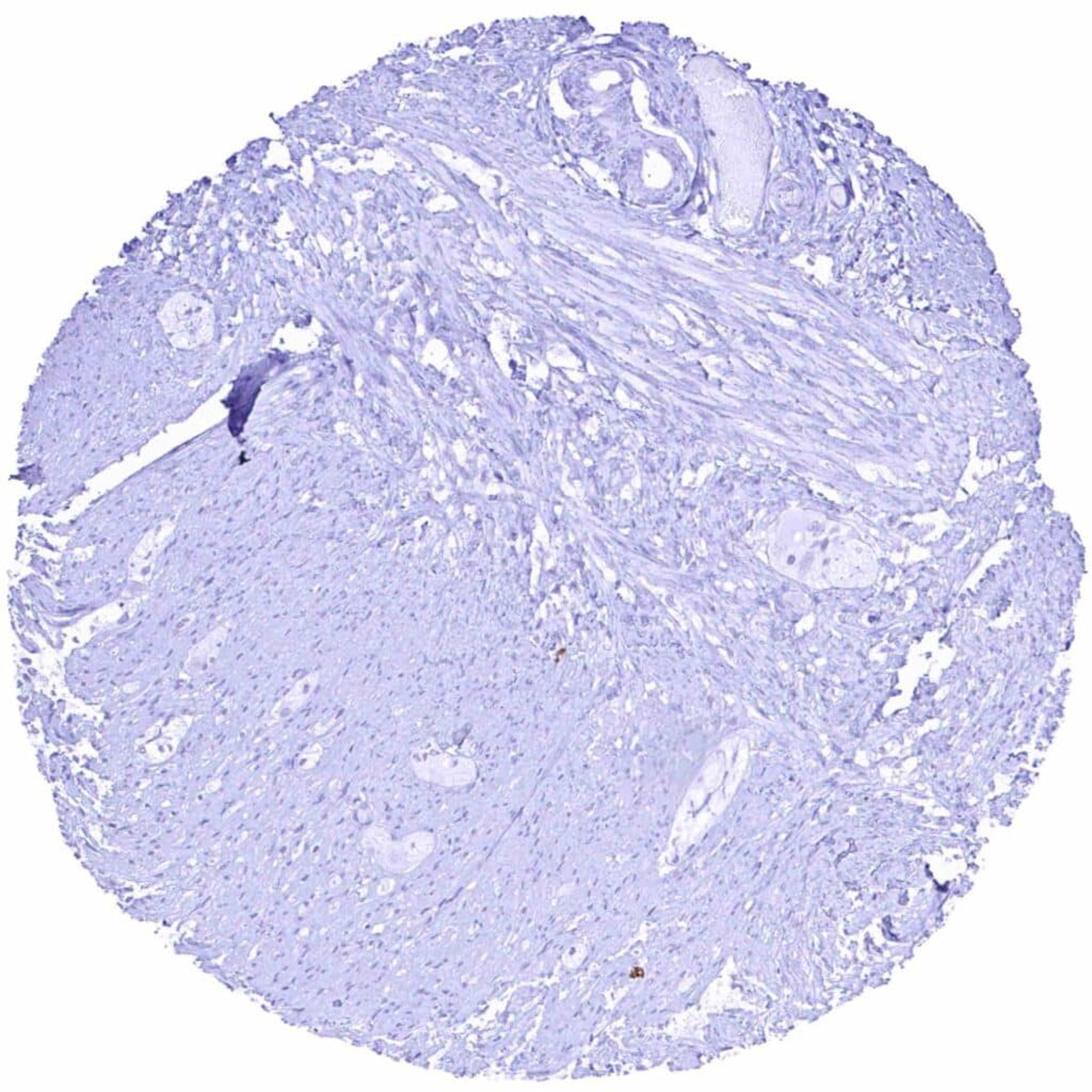
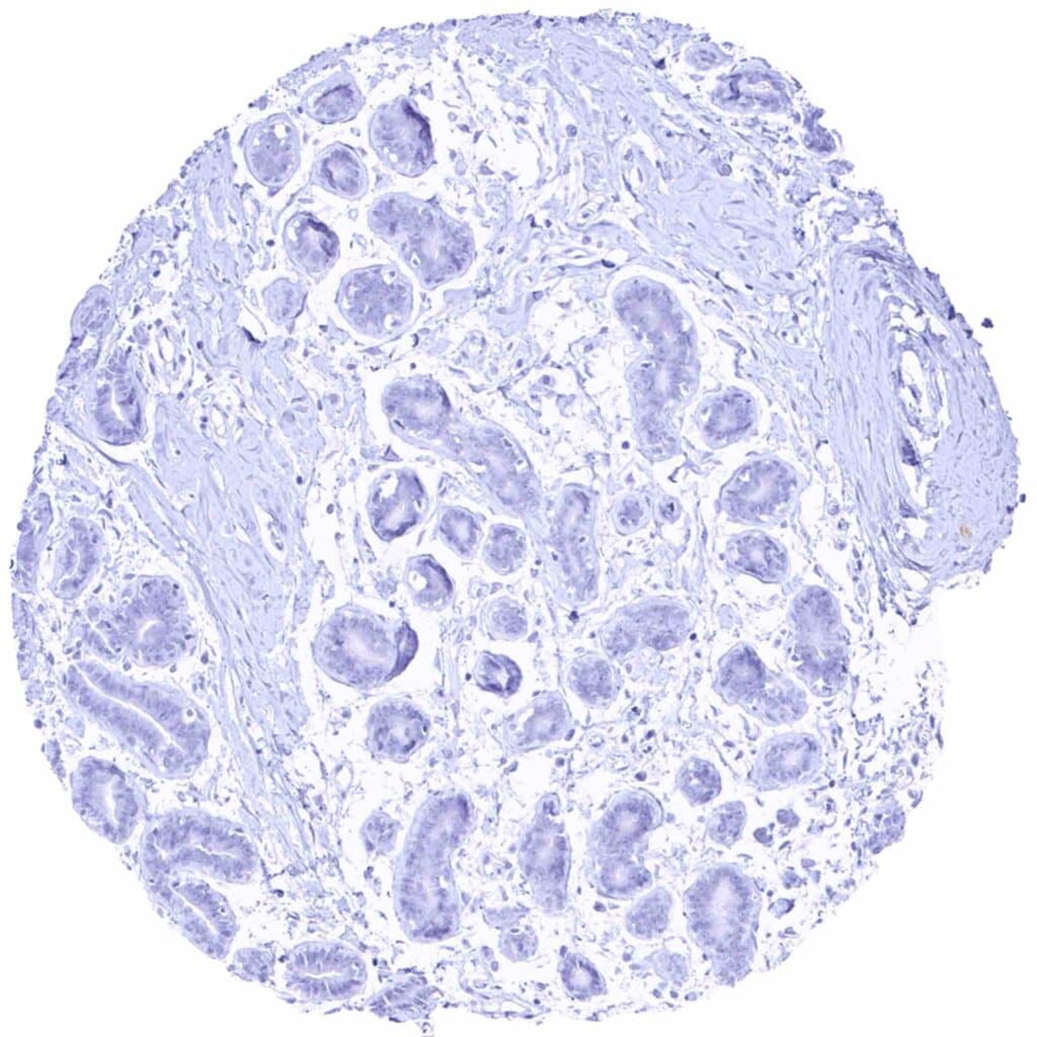
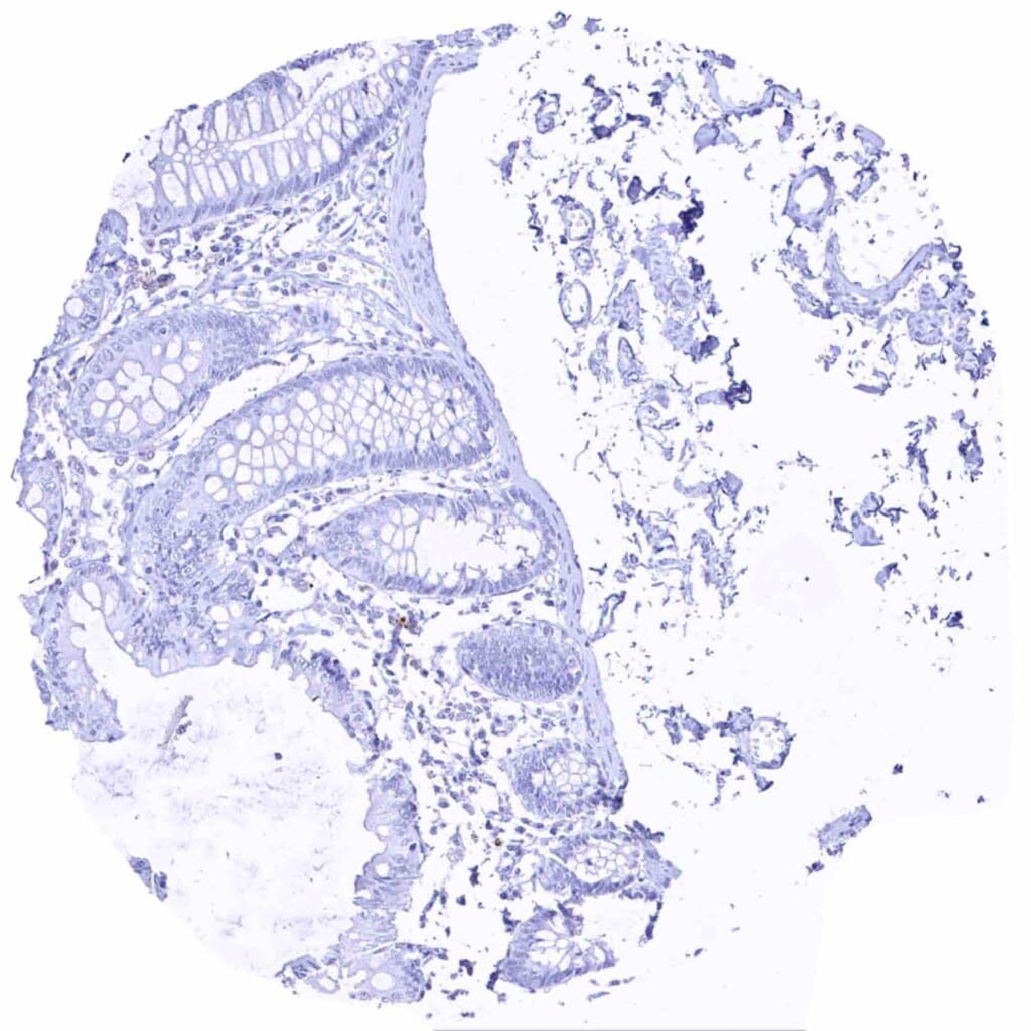
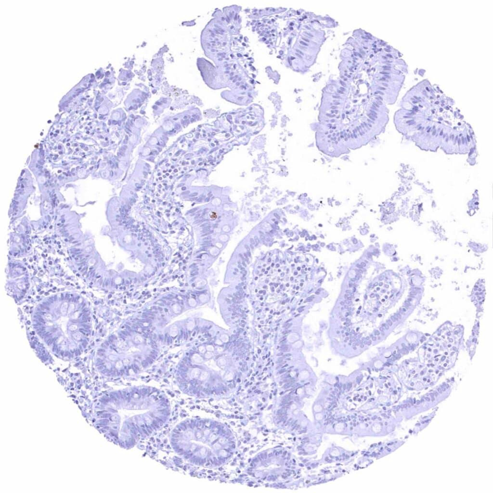
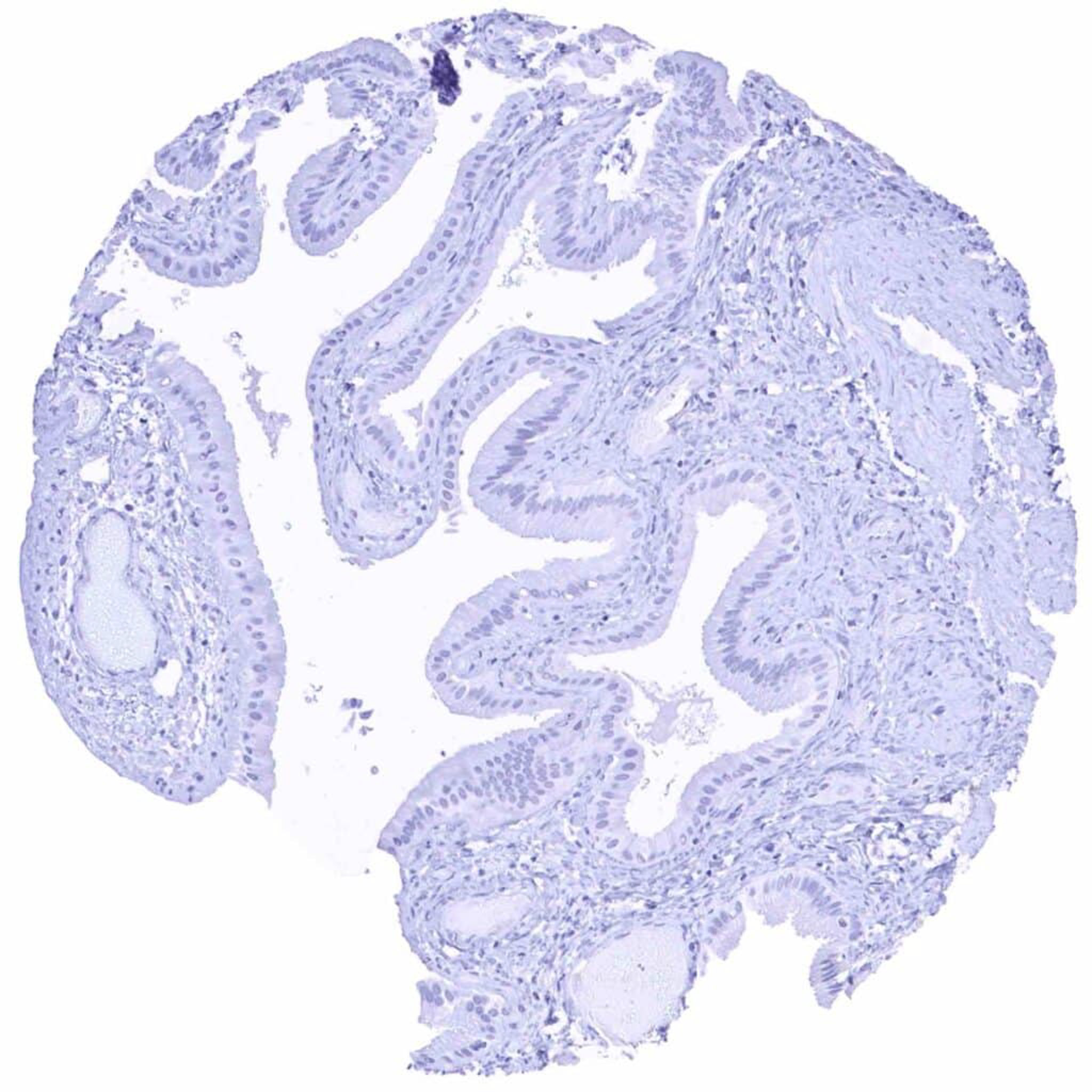
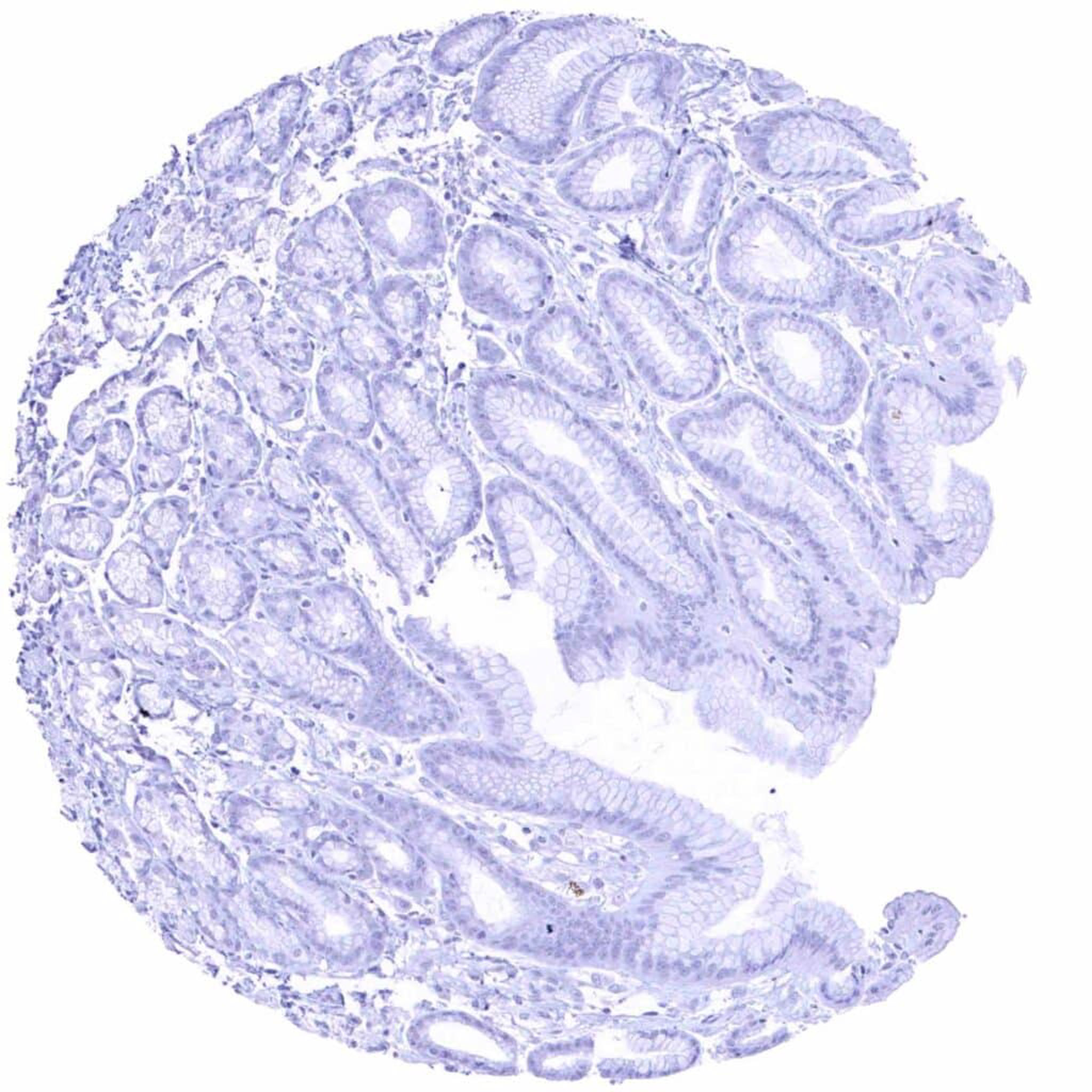
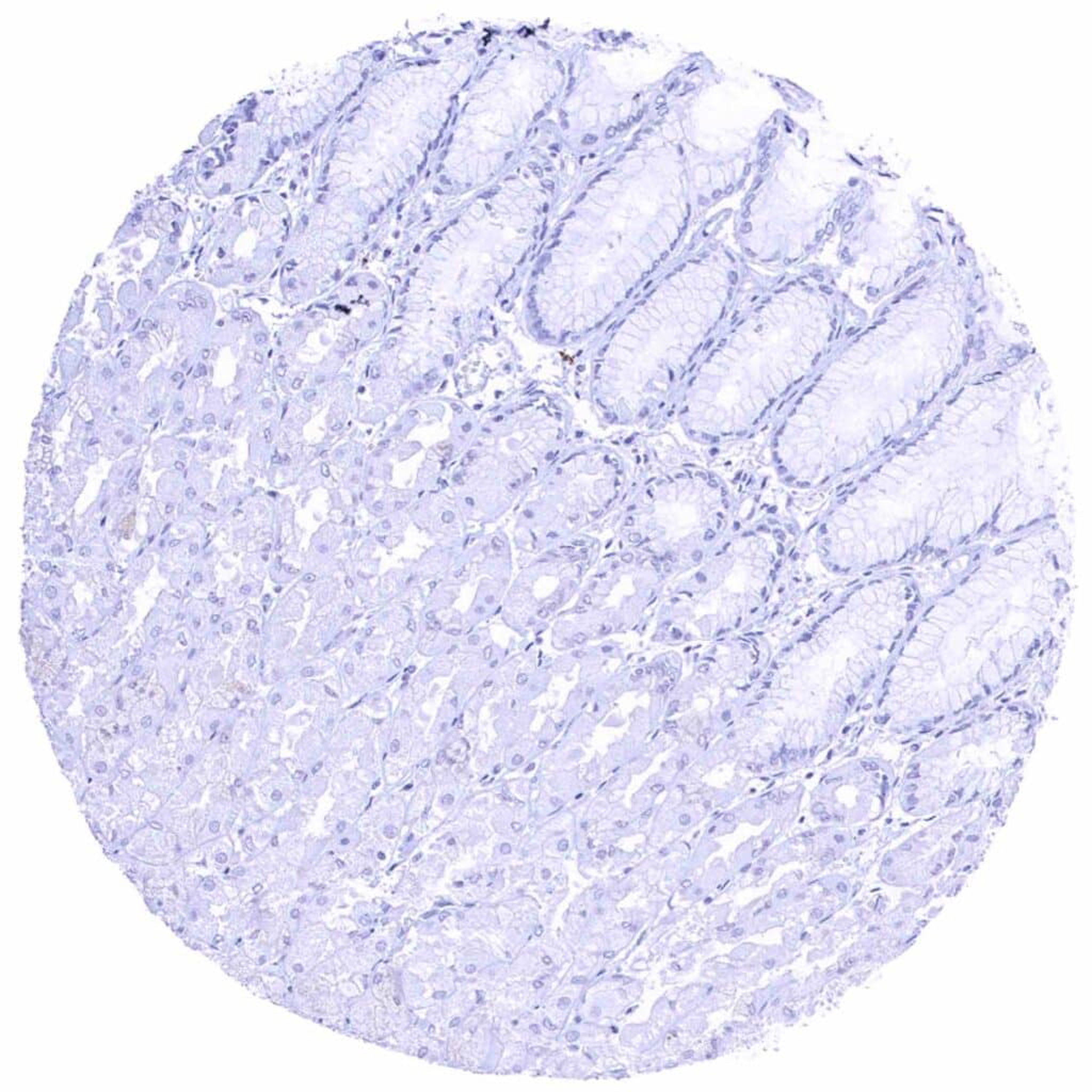
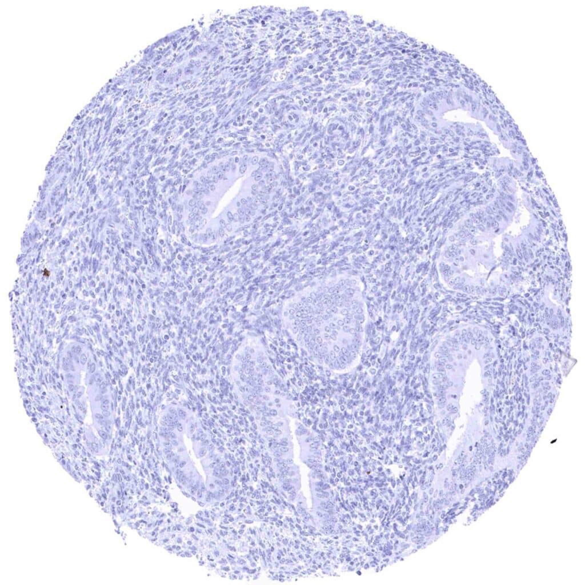
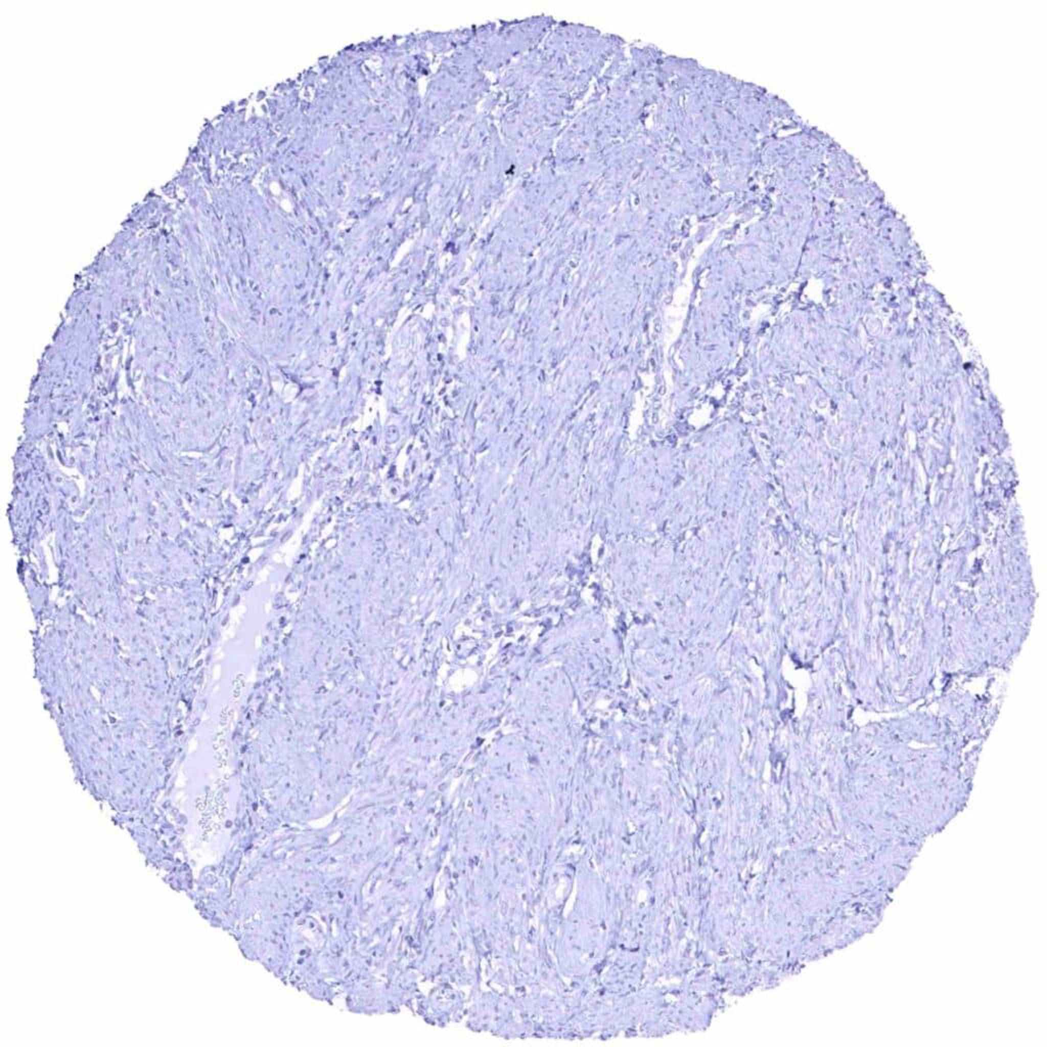
C-Peptide antibody [HMV363] HistoMAXTM


Go to Cancer Tissue Gallery
  
 Go back to product page
  

Go to HistoMAX page


Adrenal gland

Aorta, media

Appendix, mucosa

Appendix, mucosa


Appendix, muscular wall

Bone marrow

Breast

Bronchus, mucosa


Cerebellum (white matter)

Cerebellum, cortex (molecular layer, Purkinje cell layer, granule cell layer, white matter)

Cerebrum, grey matter

Cerebrum, white matter


Colon descendens, mucosa

Colon descendens, muscular wall

Duodenum, Brunner gland

Duodenum, mucosa


Epididymis, caput

Epididymis, cauda

Esophagus, squamous epithelium

Fallopian tube, mucosa


Fat

Gallbladder, epithelium

Heart muscle

Ileum, mucosa


Kidney, cortex

Kidney, medulla

Liver

Lung


Lymph node

Lymph node

Ovary, corpus luteum

Ovary, stroma


Pancreas – Intense cytoplasmic C-peptide staining of most islet cells. Faint staining of acinar cells in the immediate vicinity of pancreatic islets represents a „contamination artifact“ caused by diffusion of C-peptide into „islet-adjacent“ tissue

Pancreas – Strong cytoplasmic C-peptide positivity of the majority of islet cells

Pancreas – Strong cytoplasmic C-peptide staining of most islet cells

Parathyroid gland


Parotid gland

Pituitary gland, anterior lobe

Pituitary gland, posterior lobe

Placenta (amnion and chorion)


Placenta, early

Placenta, mature

Prostate

Rectum, mucosa


Seminal vesicle

Sinus paranasales

Skeletal muscle

Skin


Spleen

Stomach, antrum

Stomach, corpus

Testis


Thymus

Thyroid gland

Tonsil, surface epithelium

Tonsil


Urinary bladder, muscular wall

Urinary bladder, urothelium

Uterus, ectocervix

Uterus, endocervix


Uterus, endometrium (pregnancy)

Uterus, endometrium (proliferation)

Uterus, endometrium (secretion)

Uterus, myometrium


| TOP |