Normal Tissue Gallery- GTX04389

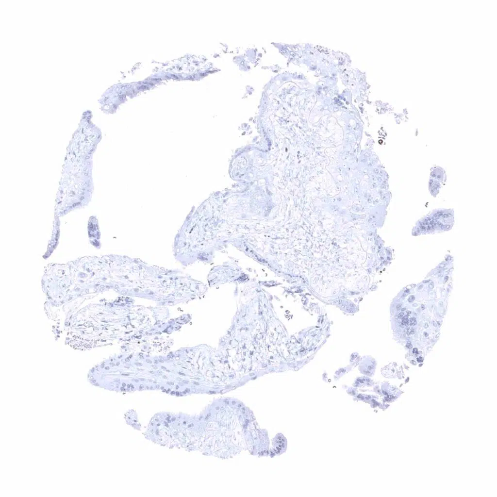
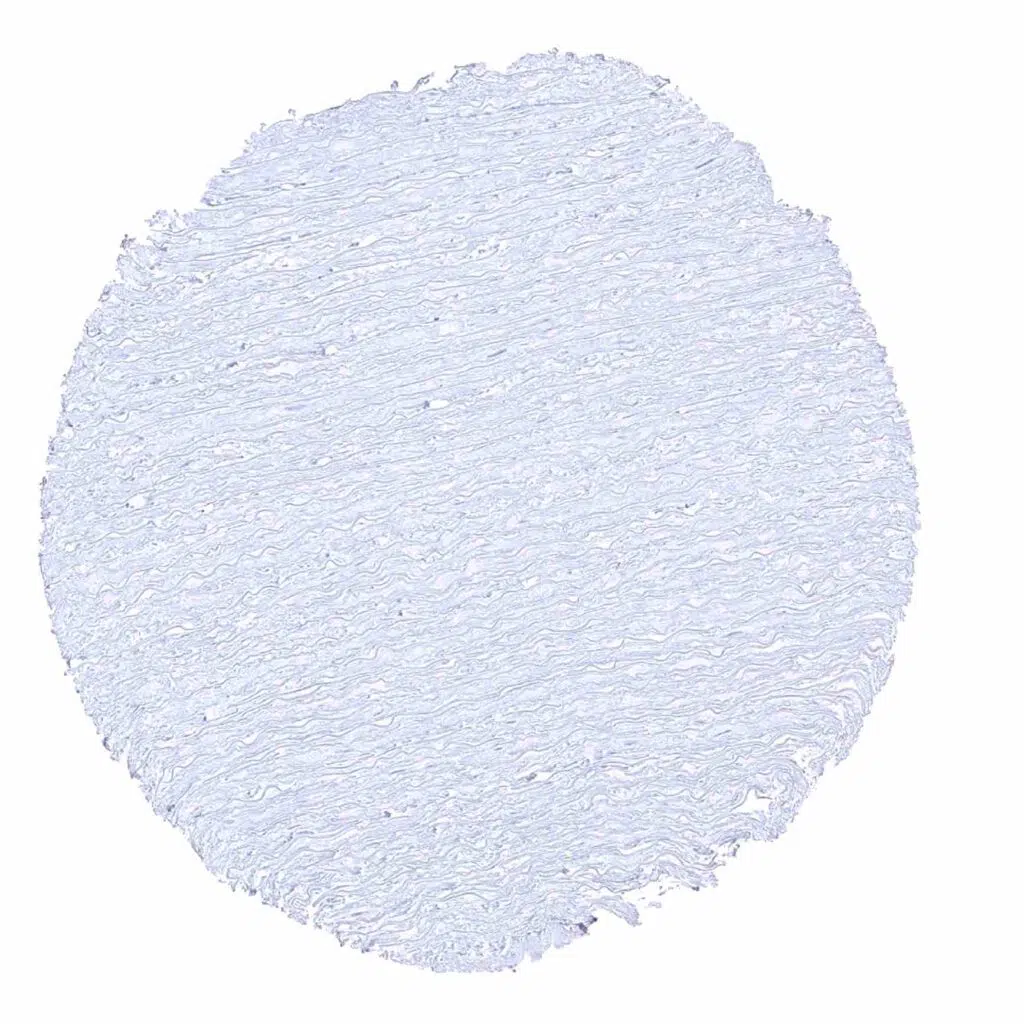
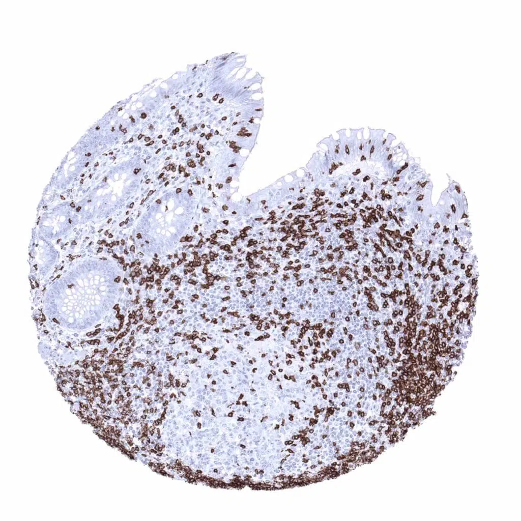
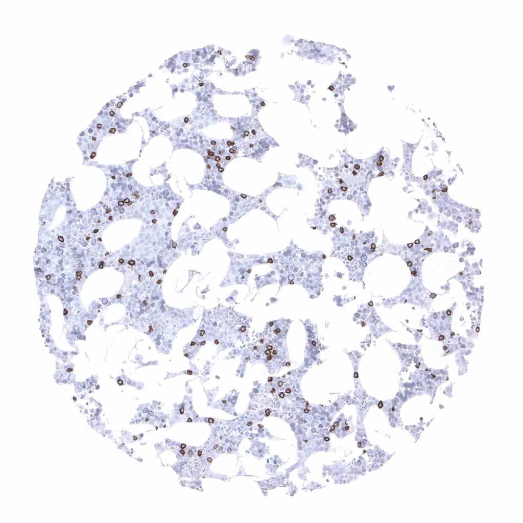
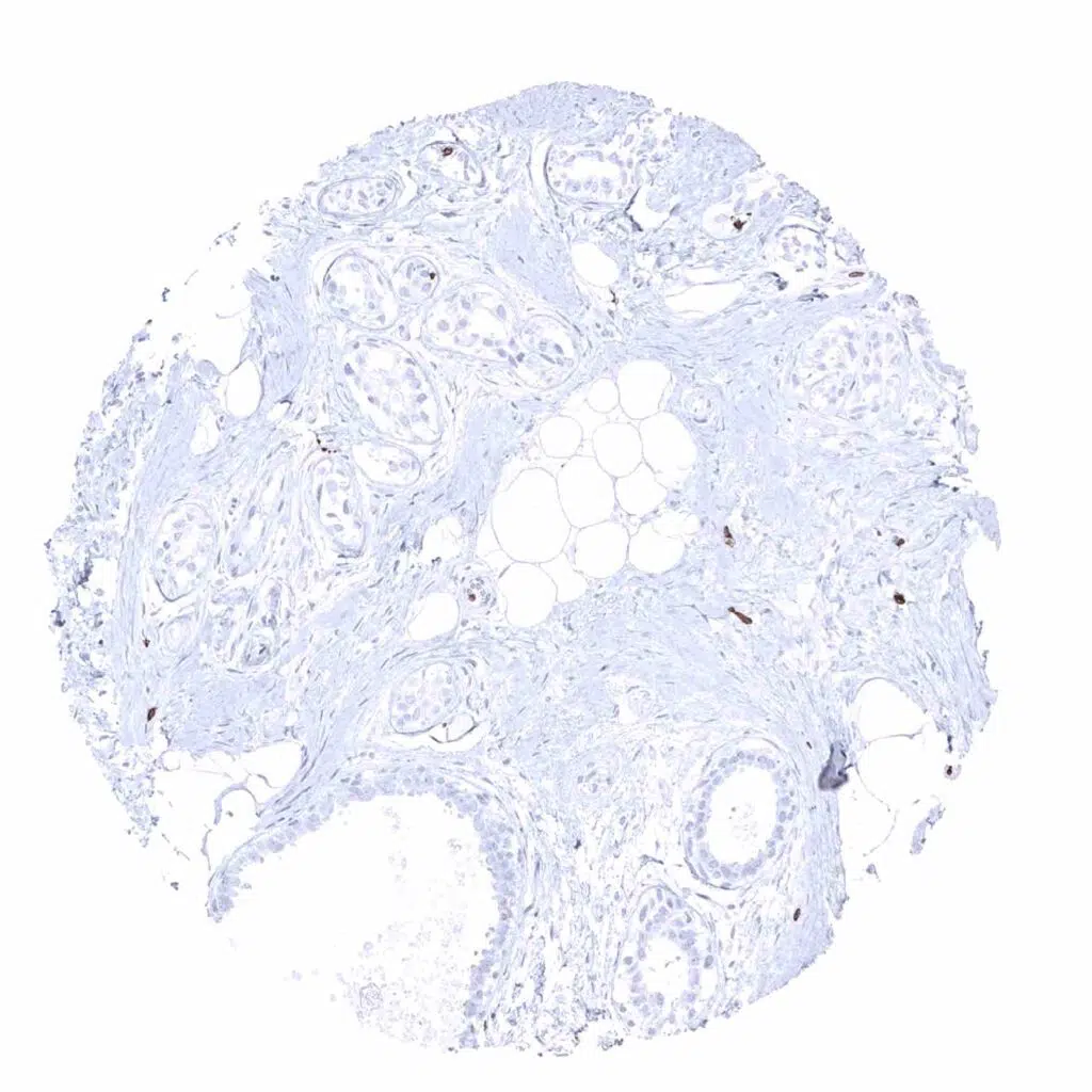
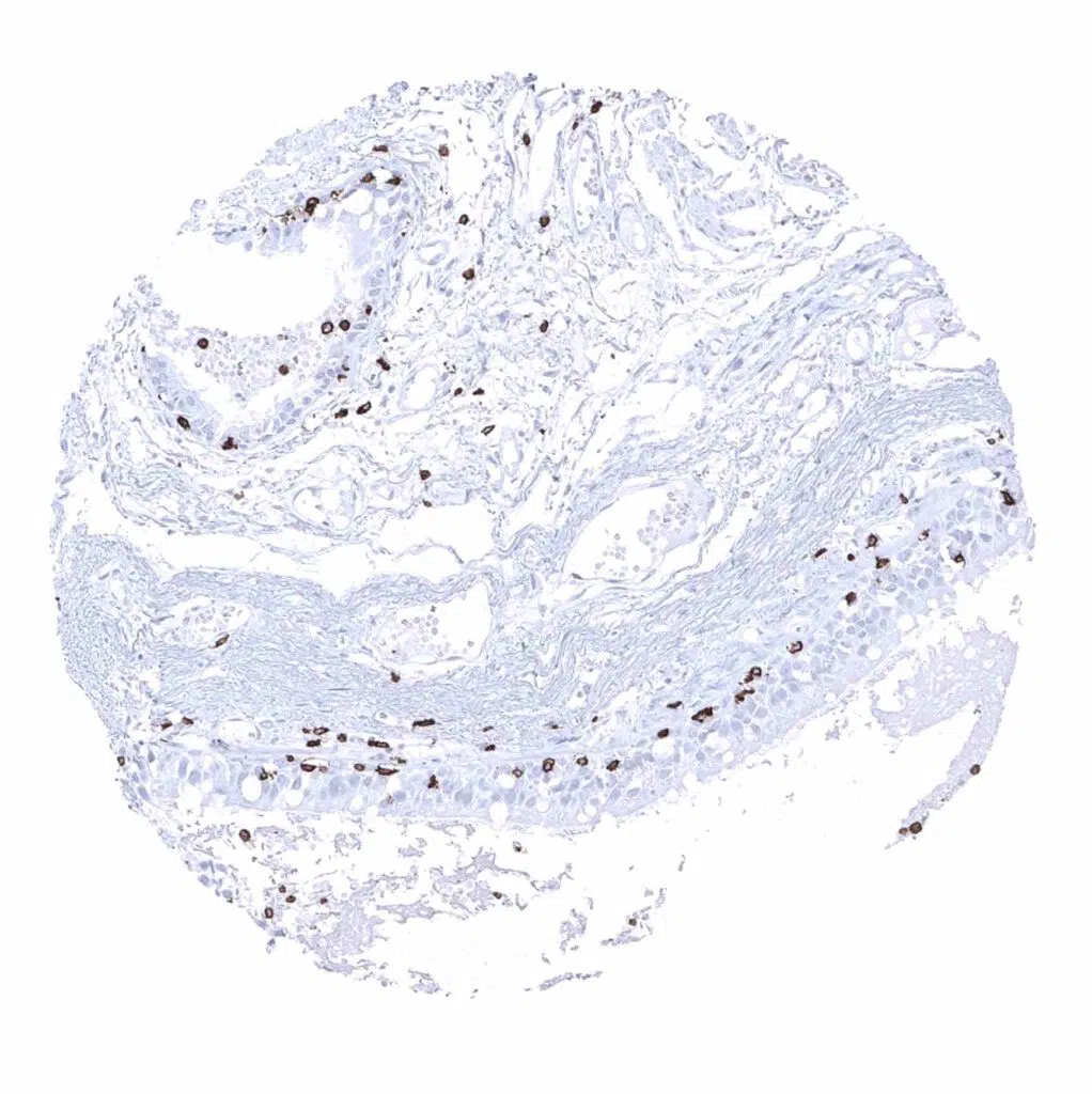
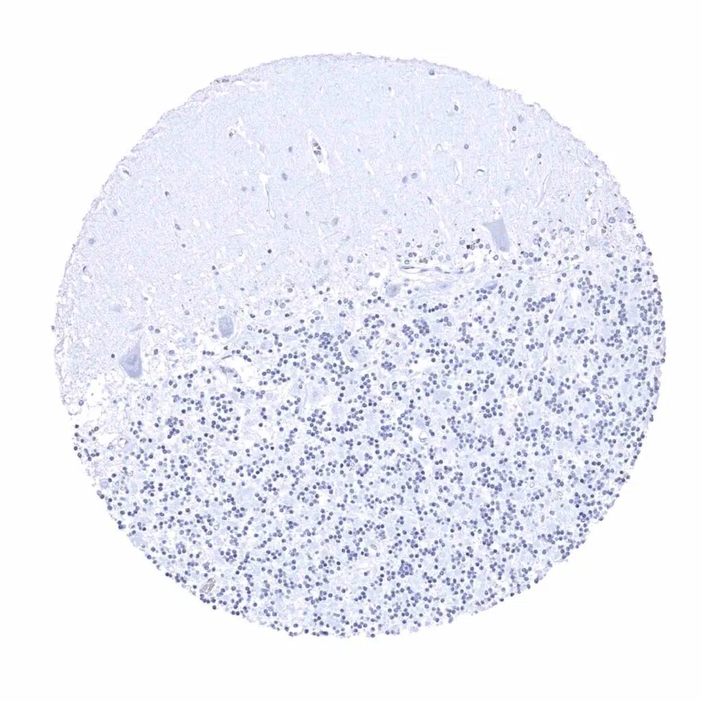
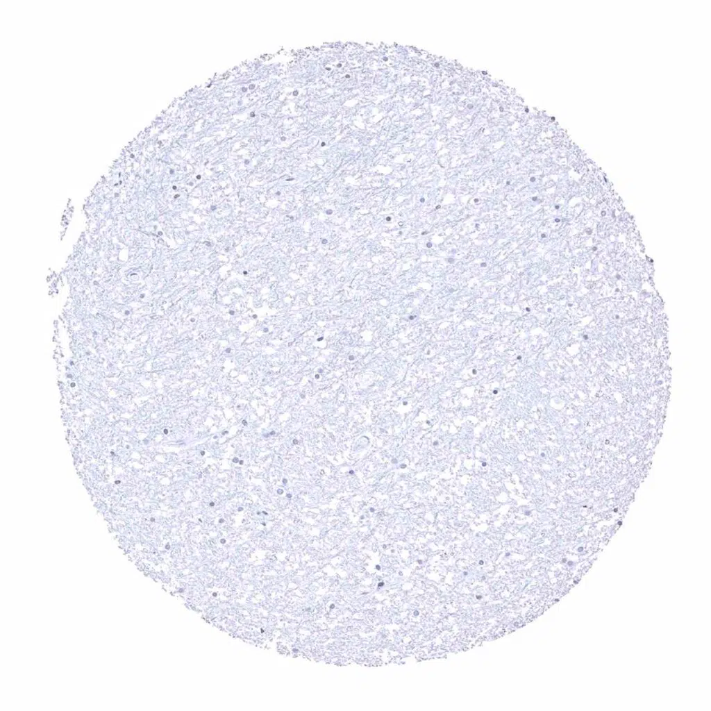
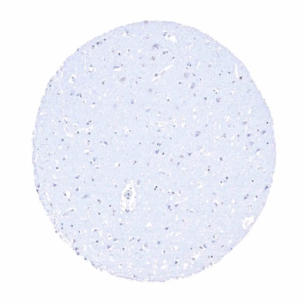
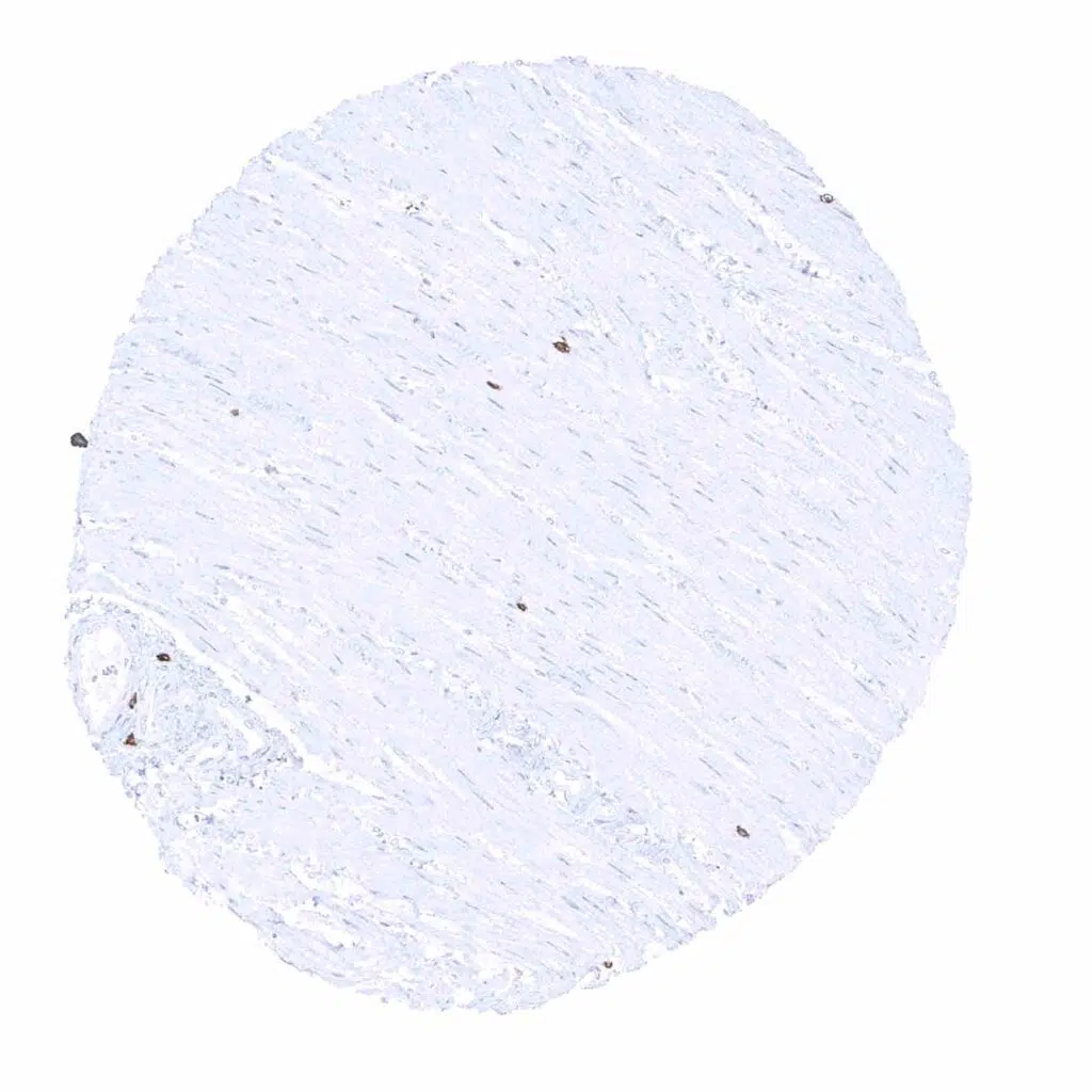
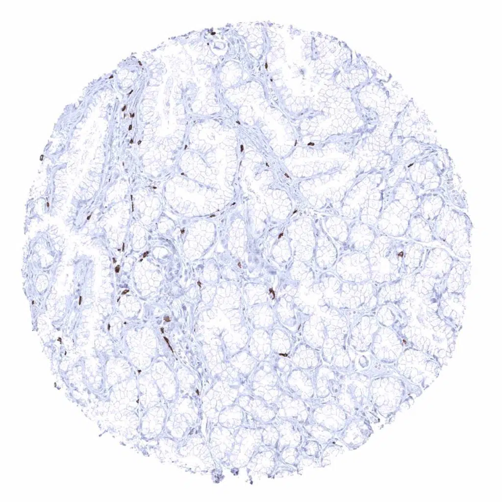
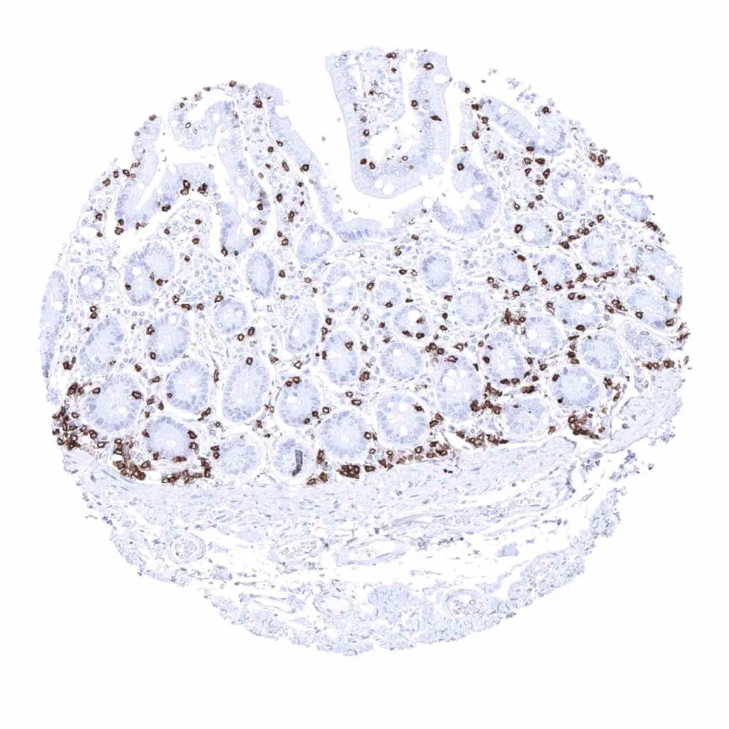
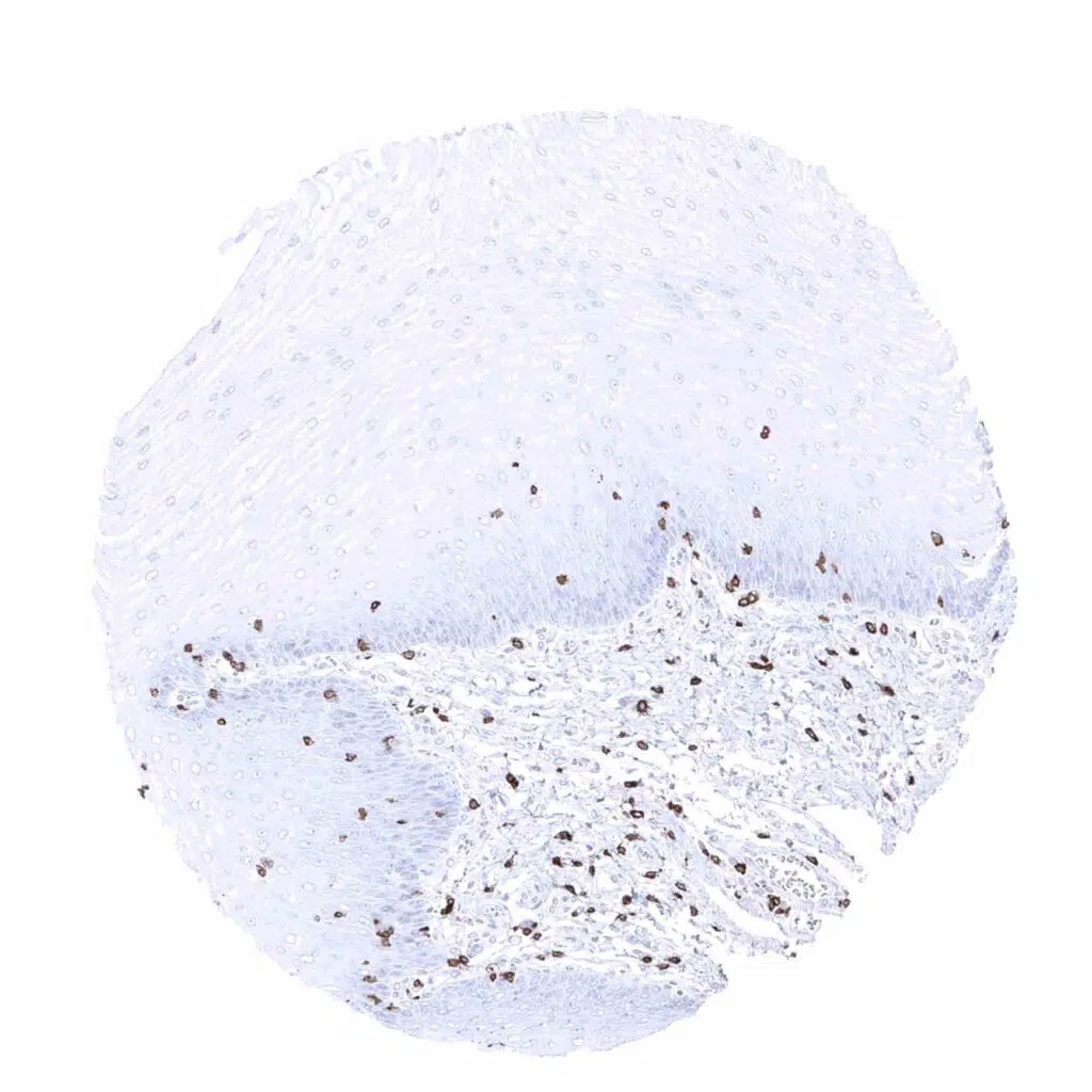
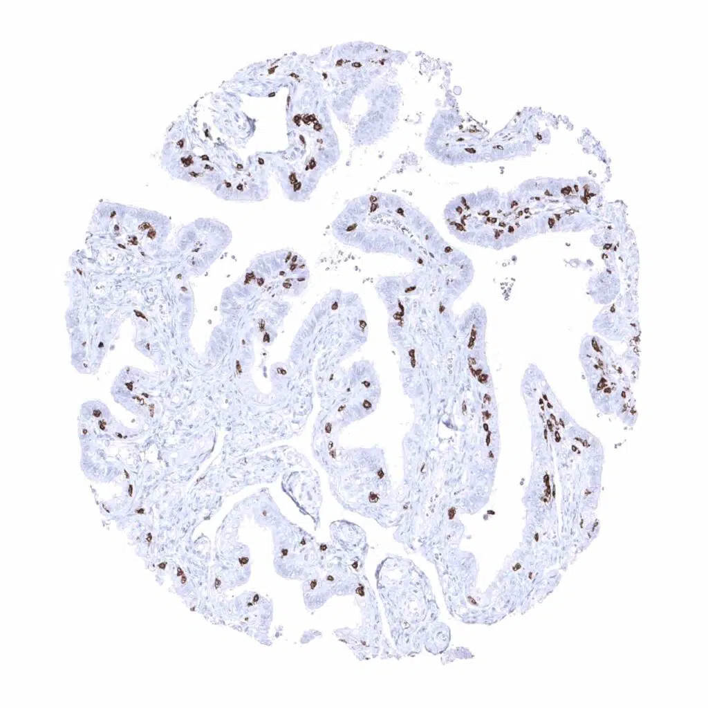
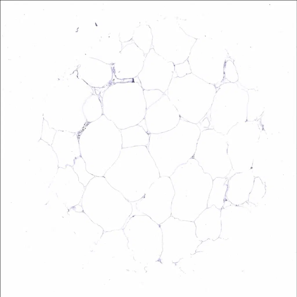
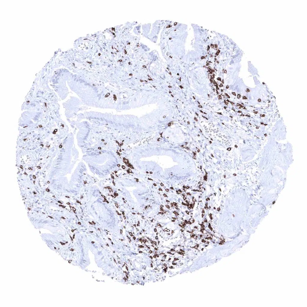
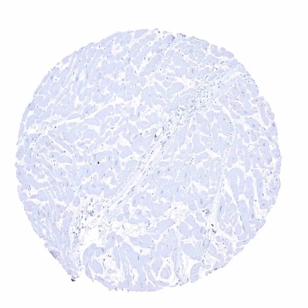
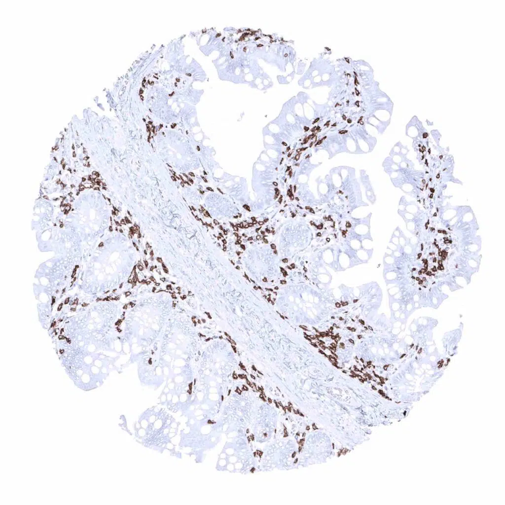
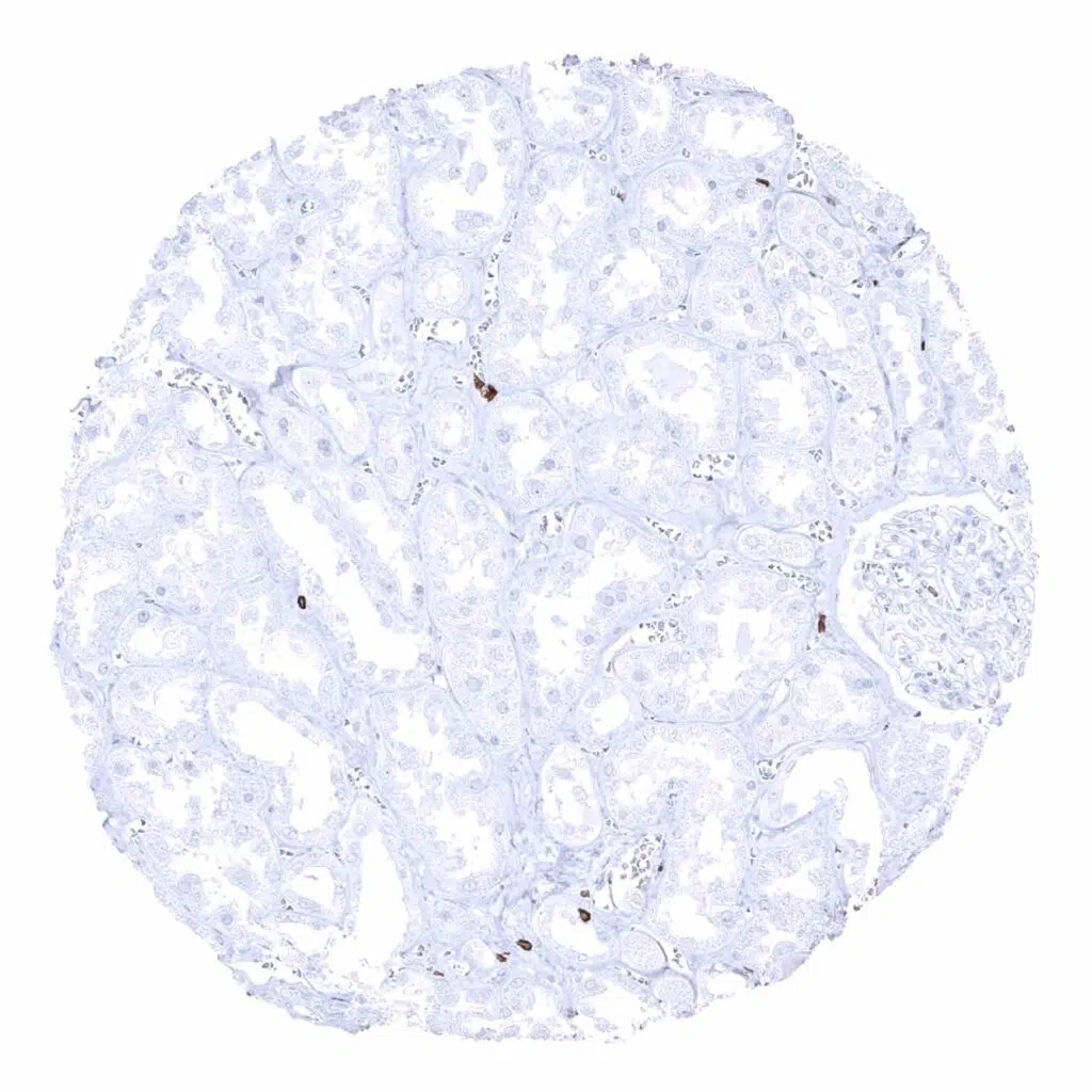
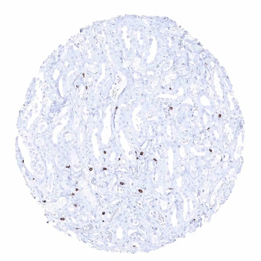
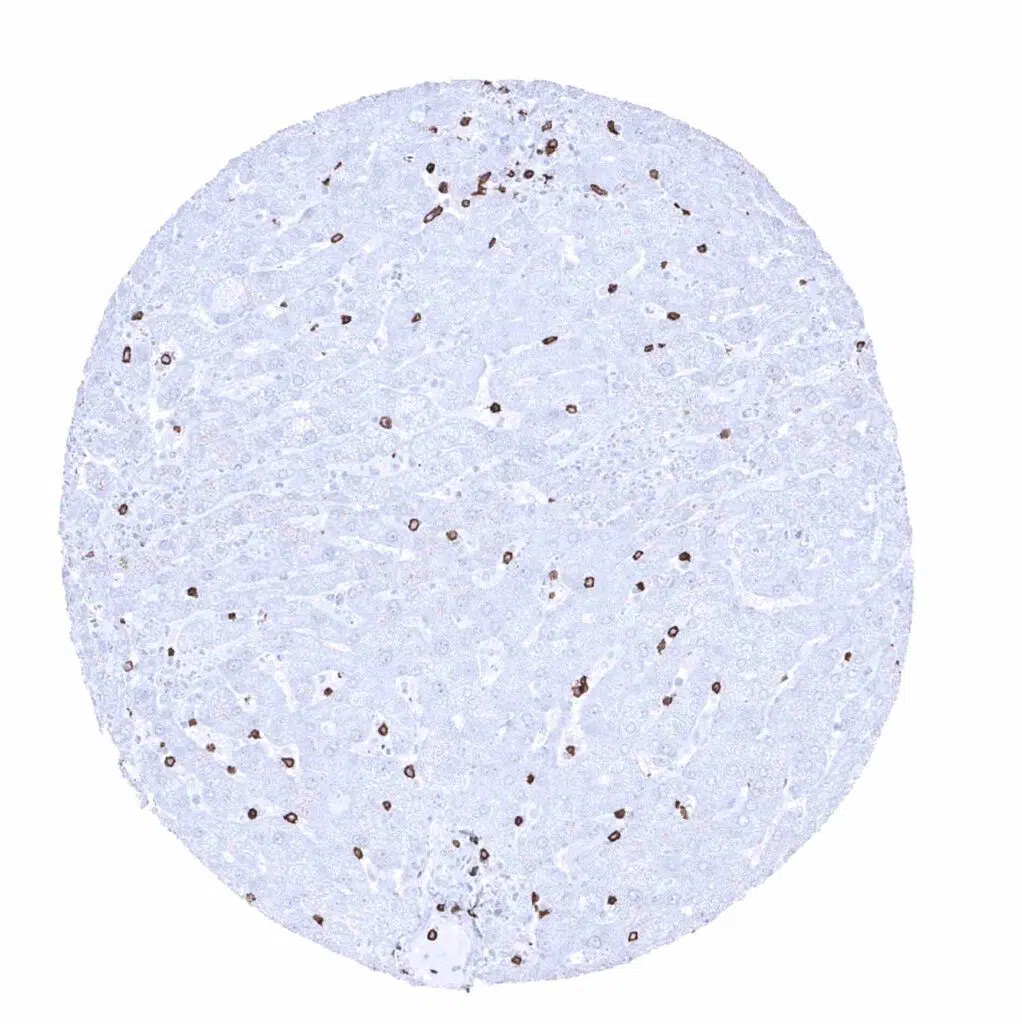
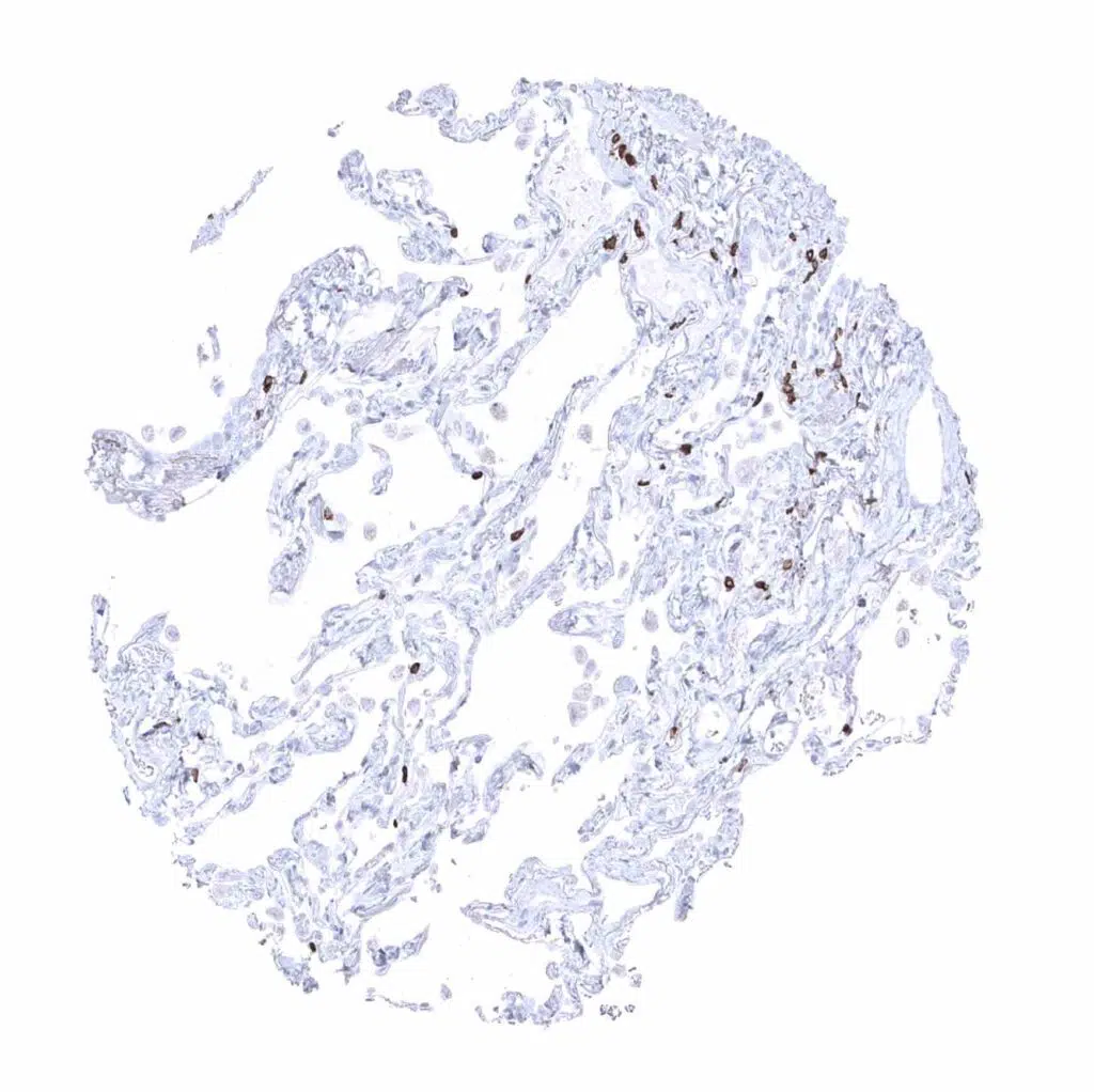
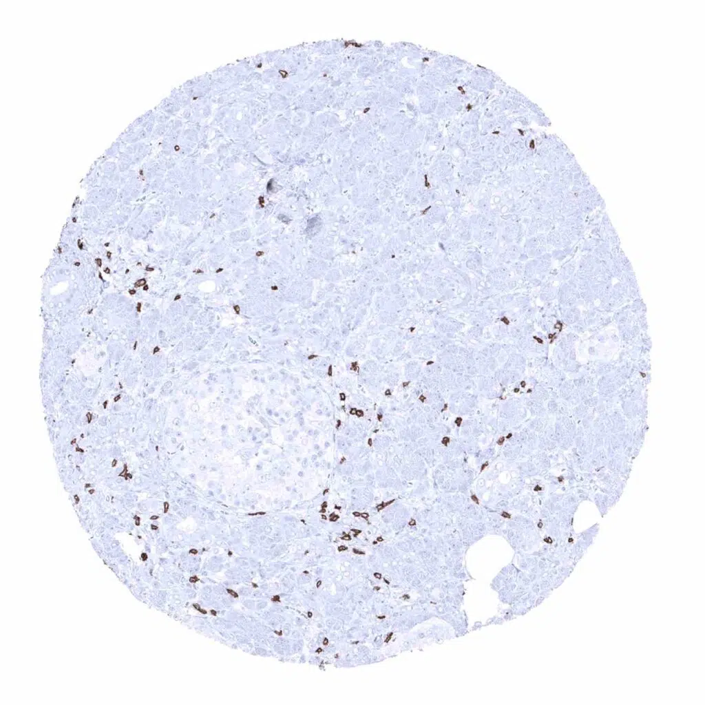
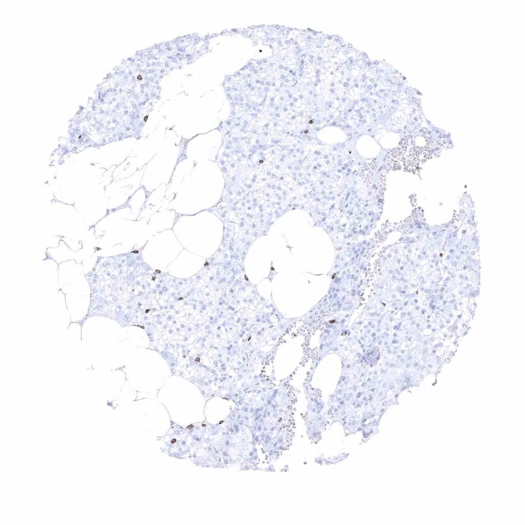
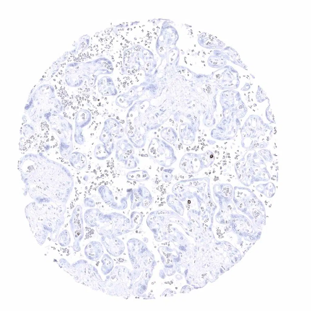
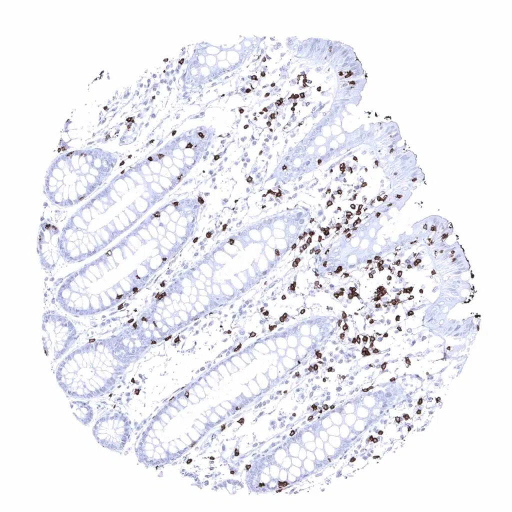
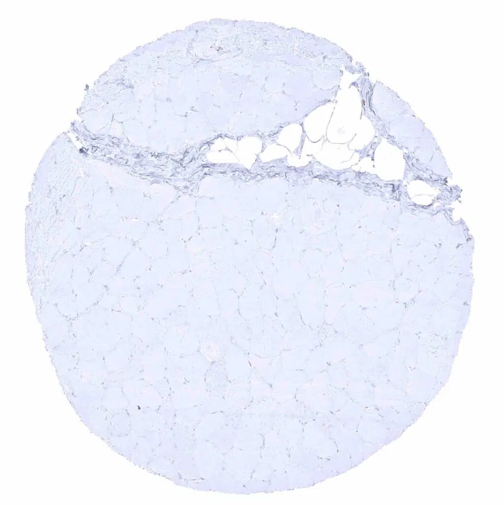
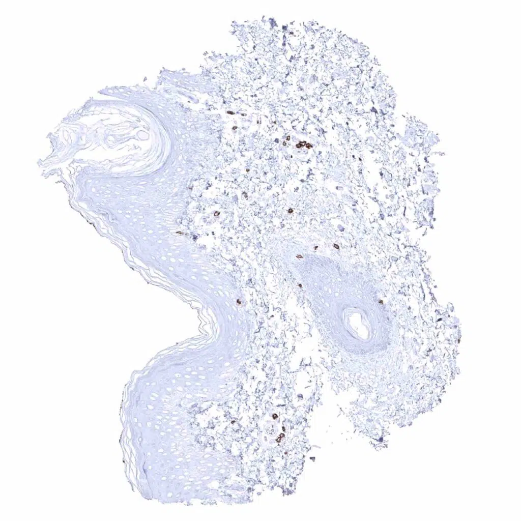
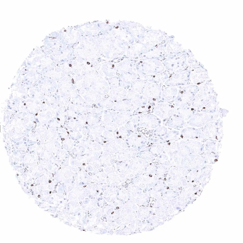
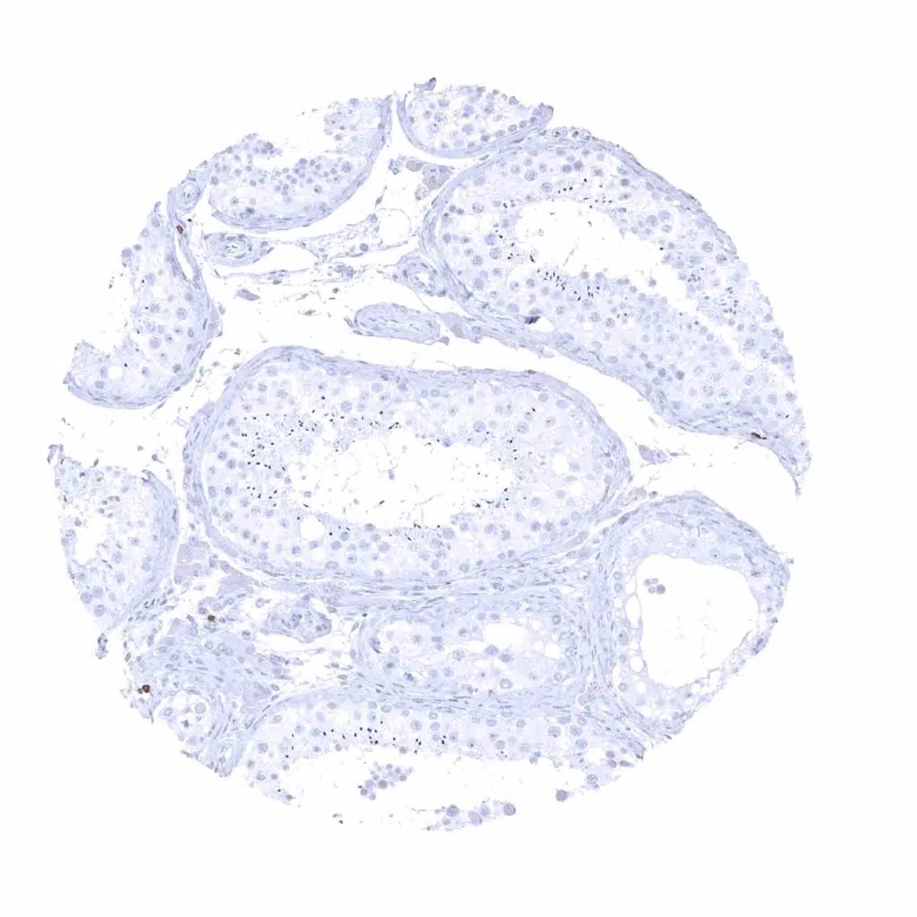
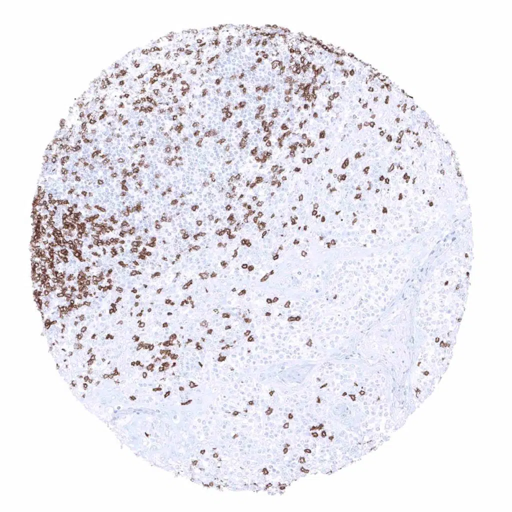
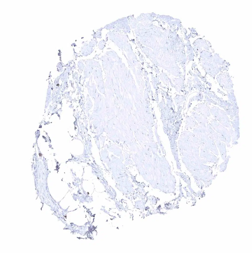
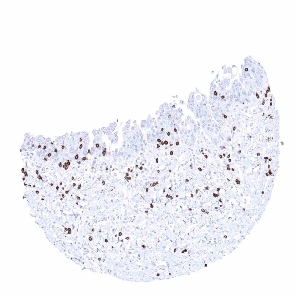
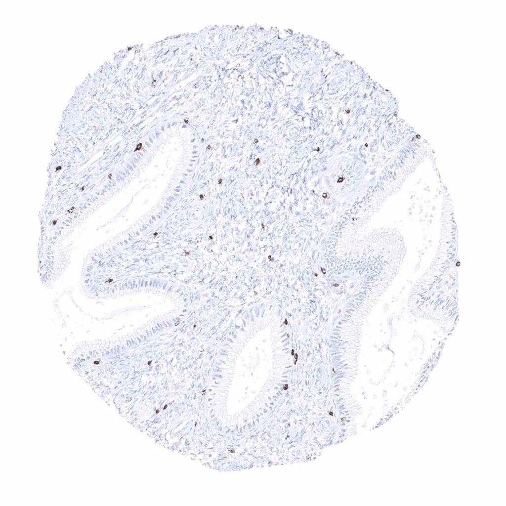
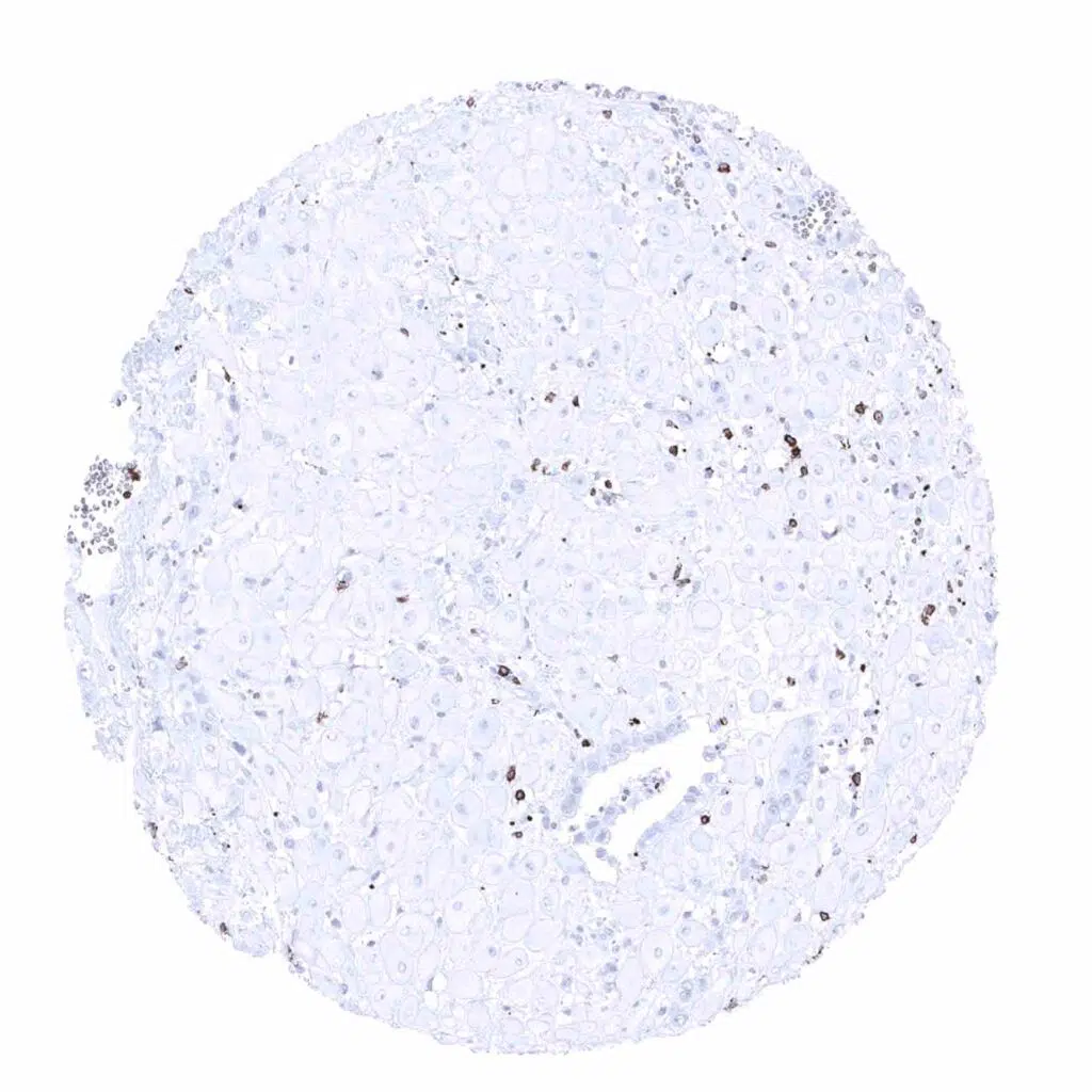
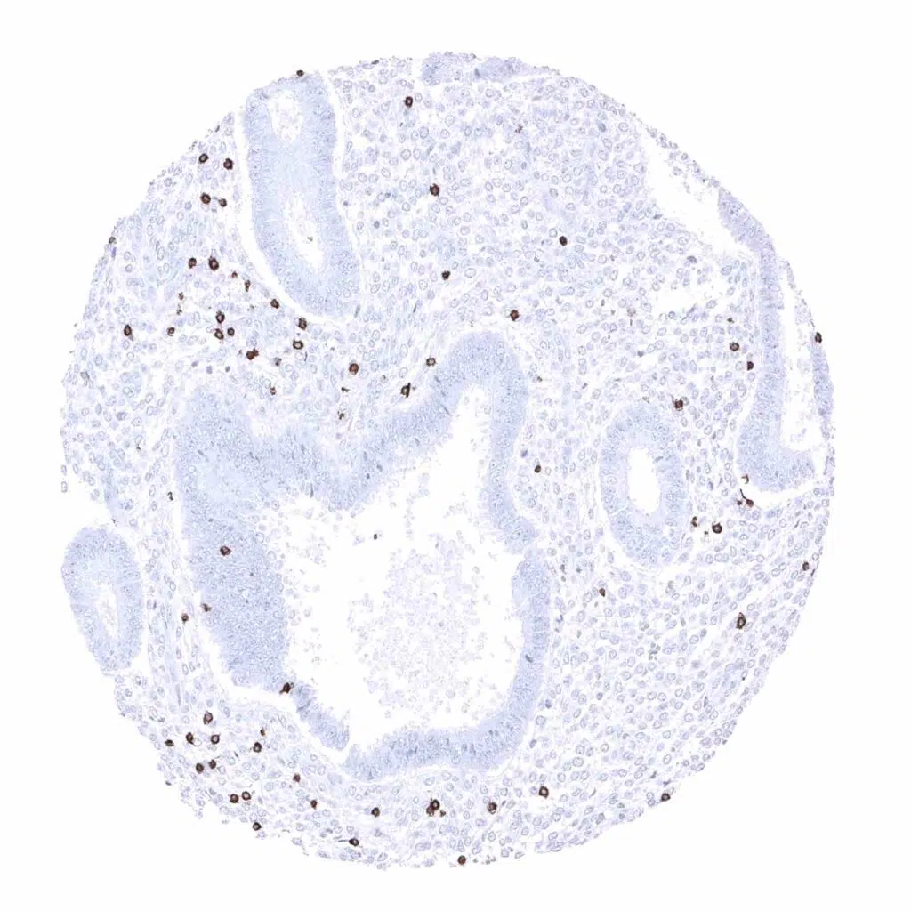
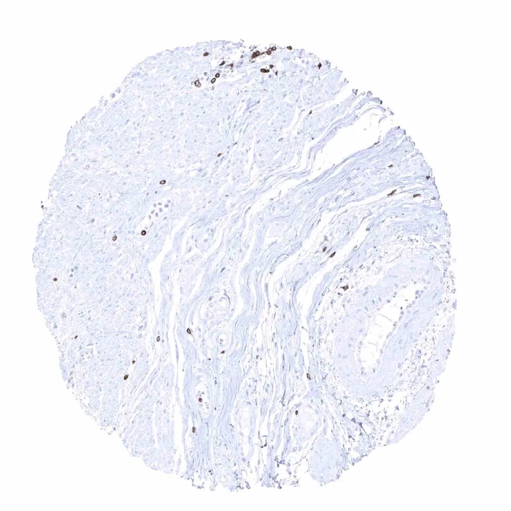
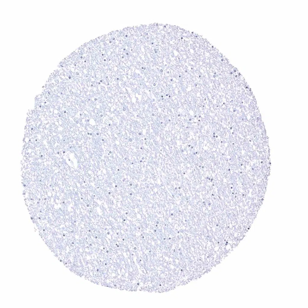
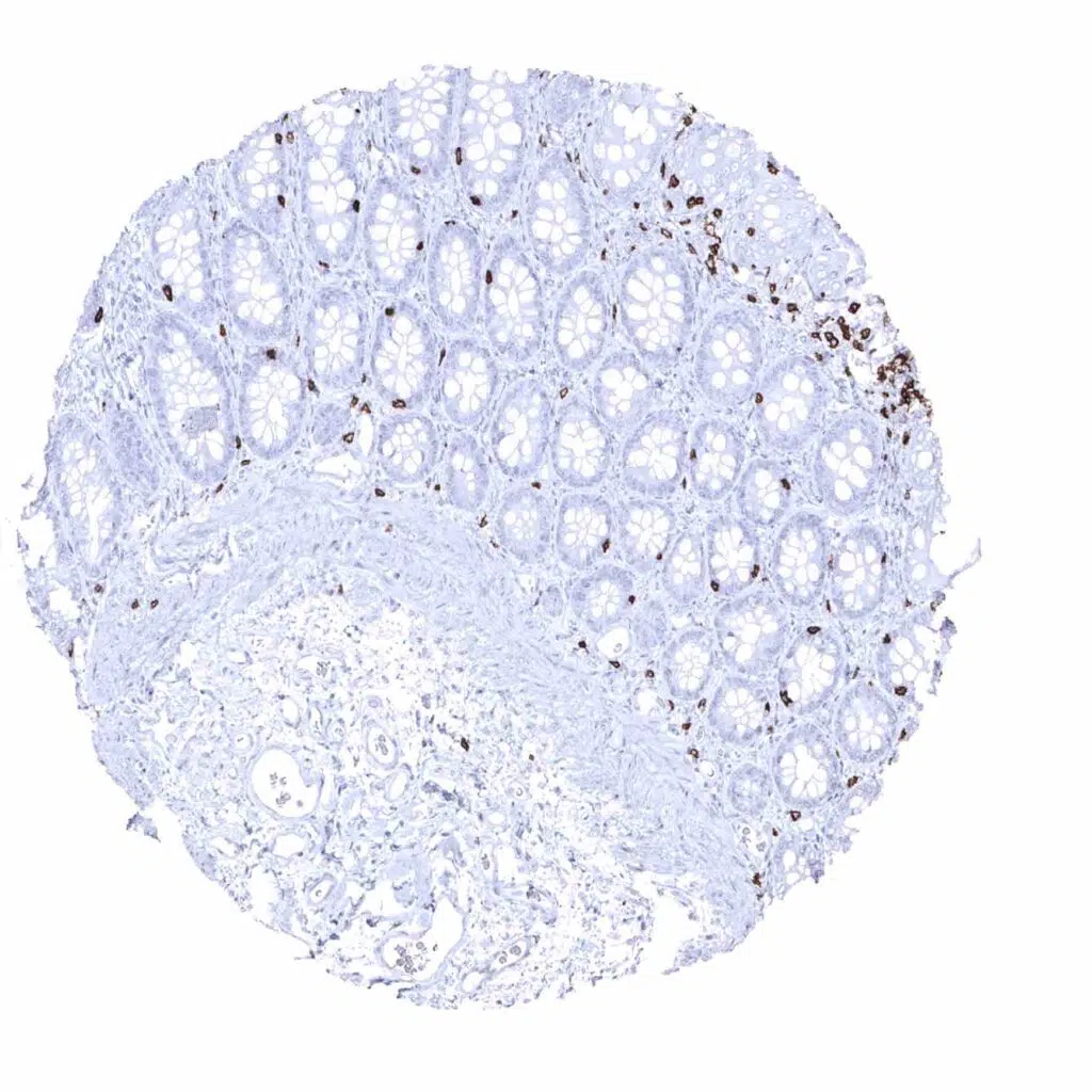
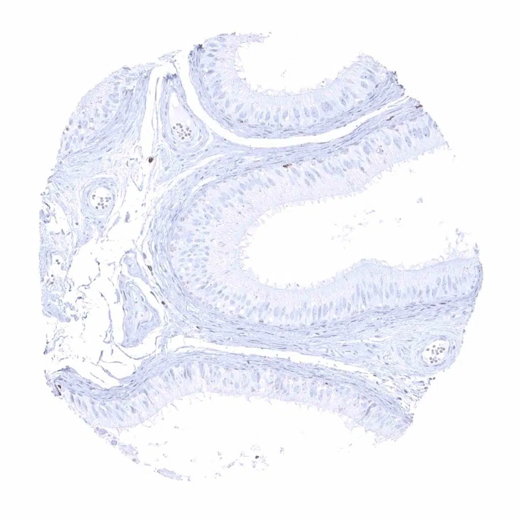
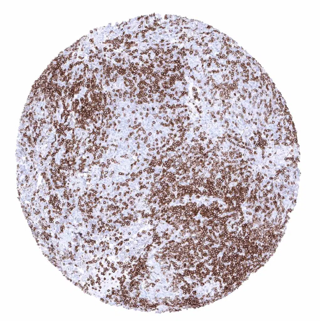
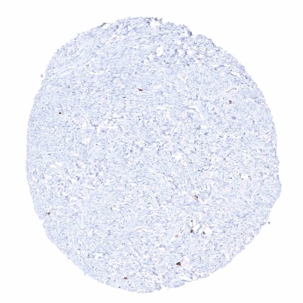
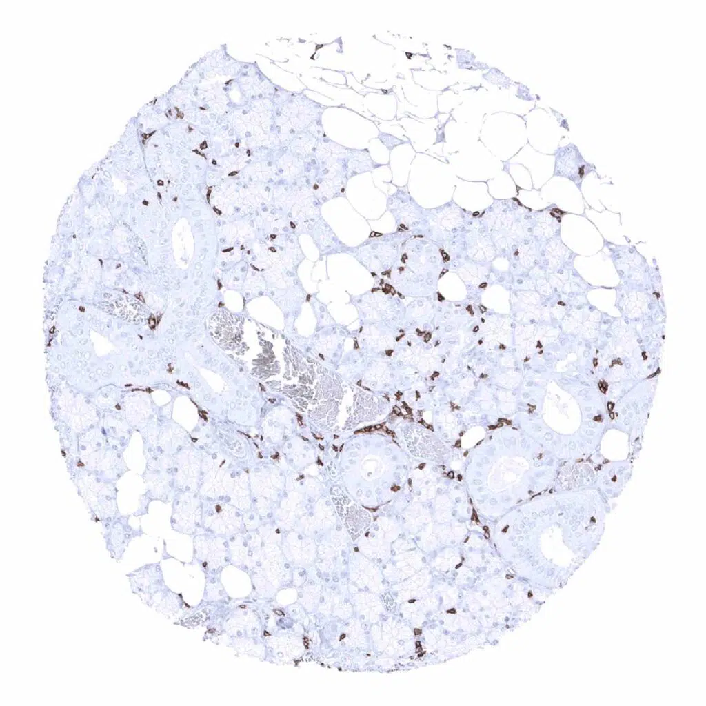
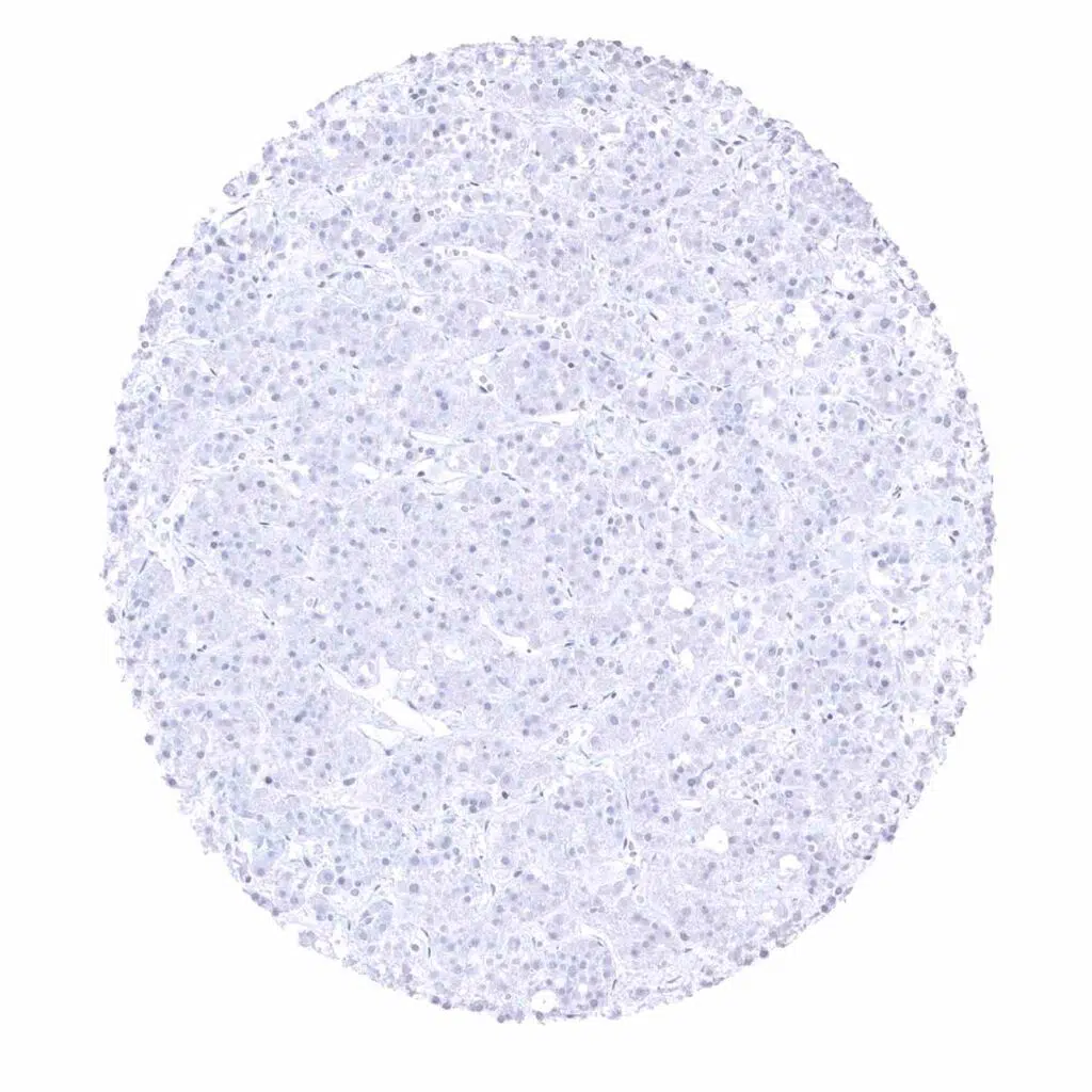
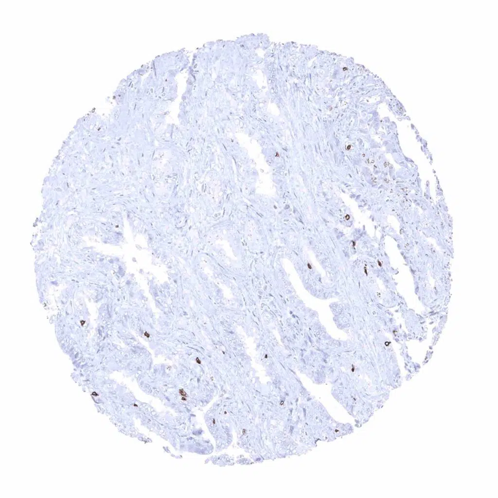
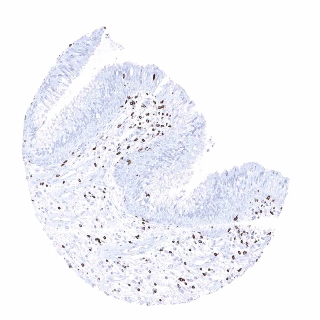
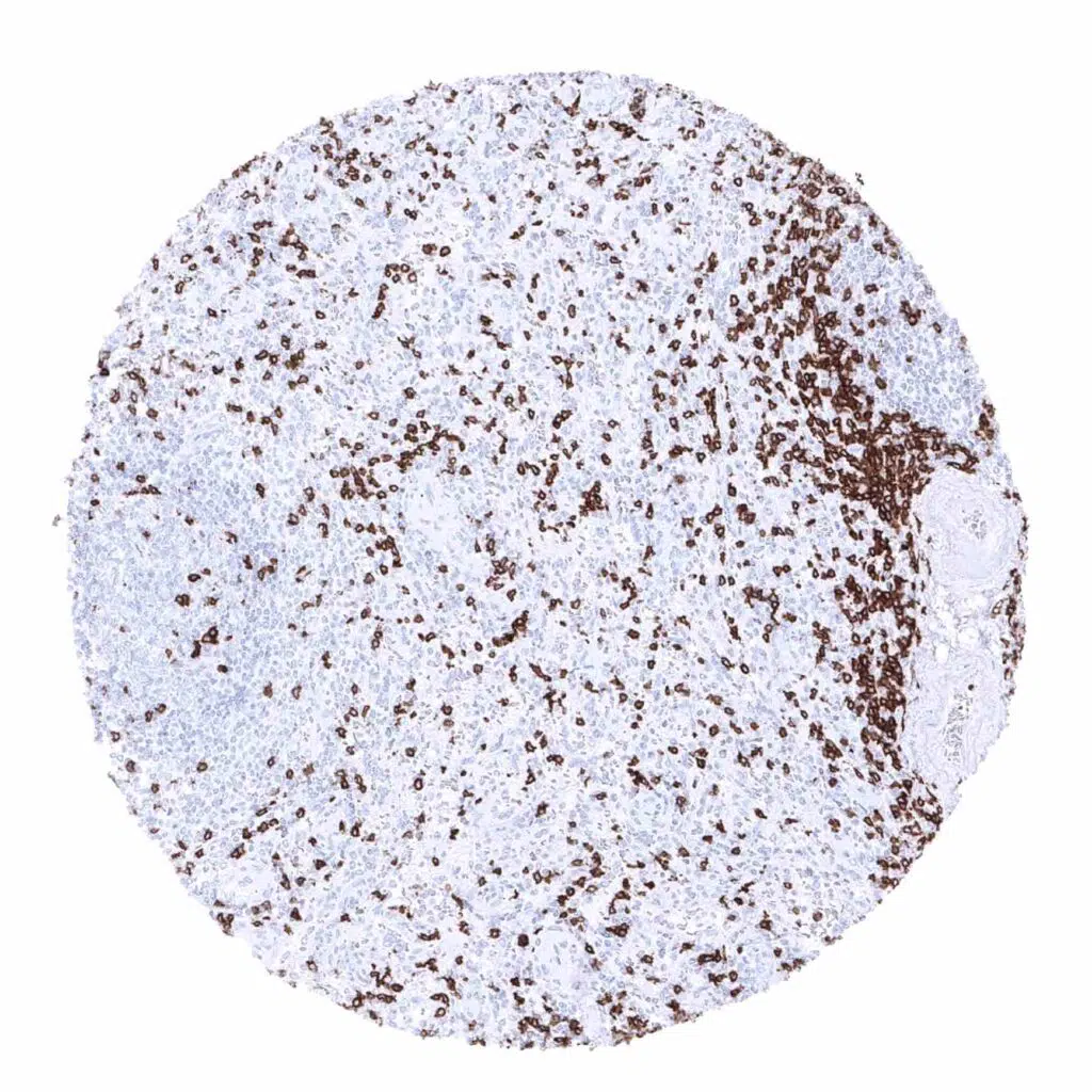
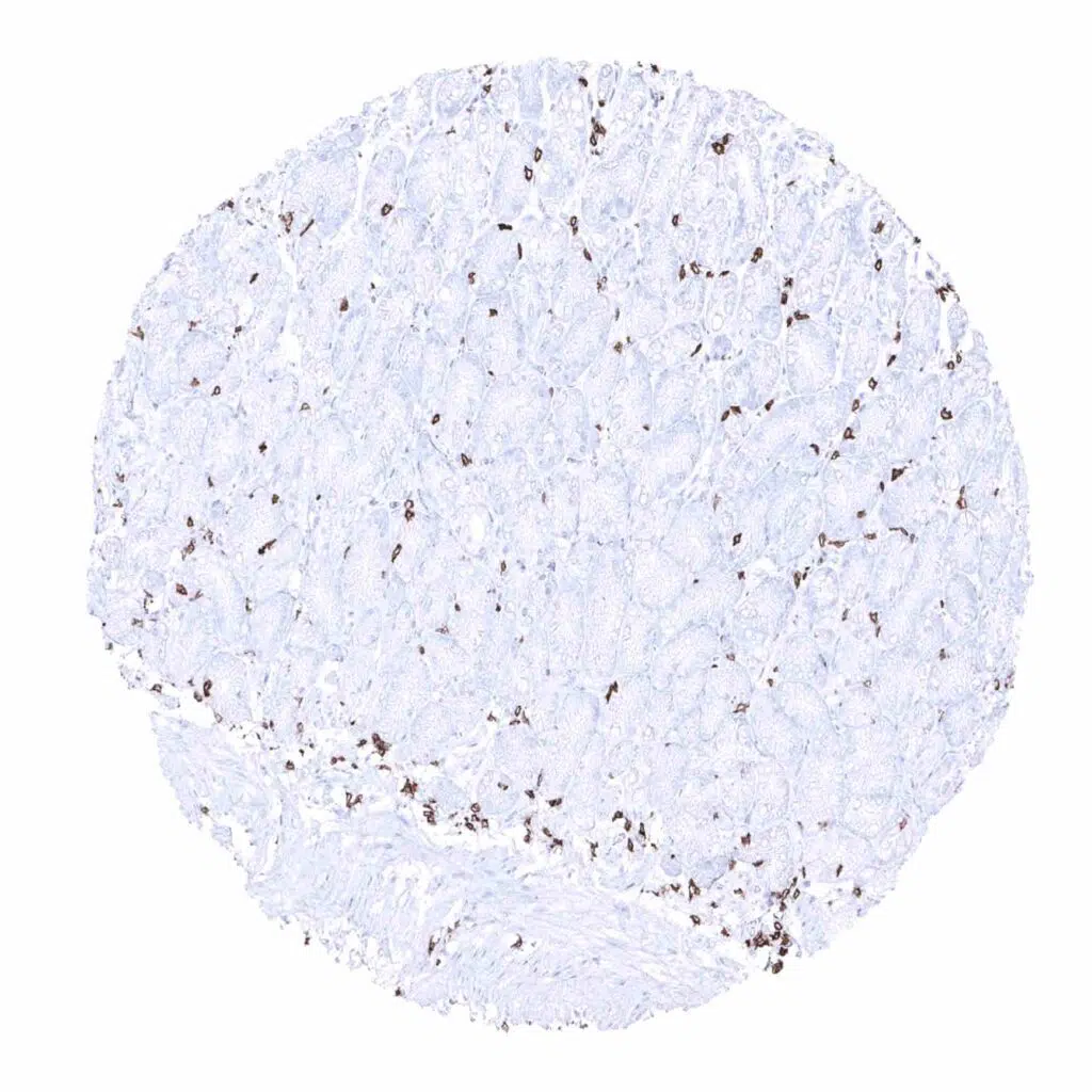
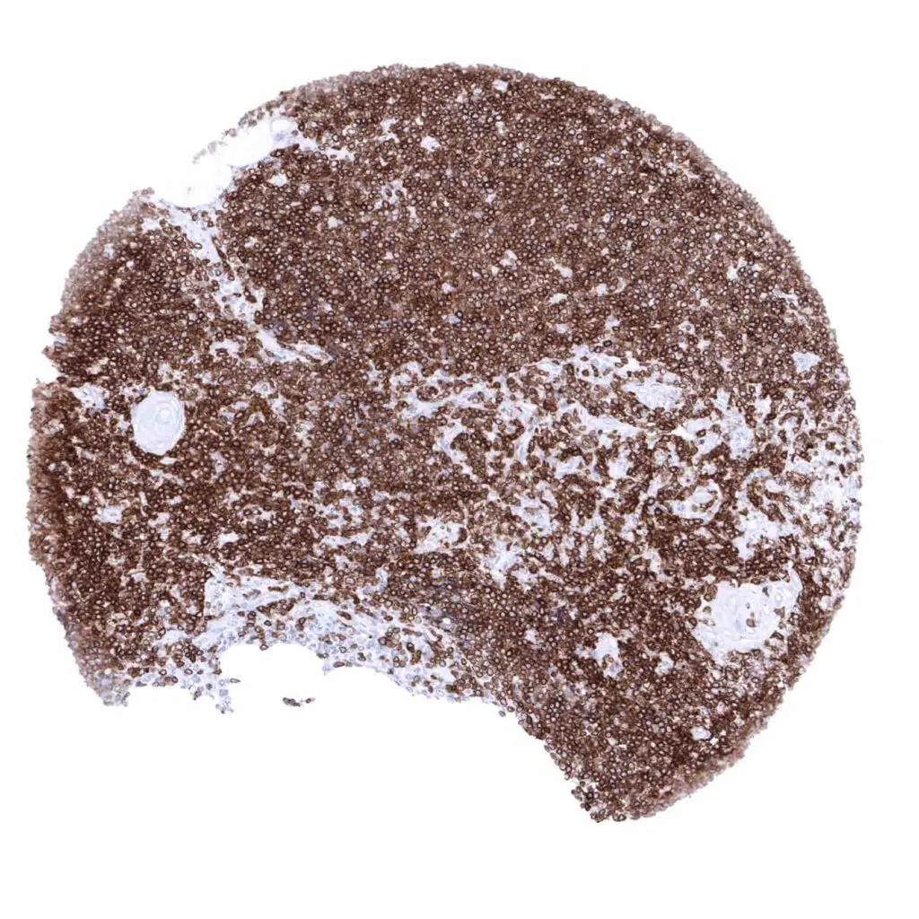
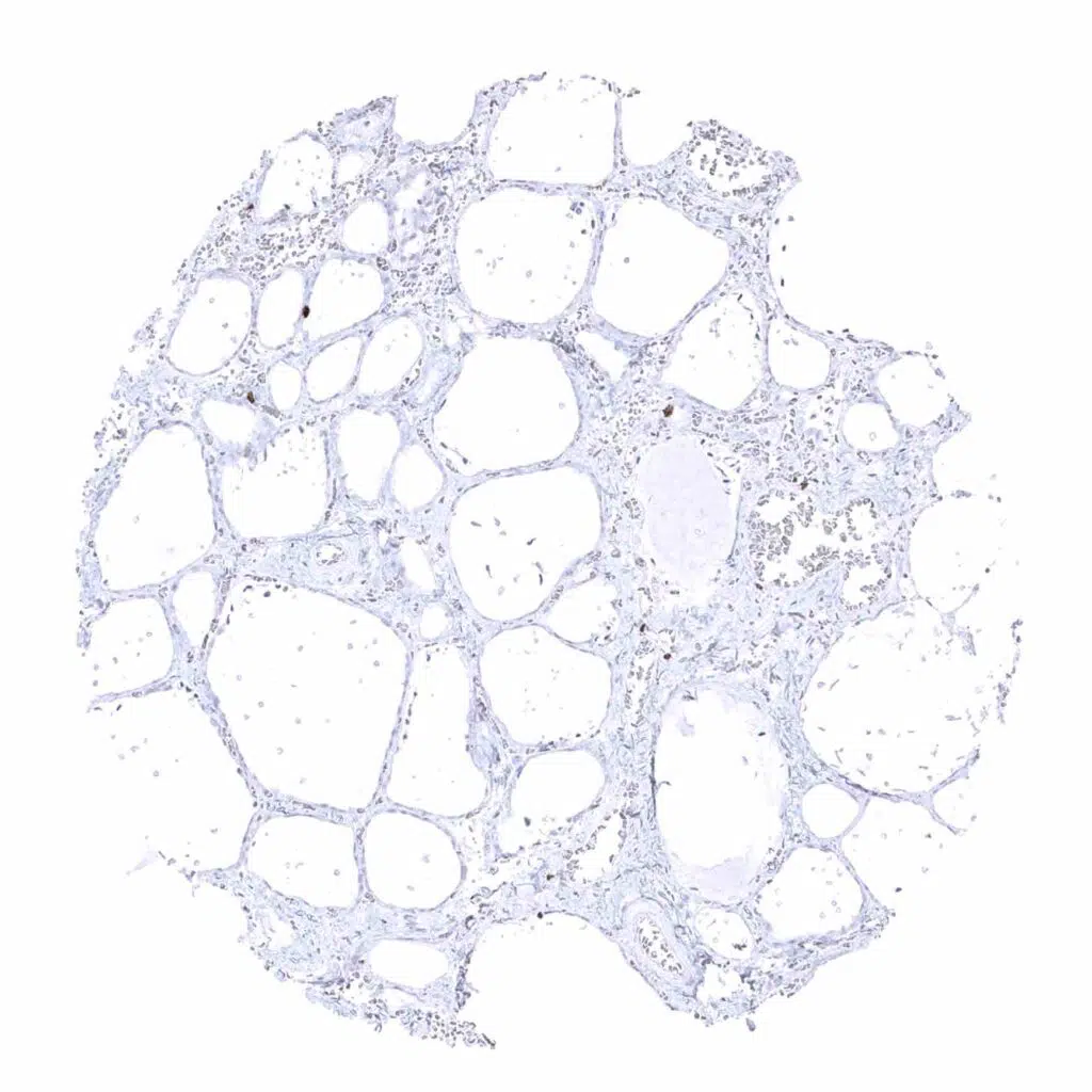
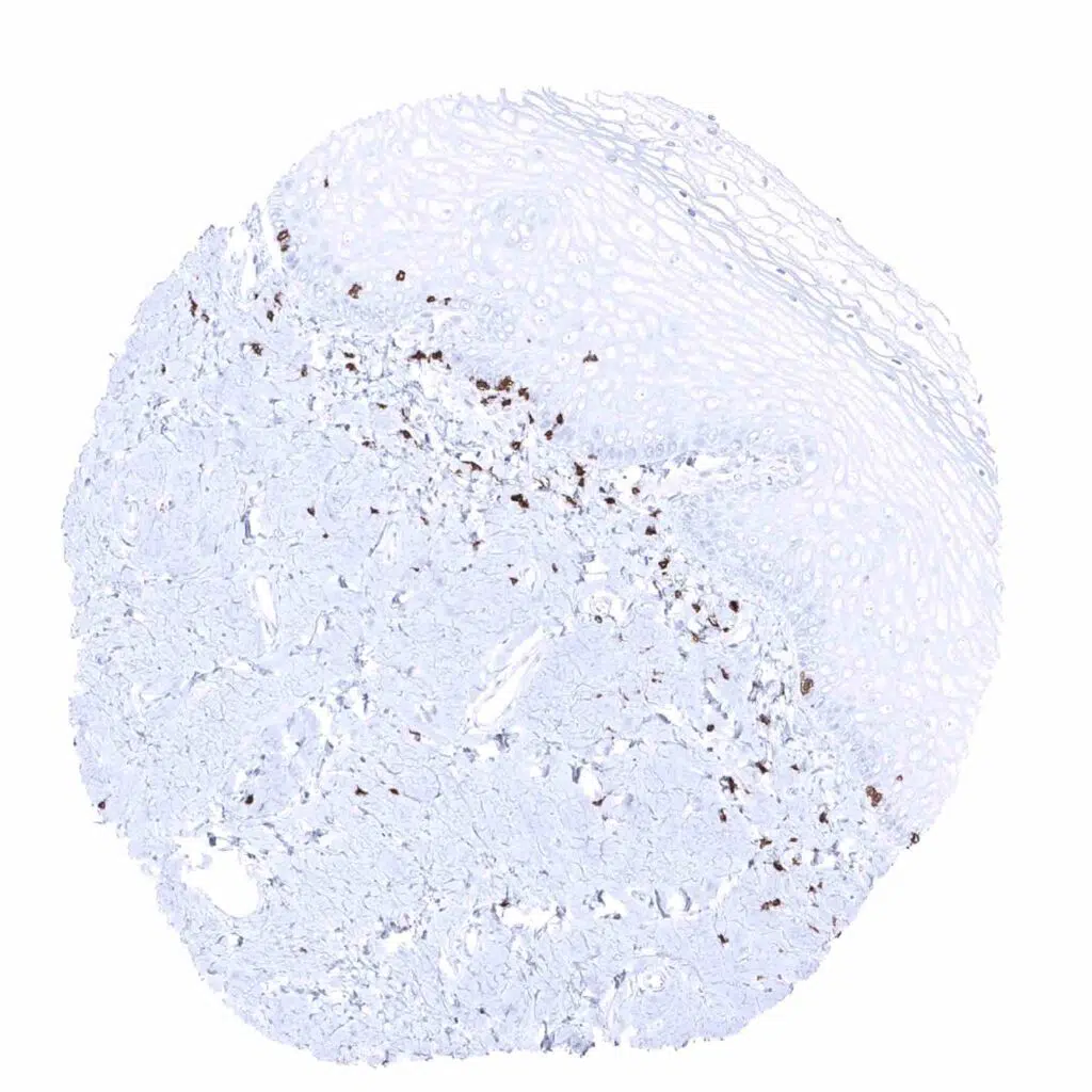
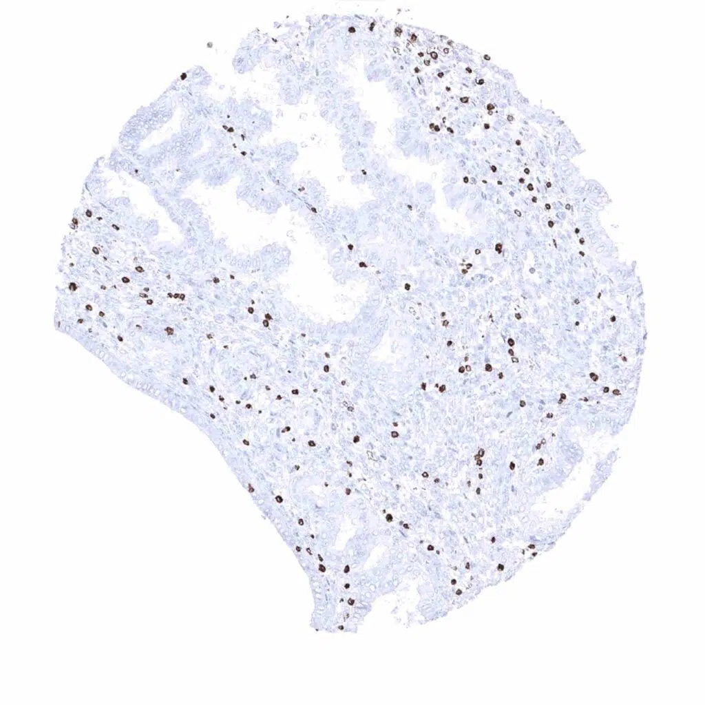
CD3 epsilon antibody [MSVA-003R] HistoMAX


Go to Cancer Tissue Gallery
  
 Go back to product page
  

Go to HistoMAX page


Adrenal gland

Aorta, media

Appendix, mucosa - In the appendix, numerous CD3 positive T-lymphocytes are seen. They occur within the epithelium, in the lamina propria, and - at lower numbers - in germinal centres

Appendix, muscular wall


Bone marrow

Breast

Bronchus, mucosa

Cerebellum (molecular layer, Purkinje cell layer, granule cell layer)


Cerebellum (white matter)

Cerebrum, grey matter

Cerebrum, white matter

Colon descendens, mucosa


Colon descendens, muscular wall

Duodenum, Brunner gland

Duodenum, mucosa - A considerable number of CD3 positive T-lymphocytes is regularly seen in the lamina propria of the duodeum

Epididymis


Esophagus, squamous epithelium

Fallopian tube, mucosa

Fat

Gallbladder, epithelium


Heart muscle

Ileum, mucosa

Kidney, cortex

Kidney, medulla


Liver

Lung

Lymph node

Ovary, stroma


Pancreas

Parathyroid gland

<

Parotid gland

Pituitary gland, anterior lobe


Pituitary gland, posterior lobe

Placenta (amnion)

Placenta (chorion)

Placenta, early


Placenta, mature

Prostate

Rectum, mucosa

Seminal vesicle


Sinus paranasales

Skeletal muscle

Skin

Spleen


Stomach, antrum

Stomach, corpus

Testis

Thymus - The highest density of CD3 positive T-lymphocytes is seen in the thymus


Thyroid gland

Tonsil

Urinary bladder, muscular wall

Urinary bladder, urothelium


Uterus, ectocervix

Uterus, endocervix

Uterus, endometrium (pregnancy)

Uterus, endometrium (proliferation)


Uterus, endometrium (secretion)

Uterus, myometrium


| TOP |