Normal Tissue Gallery- GTX04374

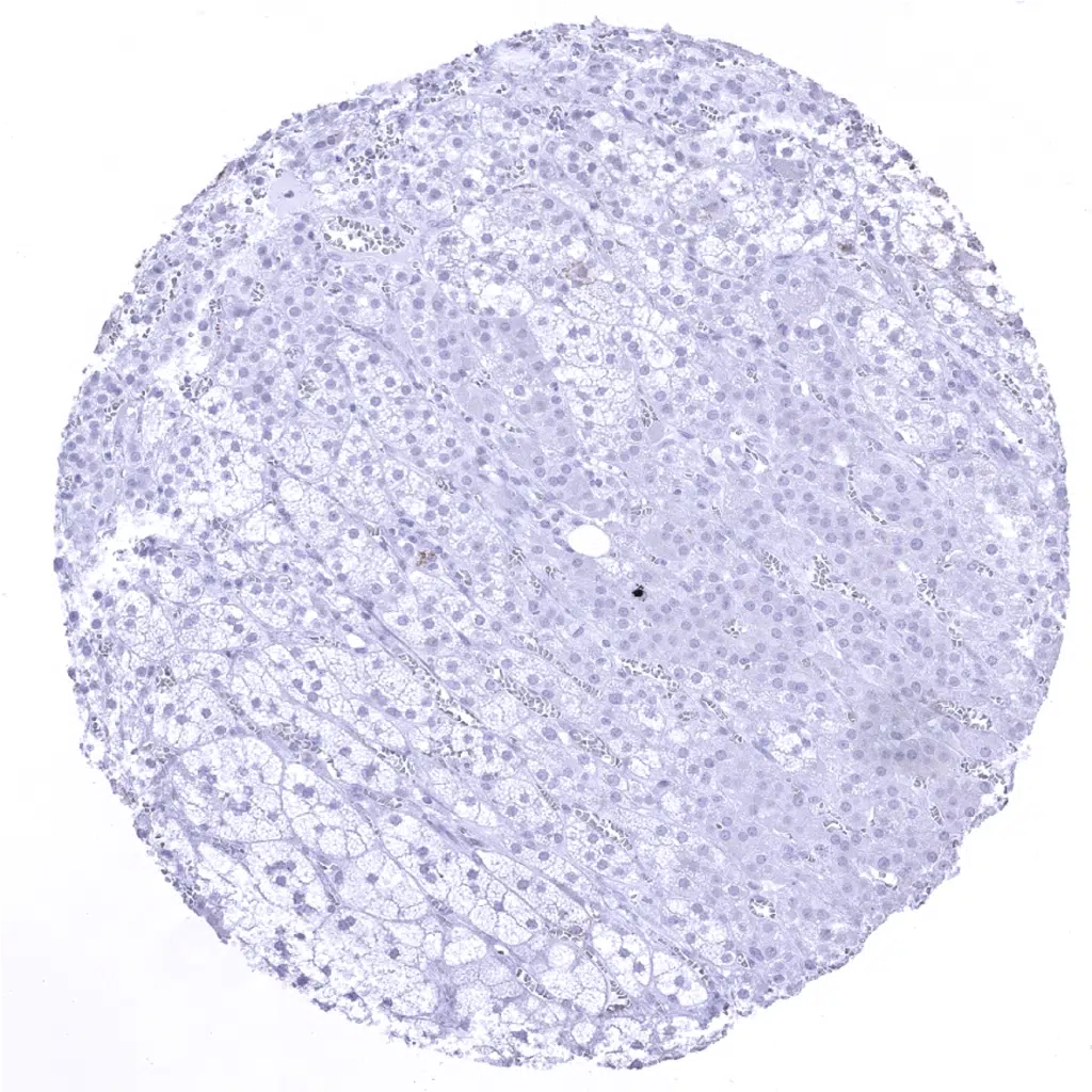
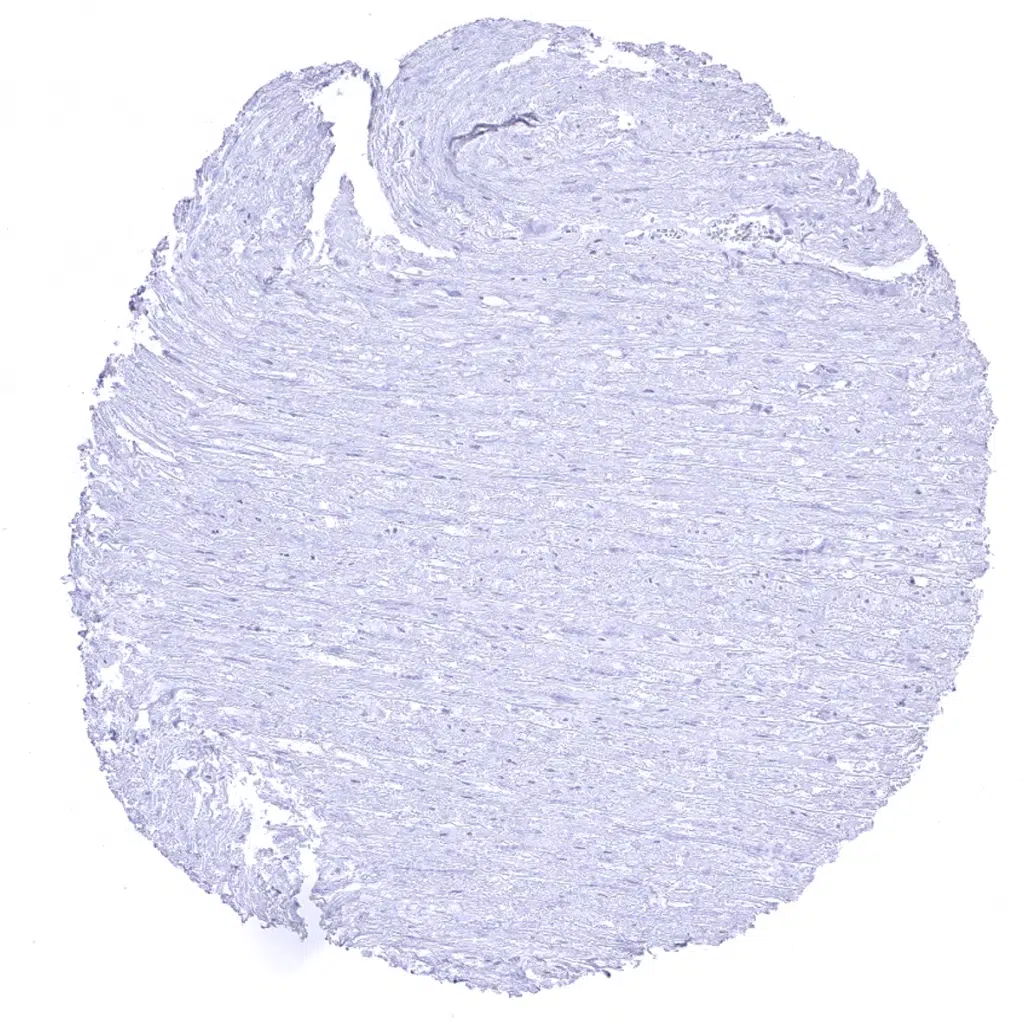
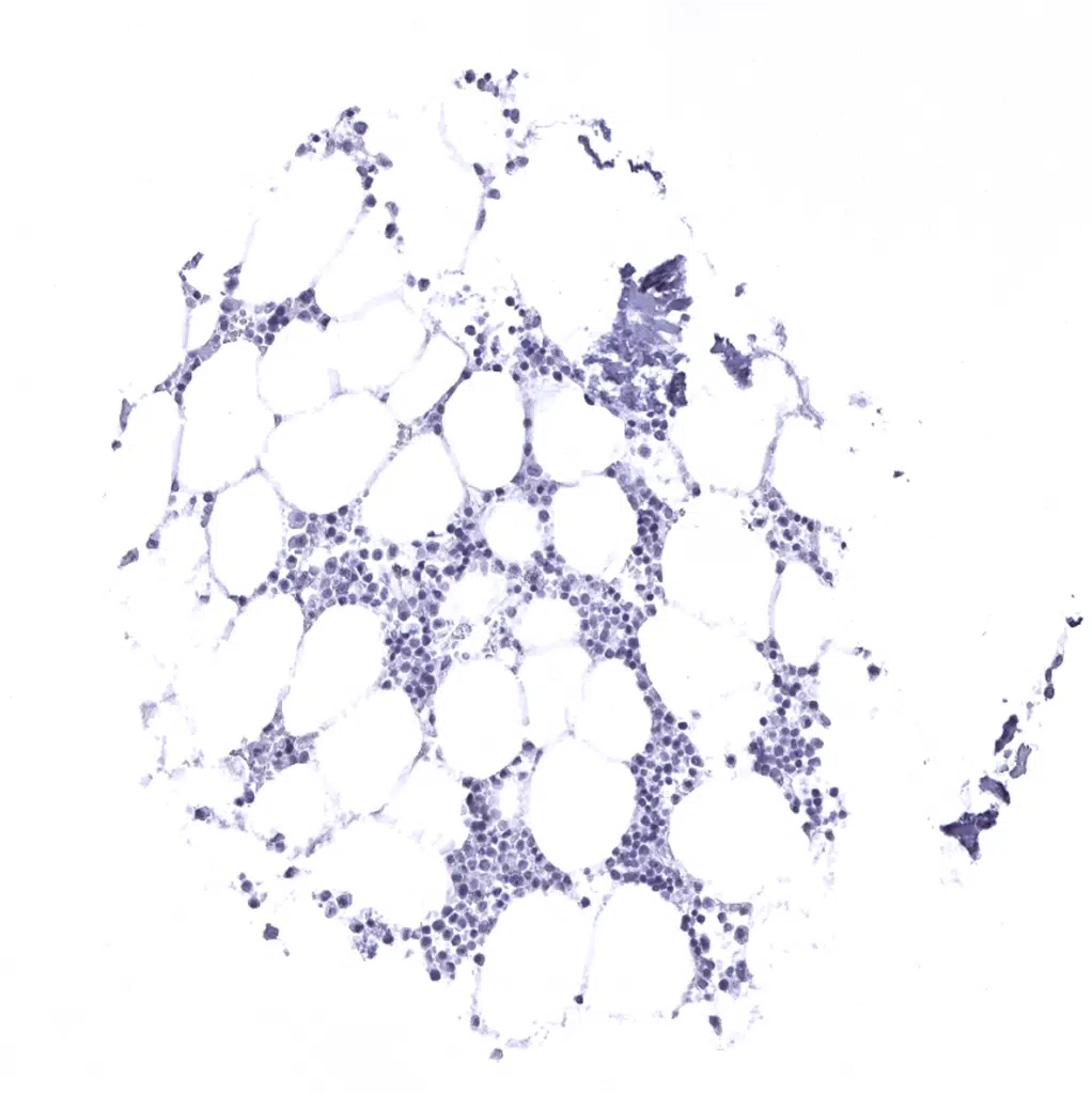
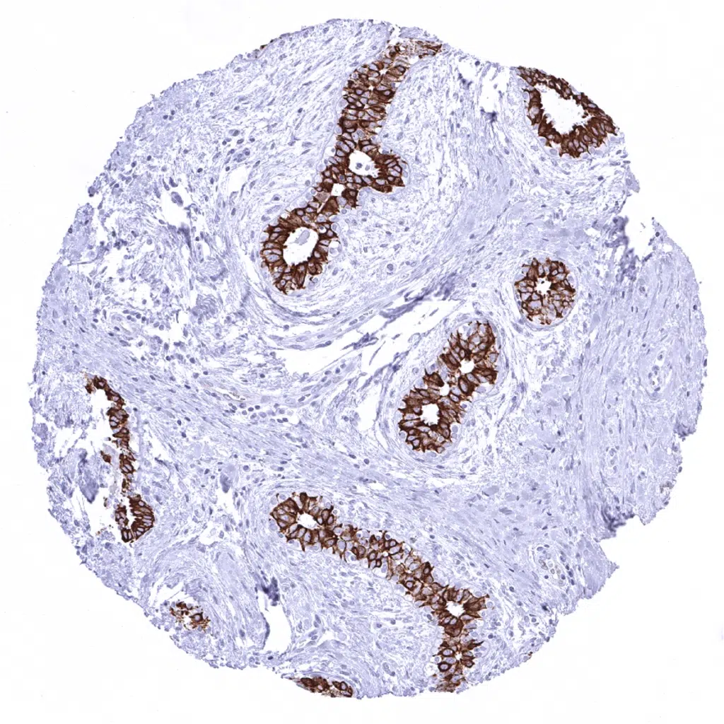
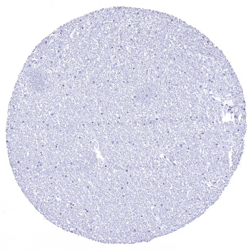
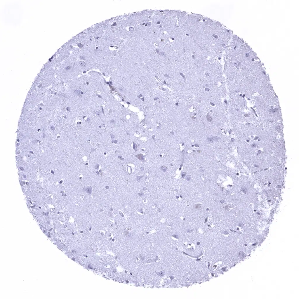
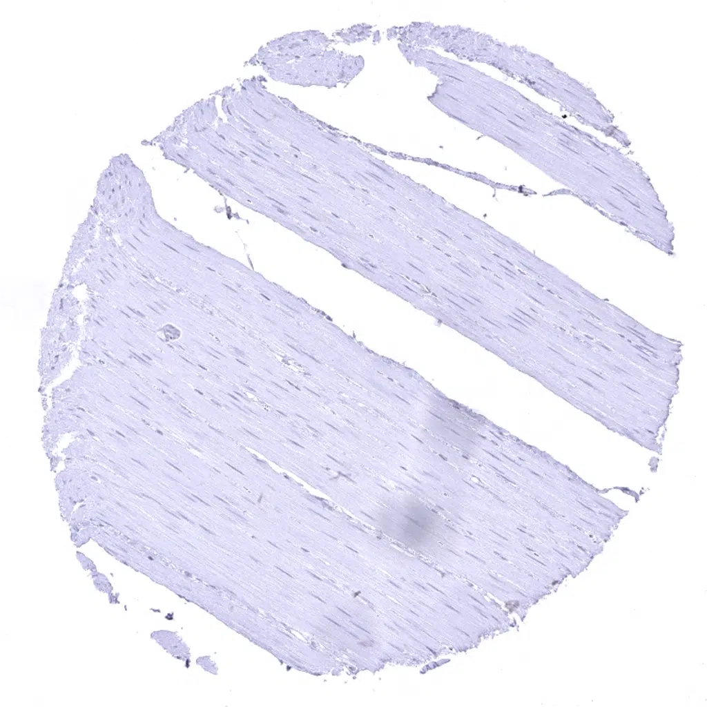
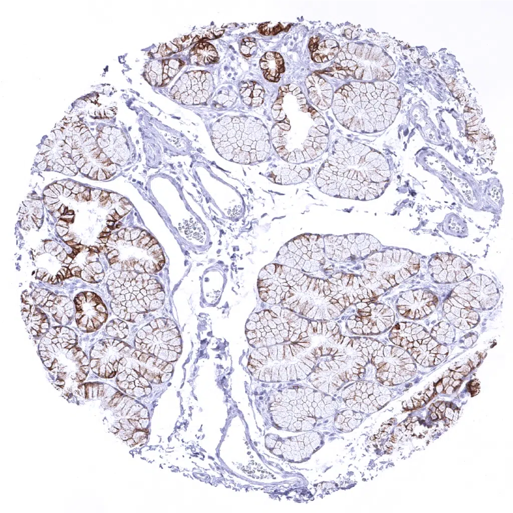
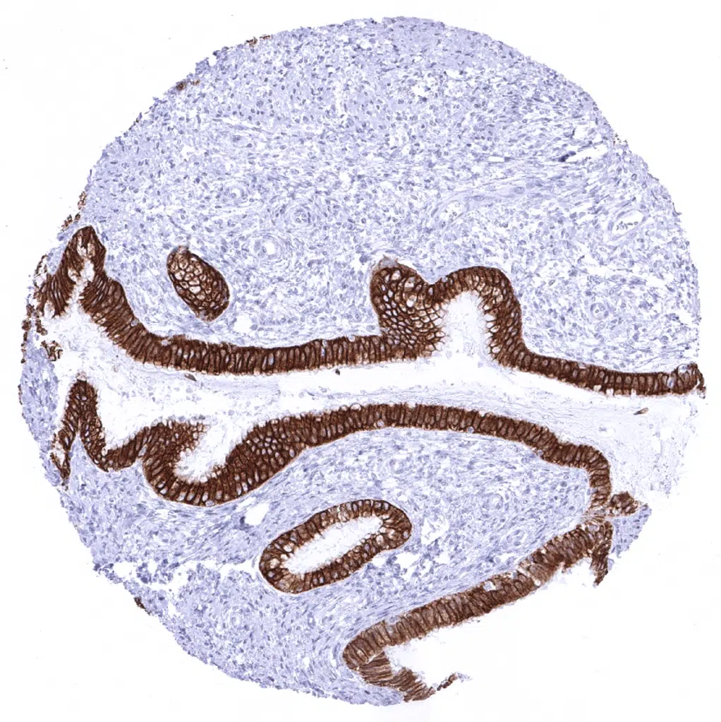
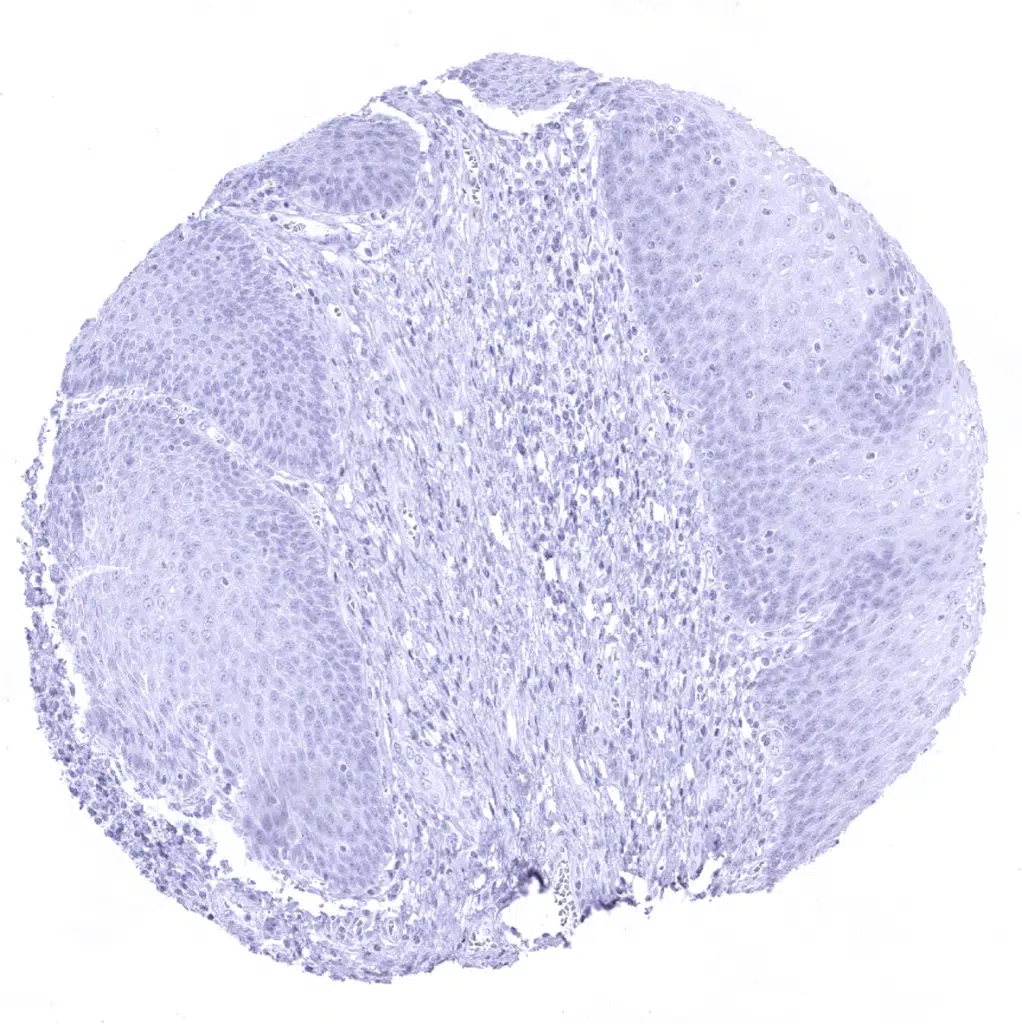
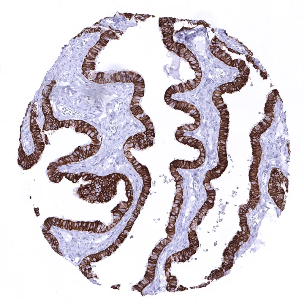
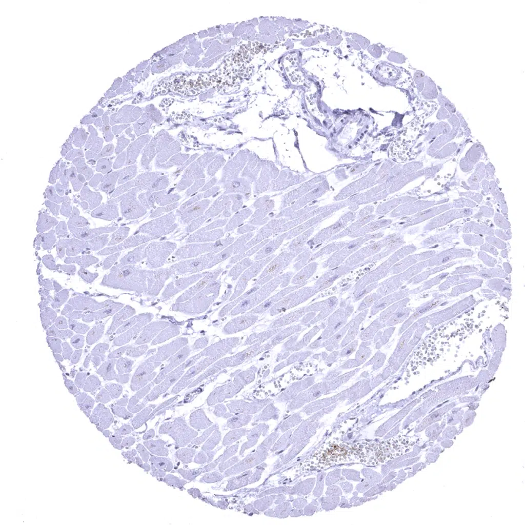
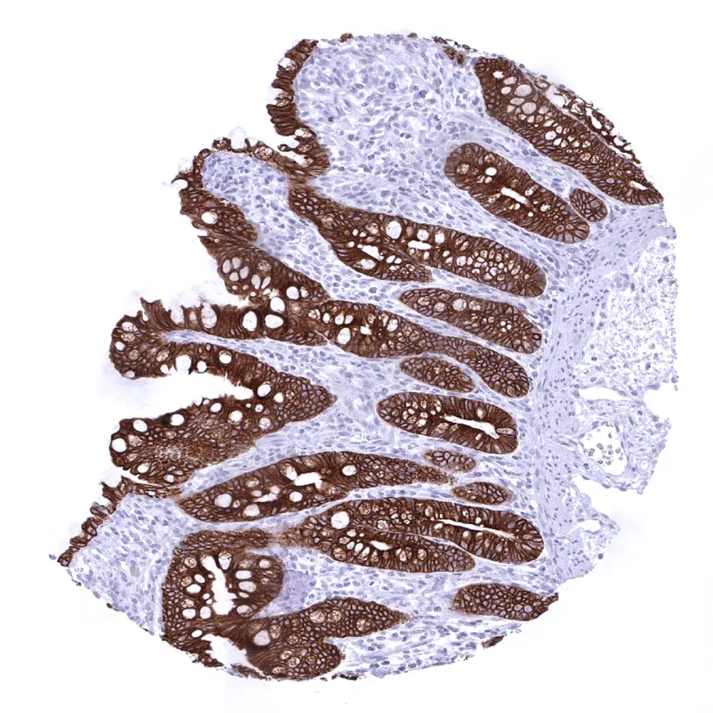
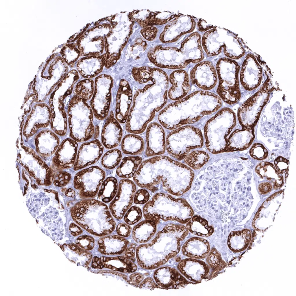
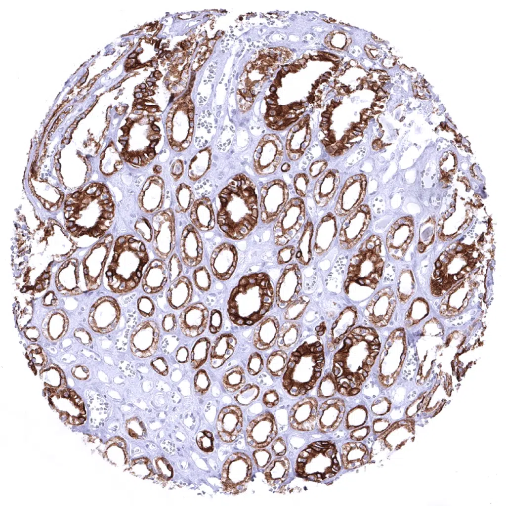
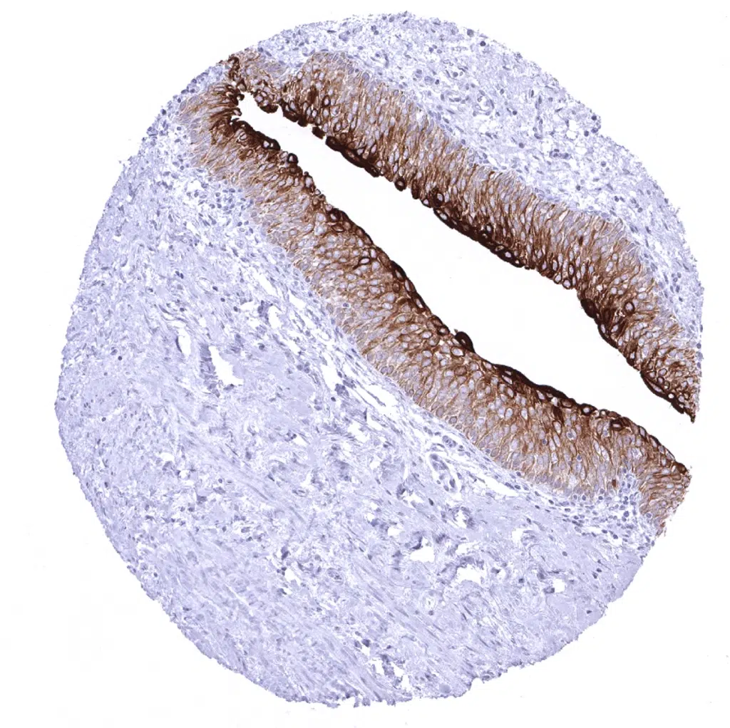
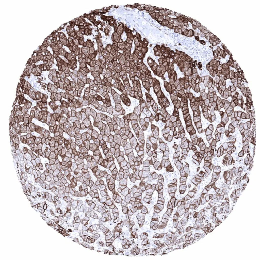
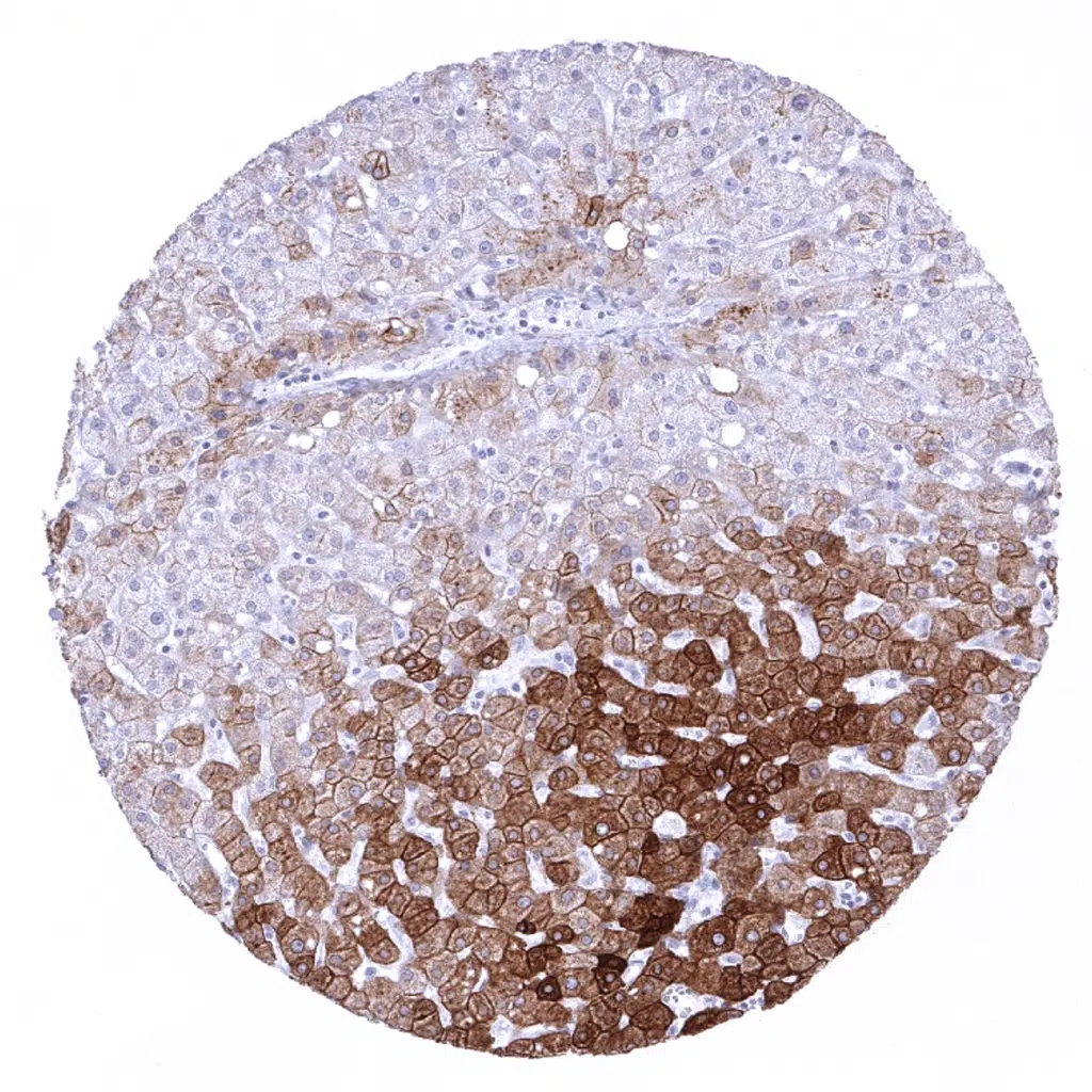
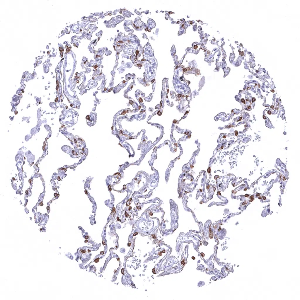
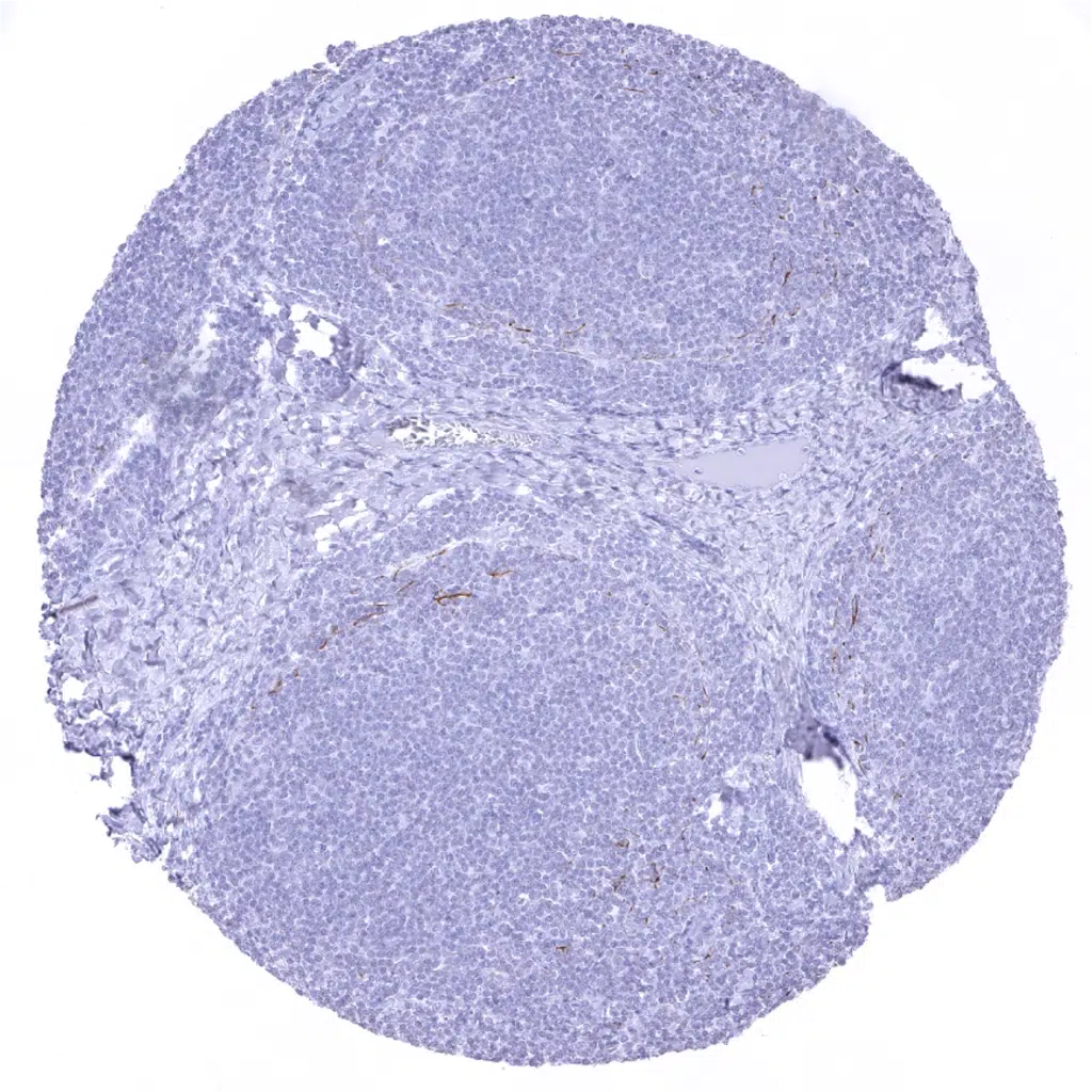
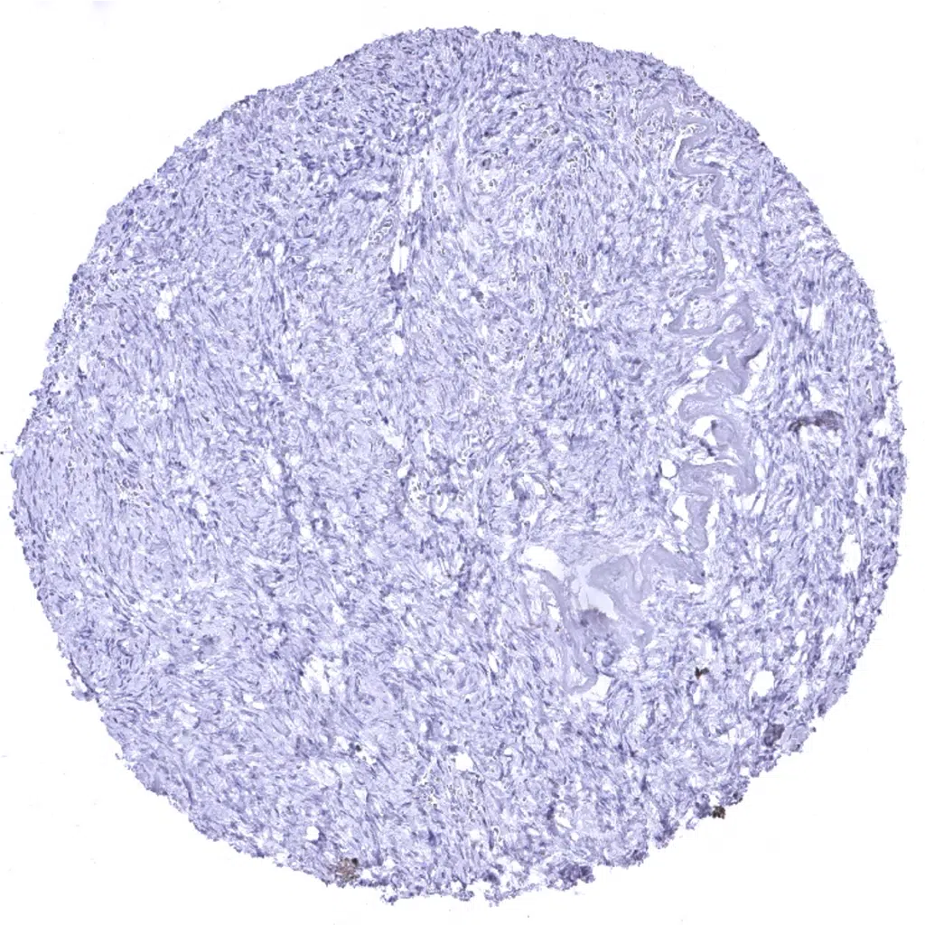
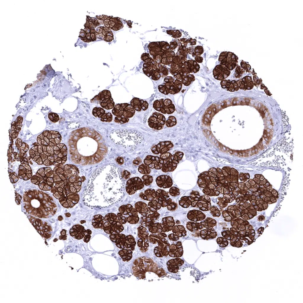
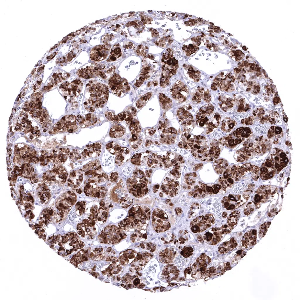
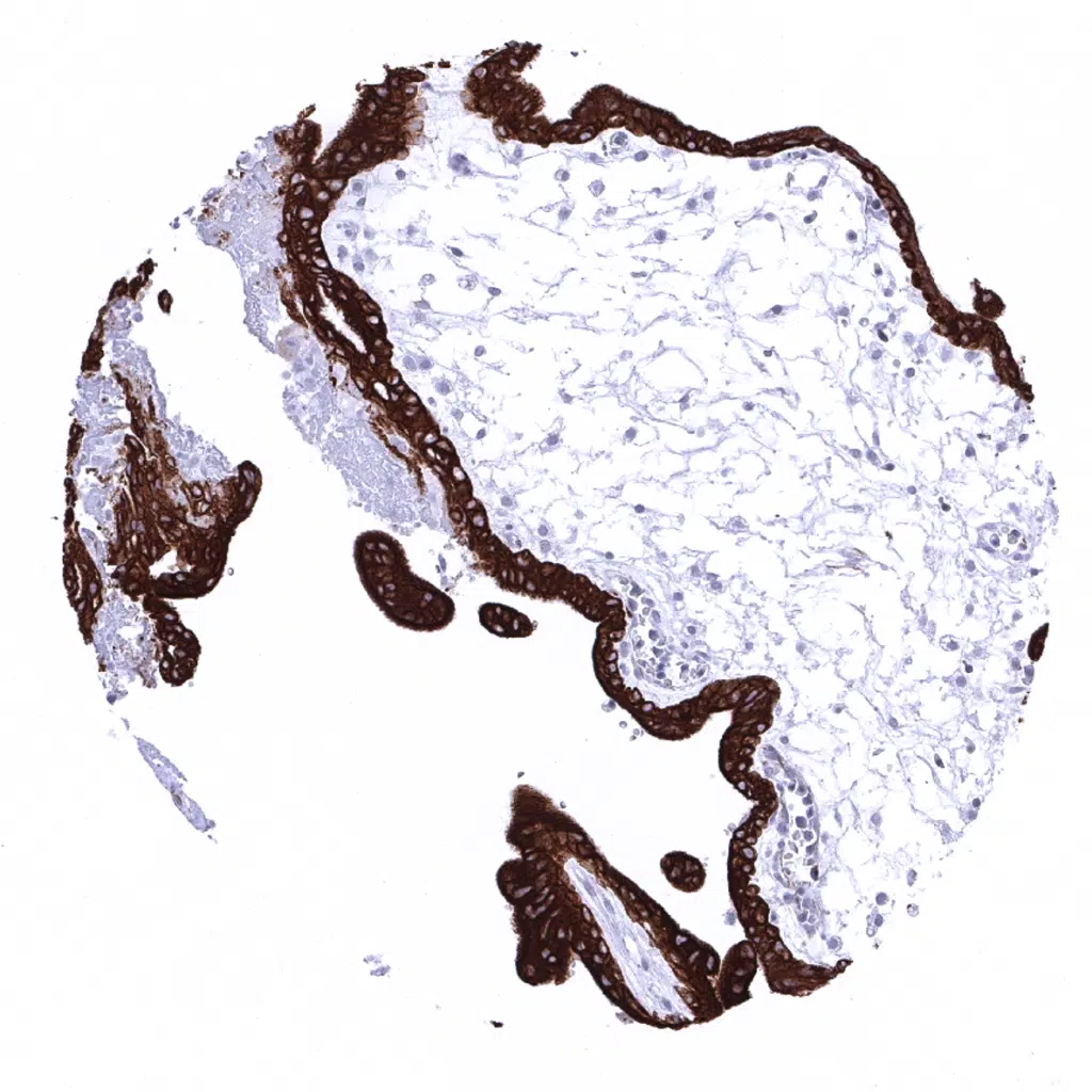
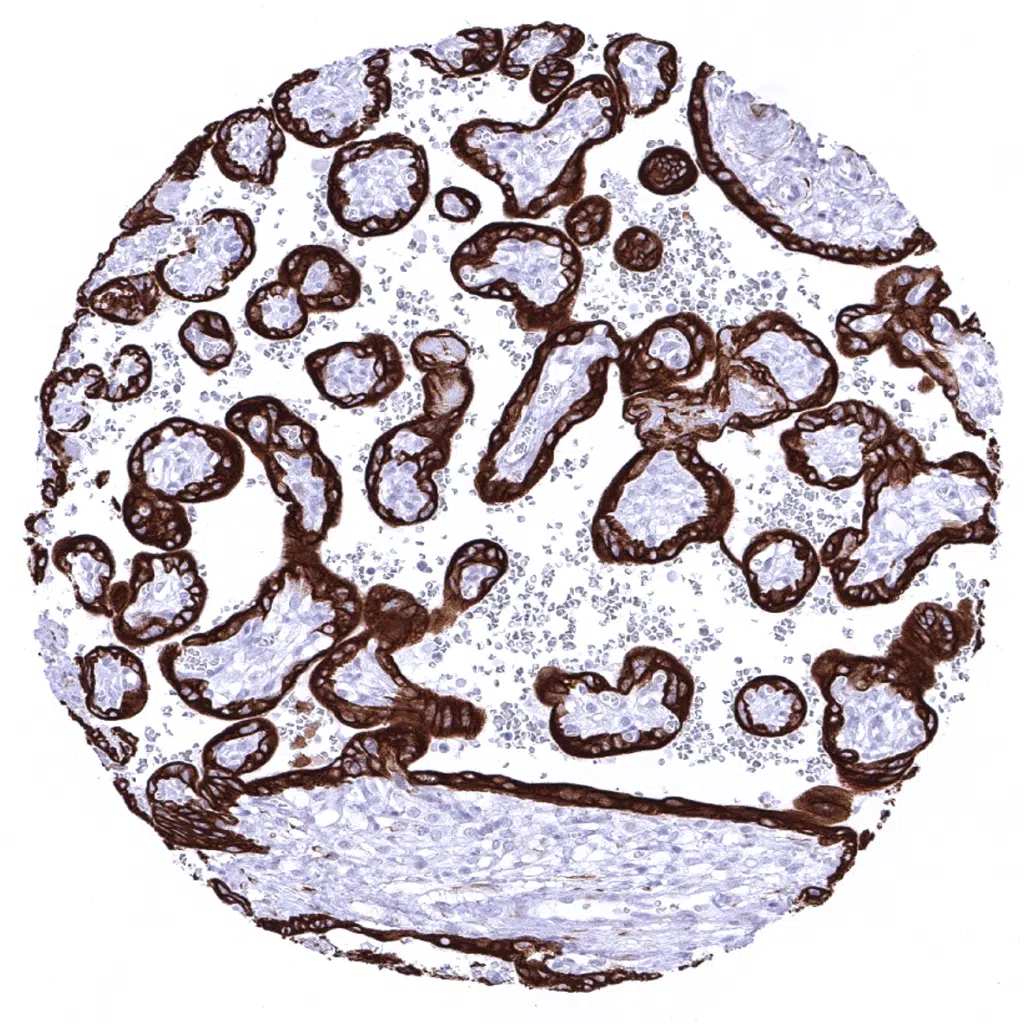
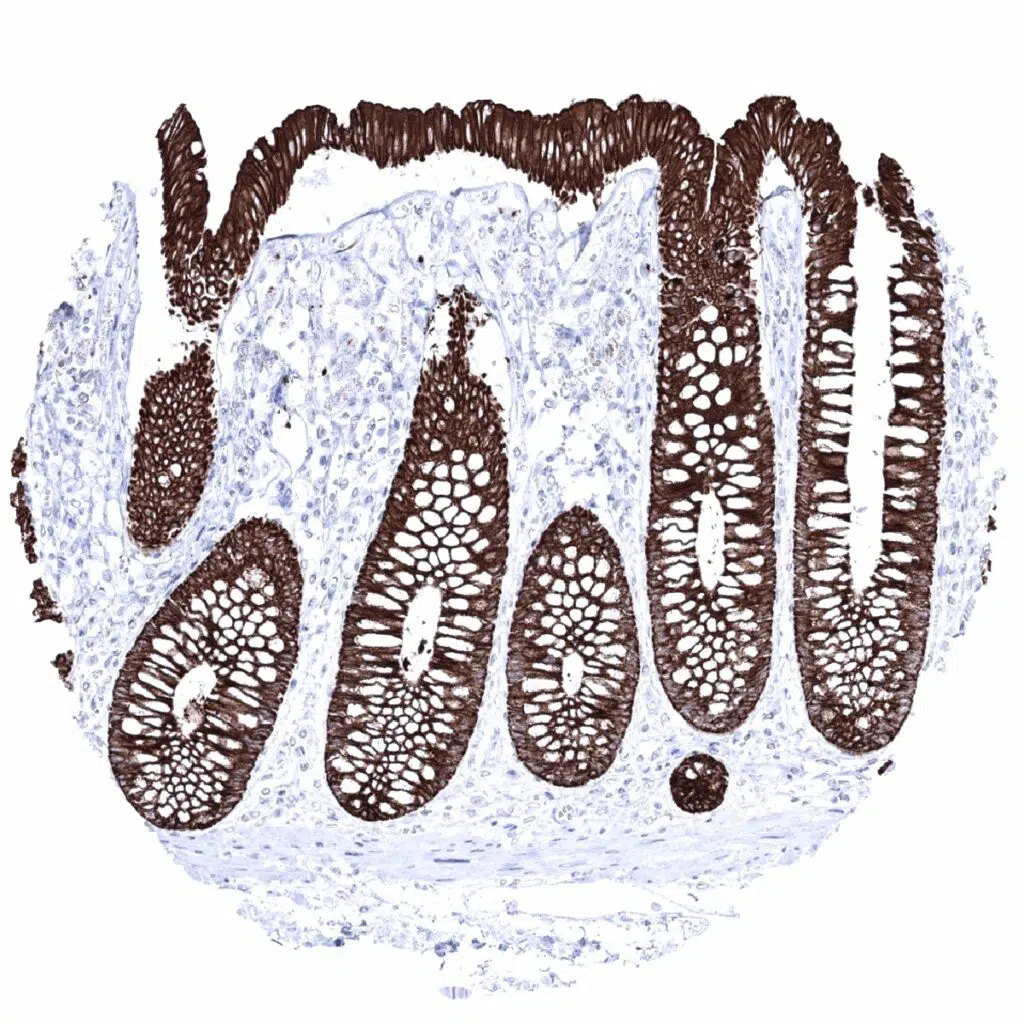
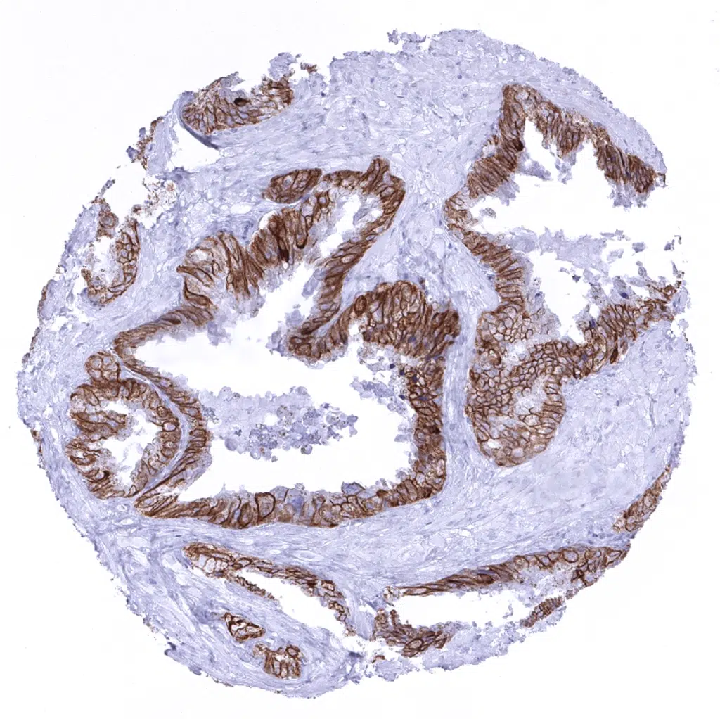
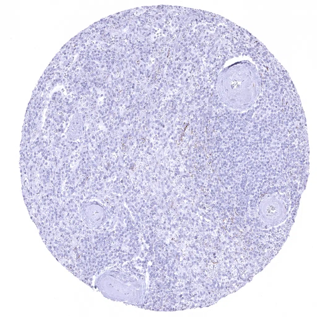
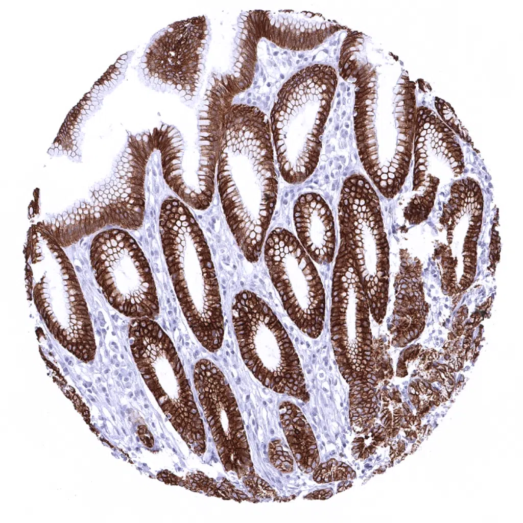
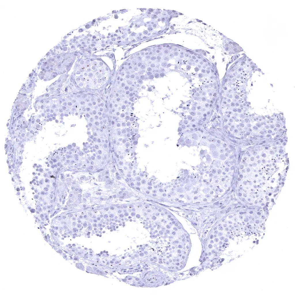
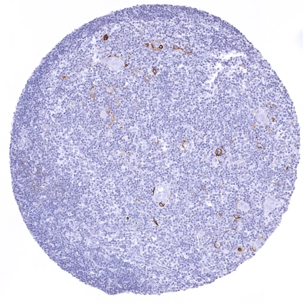
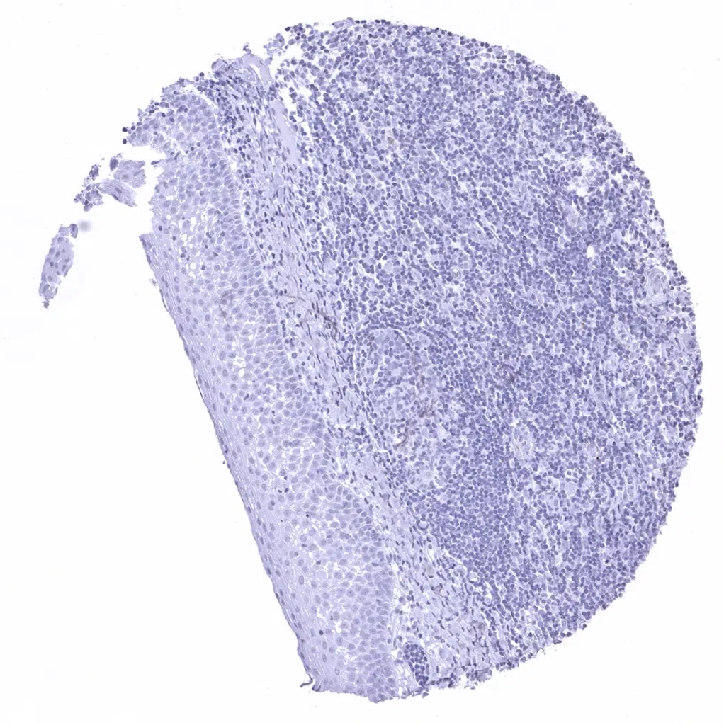
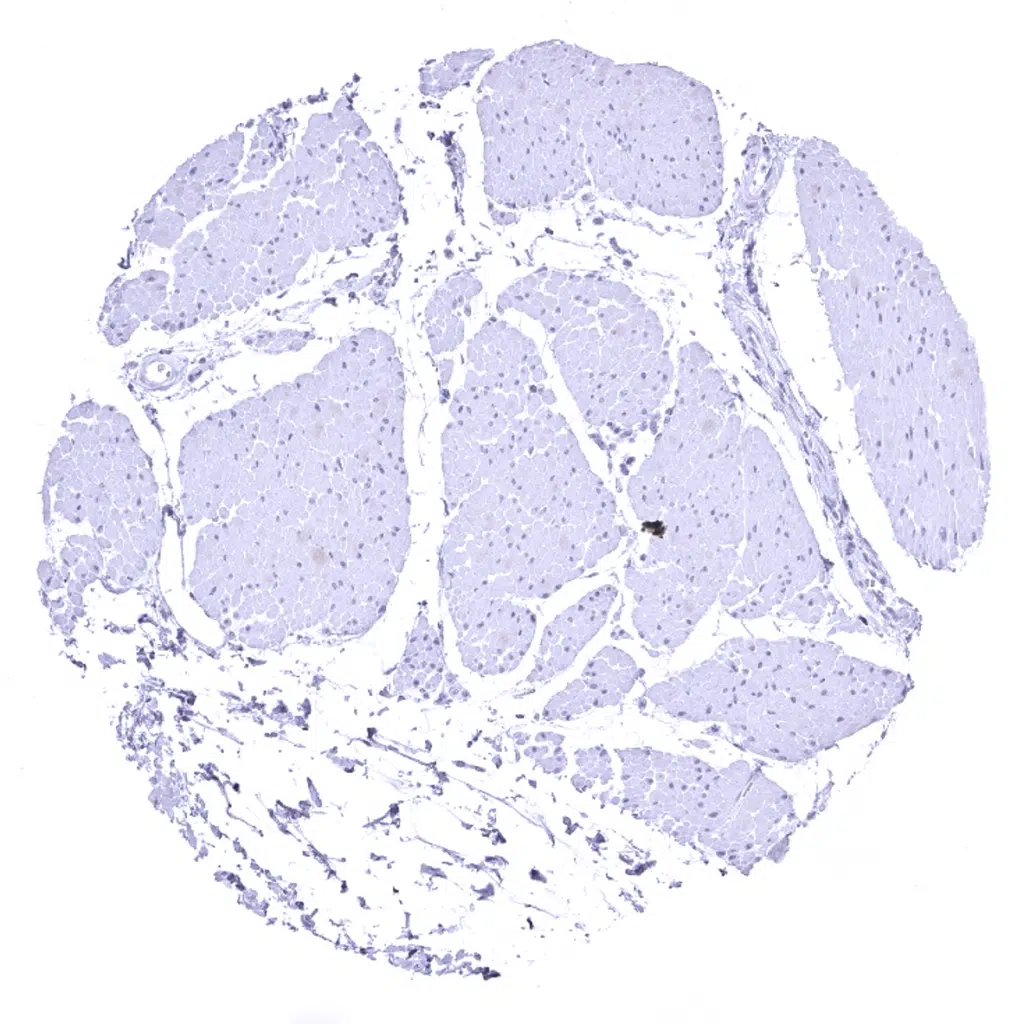
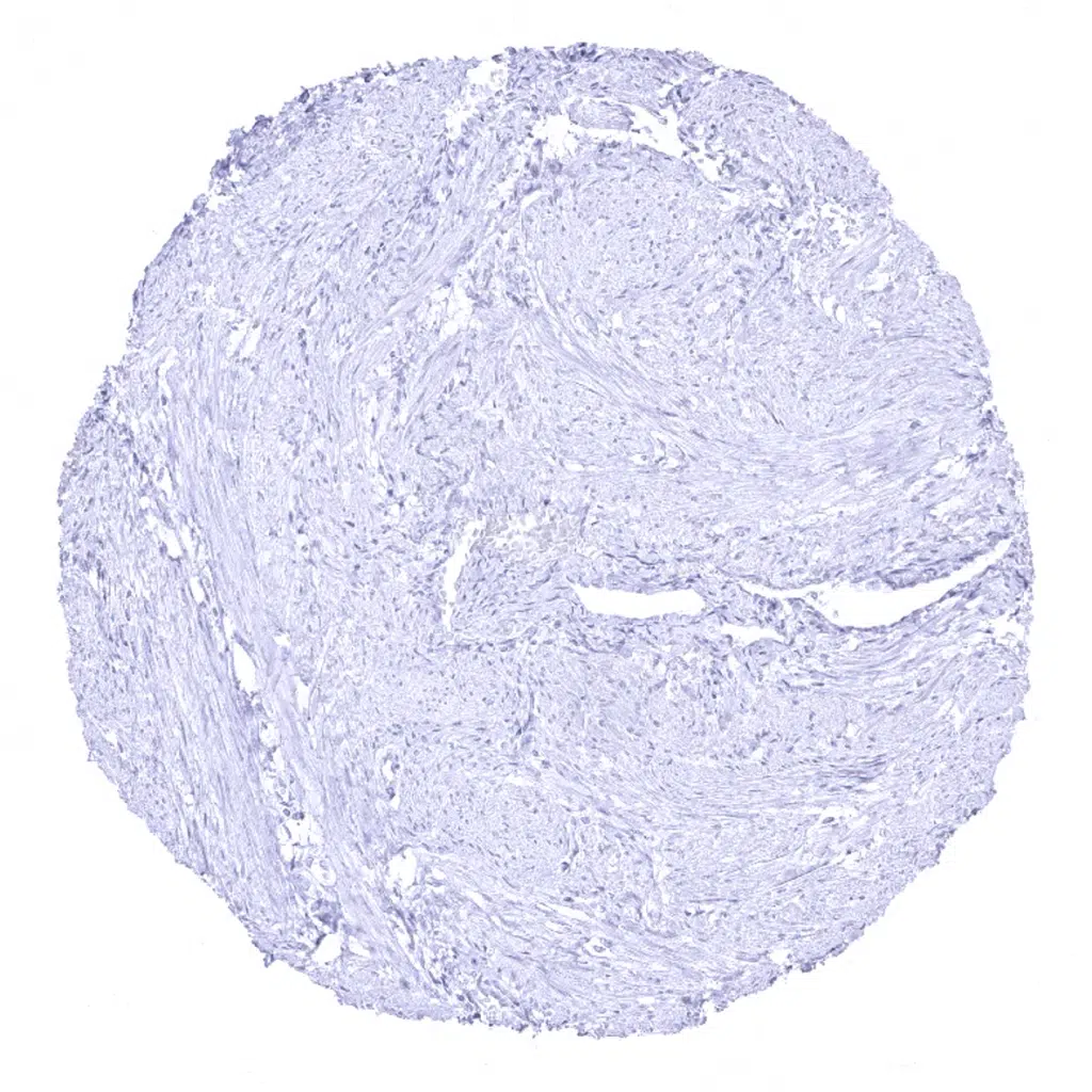
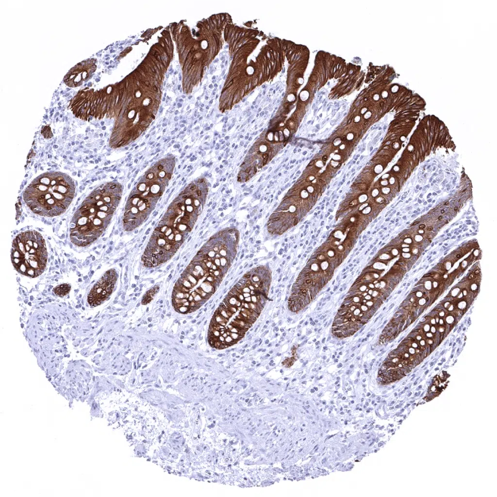
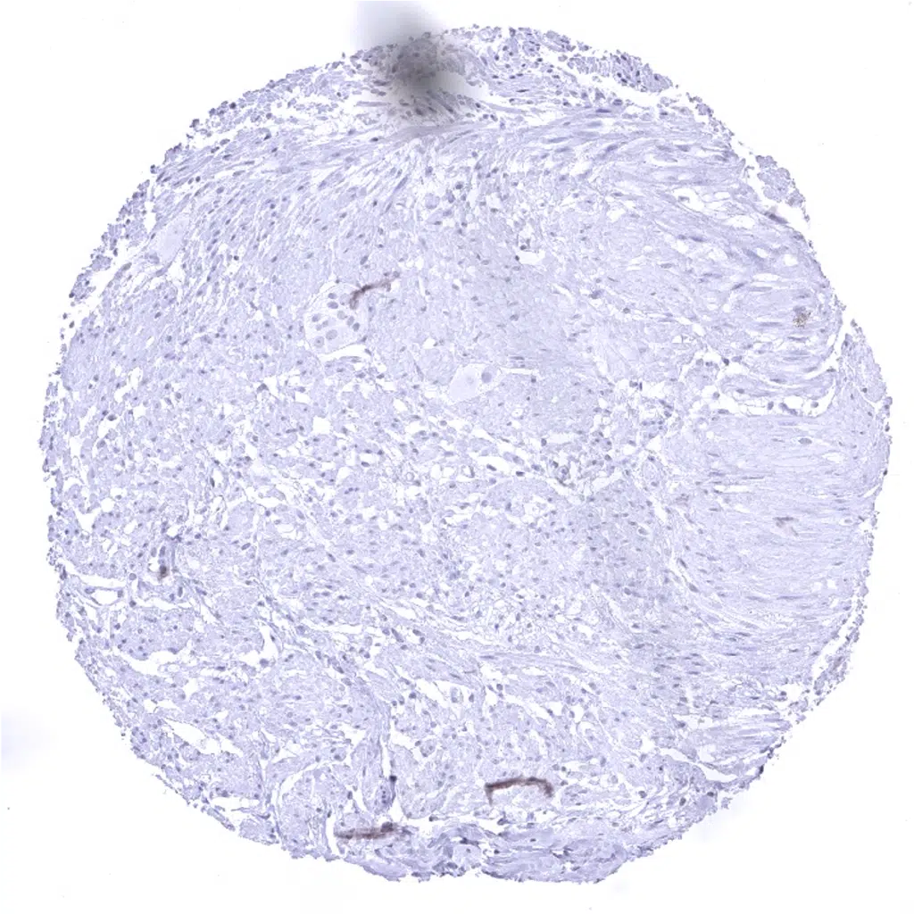
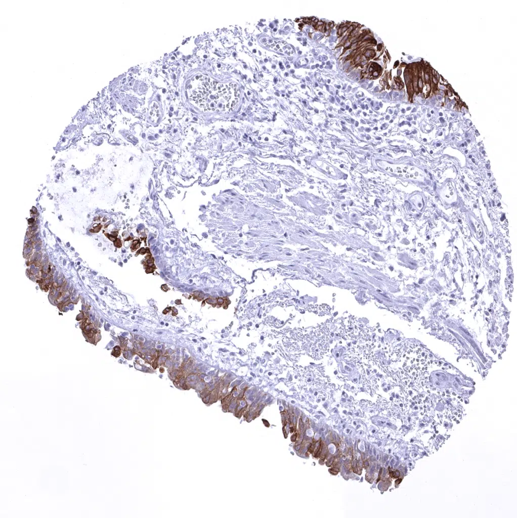
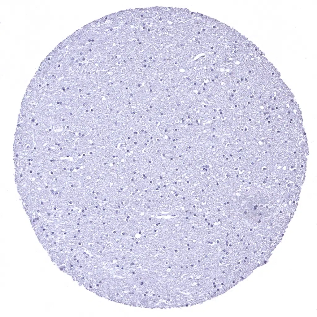
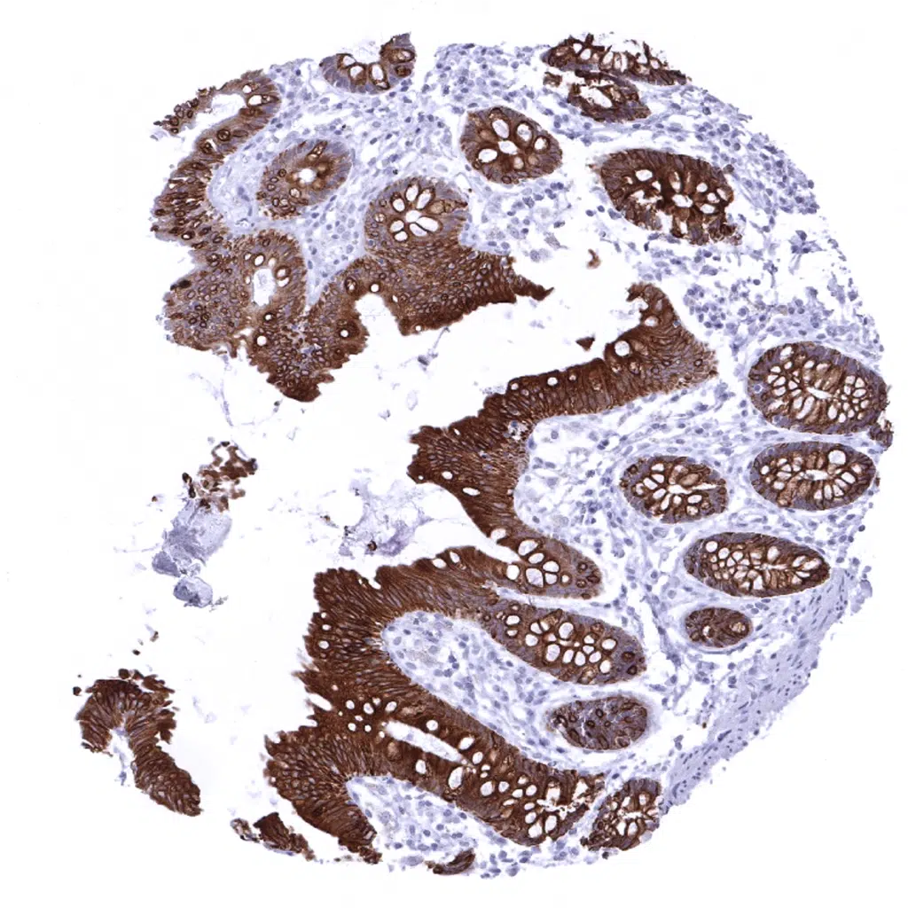
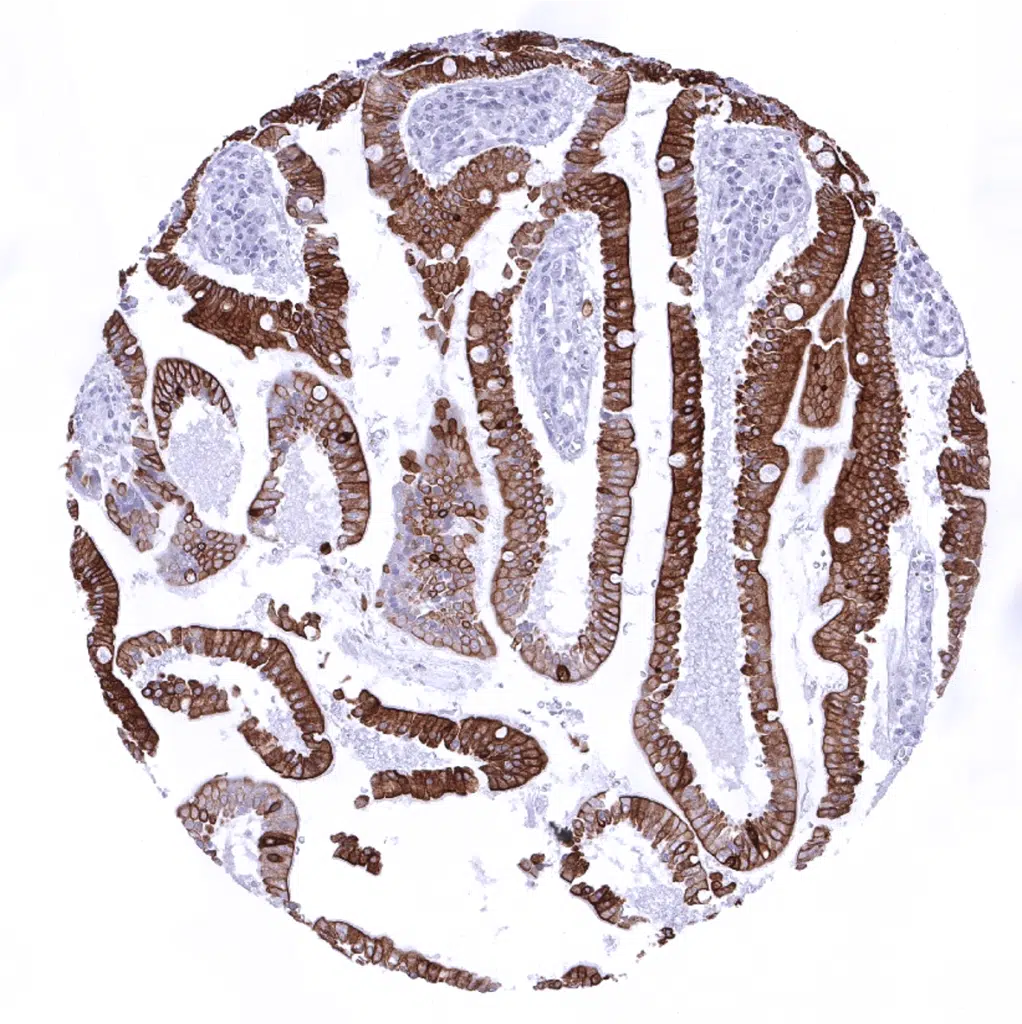
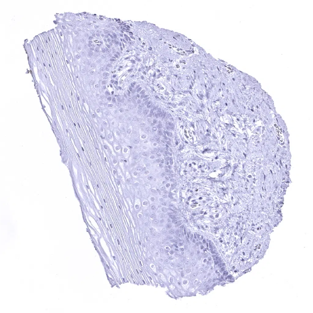
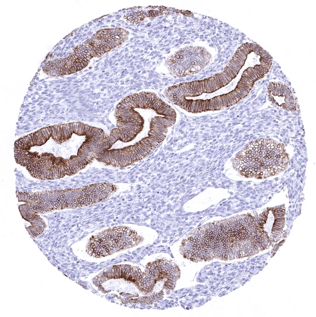
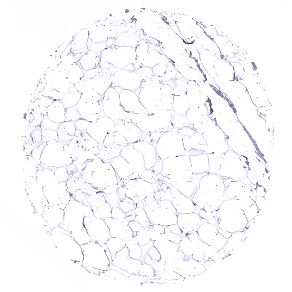
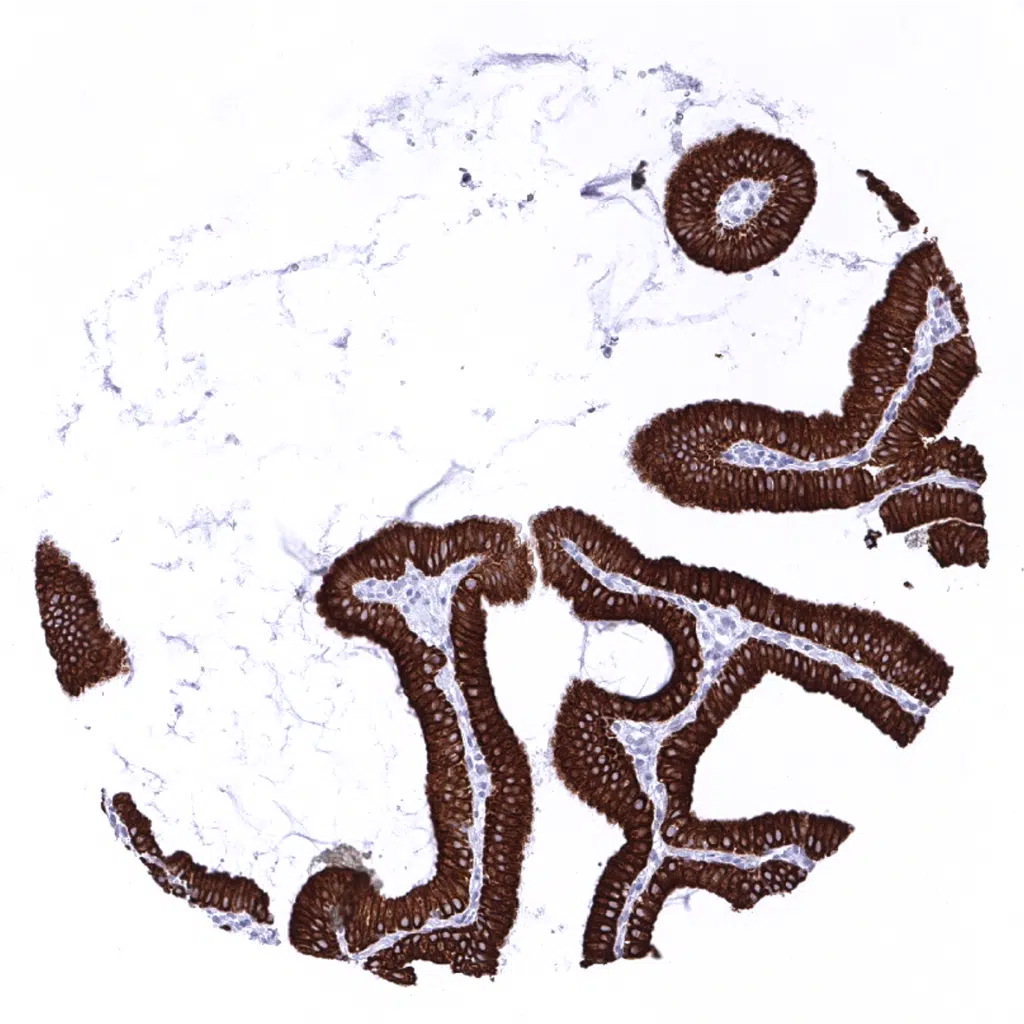
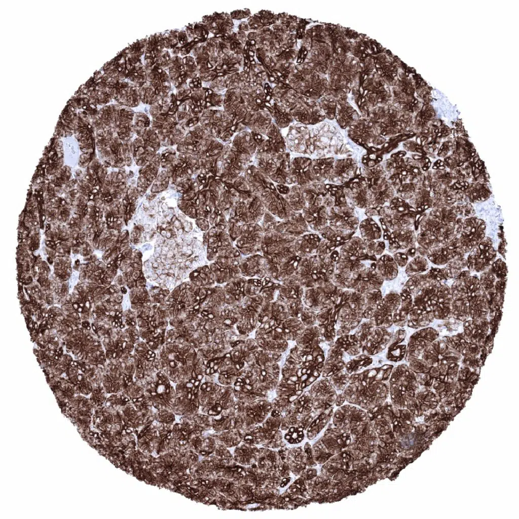
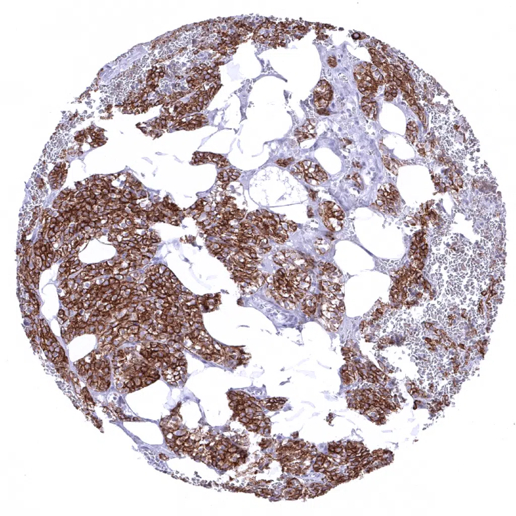
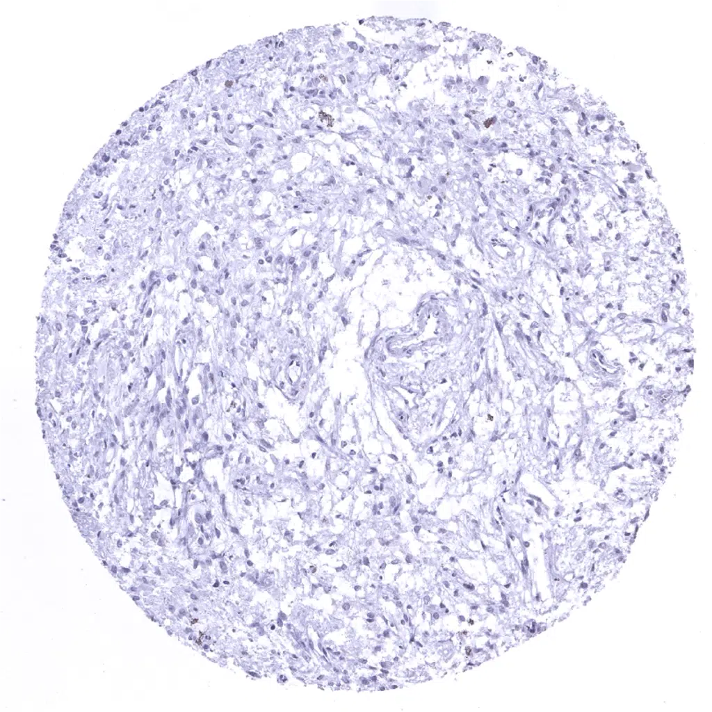
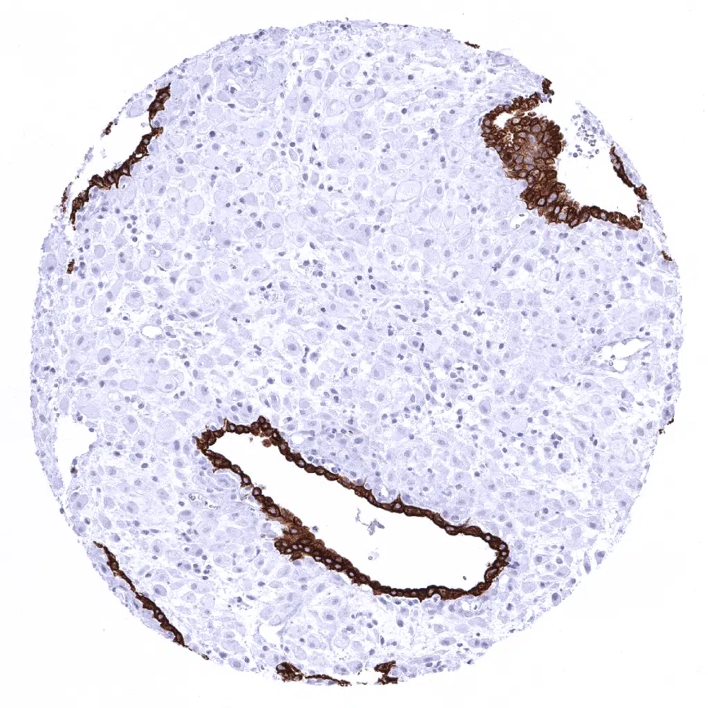
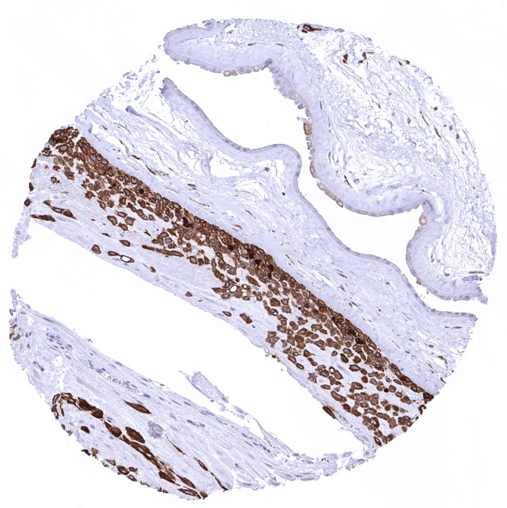
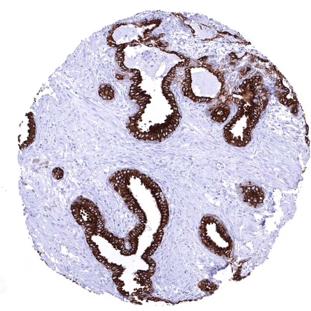
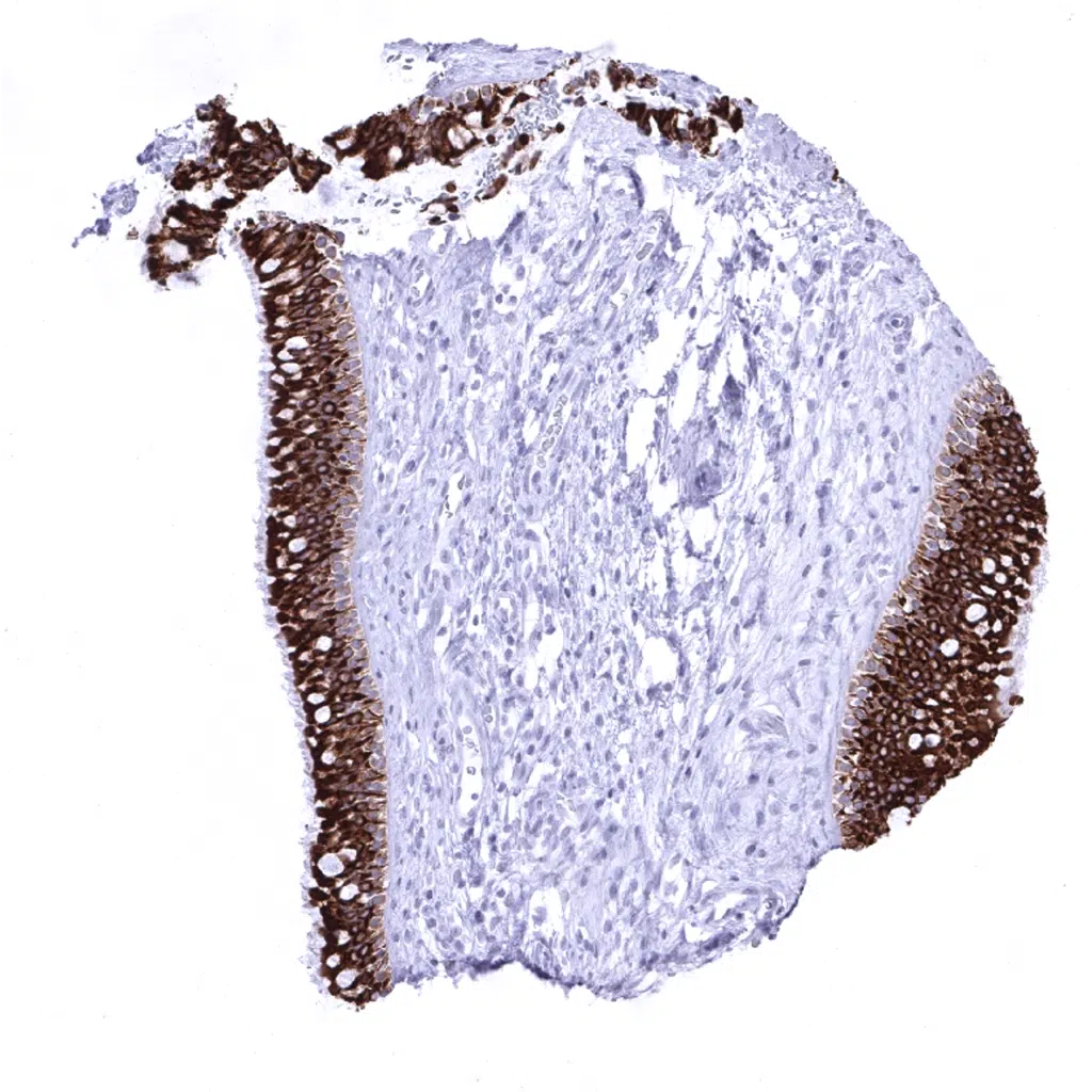
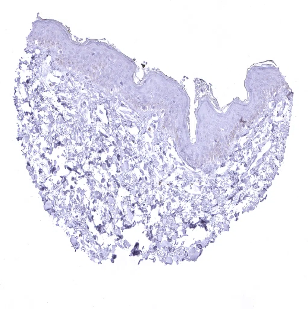
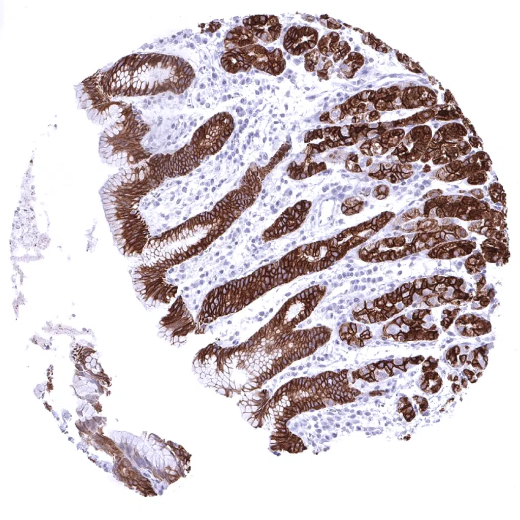
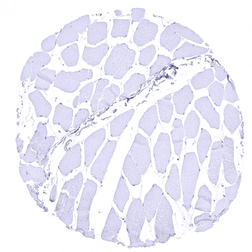
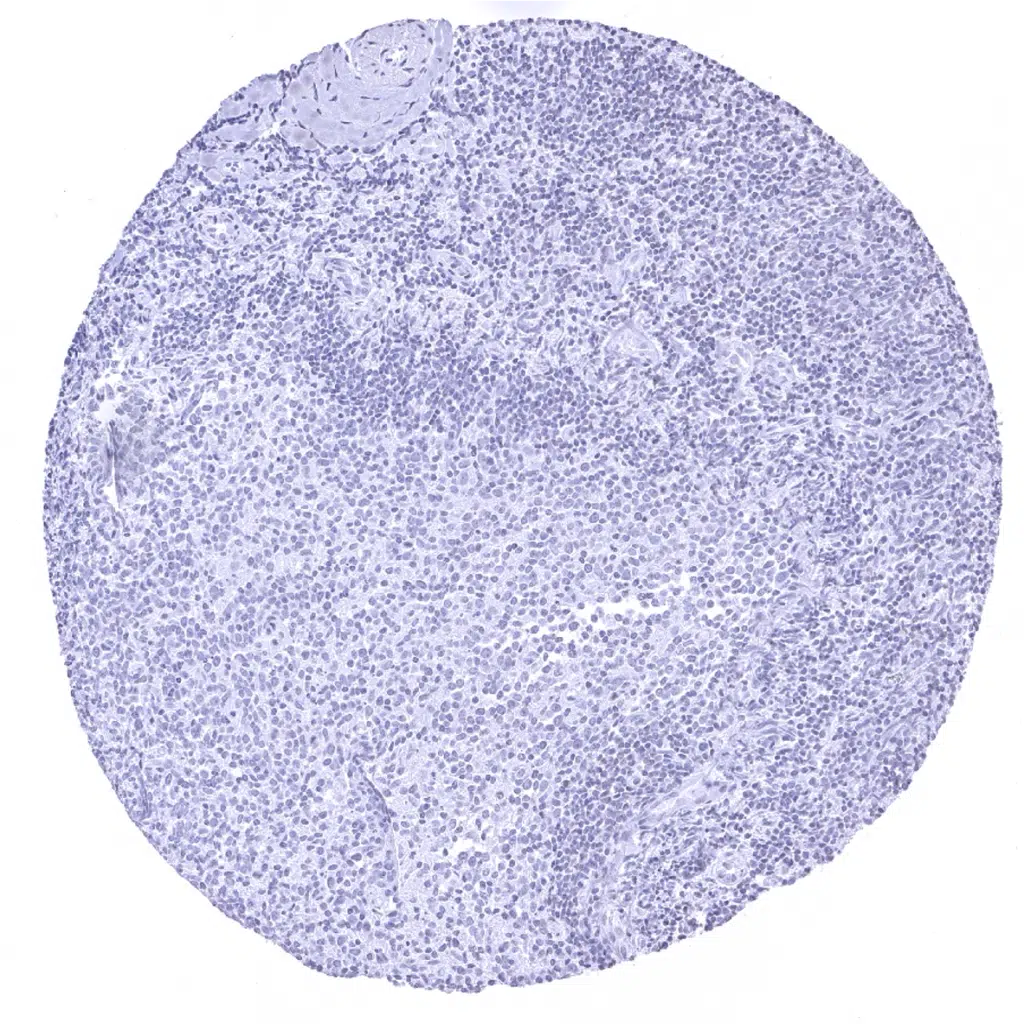
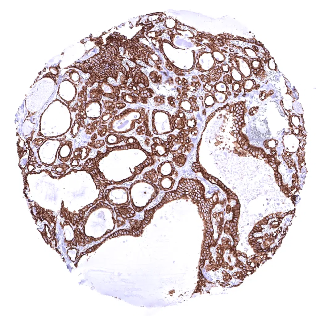
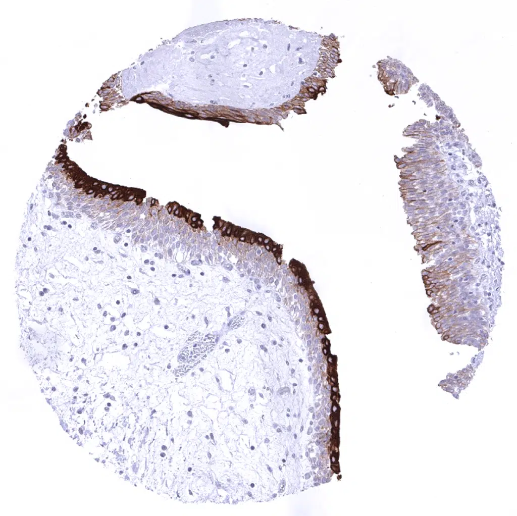
Cytokeratin 18 antibody [MSVA-118R] HistoMAX


Go to Cancer Tissue Gallery
  
 Go back to product page
  

Go to HistoMAX page


Adrenal gland

Aorta, media

Appendix, mucosa - All epithelial cells are Cytokeratin 18 positive.

Appendix, muscular wall


Bone marrow

Breast - Luminal cells but not myoepithelial cells stain Cytokeratin 18 positive in normal breast.

Bronchus, mucosa

Cerebellum (molecular layer, Purkinje cell layer, granule cell layer, white matter)


Cerebellum (white matter)

Cerebrum, grey matter

Cerebrum, white matter

Colon descendens, mucosa - All epithelial cells are Cytokeratin 18 positive.


Colon descendens, muscular wall

Duodenum, Brunner gland - Weak Cytokeratin 18 staining of Brunner glands.

Duodenum, mucosa

Ektocervix


Endocervix - Strong Cytokeratin 18 staining of all epithelial cells.

Endometrium, proliferation

Endometrium, secretion

Epididymis - All epithelial cells are Cytokeratin 18 positive.


Esophagus, squamous epithelium

Fallopian tube, mucosa

Fat

Gallbladder, epithelium


Heart

Ileum, mucosa - All epithelial cells are Cytokeratin 18 positive.

Kidney, cortex

Kidney, medulla


Kidney, pelvis, urothelium - The urothelium shows the strongest Cytokeratin 18 staining in umbrella cells. Staining intensity decreases gradually from the top to the bottom layers of the urothelium.

Liver - Liver tissue shows a zonal variability of Cytokeratin 18 expression in hepatocytes ranging from weak to strongly positive while bile ducts are always strongly positive.

Liver - Not all hepatocytes stain positive for Cytokeratin 18 (zonal distribution).

Lung


Lymph node - A delicate Cytokeratin 18 staining occurs in fibroblastic reticulum cells of lymphoid organs.

Ovary, stroma

Pancreas

Parathyroid


Parotid gland

Pituitary gland, anterior lobe

Pituitary gland, posterior lobe

Pregnant uterus (decidua)


Placenta, early

Placenta, mature

Placenta (amnion and chorion) - Strong Cytokeratin 18 staining in chorion cells while the amnion cells are only weakly Cytokeratin 18 positive.

Prostate


Rectum, mucosa

Seminal vesicle

Sinus paranasales

Skin


Spleen

Stomach, antrum - Strong Cytokeratin 18 staining of all epithelial cells.

Stomach, corpus

Striated muscle


Testis

Thymus - Some elements (few cells) of Hassal bodies are Cytokeratin 18 positive.

Thymus

Thyroid gland


Tonsil, surface epithelium

Urinary bladder, muscular wall

Urinary bladder, urothelium - Urothelium with particularly strong staining in umbrella cells. Cytokeratin 18 staining is weaker in urothelial cells and gradually decreases from the top to the bottom layers.

Uterus, myometrium


| TOP |