Normal Tissue Gallery- GTX04464

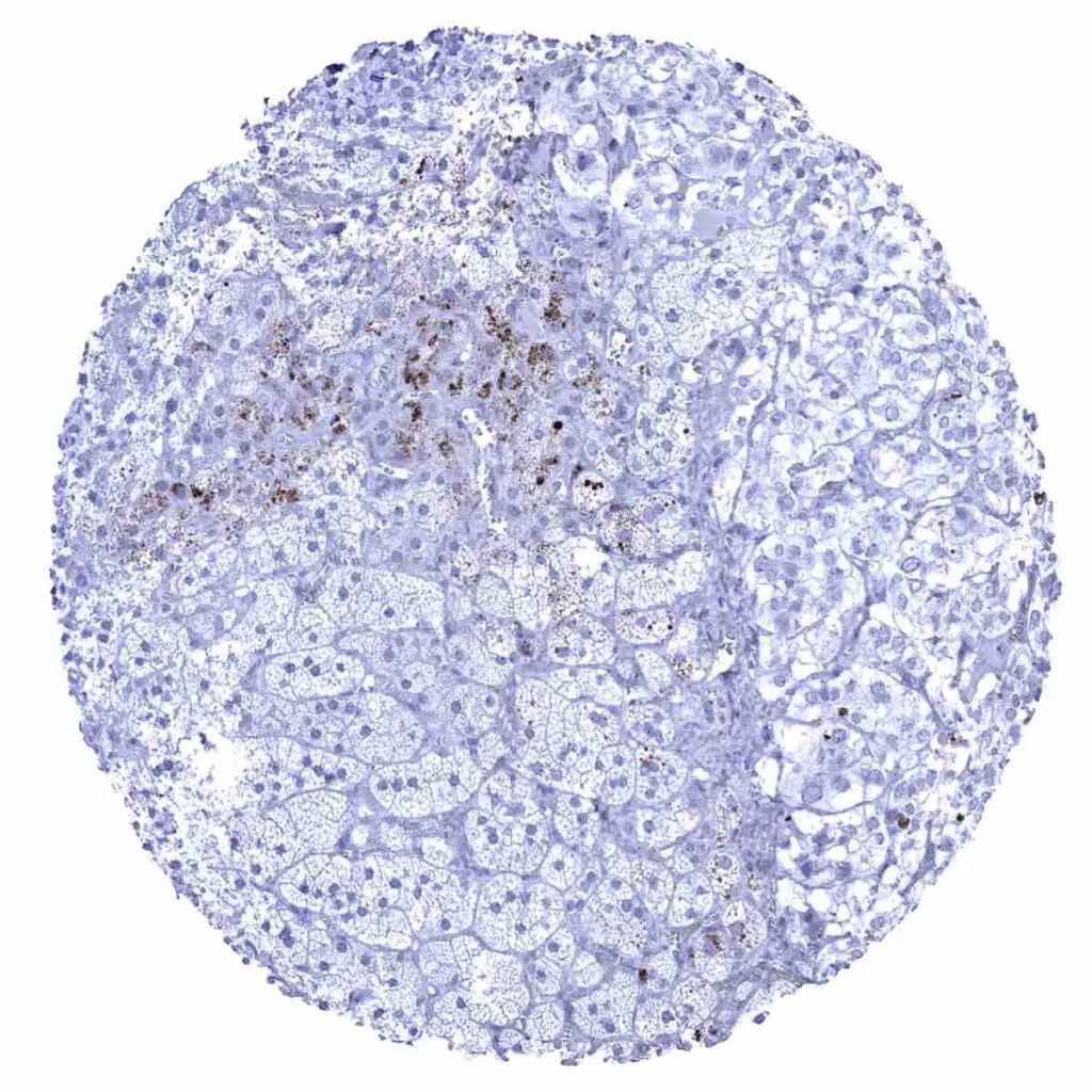
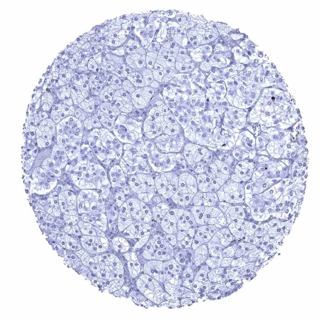
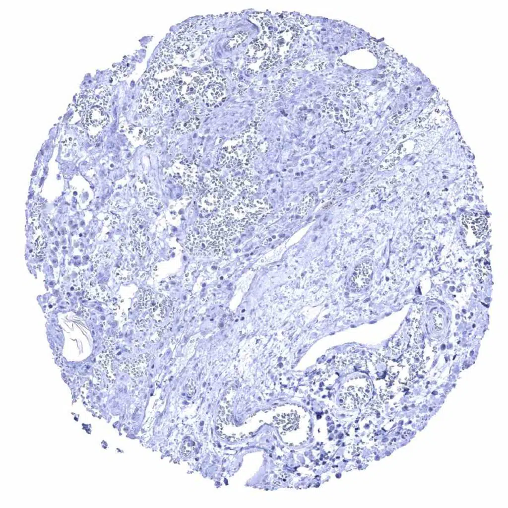
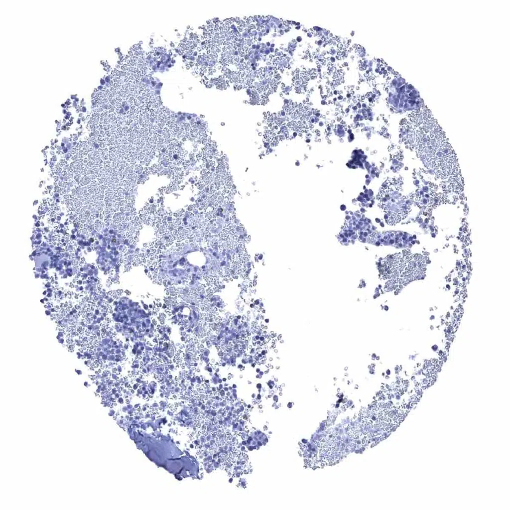
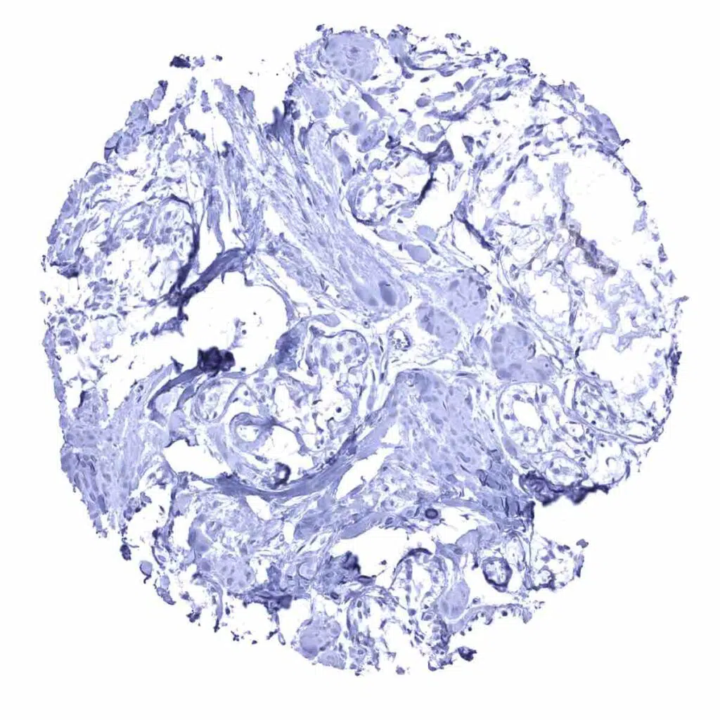
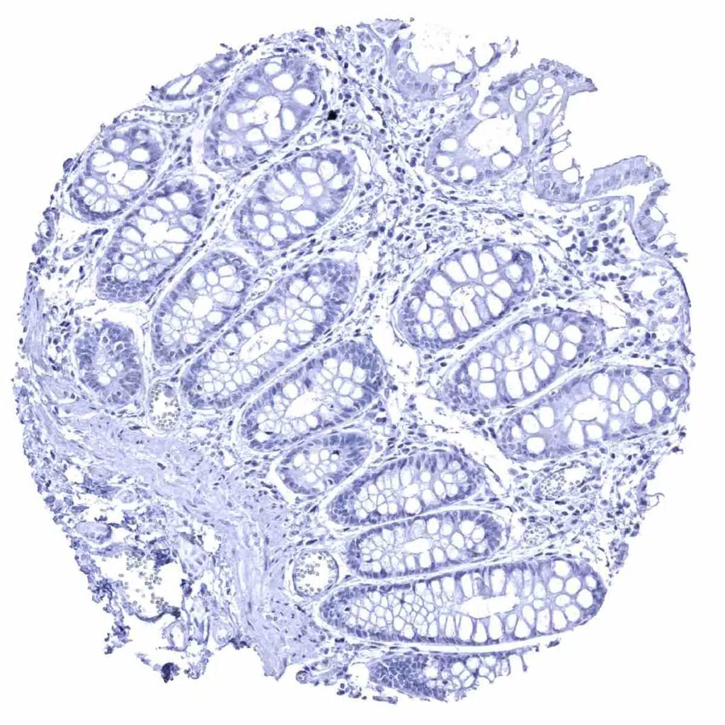
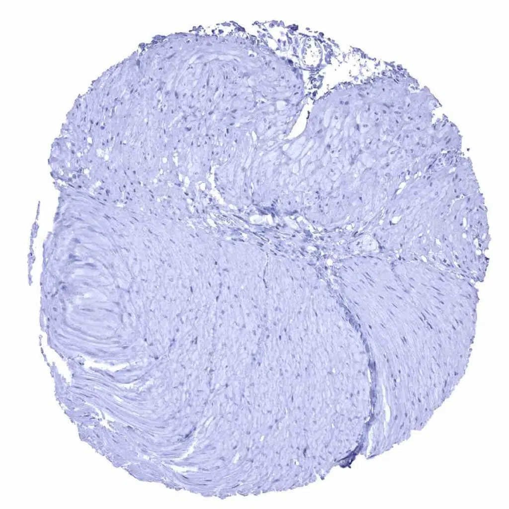
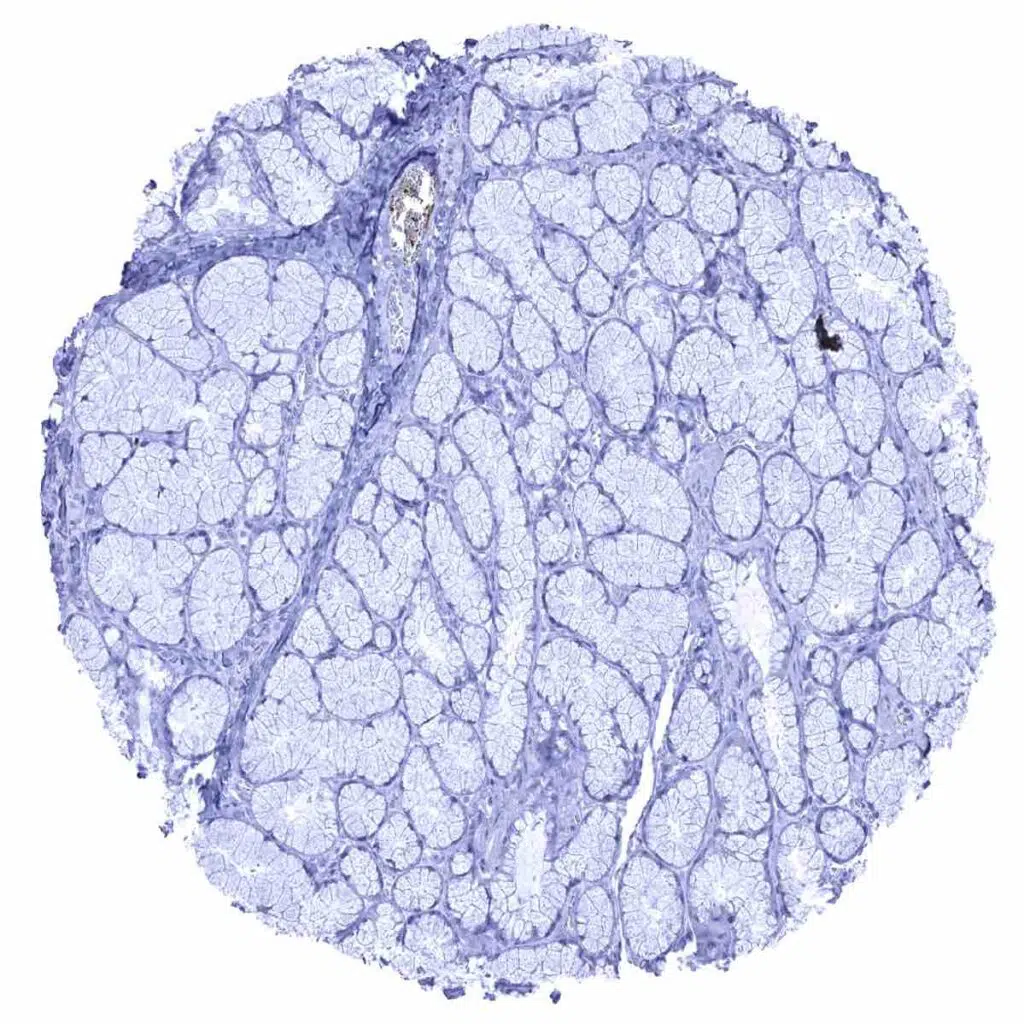
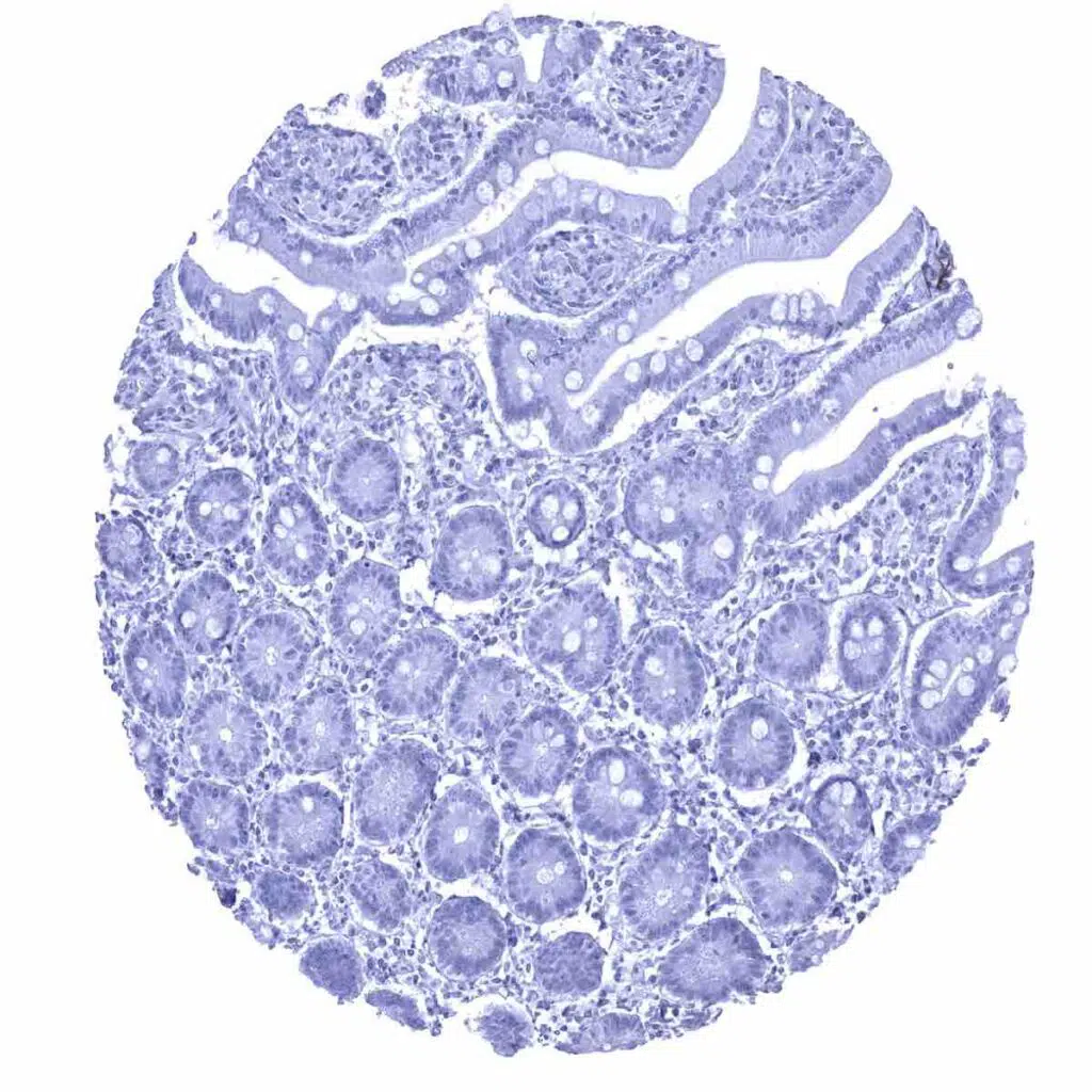
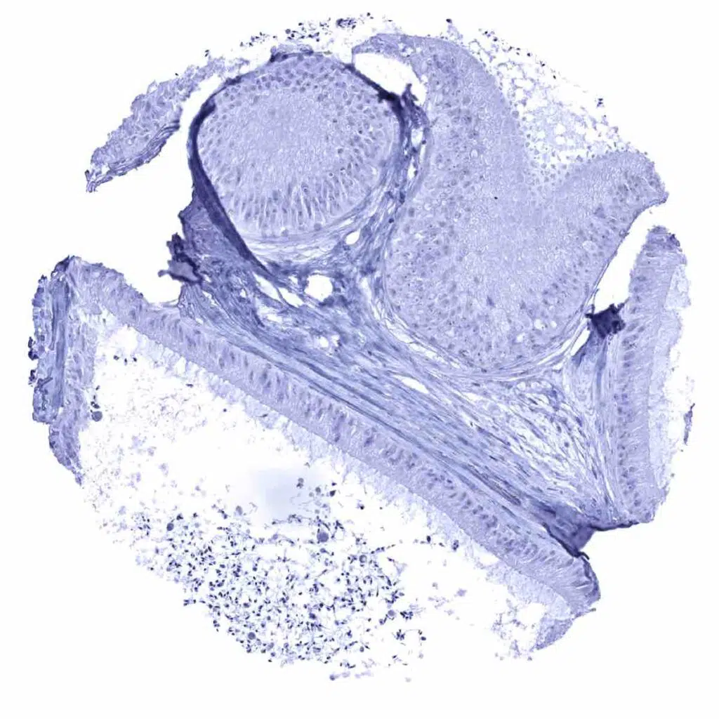
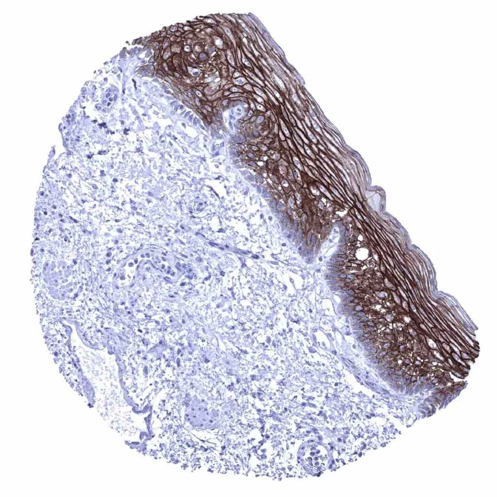
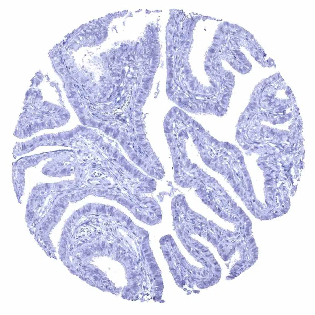
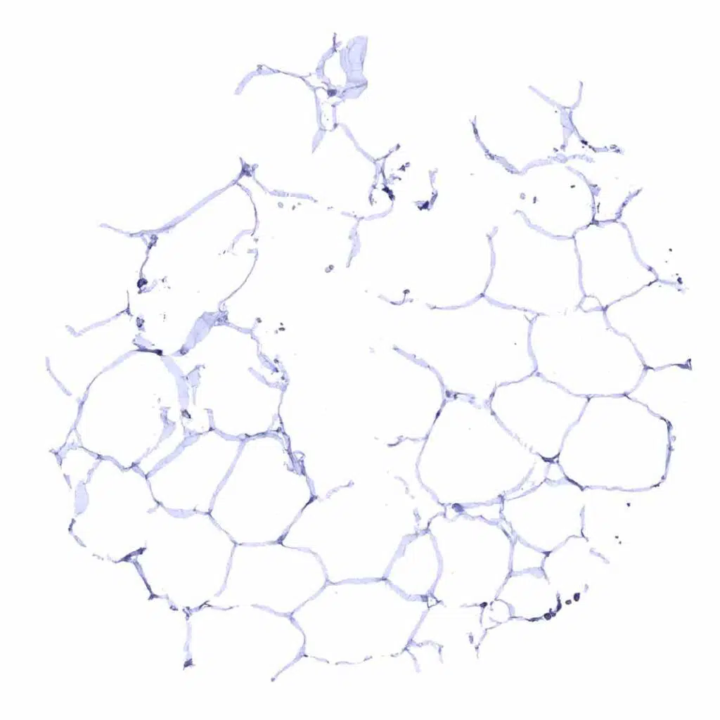
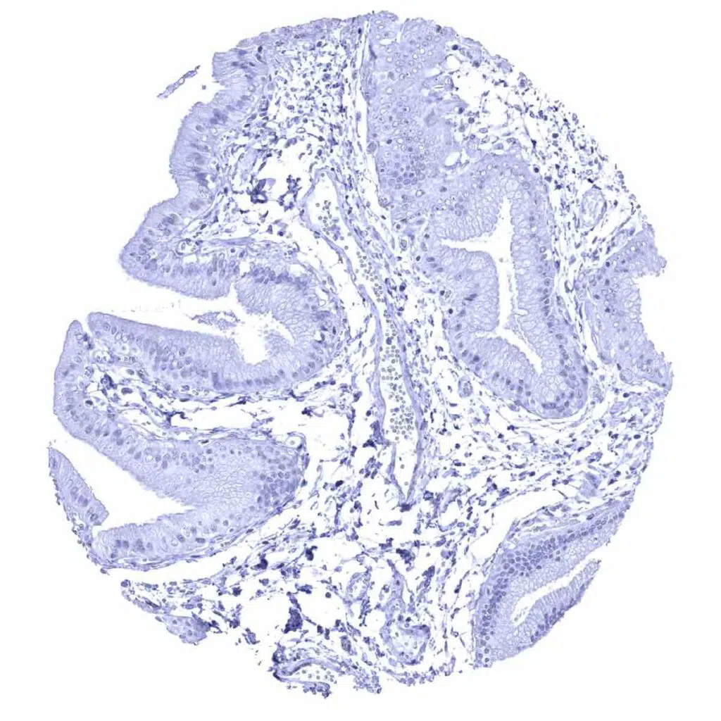
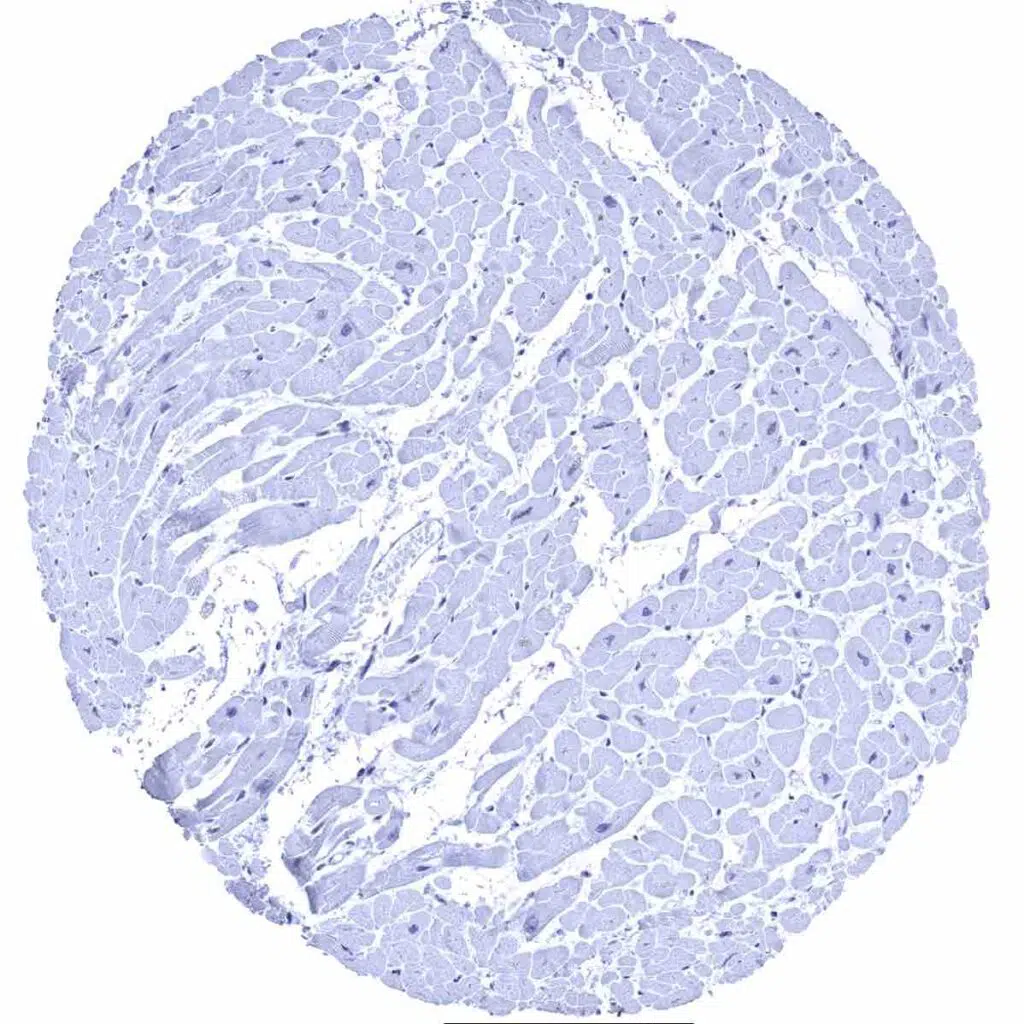
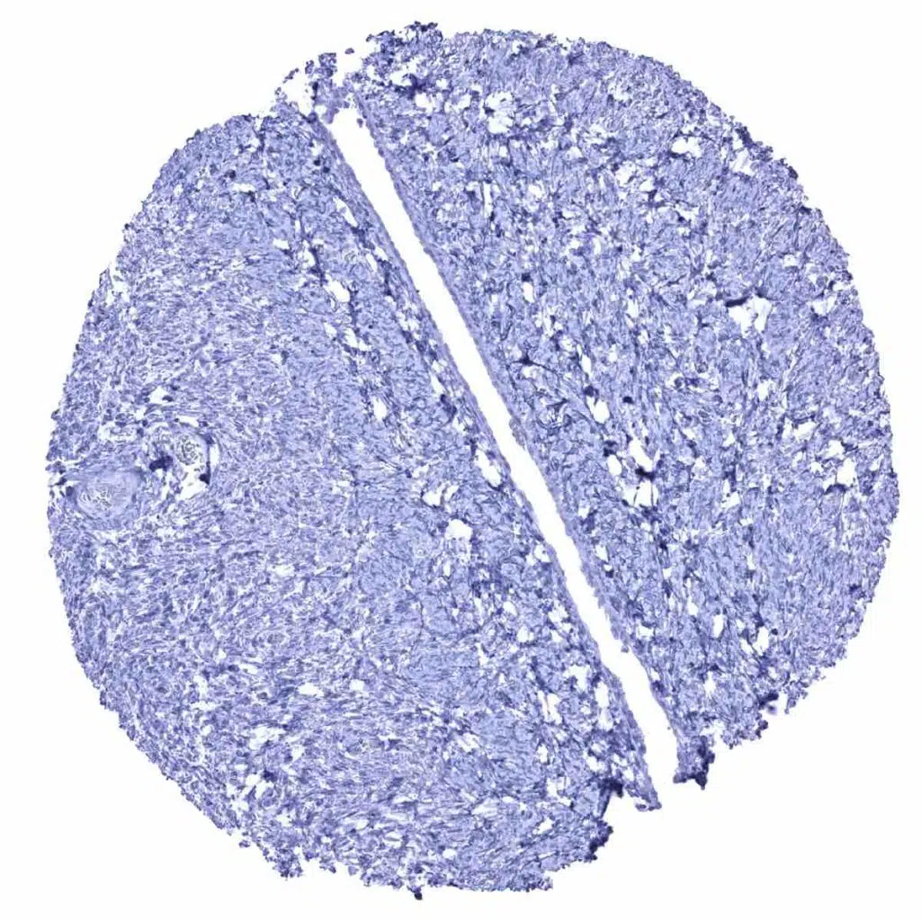
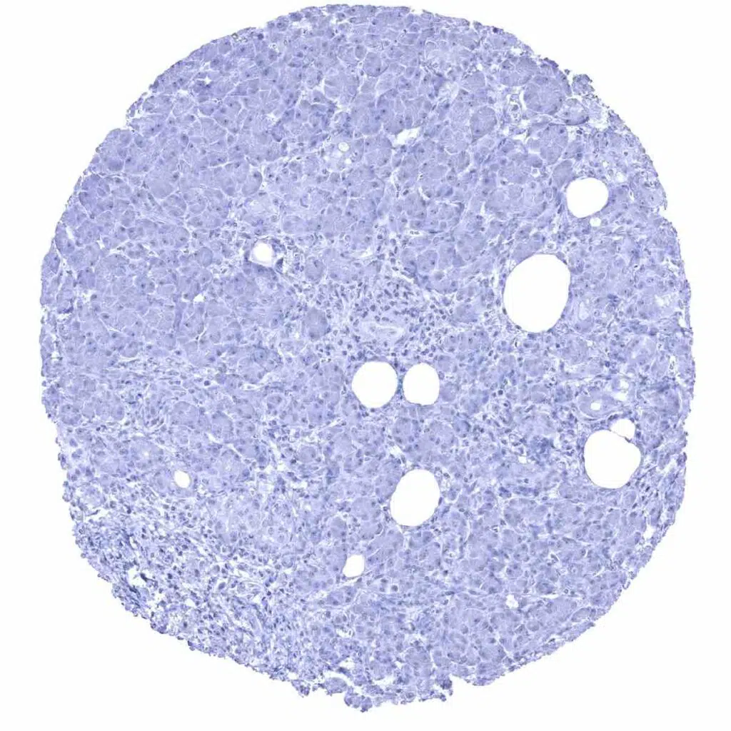
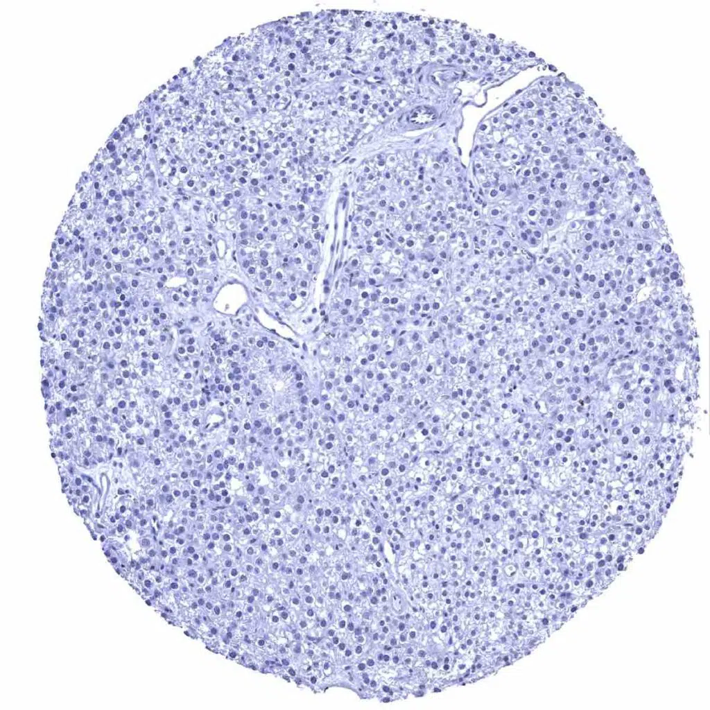
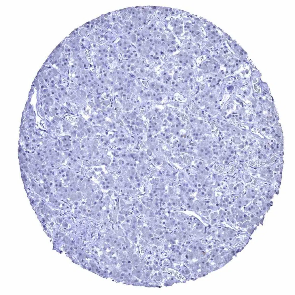
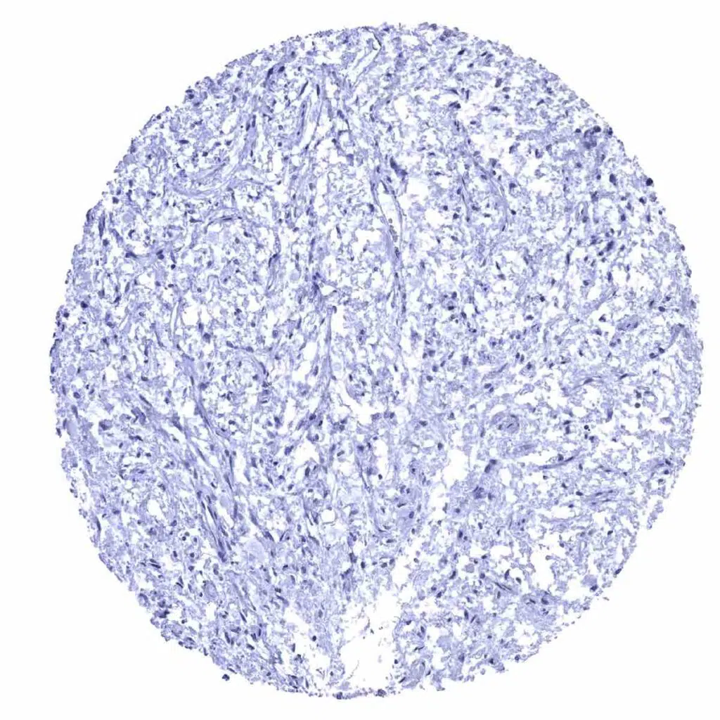
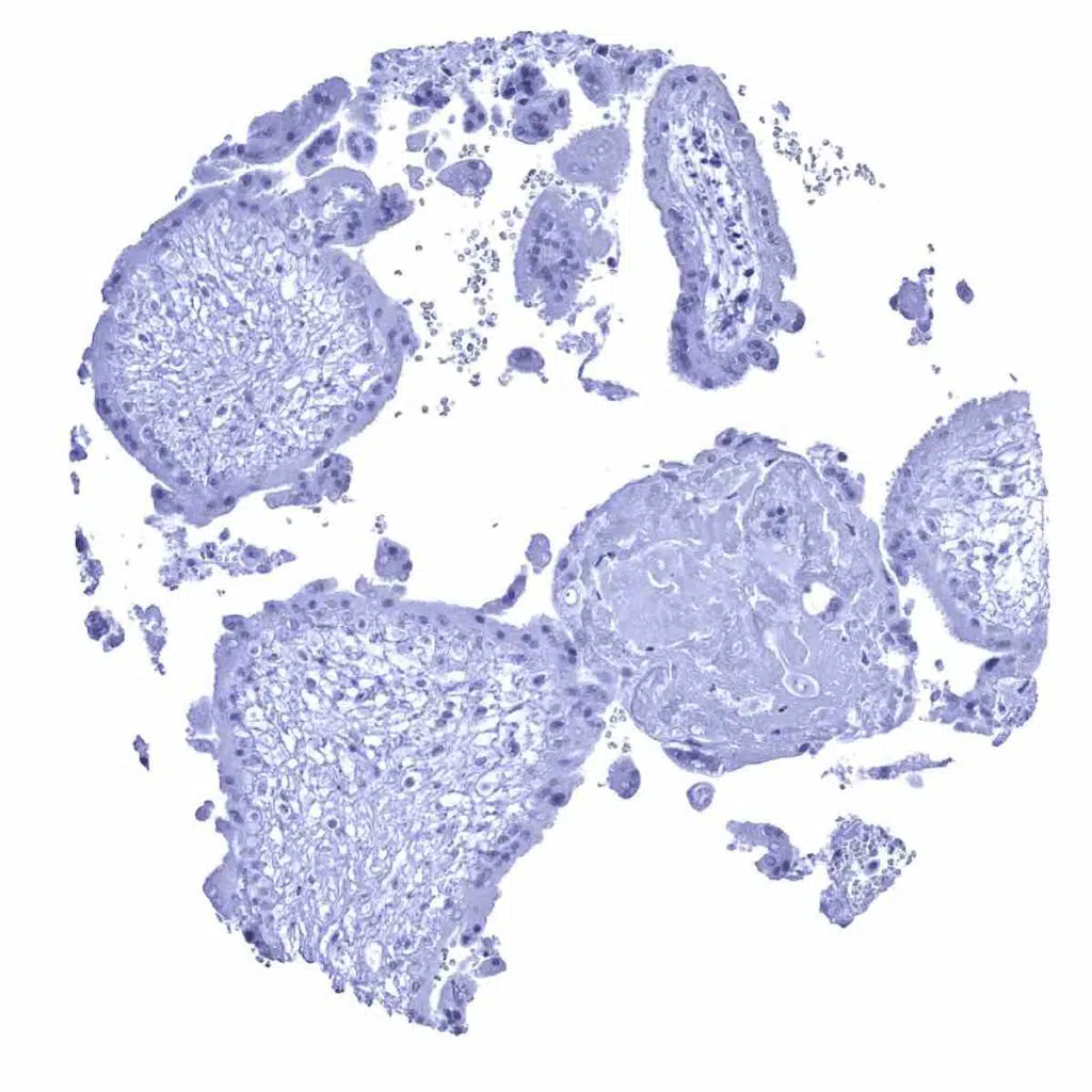
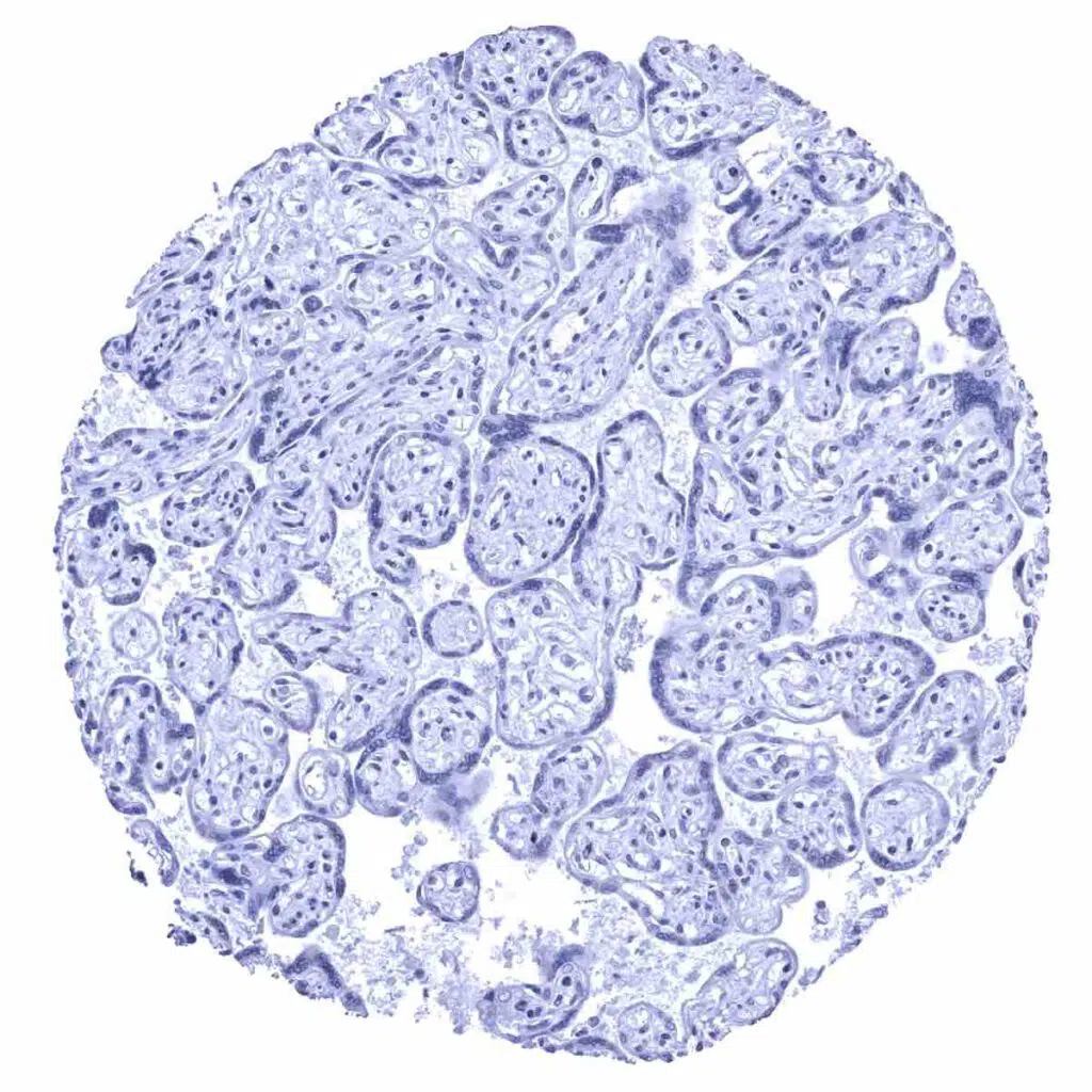
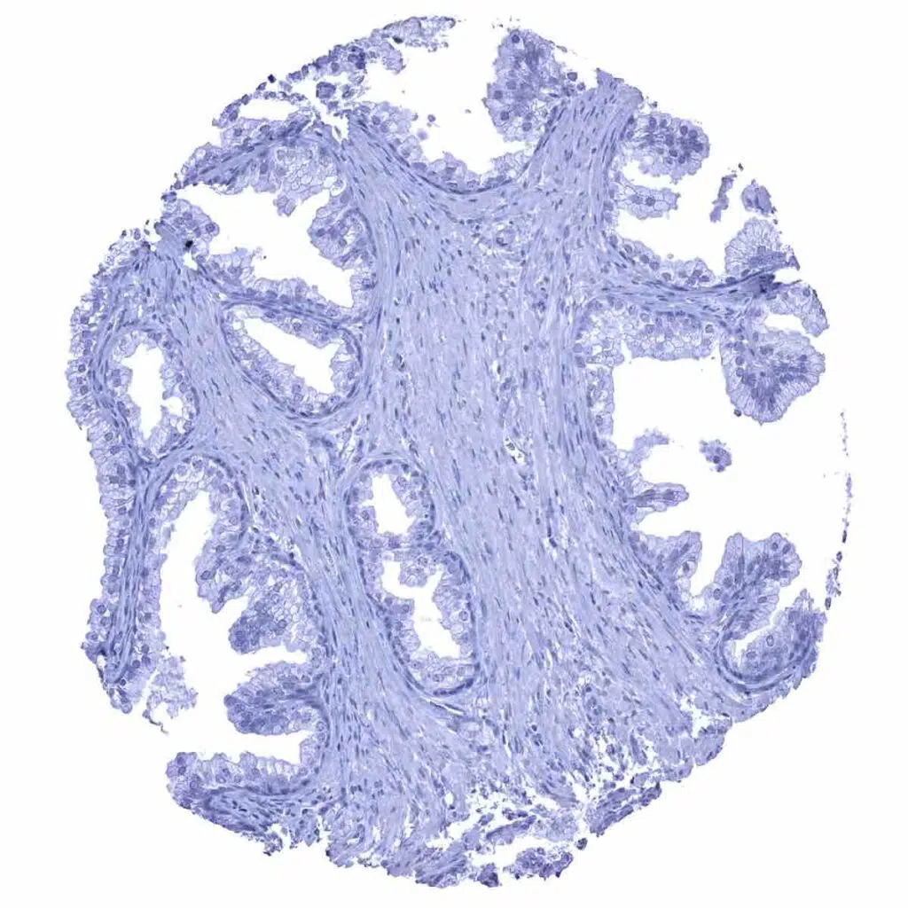
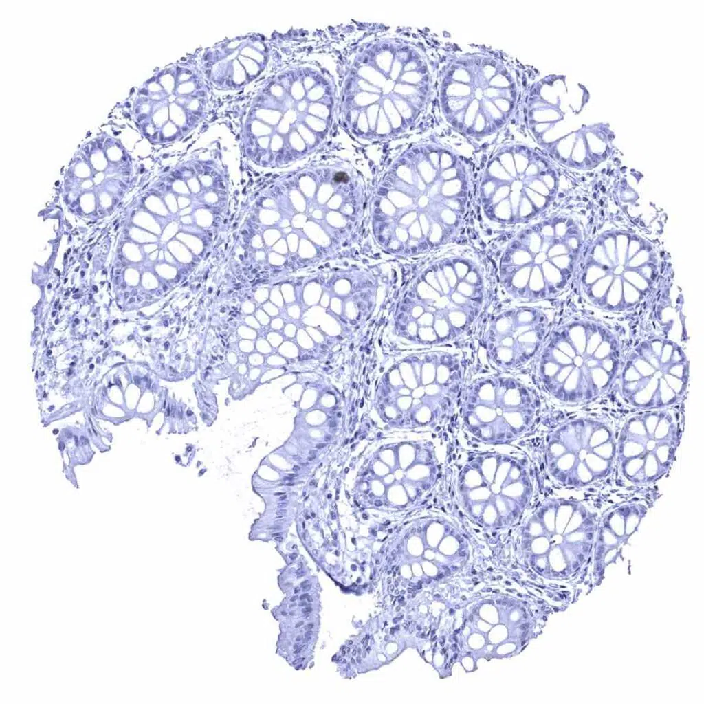
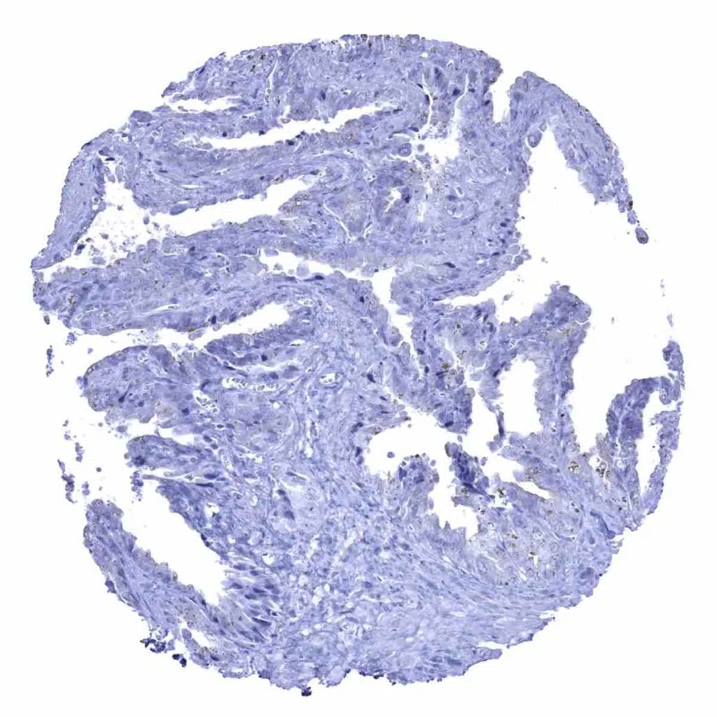
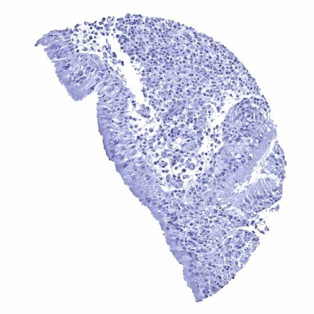
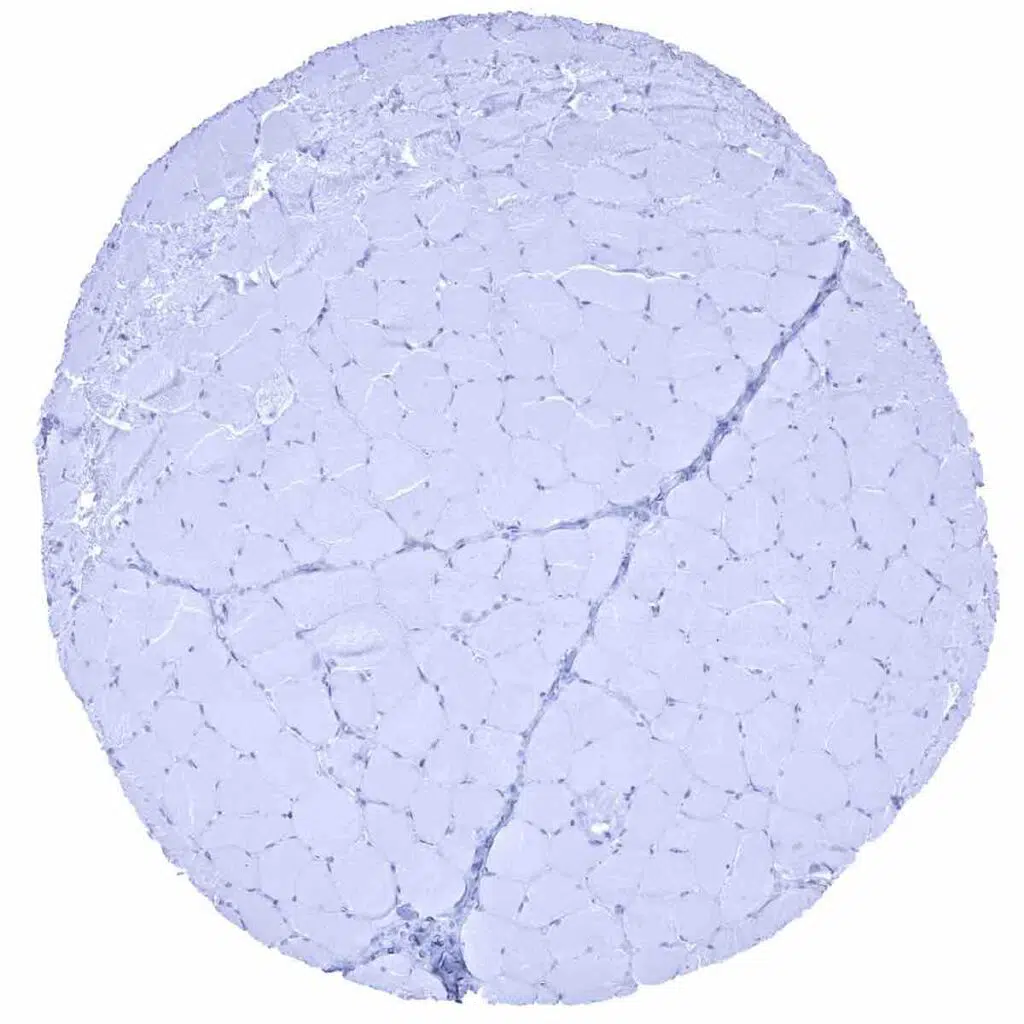
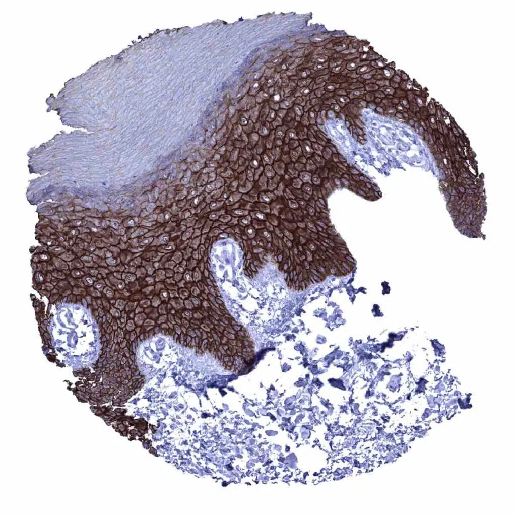
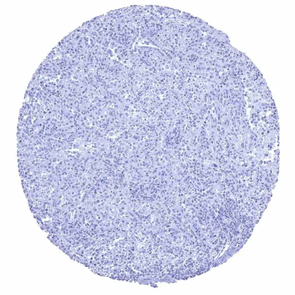
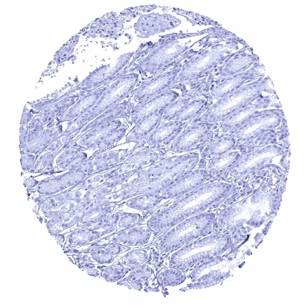
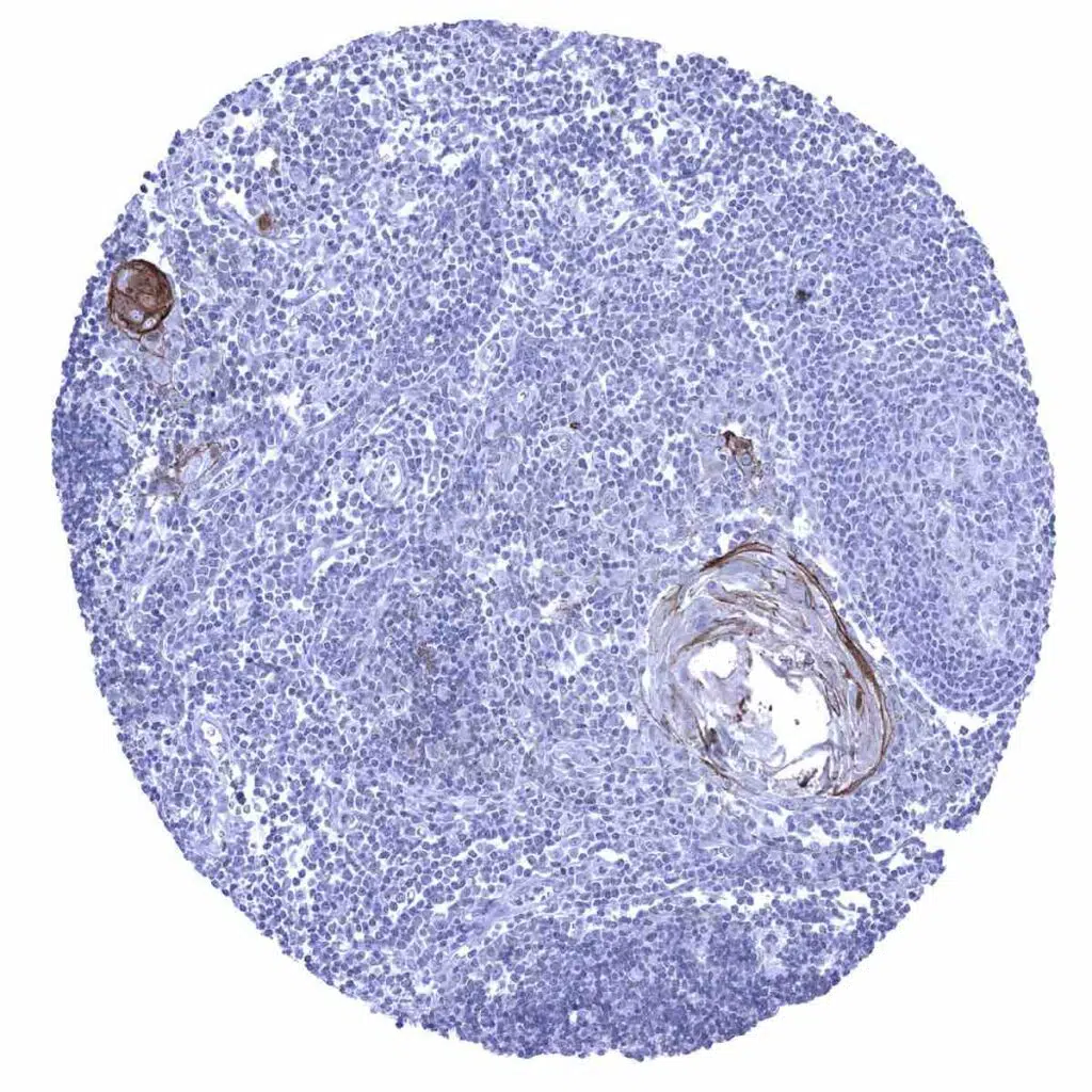
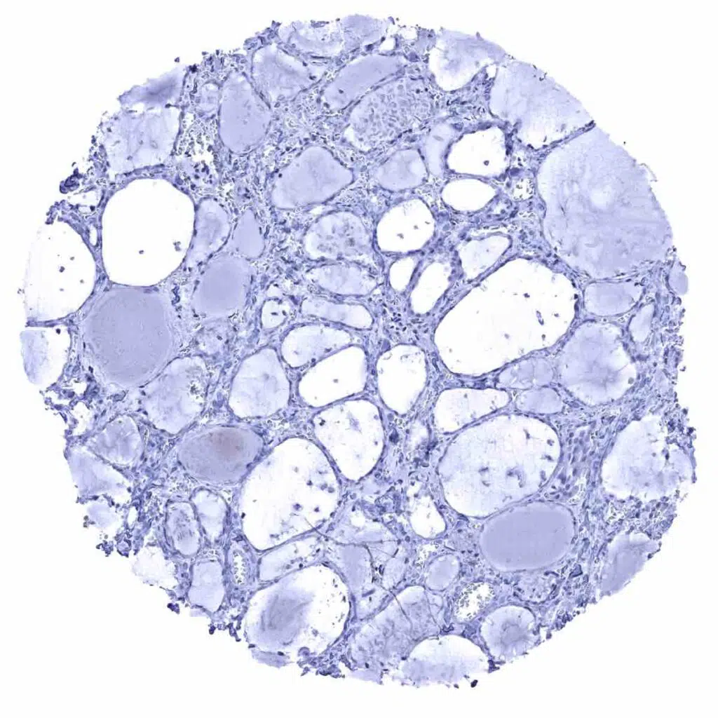
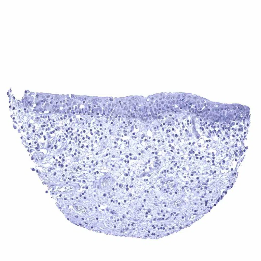
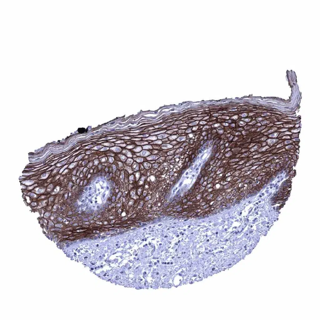
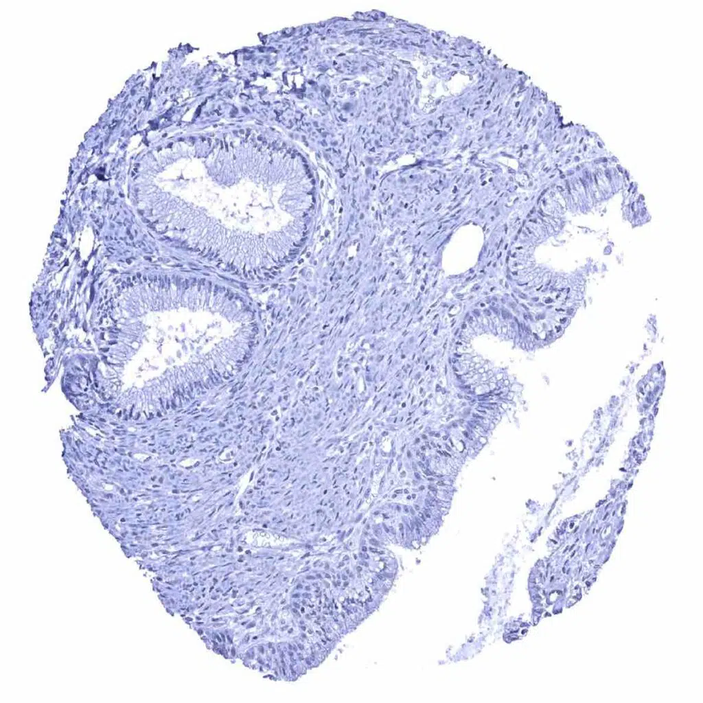
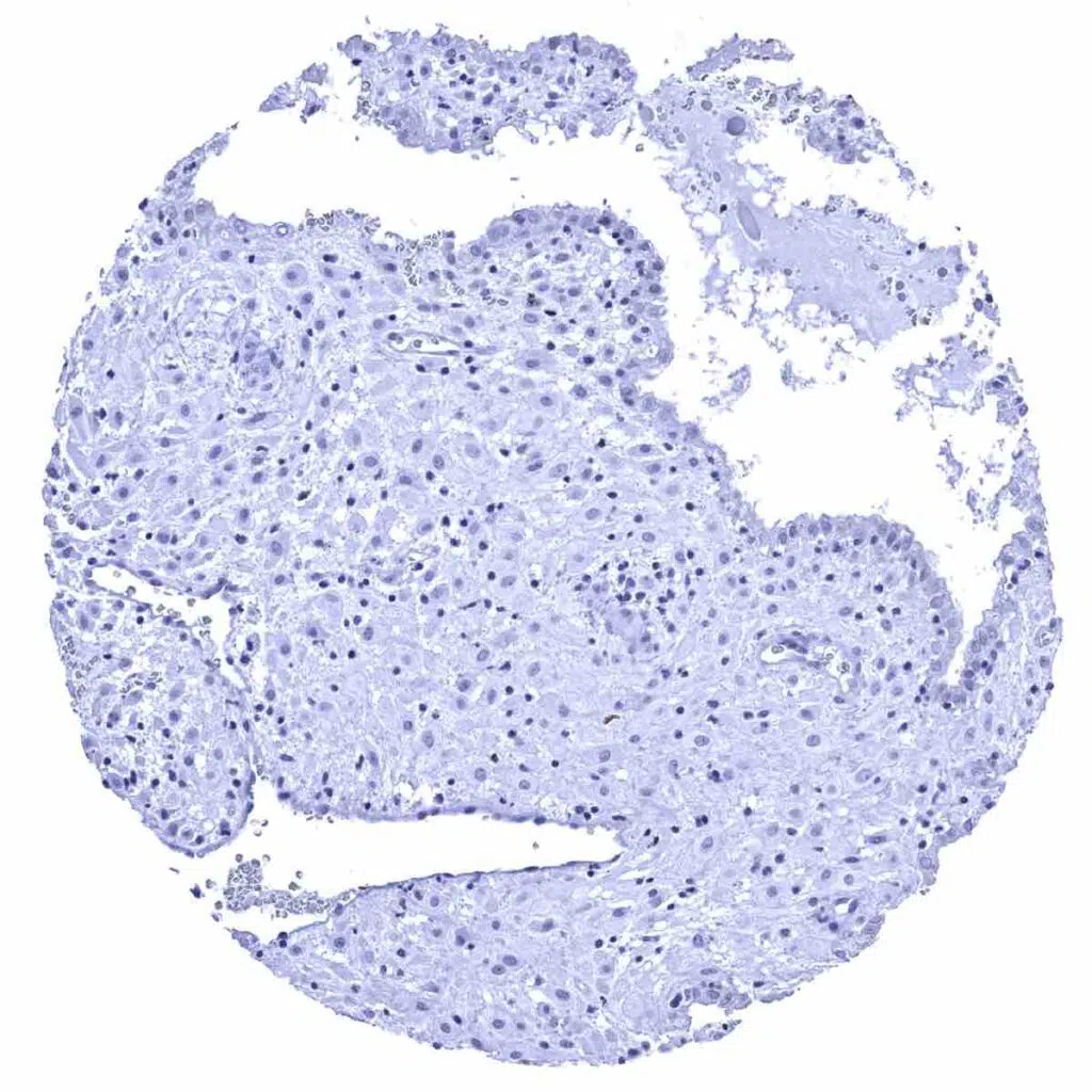
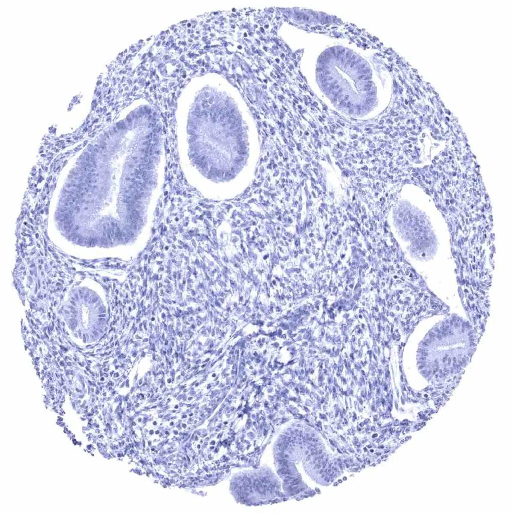
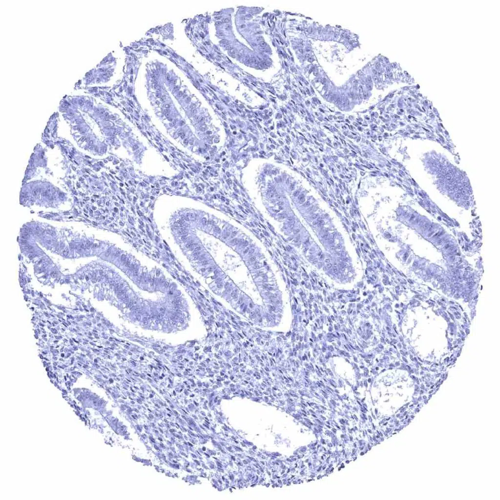
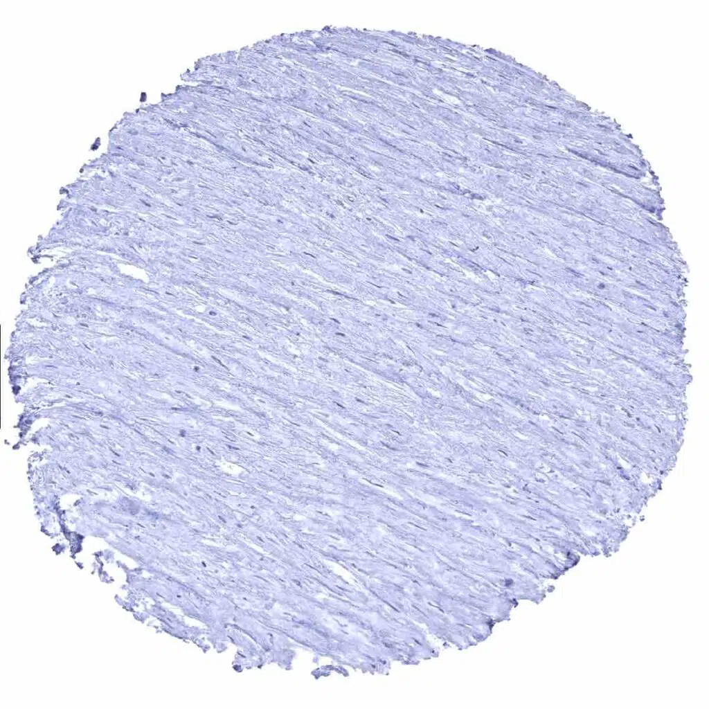
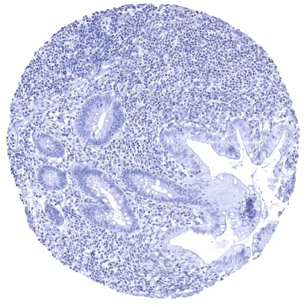
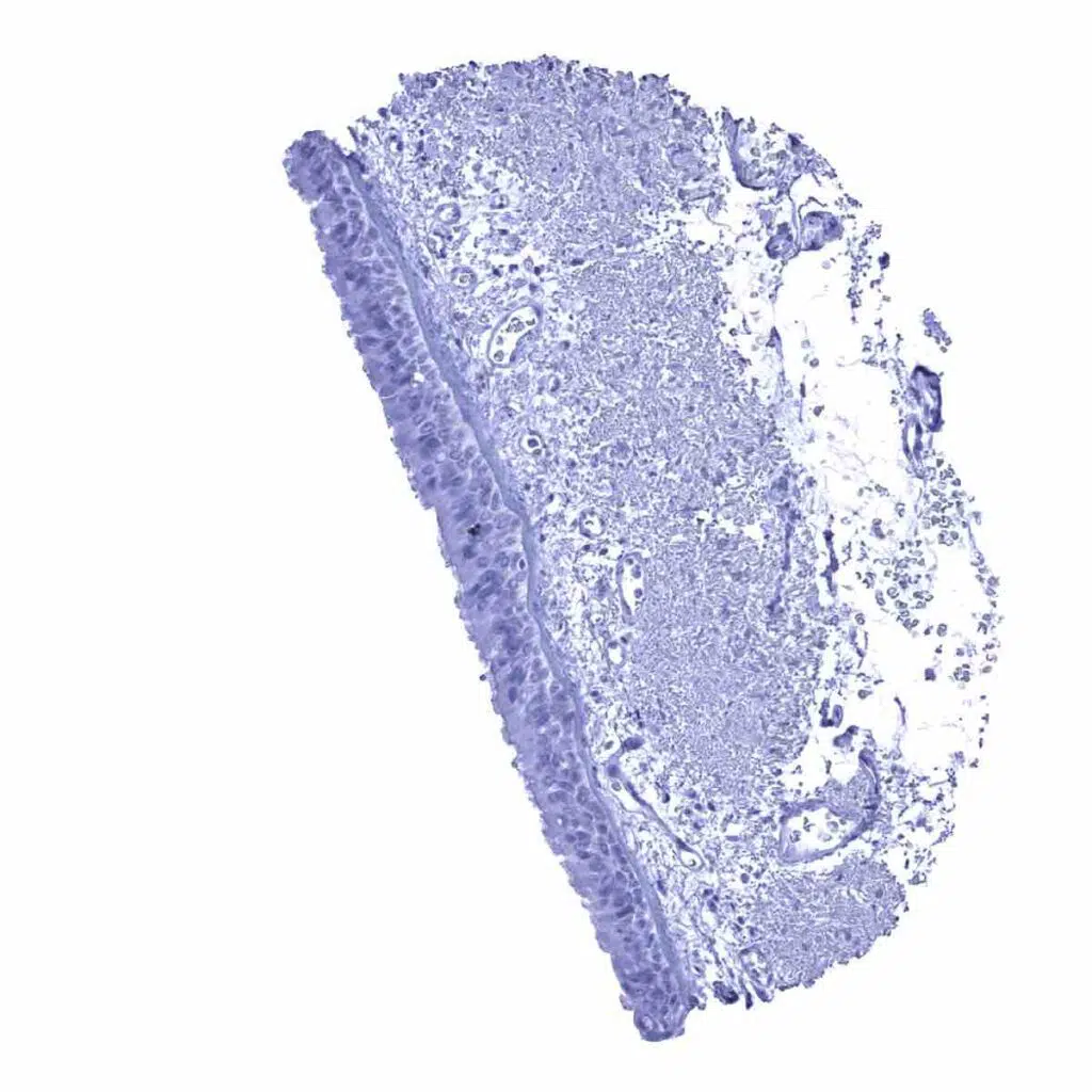
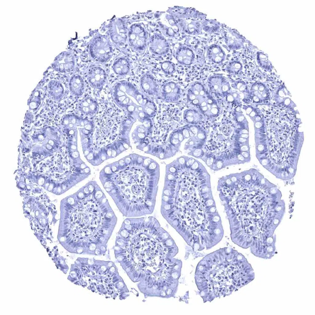
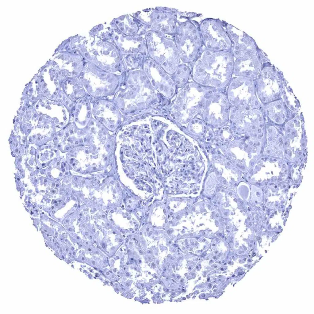
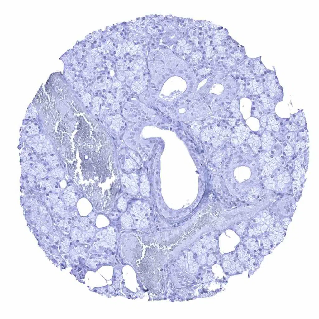
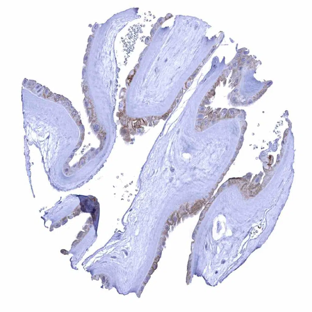
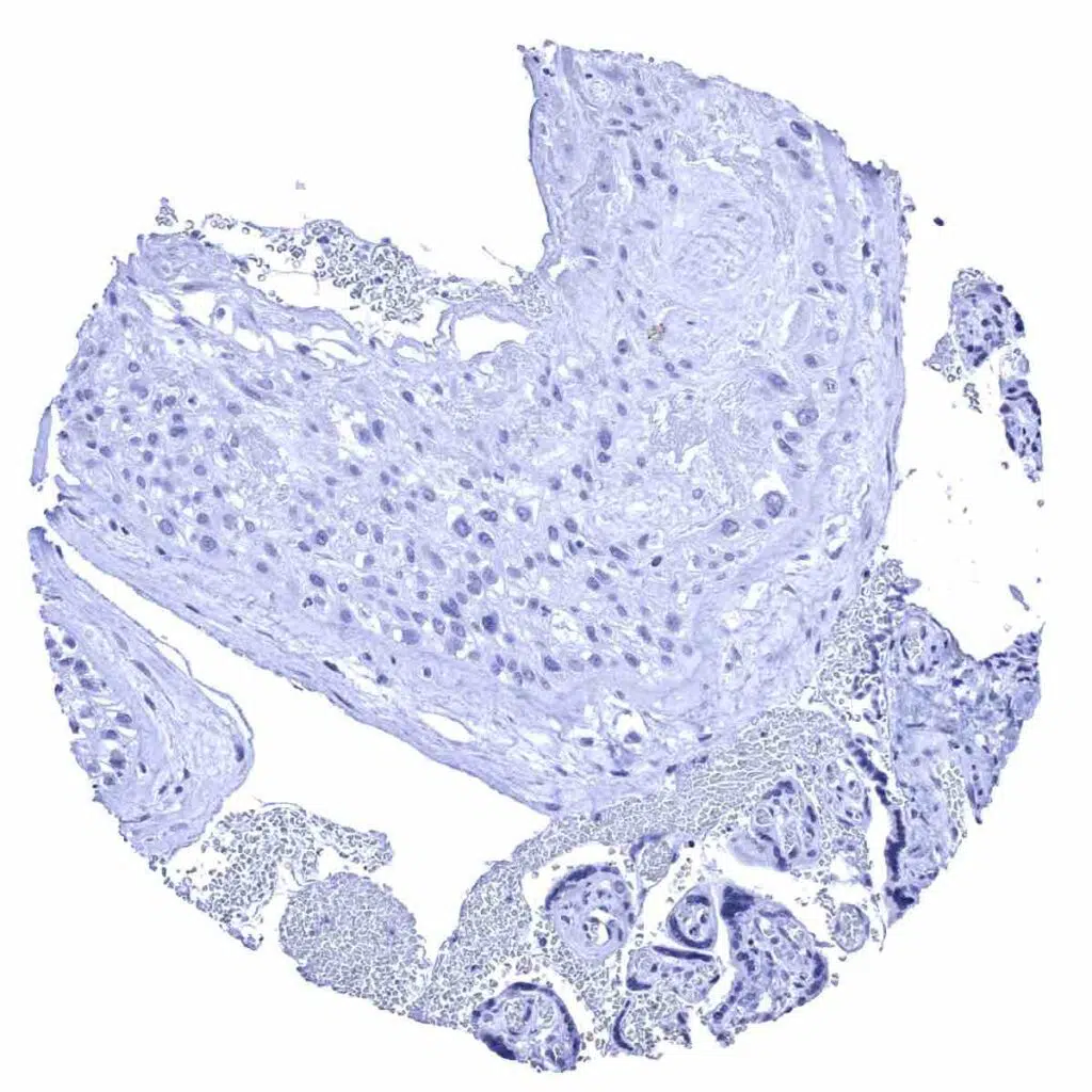
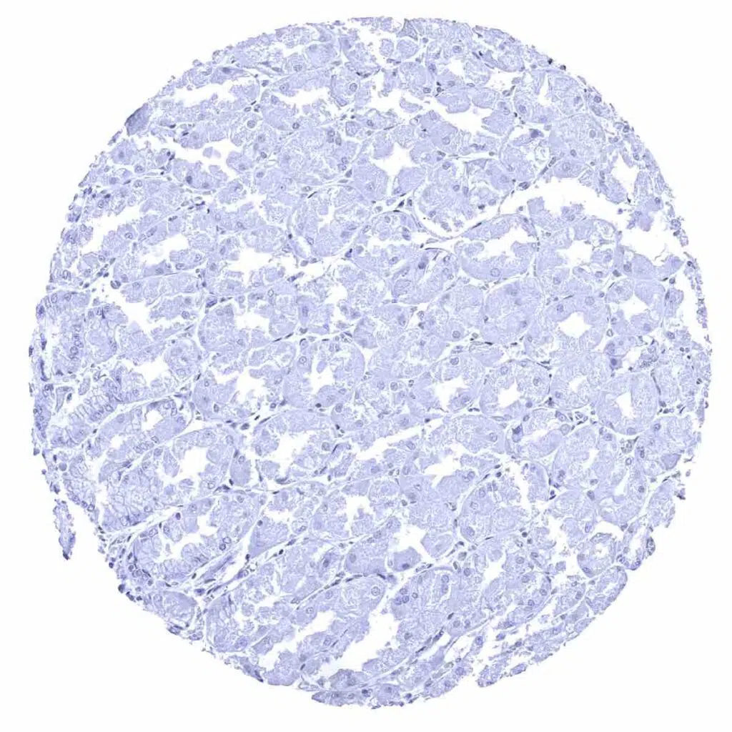
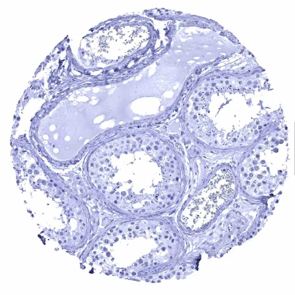
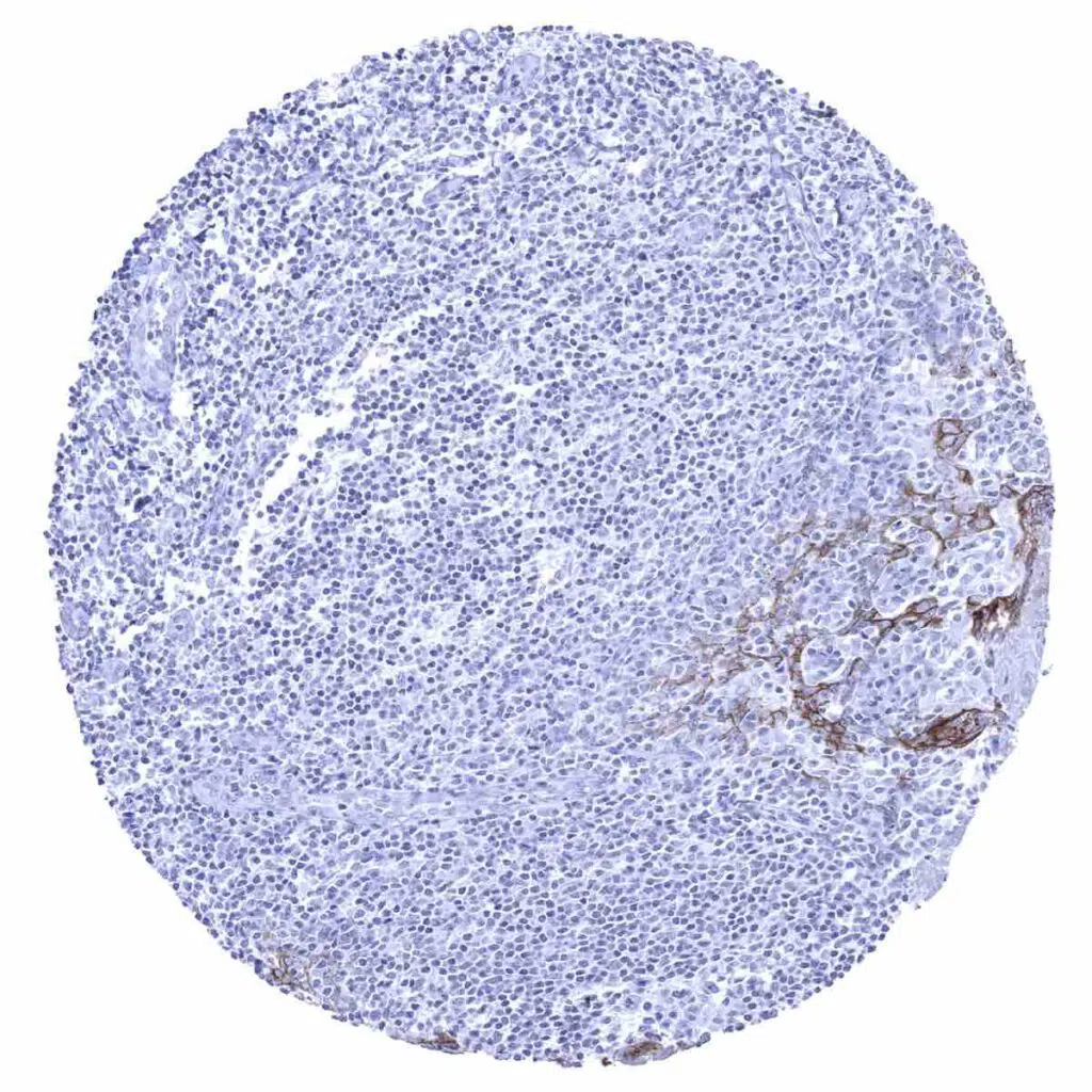
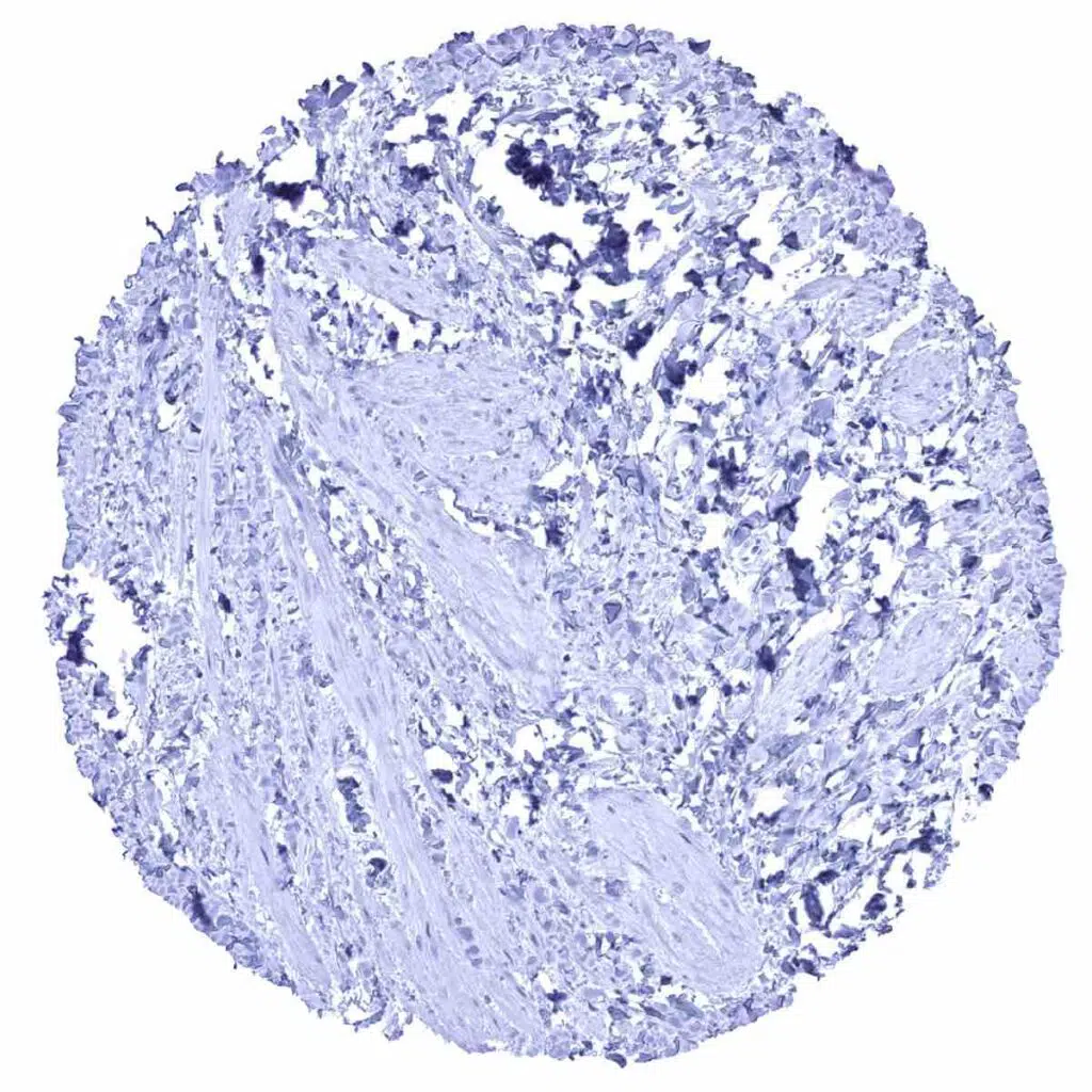
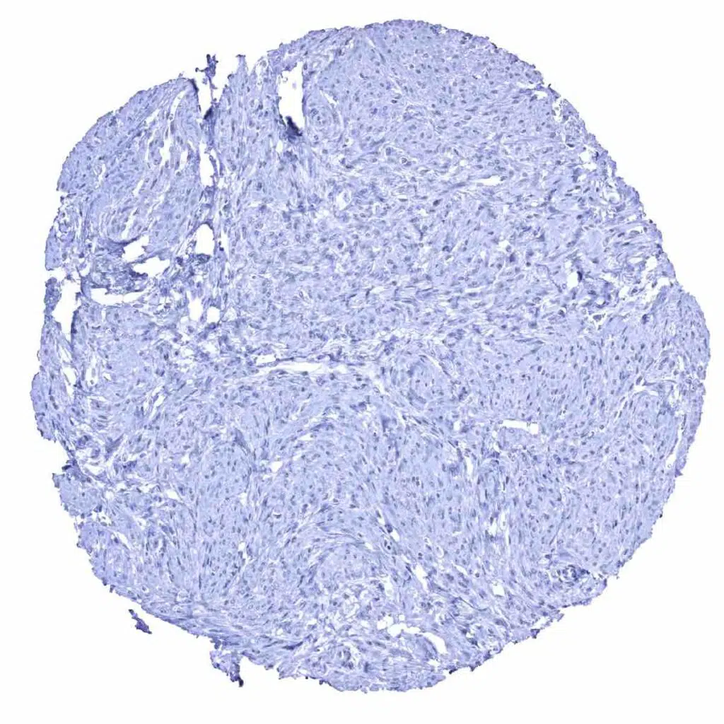
Desmoglein 1 antibody [MSVA-544M] HistoMAX


Go to Cancer Tissue Gallery
  
 Go back to product page
  

Go to HistoMAX page


Adrenal gland – This sample contains pigment , no true staining

Adrenal gland

Aorta, media

Appendix, mucosa


Appendix, muscular wall

Bone marrow

Breast

Bronchus, mucosa


Cerebellum (molecular layer, Purkinje cell layer, granule cell layer, white matter)

Cerebellum (molecular, Purkinje cell, granule cell layers; white matter)

Cerebrum, grey matter

Cerebrum, white matter


Colon descendens, mucosa

Colon descendens, muscular wall

Duodenum, Brunner gland

Duodenum, mucosa


Epididymis

Esophagus, squamous epithelium – Moderate to strong membranous desmoglein 1 immunostaining of the squamous epithelium. The staining is less intense in the basal cell layers

Fallopian tube, mucosa

Fat


Gallbladder, epithelium

Heart muscle

Ileum, mucosa

Kidney, cortex


Kidney, medulla

Liver

Lung

Lymph node


Ovary, stroma

Pancreas

Parathyroid gland

Parotid gland


Pituitary gland, anterior lobe

Pituitary gland, posterior lobe

Placenta (amnion and chorion) – Weak to moderate, membranous, predominantly apical desmoglein 1 immunostaining of amnion cells

Placenta (amnion and chorion)


Placenta, early

Placenta, mature

Prostate

Rectum, mucosa


Seminal vesicle

Sinus paranasales

Skeletal muscle

Skin – Strong membranous desmoglein 1 immunostaining of the squamous epithelium of the skin. A slight decrease of staining occurs in the most superficial cell layers


Spleen

Stomach, antrum

Stomach, corpus

Testis


Thymus – Moderate to strong membranous desmoglein 1 immunostaining of squamous epithelial cells of corpuscles of Hassall's

Thyroid gland

Tonsil – Moderate to strong desmoglein 1 immunostaining of a fraction of squamous epithelial cells in tonsil crypt epithelium

Tonsil, surface epithelium – Weak to moderate membranous desmoglein 1 immunostaining of the squamous epithelium. The staining is least intensive in the basal cell layers


Urinary bladder, muscular wall

Urinary bladder, urothelium

Uterus, ectocervix – Strong membranous desmoglein 1 immunostaining of all cell layers of the squamous epithelium

Uterus, endocervix


Uterus, endometrium (pregnancy)

Uterus, endometrium (proliferation)

Uterus, endometrium (secretion)

Uterus, myometrium


| TOP |