Normal Tissue Gallery- GTX04429

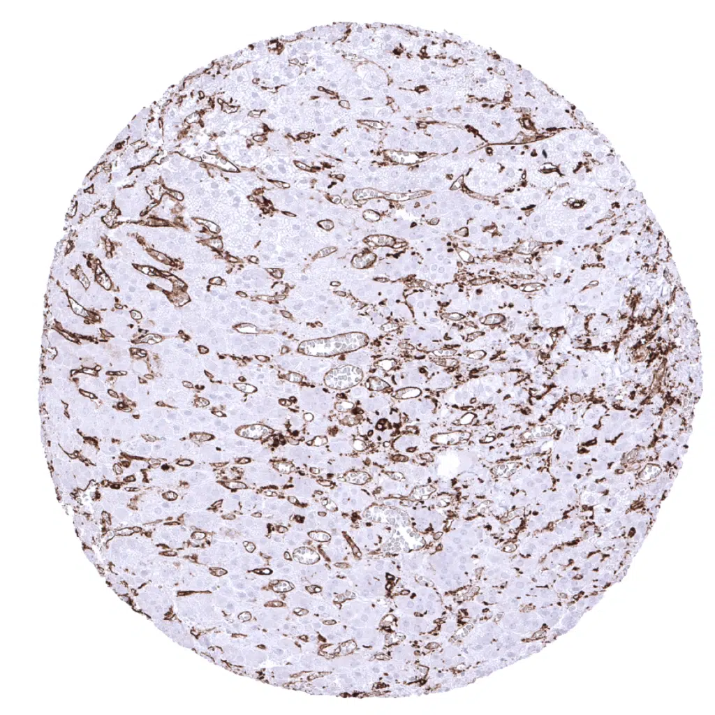
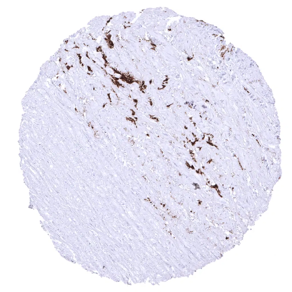
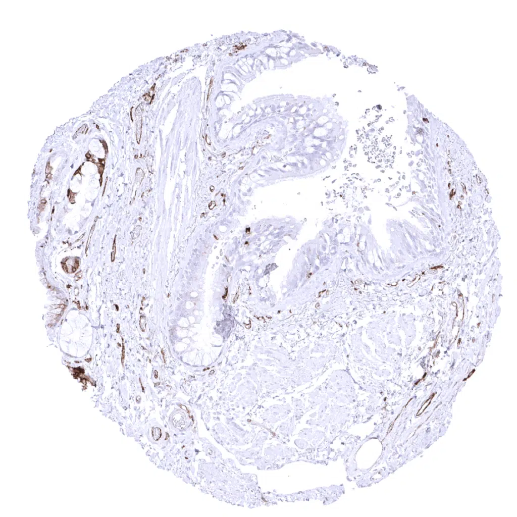
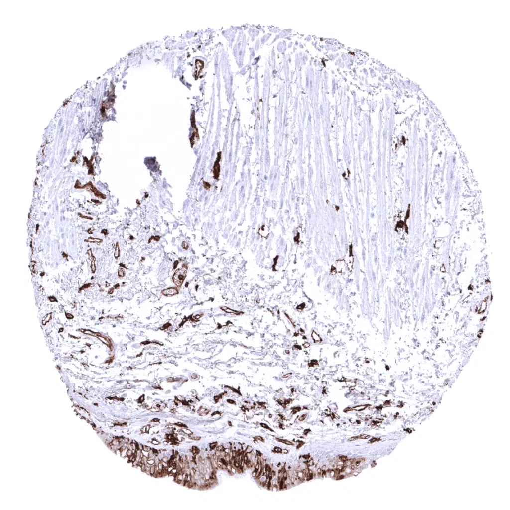
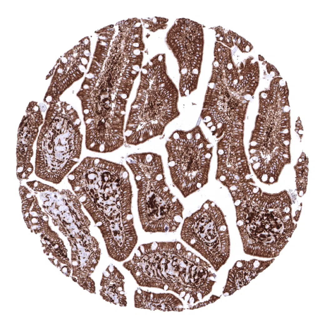
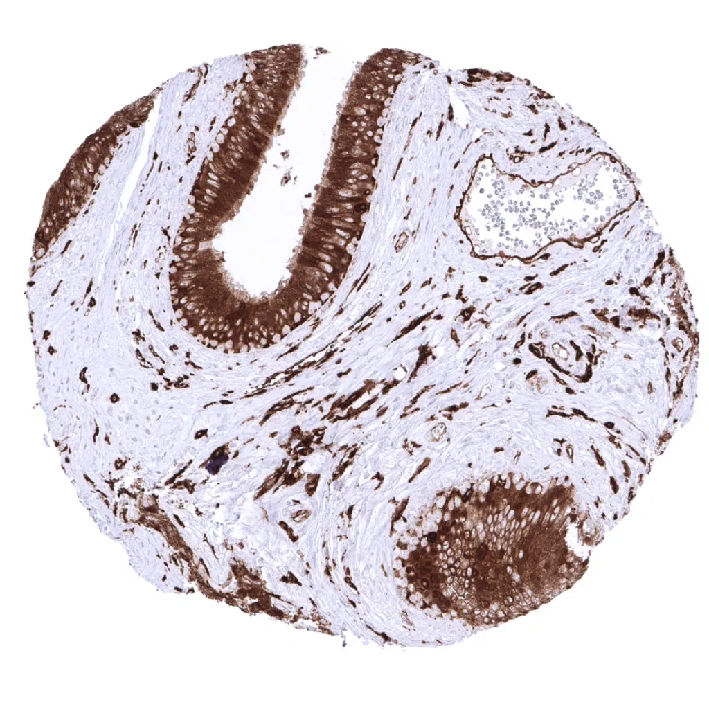
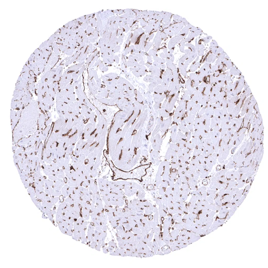
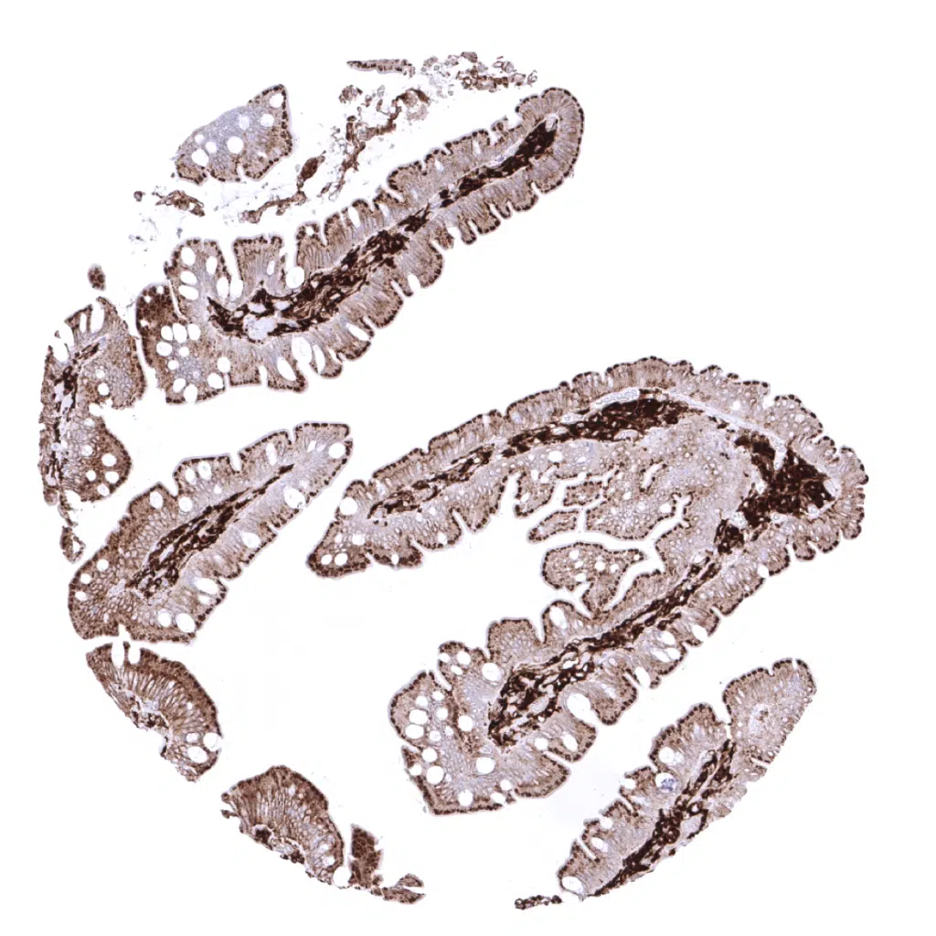
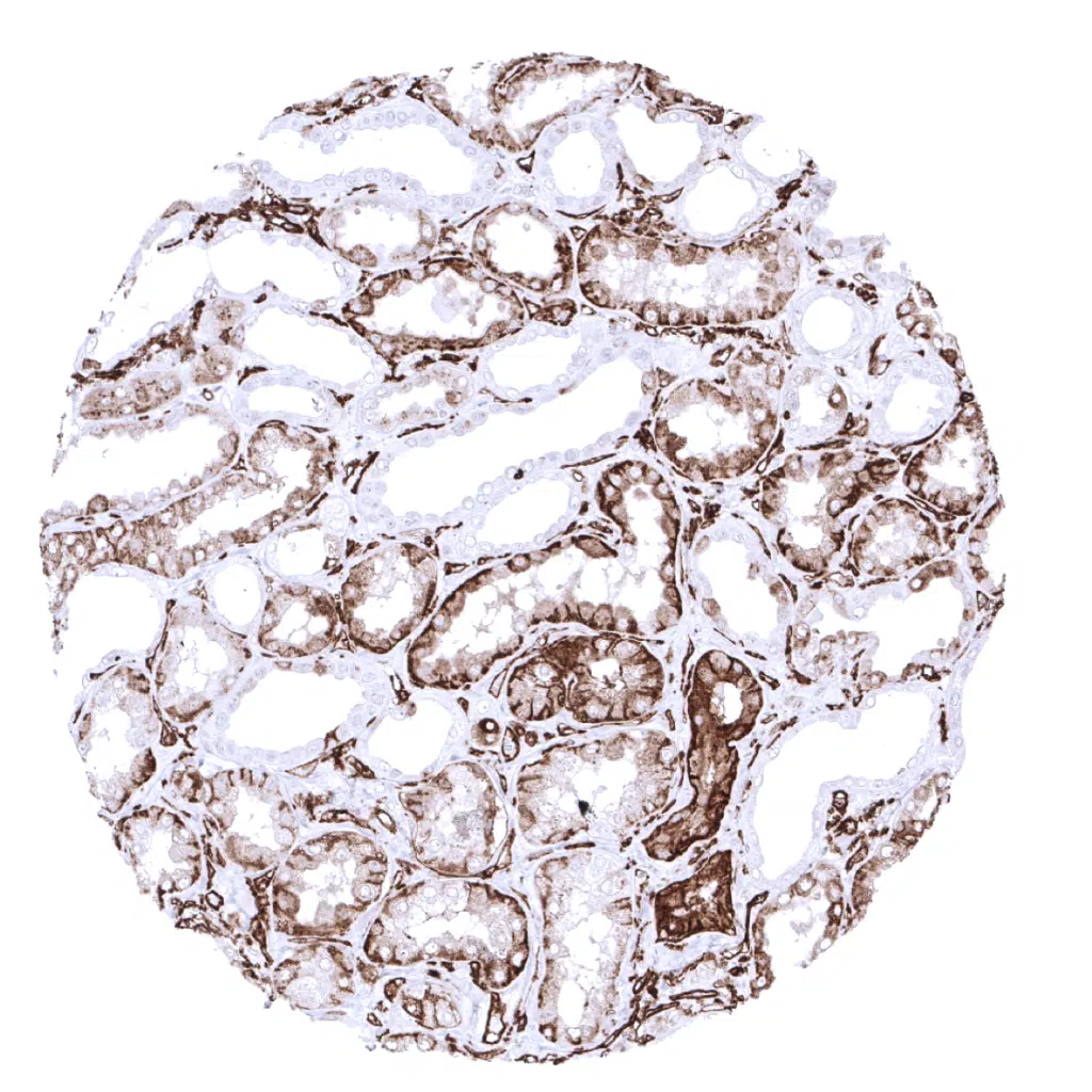
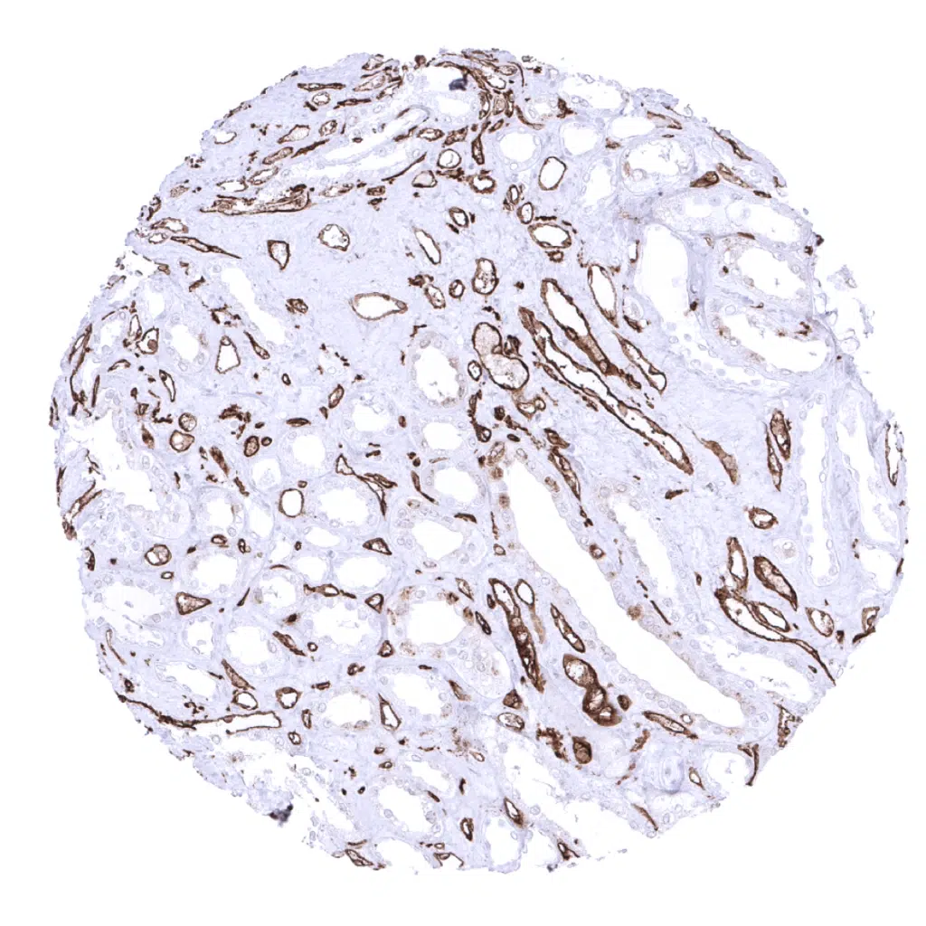
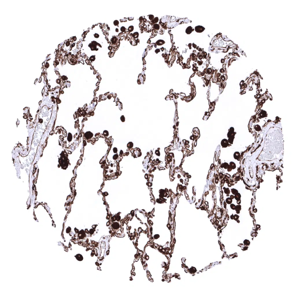
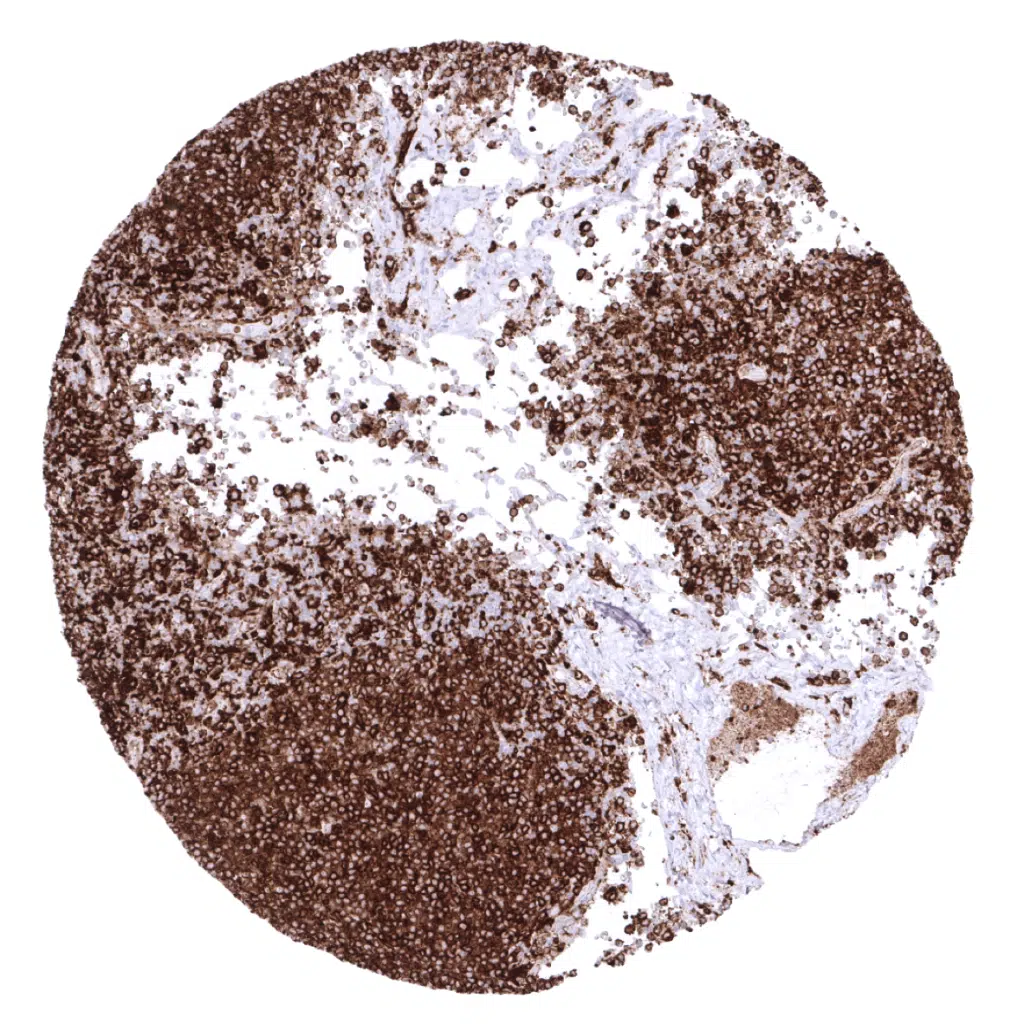
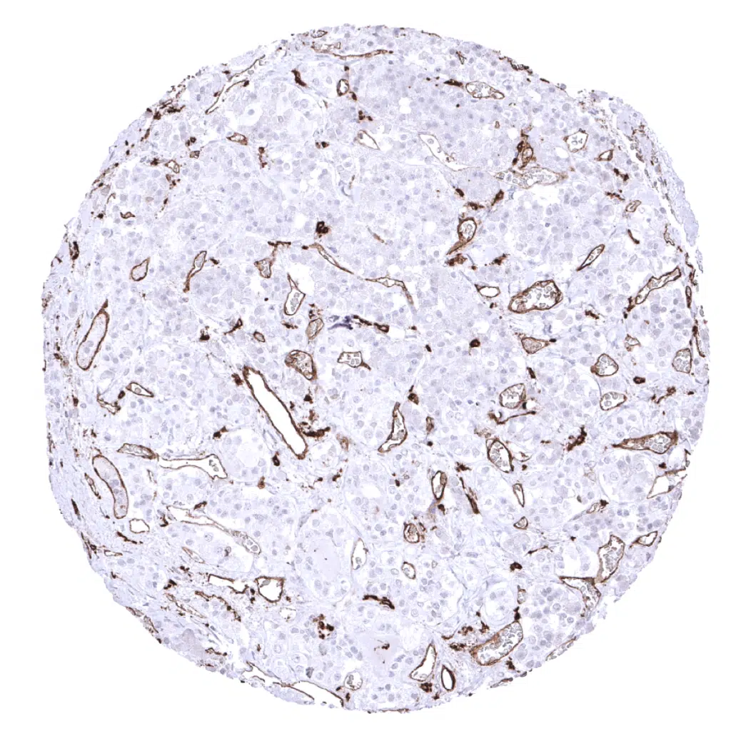
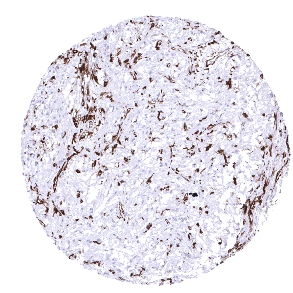
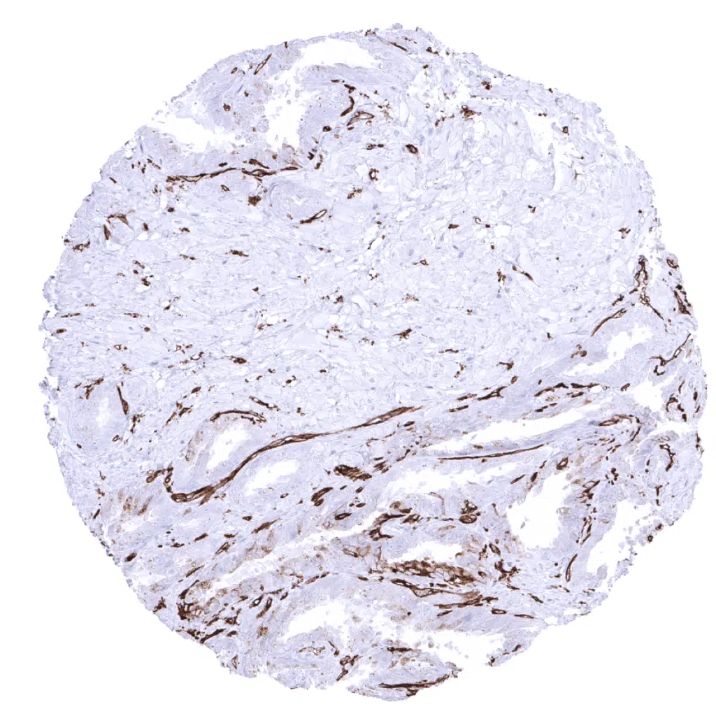
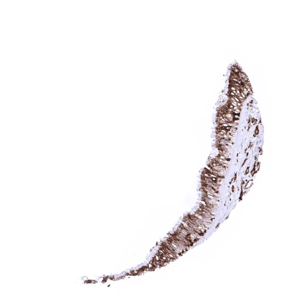
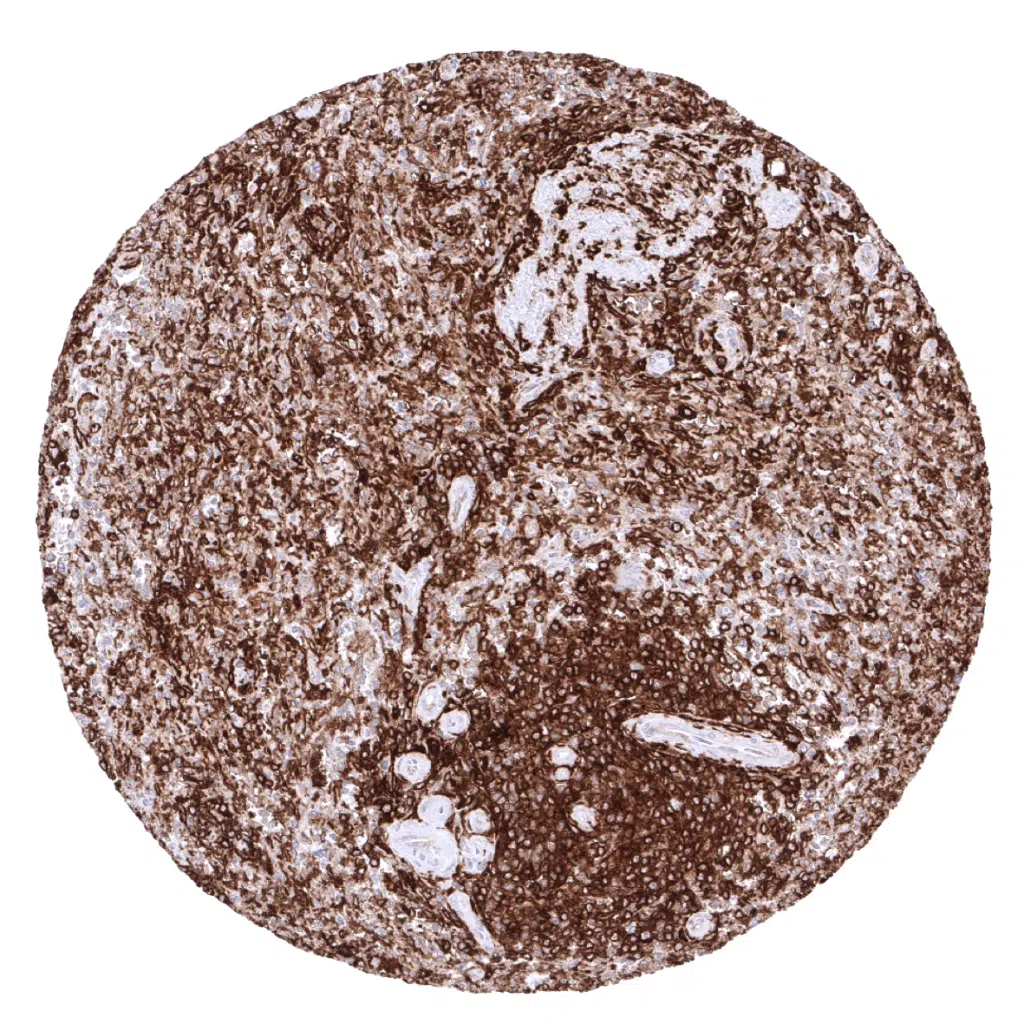
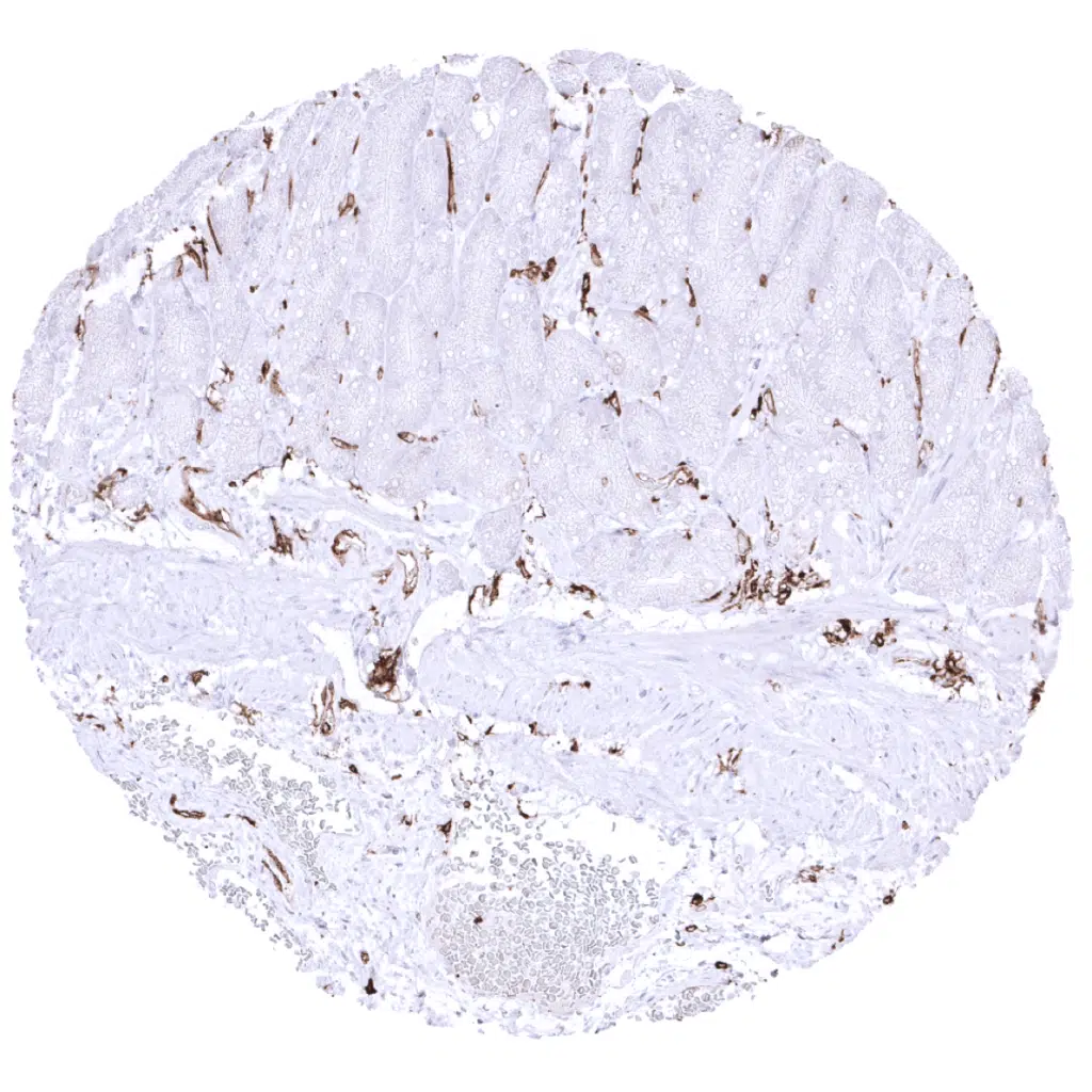
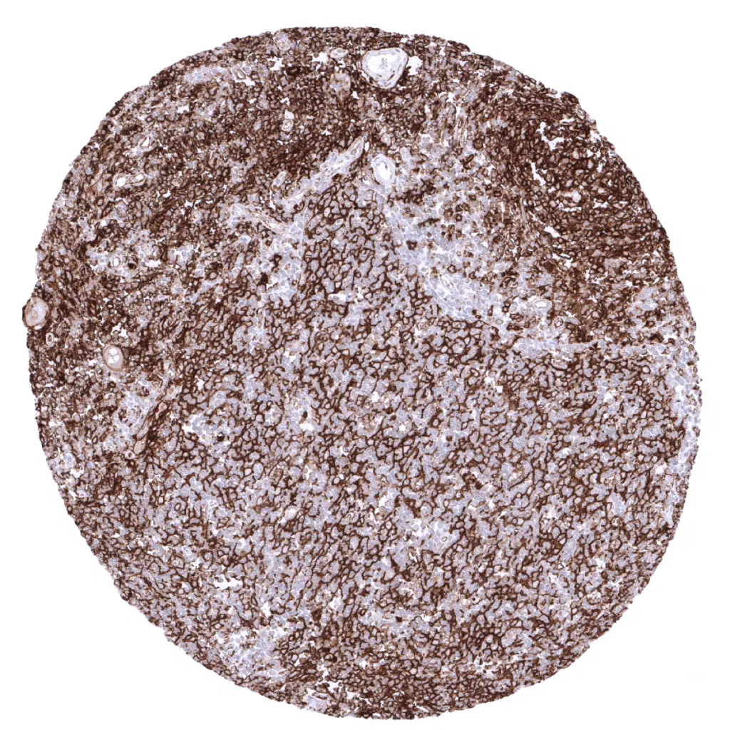
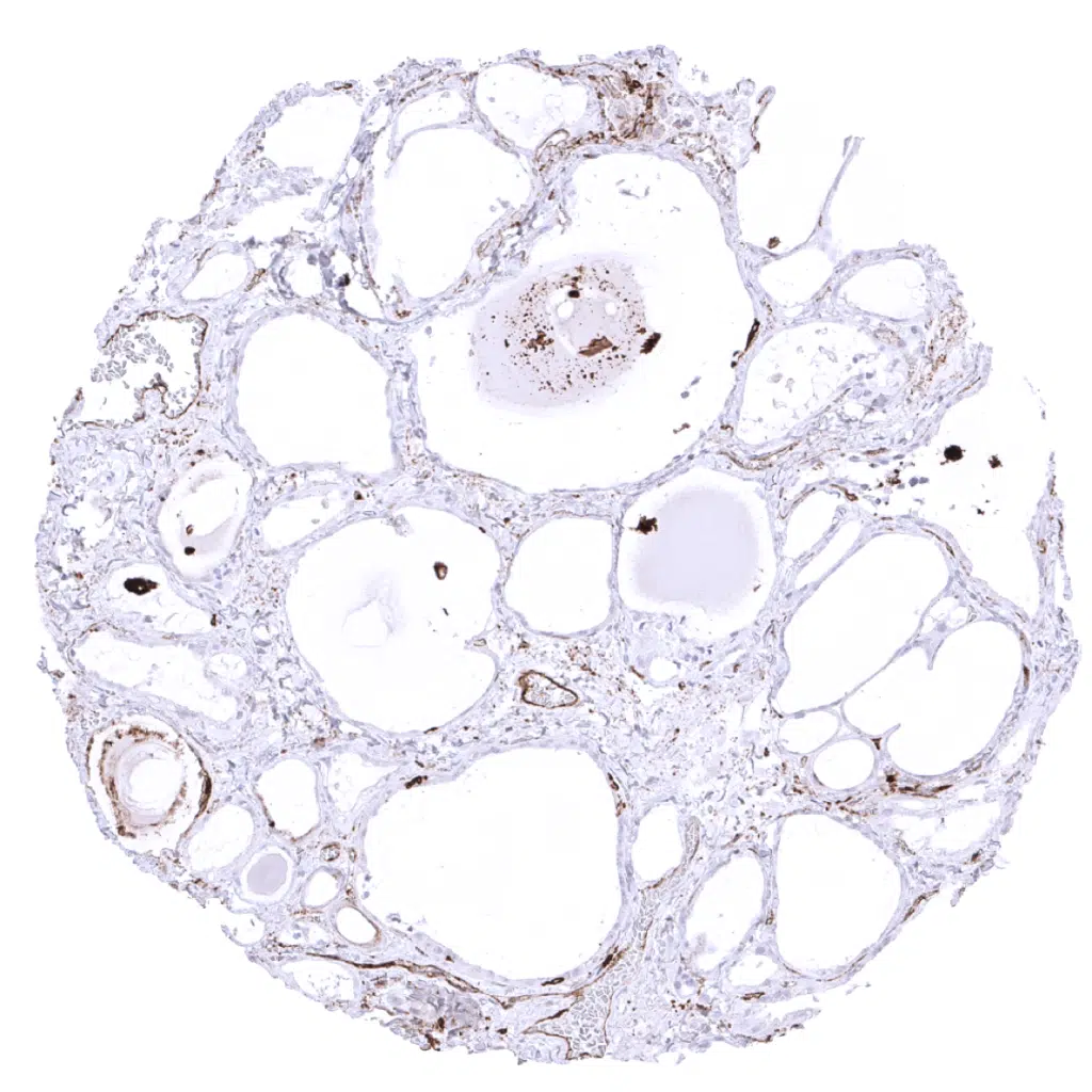
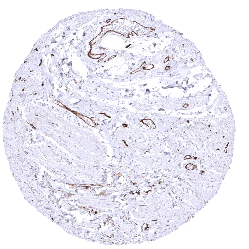
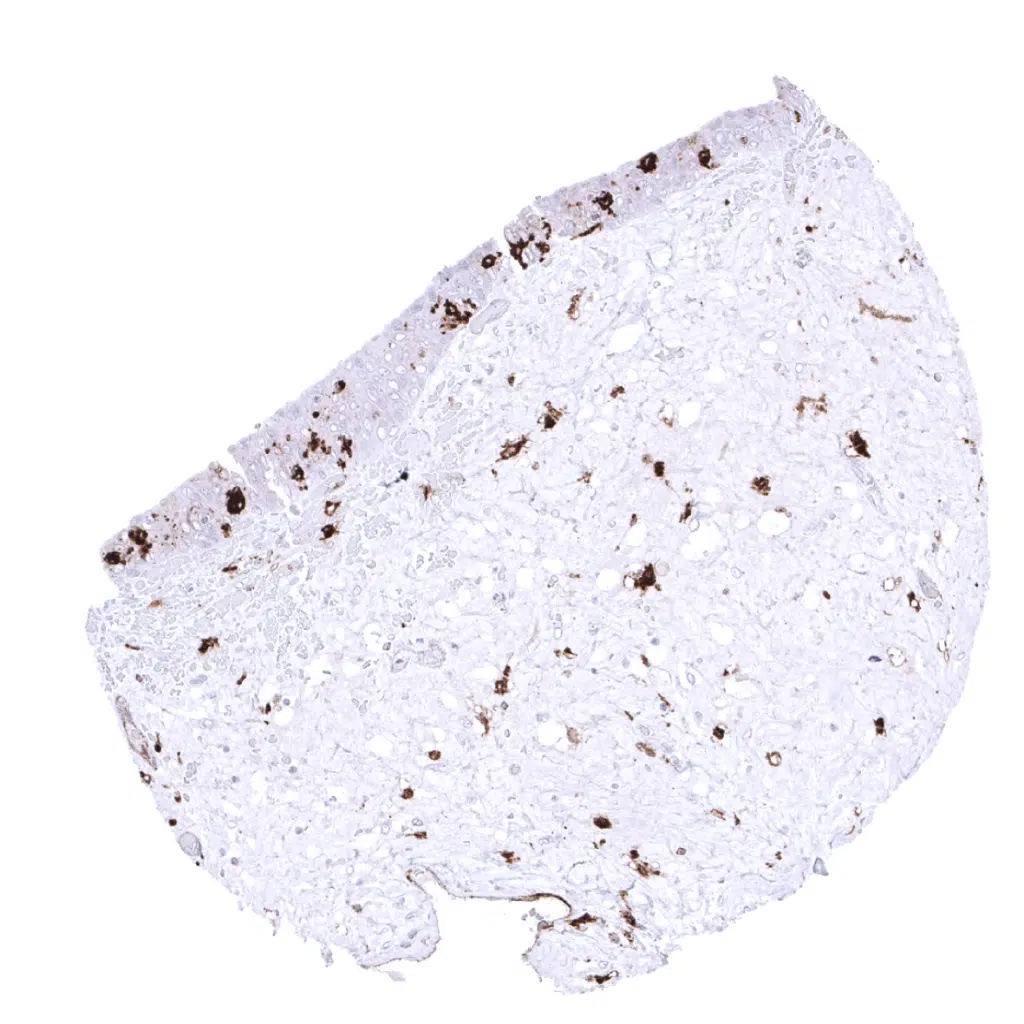
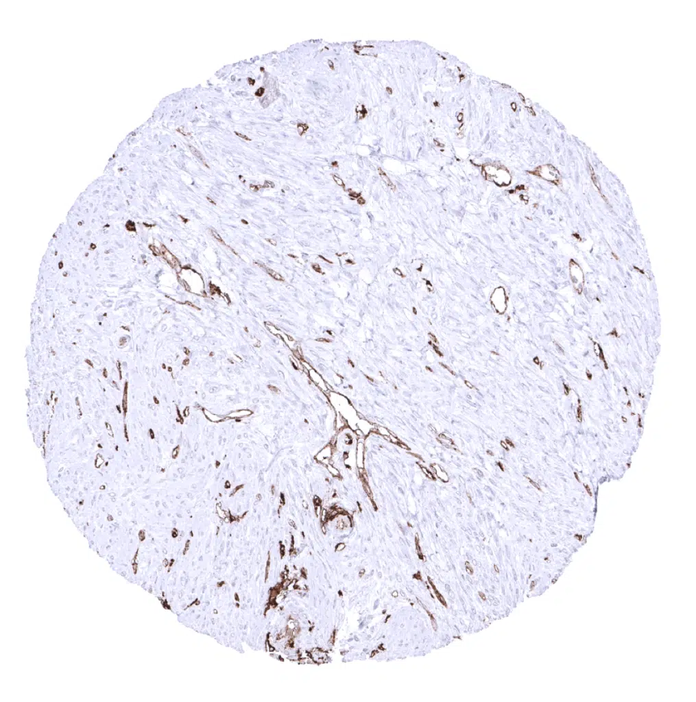
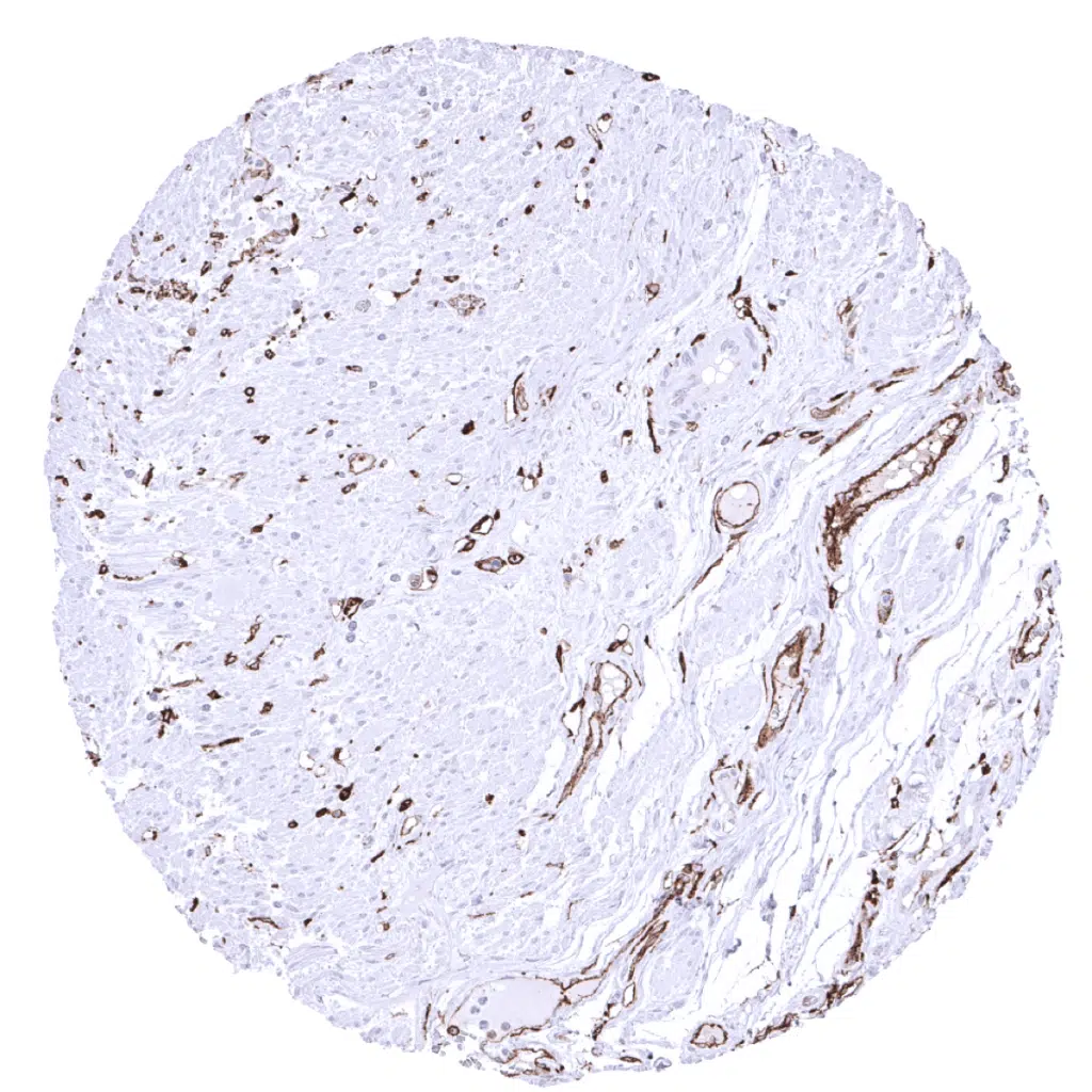
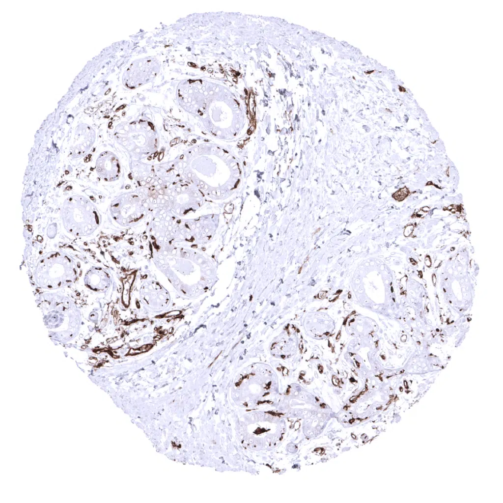
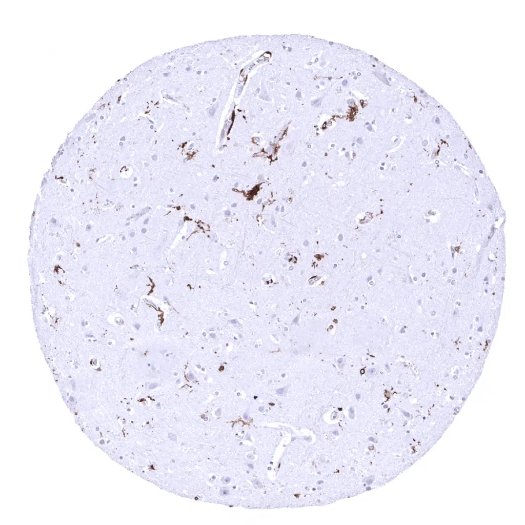
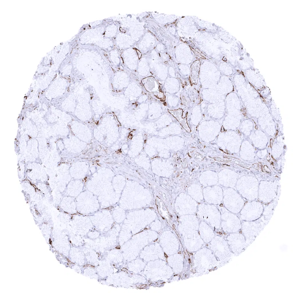
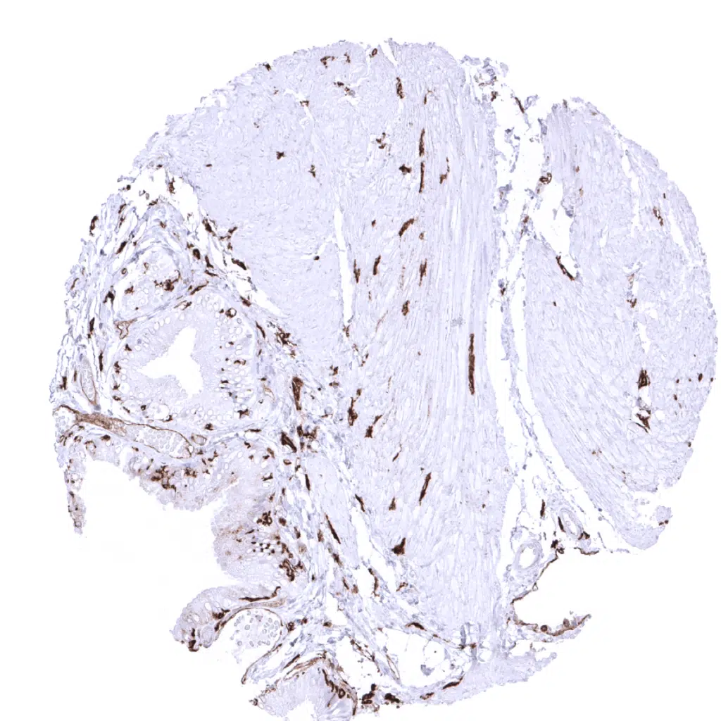
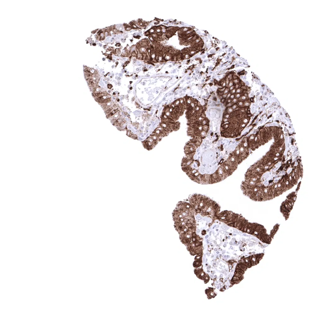
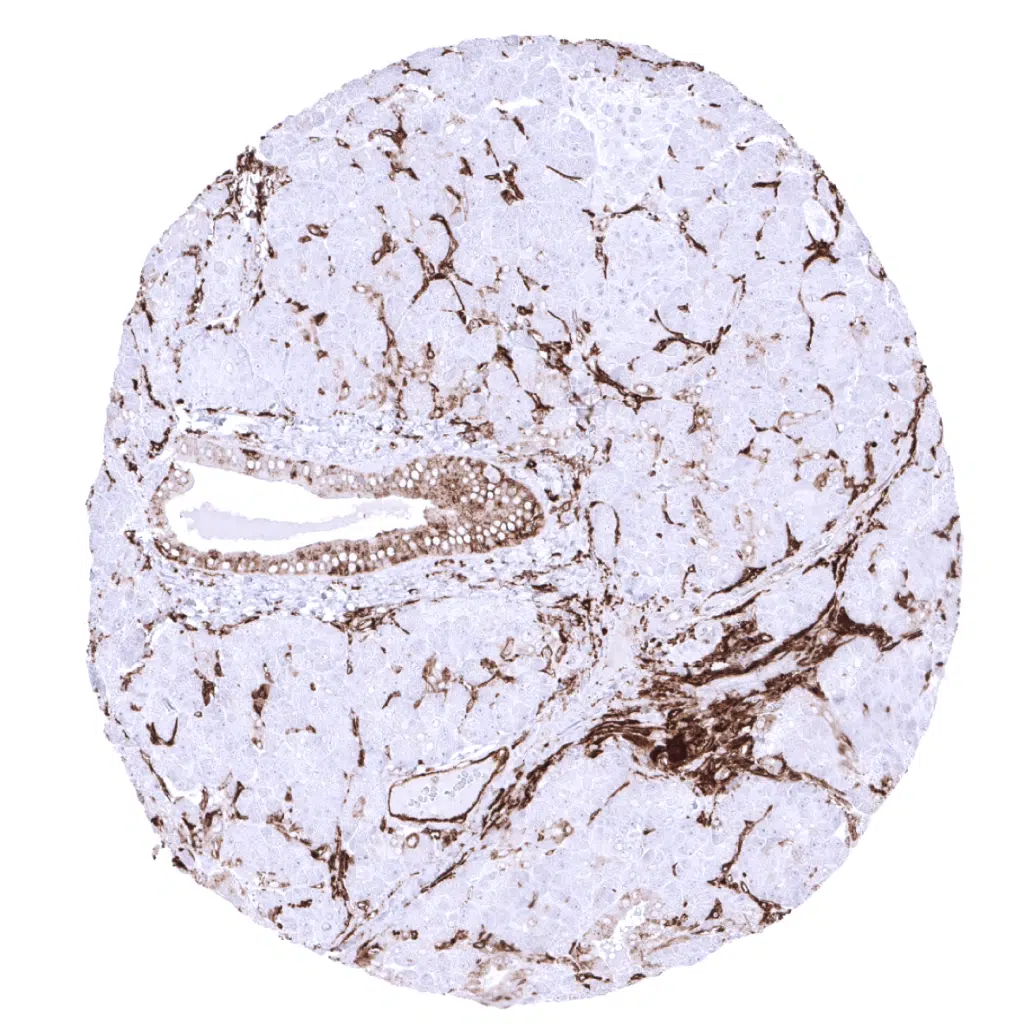
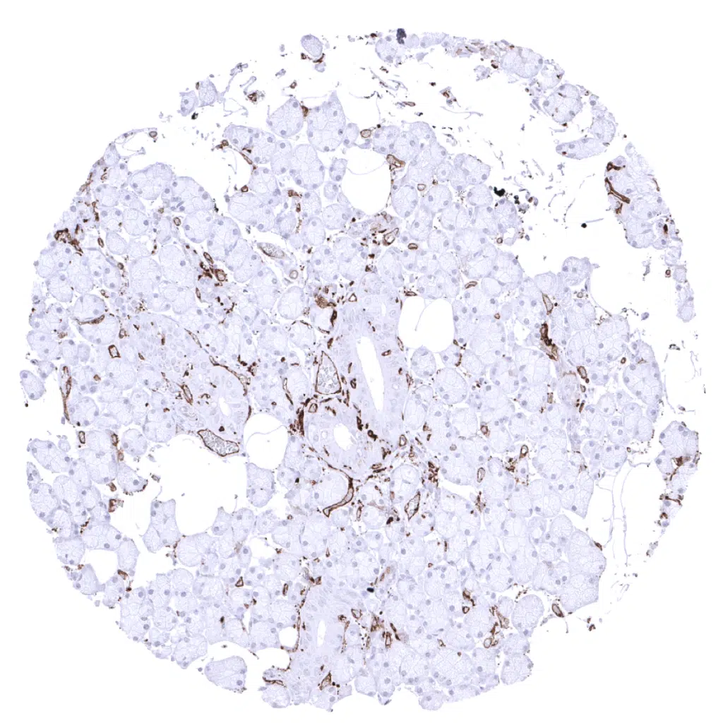
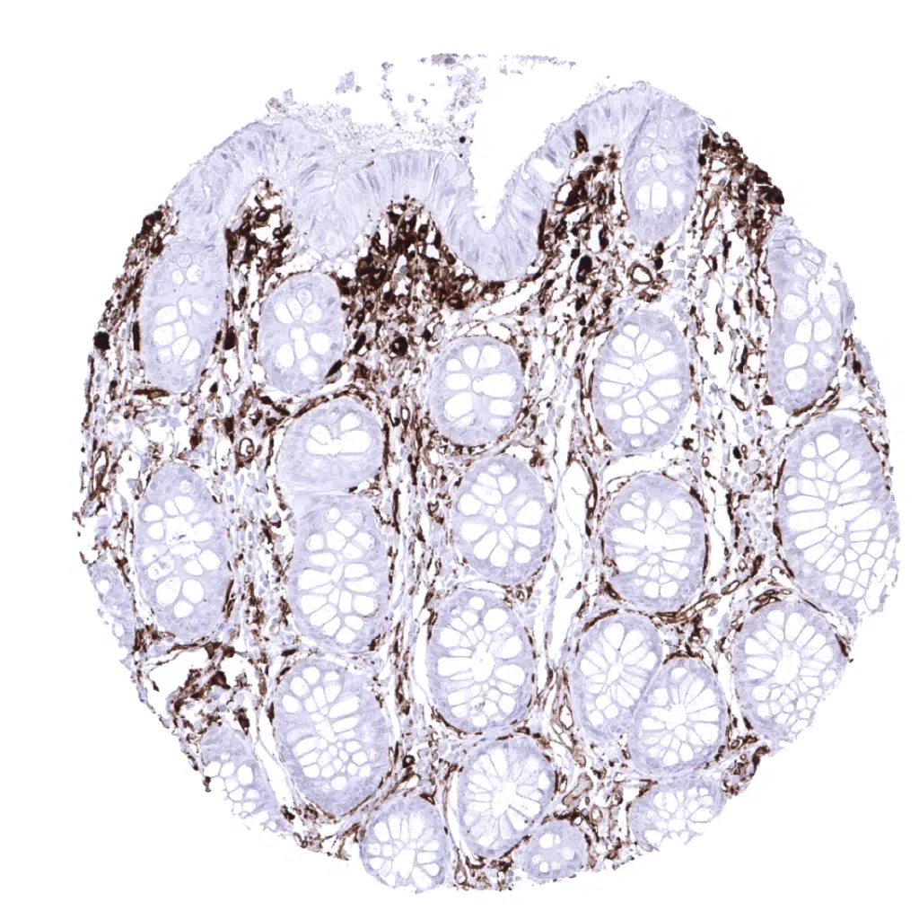
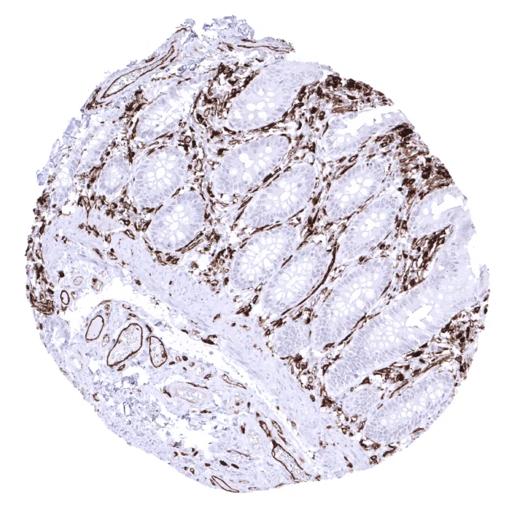
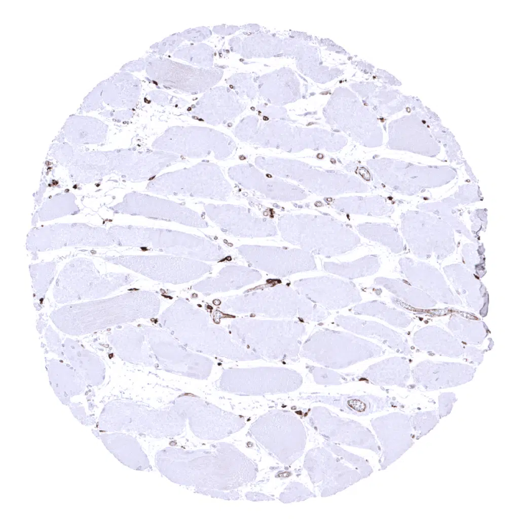
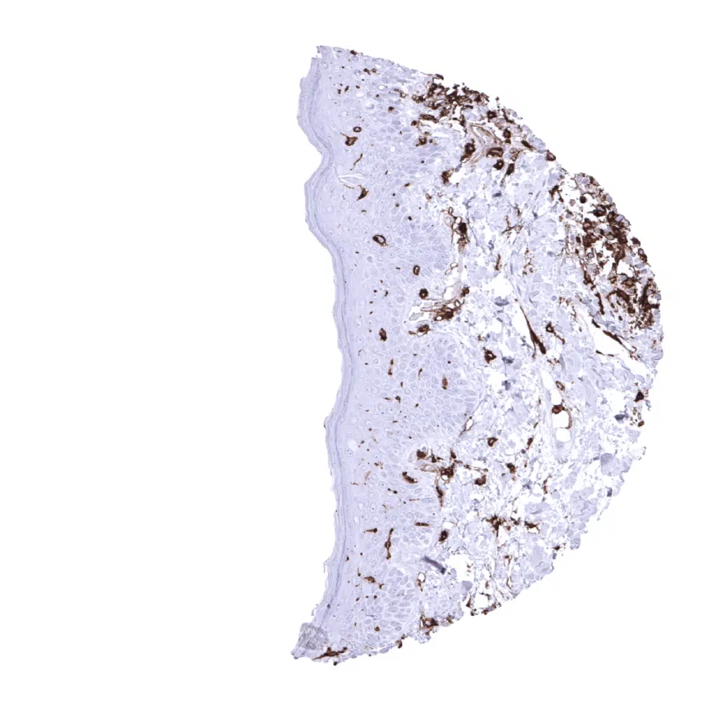
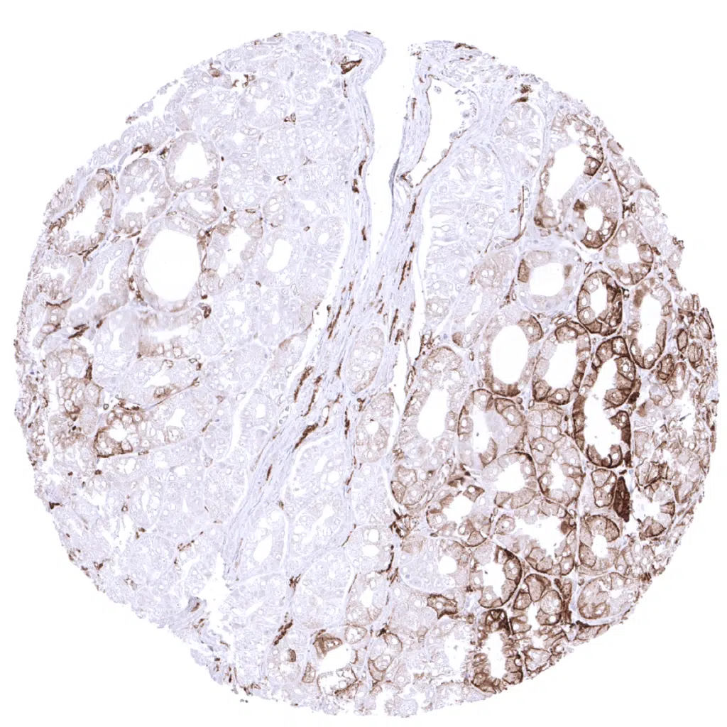
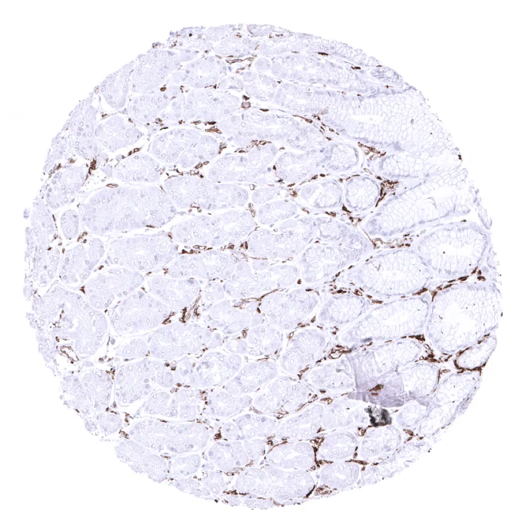
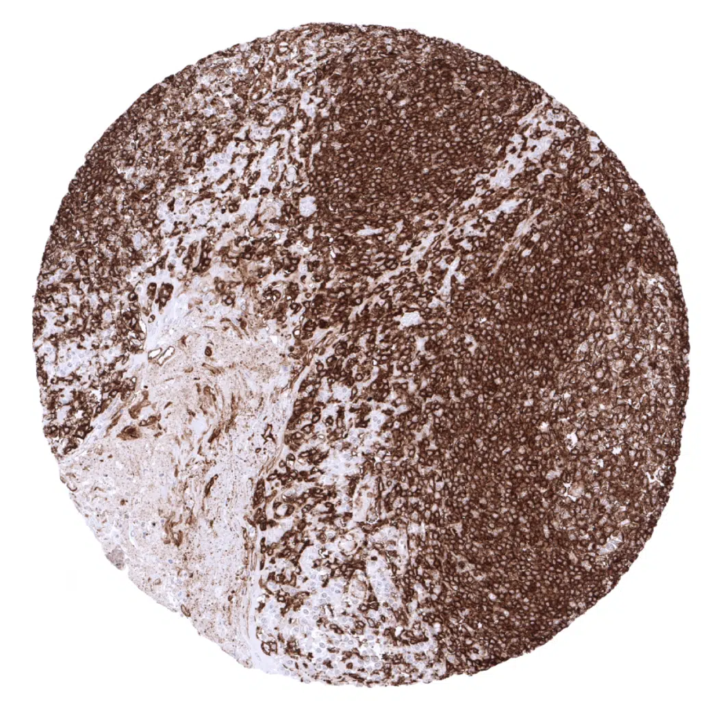
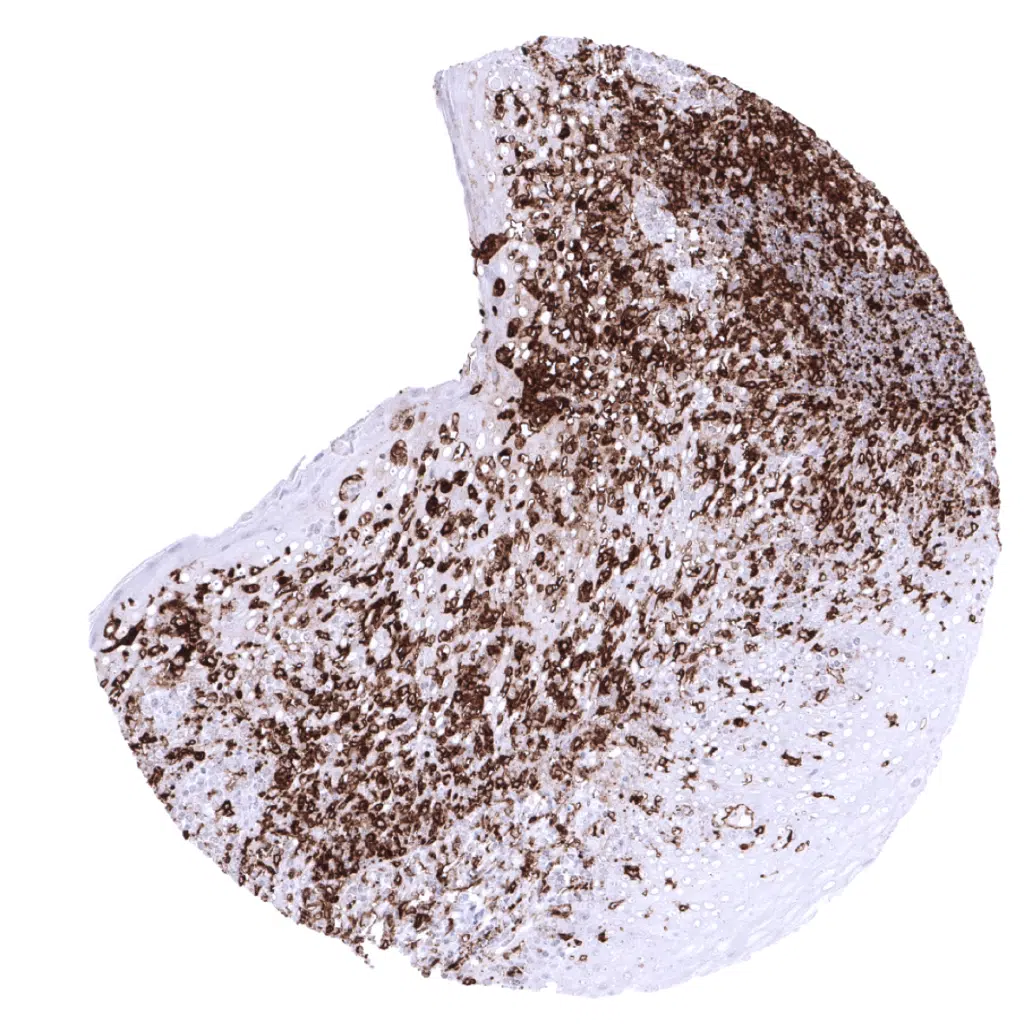
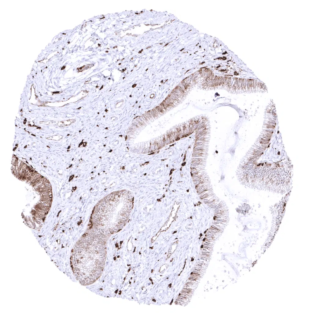
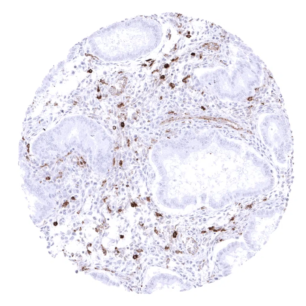
HLA-DRA antibody [MSVA-470R] HistoMAX


Go to Cancer Tissue Gallery
  
 Go back to product page
  

Go to HistoMAX page


Adrenal gland

Aorta, media

Appendix, muscular wall

Bone marrow


Breast

Bronchus, mucosa

Bronchus, mucosa: Respiratory epithelium can show a cytoplasmic and membranous HLA-DR immunostaining of variable intensity.

Cerebellum (granule cell layer, white matter)


Cerebellum (molecular, Purkinje, and granule cell layer): In the brain, HLA-DRa immunostaining occurs in cells of monocytic origin.

Cerebrum, grey matter

Cerebrum, white matter

Colon descendens, muscular wall


Duodenum, Brunner gland

Duodenum, mucosa: The strongest membranous and cytoplasmic epithelial cell HLA-DRa staining occurs in the duodenum.

Epididymis: A strong cytoplasmic and membranous HLA-DRa immunostaining can be seen in epithelial cells of the epididymis.

Epididymis: In some samples, epithelial cells of the epididymis are lacking HLA-DRa immunostaining.


Esophagus, squamous epithelium

Fallopian tube, mucosa: A strong, predominantly membranous HLA-DRa immunostaining is regularly seen in a subset of the surface epithelial cells of the fallopian tube (mosaic staining pattern).

Fat

Gallbladder, epithelium


Gallbladder, epithelium: A strong cytoplasmic and membranous HLA-DRa staining can occur in the surface epithelium of the gallbladder.

Heart muscle: In the heart, HLA-DRa staining is seen in endothelial cells of blood vessels.

Ileum, mucosa: A membranous and cytoplasmic epithelial cell HLA-DRa staining of variable intensity is regularly seen in the ileum.

Kidney, cortex: Glomeruli and capillaries show intense HLA-DRa staining.


Kidney, cortex: A cytoplasmic and membranous HLA-DRa immunostaining can occur in a fraction of tubuli in the kidney.

Kidney, medulla

Liver: Sinusoids and Kupffer cells show strong HLA-DRa immunostaining in the liver.

Lung: Intense HLA-DRa immunostaining of alveolar macrophages and alveolar capillaries.


Lymph node: A strong HLA-DRa immunostaining is regularly seen on the majority of inflammatory cells including dendritic cells and macrophages.

Ovary, stroma

Pancreas: Excretory ducts may show cytoplasmic and membranous HLA-DRa staining in the pancreas.

Parathyroid gland


Parotid gland

Pituitary gland, anterior lobe

Pituitary gland, posterior lobe

Placenta (amnion and chorion)


Pregnant uterus (decidua)

Placenta, early: Focal HLA-DRa positivity regularly occurs in the early placenta.

Placenta, mature: Placenta is the only organ without any HLA-DRa positivity of blood vessels.

Prostate


Rectum, mucosa

Rectum, mucosa

Seminal vesicle

Sinus paranasales: Respiratory epithelium can show a cytoplasmic and membranous HLA-DRa immunostaining of variable intensity.


Skeletal muscle: In skeletal muscle, HLA-DRa staining is seen in endothelial cells of blood vessels

Skin

Spleen: A HLA-DRa immunostaining is regularly seen on the majority of inflammatory cells including dendritic cells and macrophages.

Stomach, antrum


Stomach, antrum: A cytoplasmic and membranous HLA-DRa staining is often seen in the surface epithelium of the stomach.

Stomach, corpus

Stomach, corpus: A cytoplasmic and membranous HLA-DRa staining is often seen in the surface epithelium of the stomach.

Testis


Thymus: A HLA-DRa immunostaining is regularly seen on the majority of inflammatory cells including dendritic cells and macrophages.

Thyroid gland

Tonsil: A HLA-DRa immunostaining is regularly seen on the majority of inflammatory cells including dendritic cells and macrophages.

Tonsil, surface epithelium


Urinary bladder, muscular wall

Urinary bladder, urothelium

Uterus, ectocervix: In the ectocervix, HLA-DRa immunostaining can be seen in endothelial cells and in some inflammatory cells. Note: Surface staining is a result of „inking“ amd not a true immunostaining.

Uterus, ectocervix: Endocervical epithelium can show a cytoplasmic and membranous HLA-DRa immunostaining of variable intensity.


Uterus, endometrium (proliferation)

Uterus, endometrium (secretion)

Uterus, myometrium


| TOP |