Normal Tissue Gallery- GTX639938

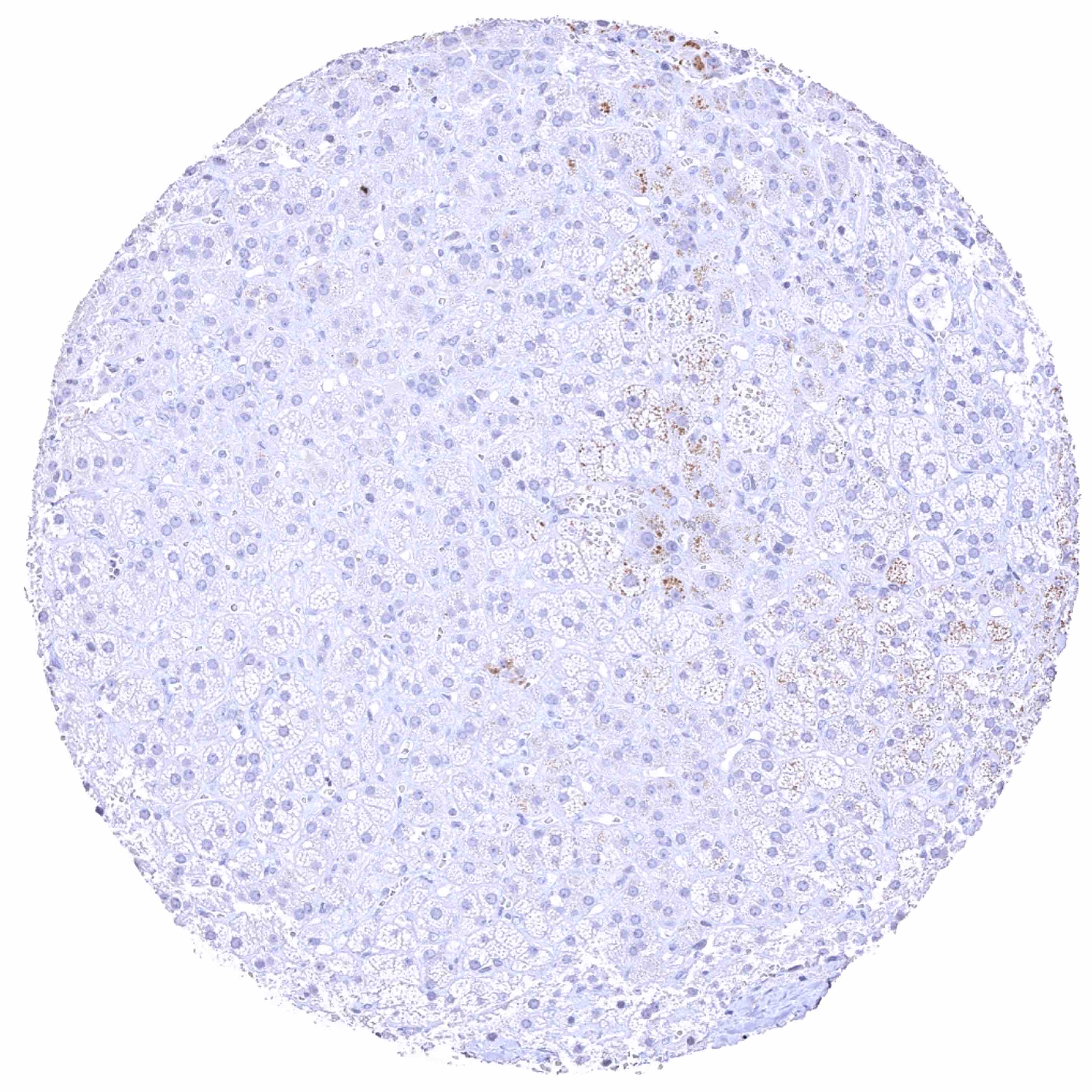
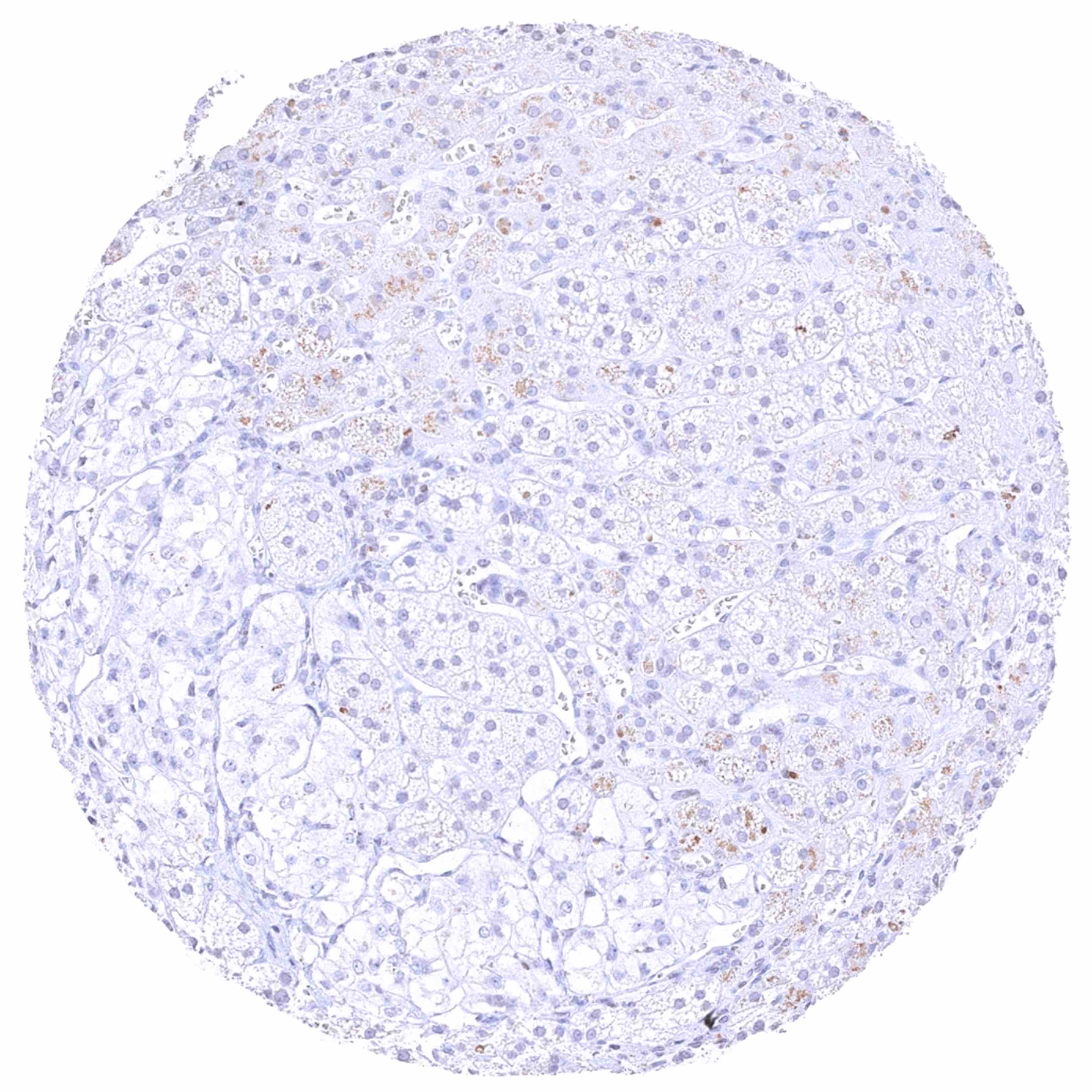
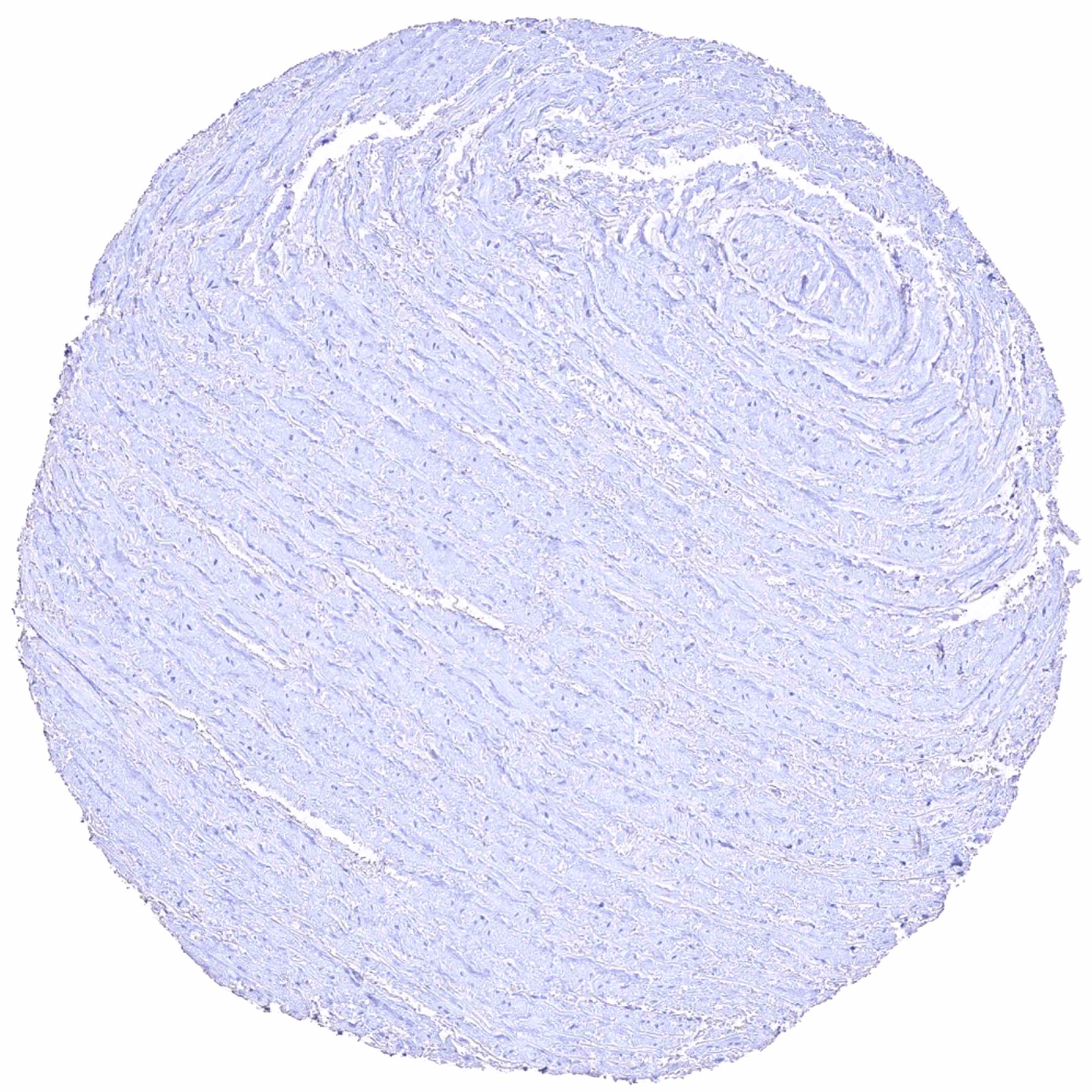
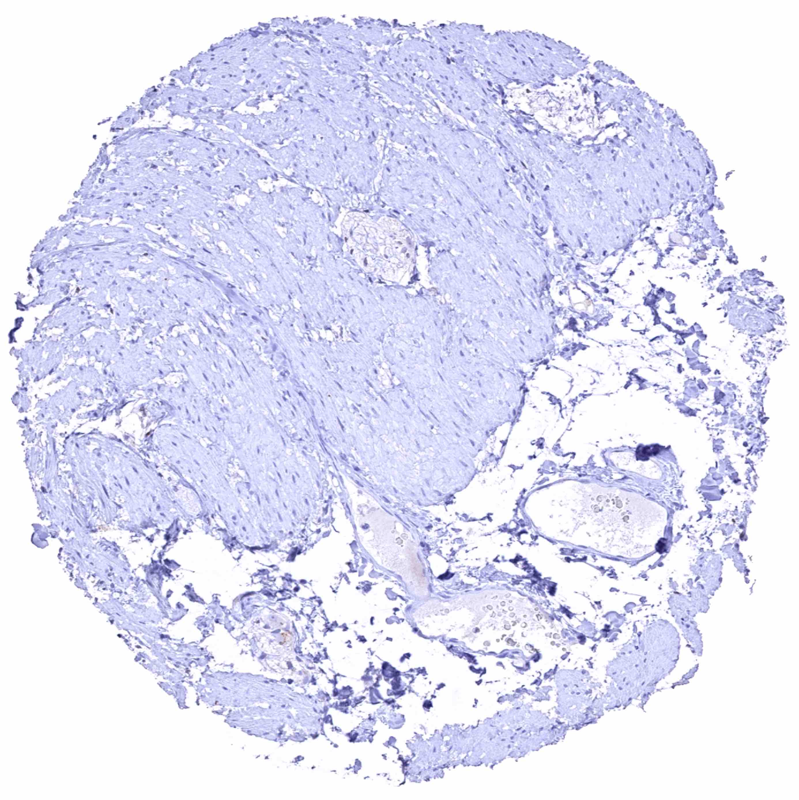
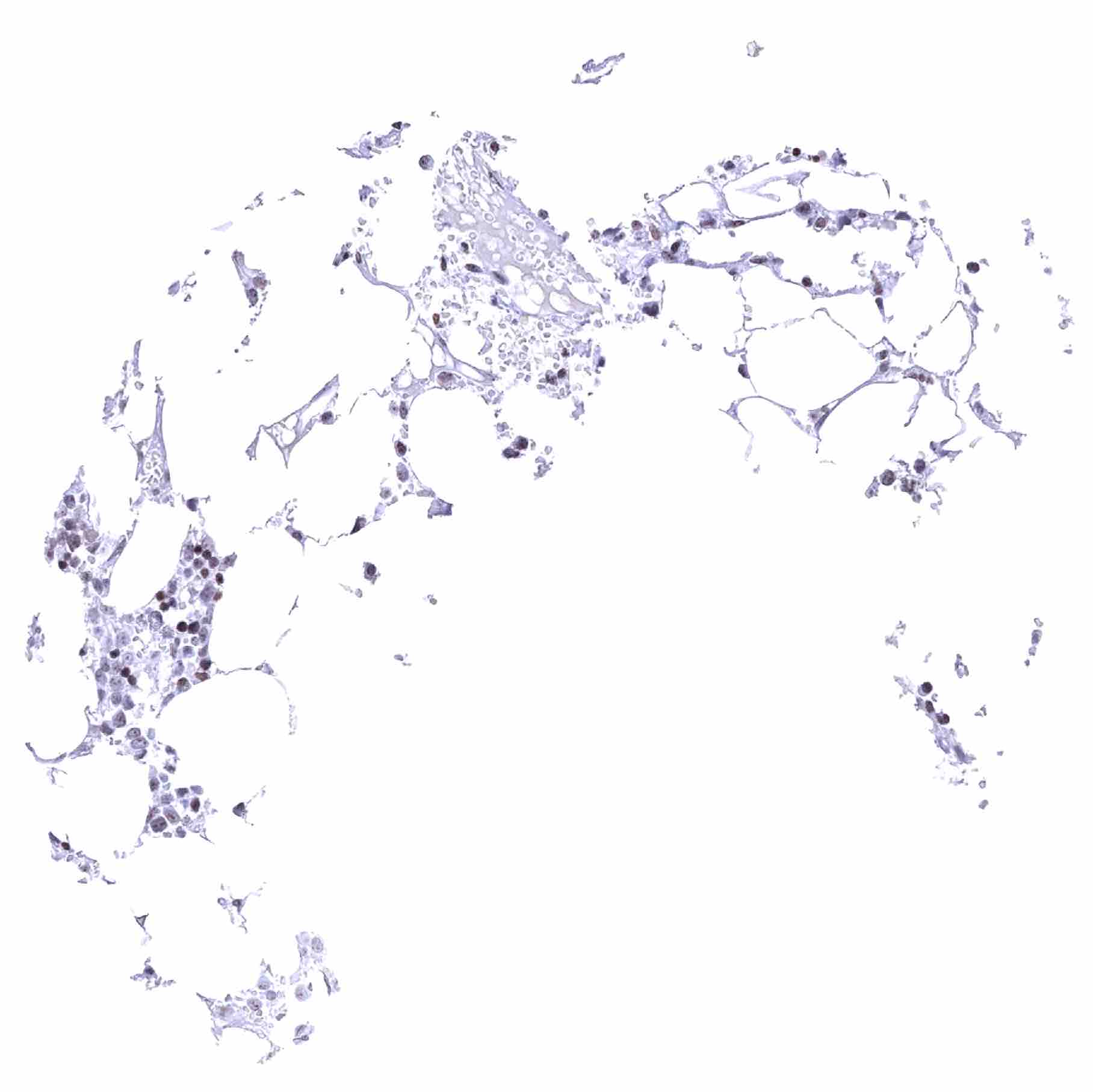
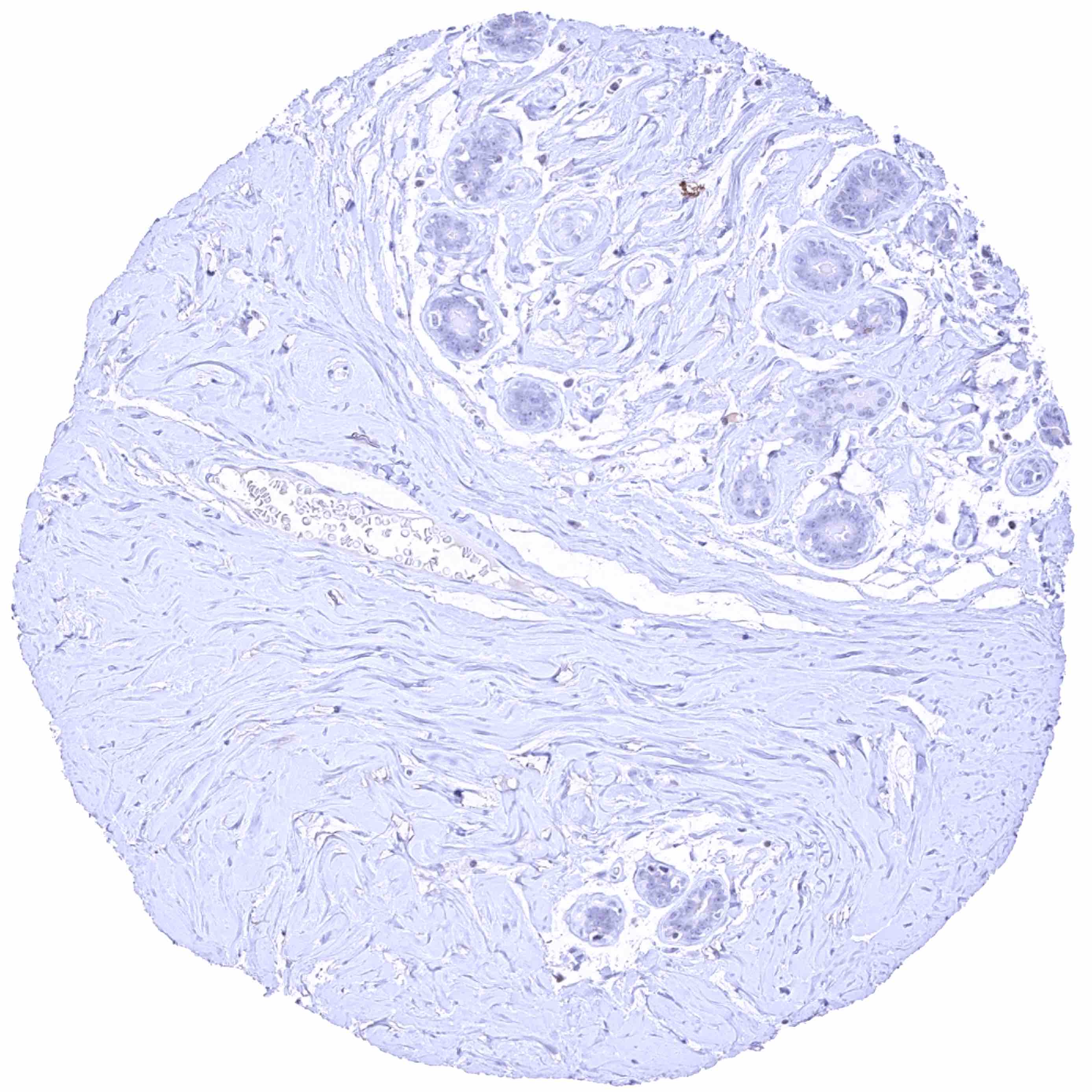
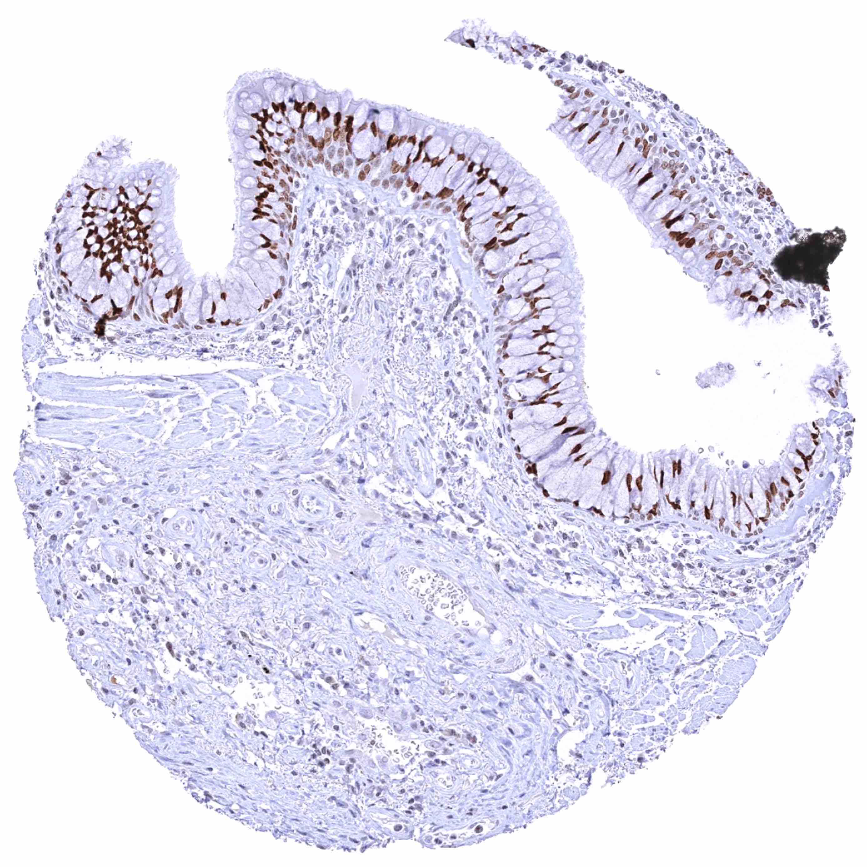
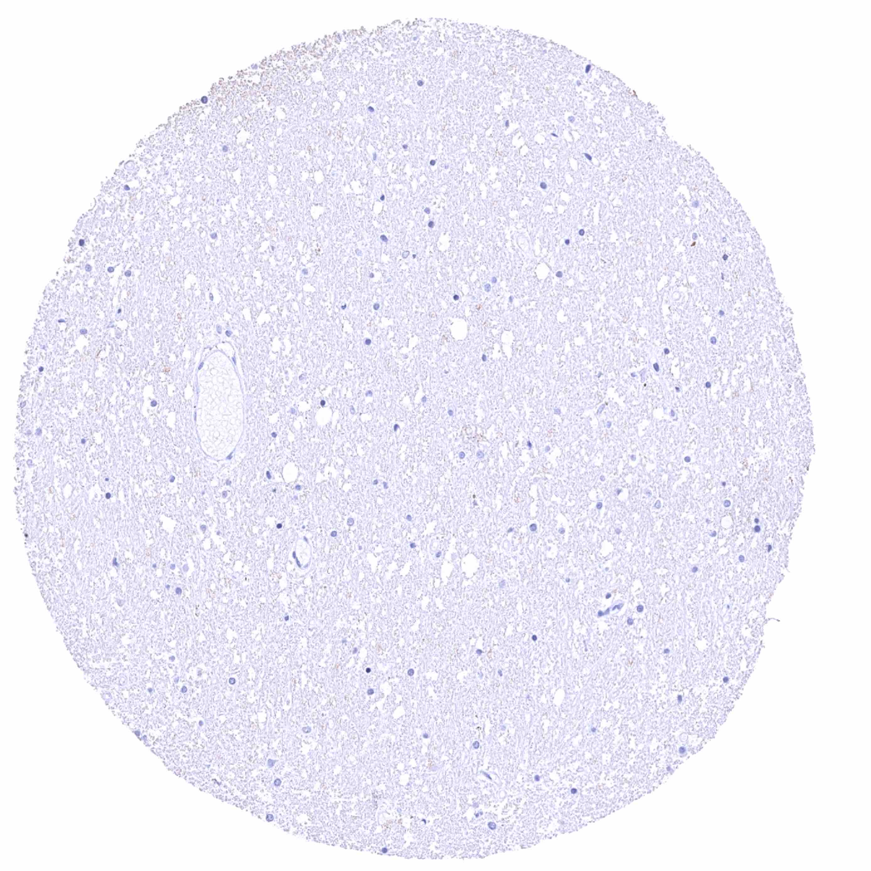
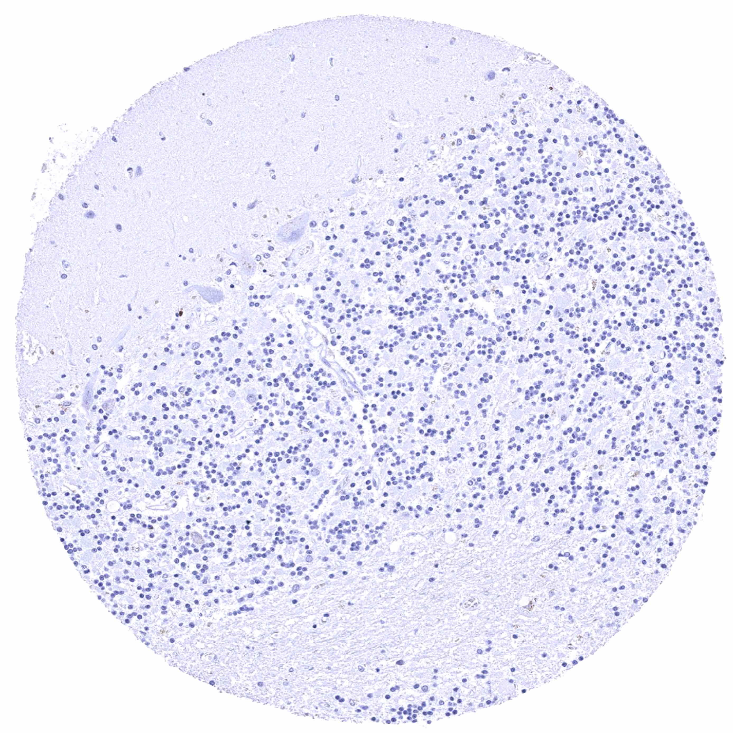
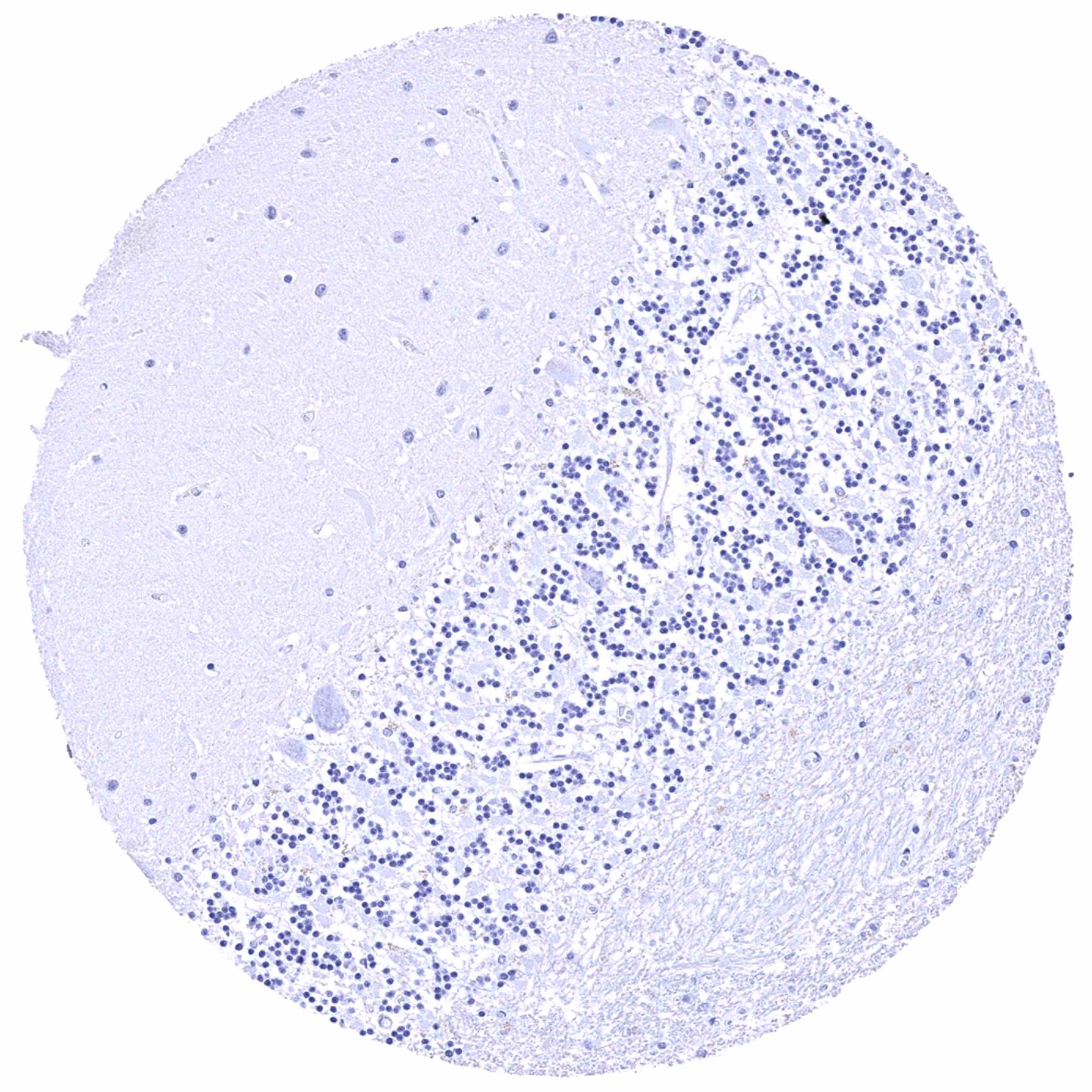
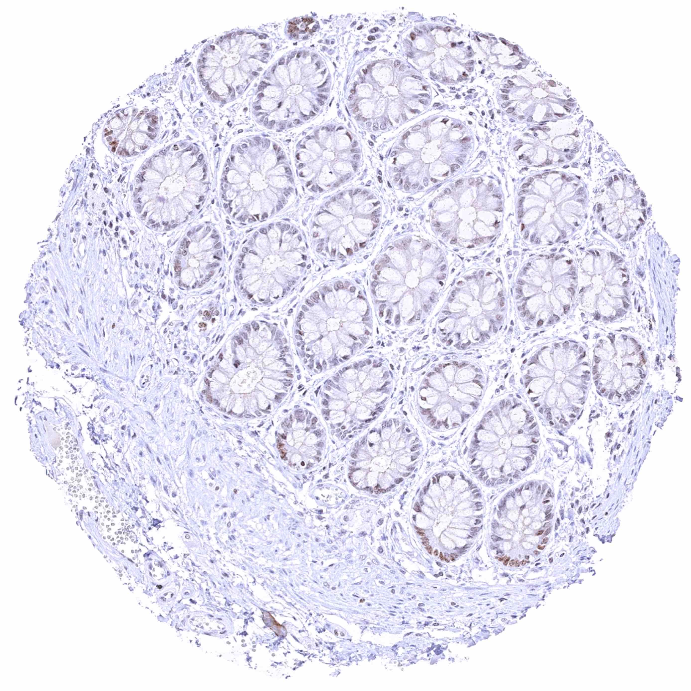
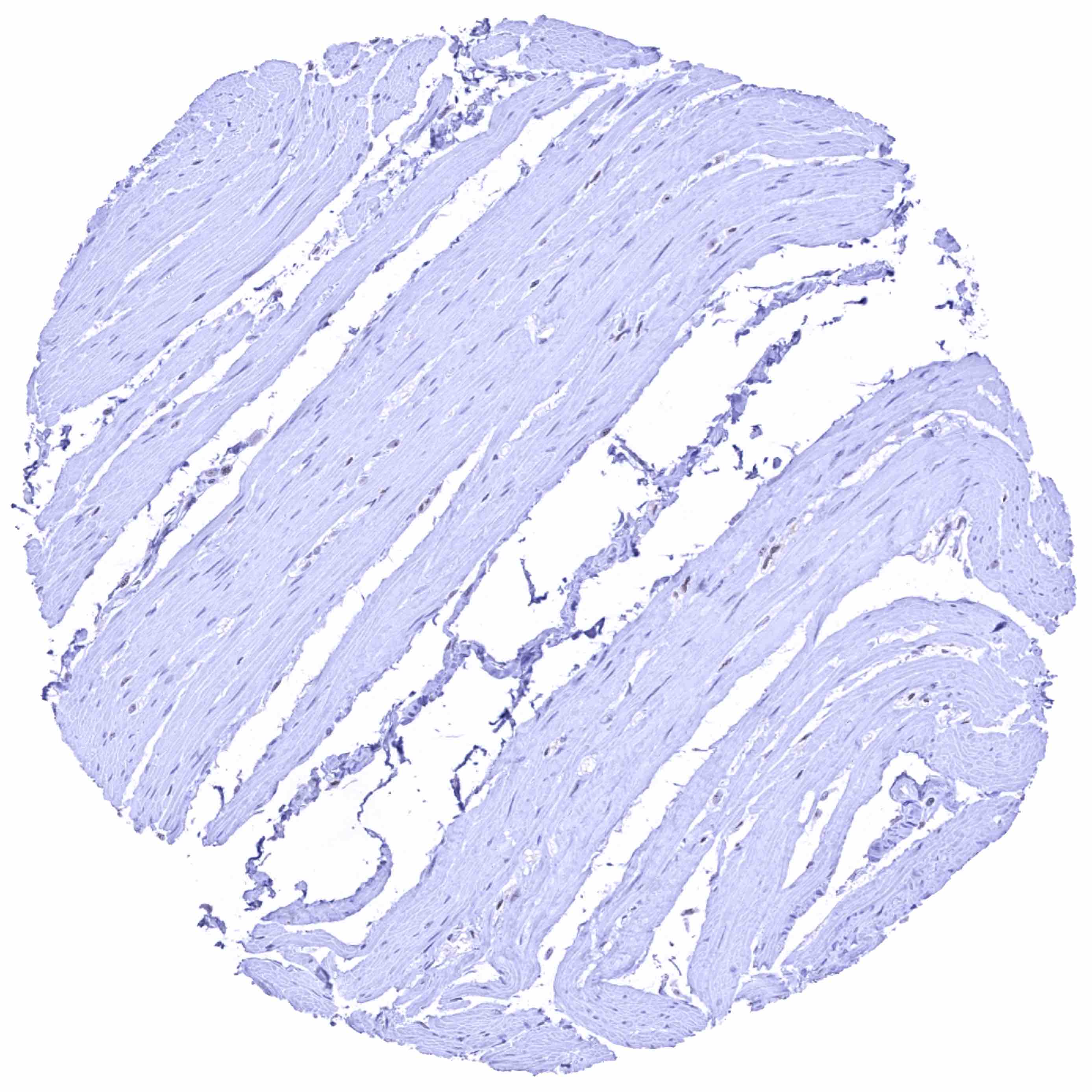
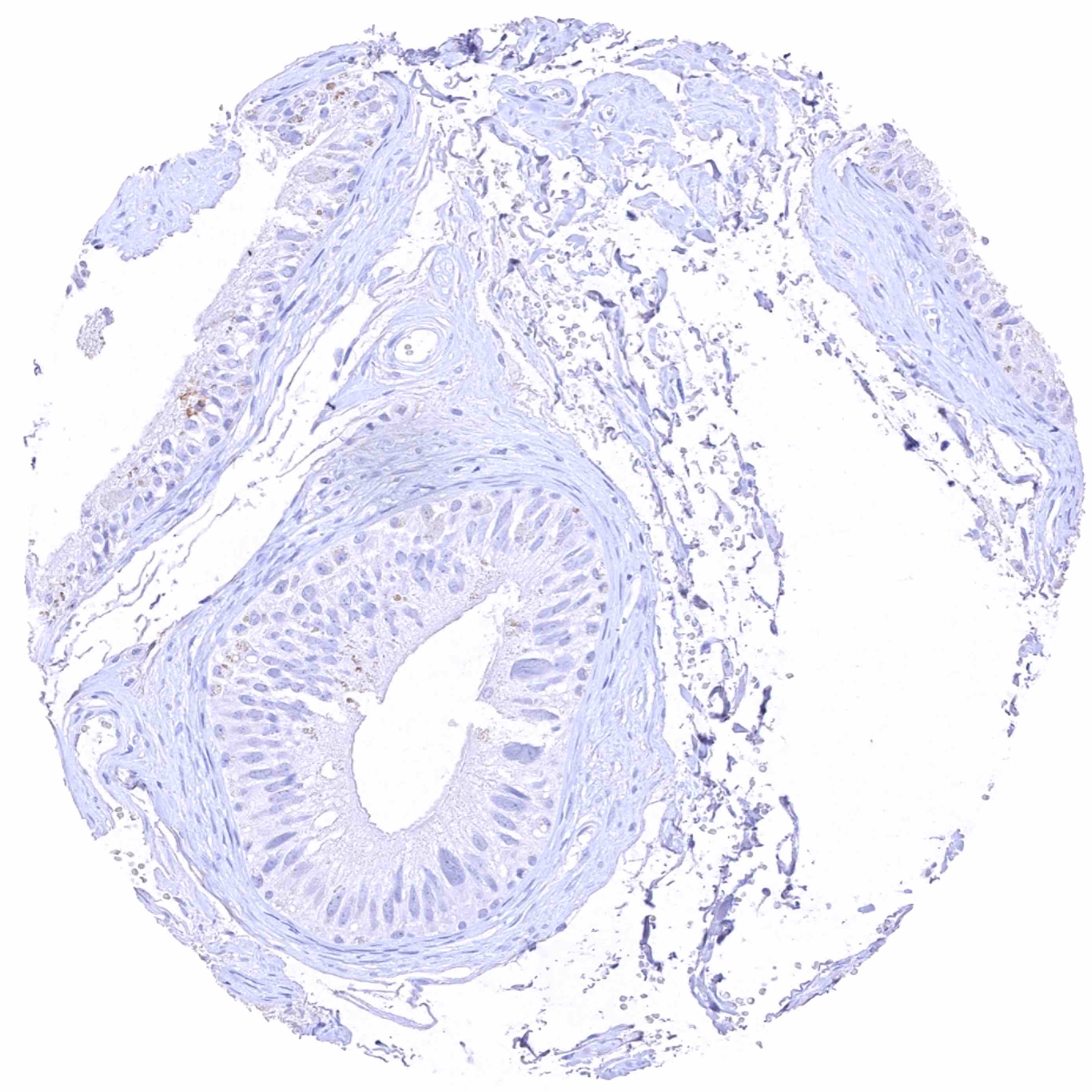
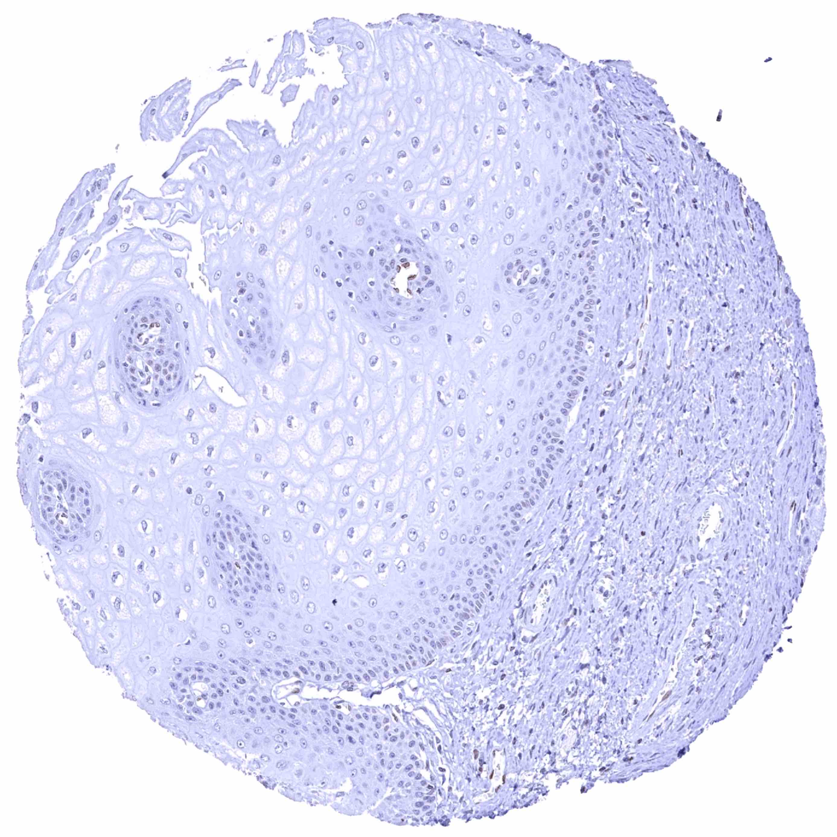
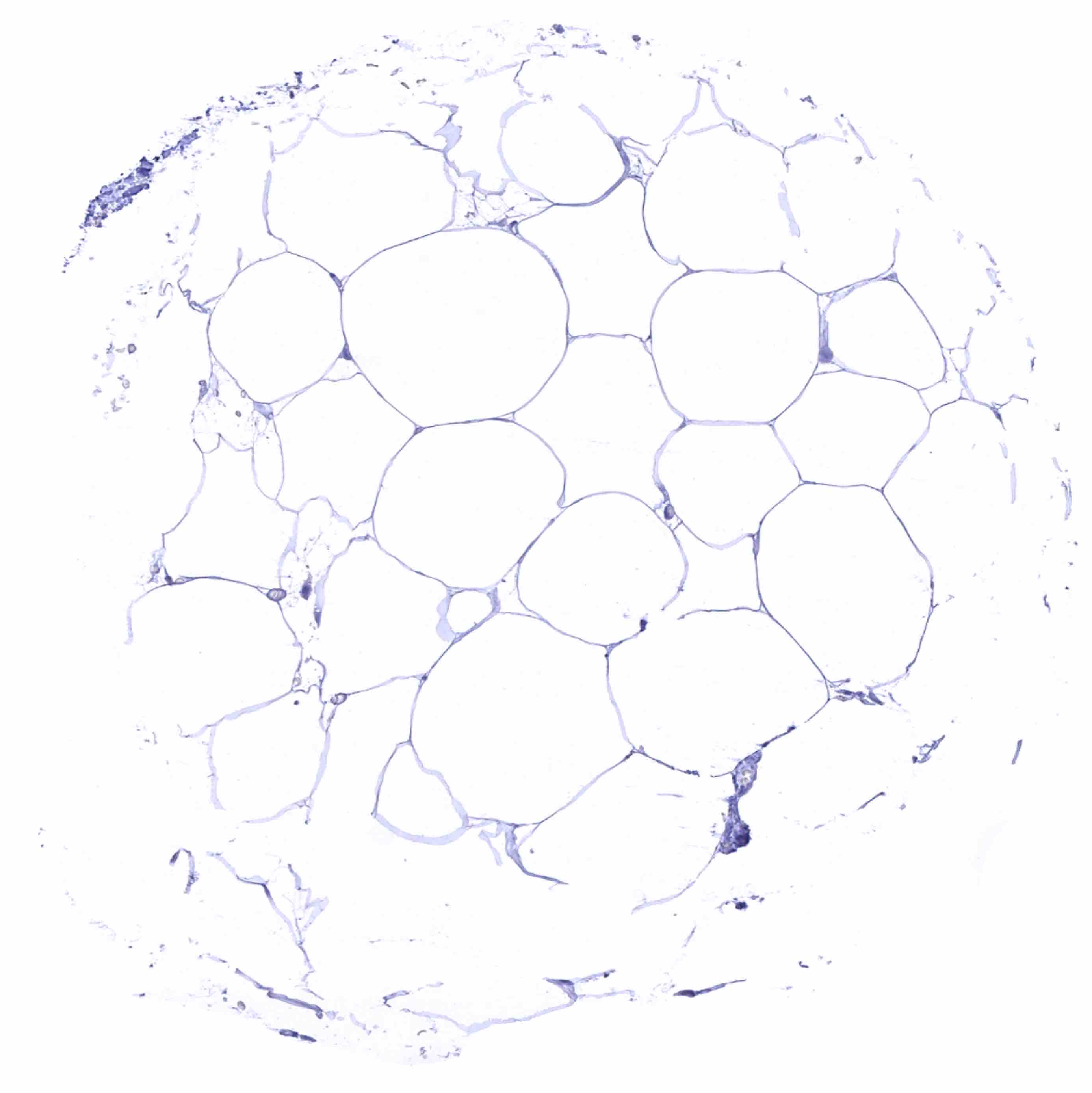
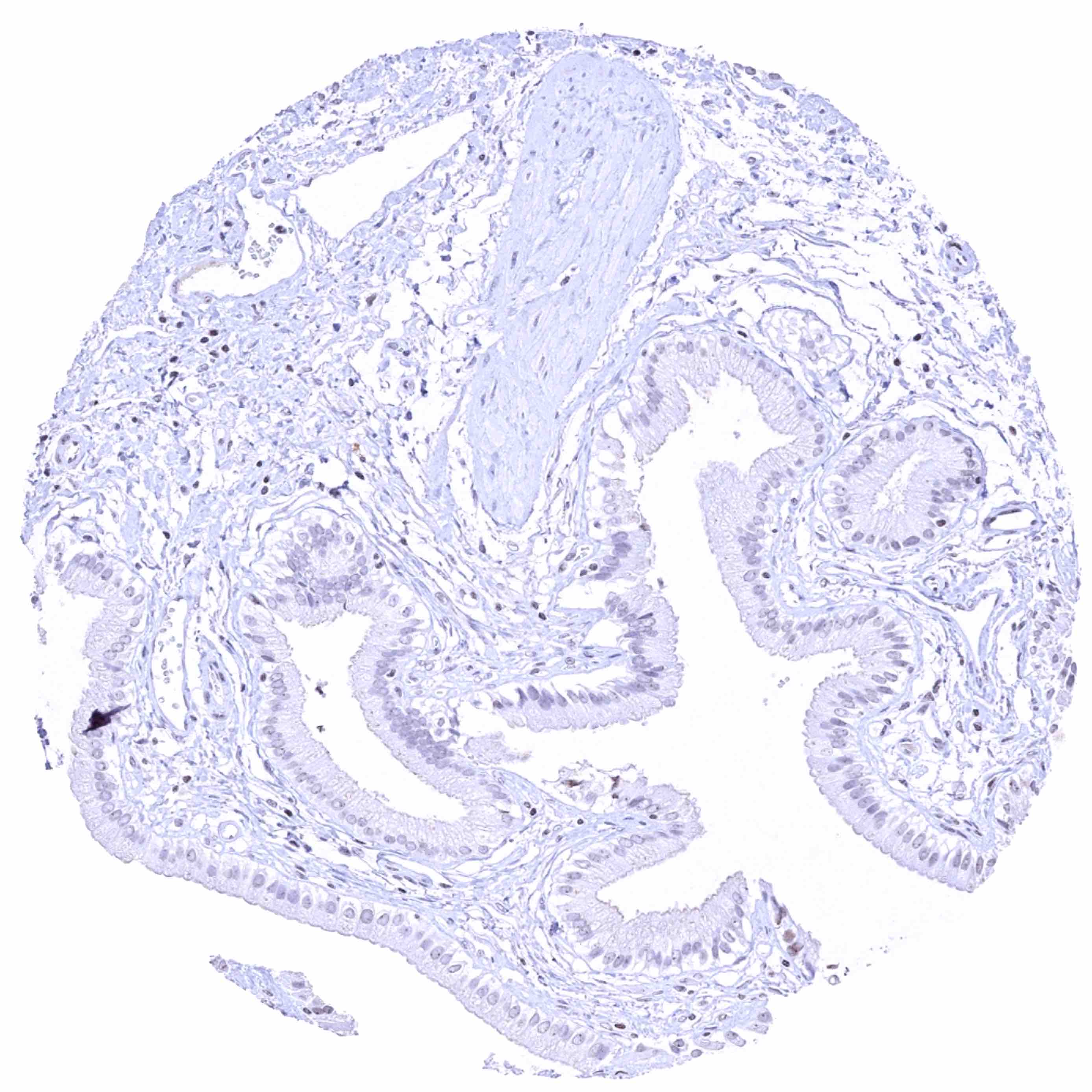
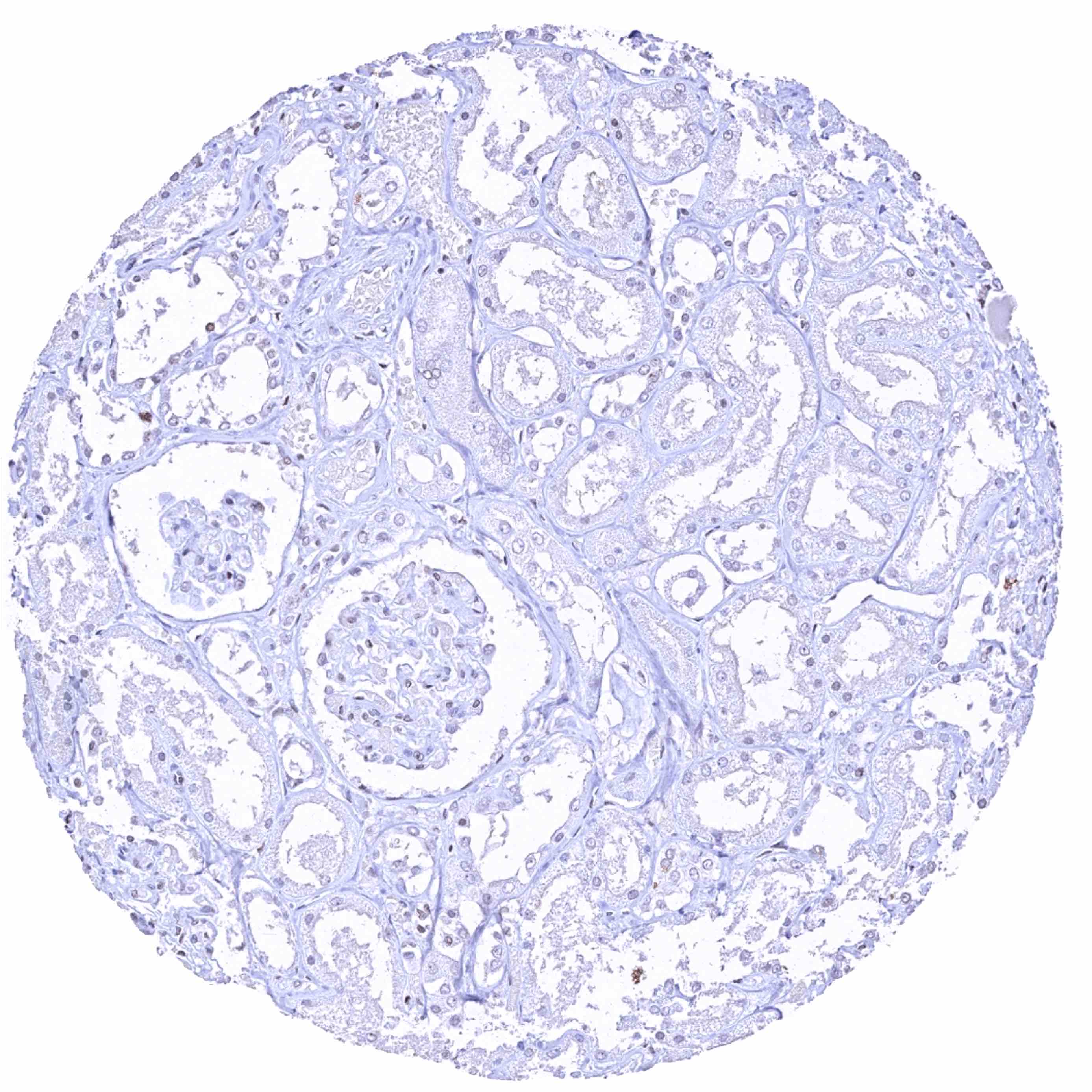
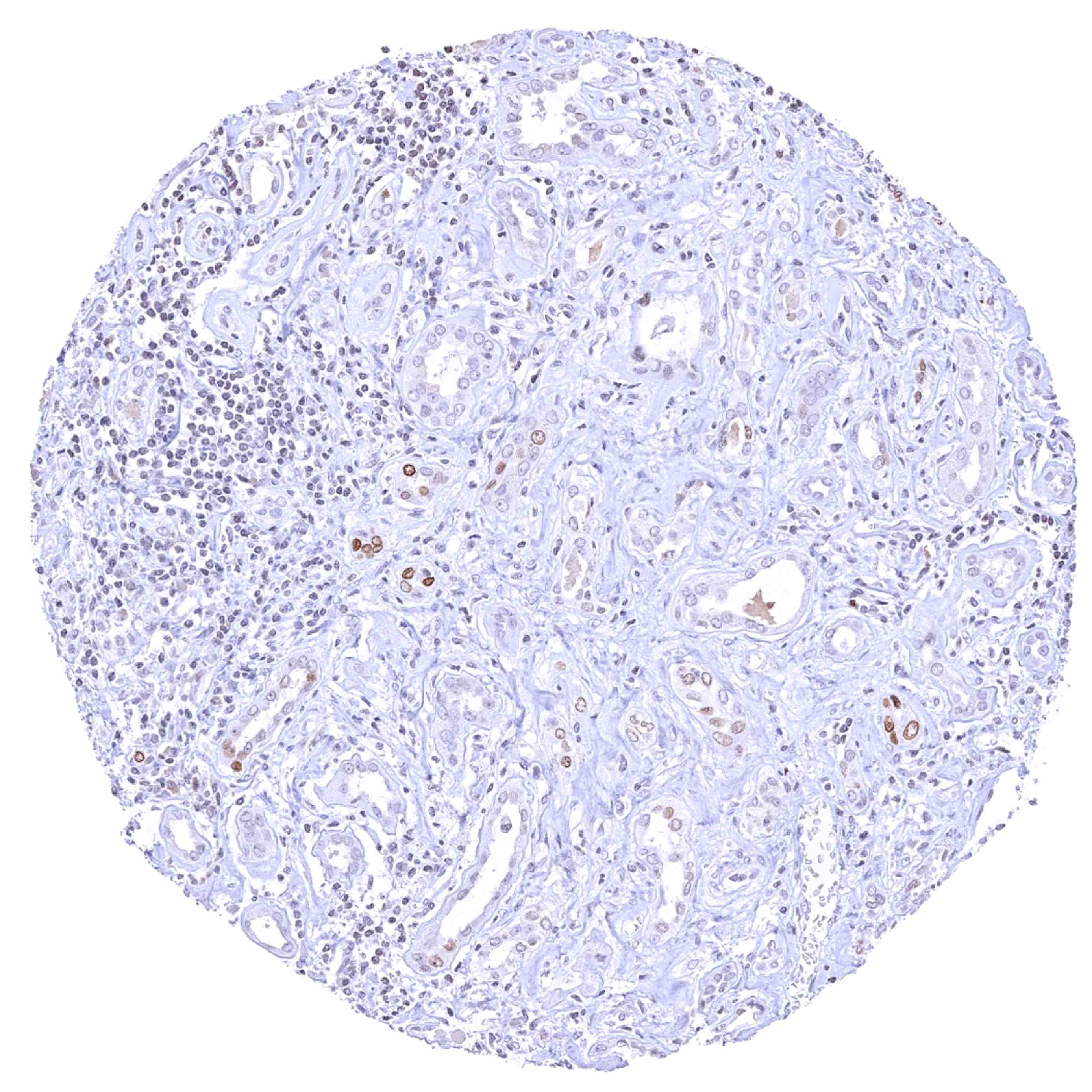
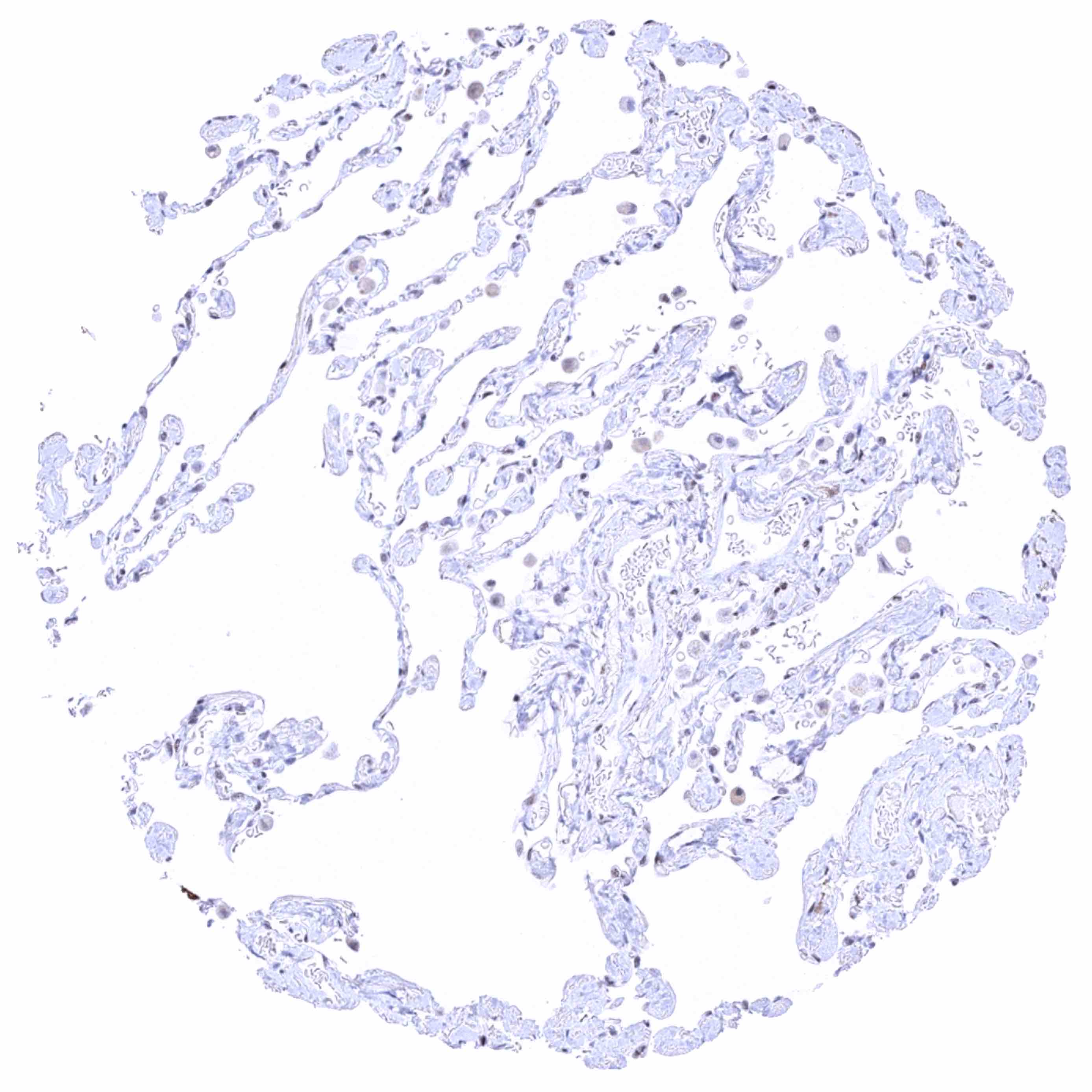
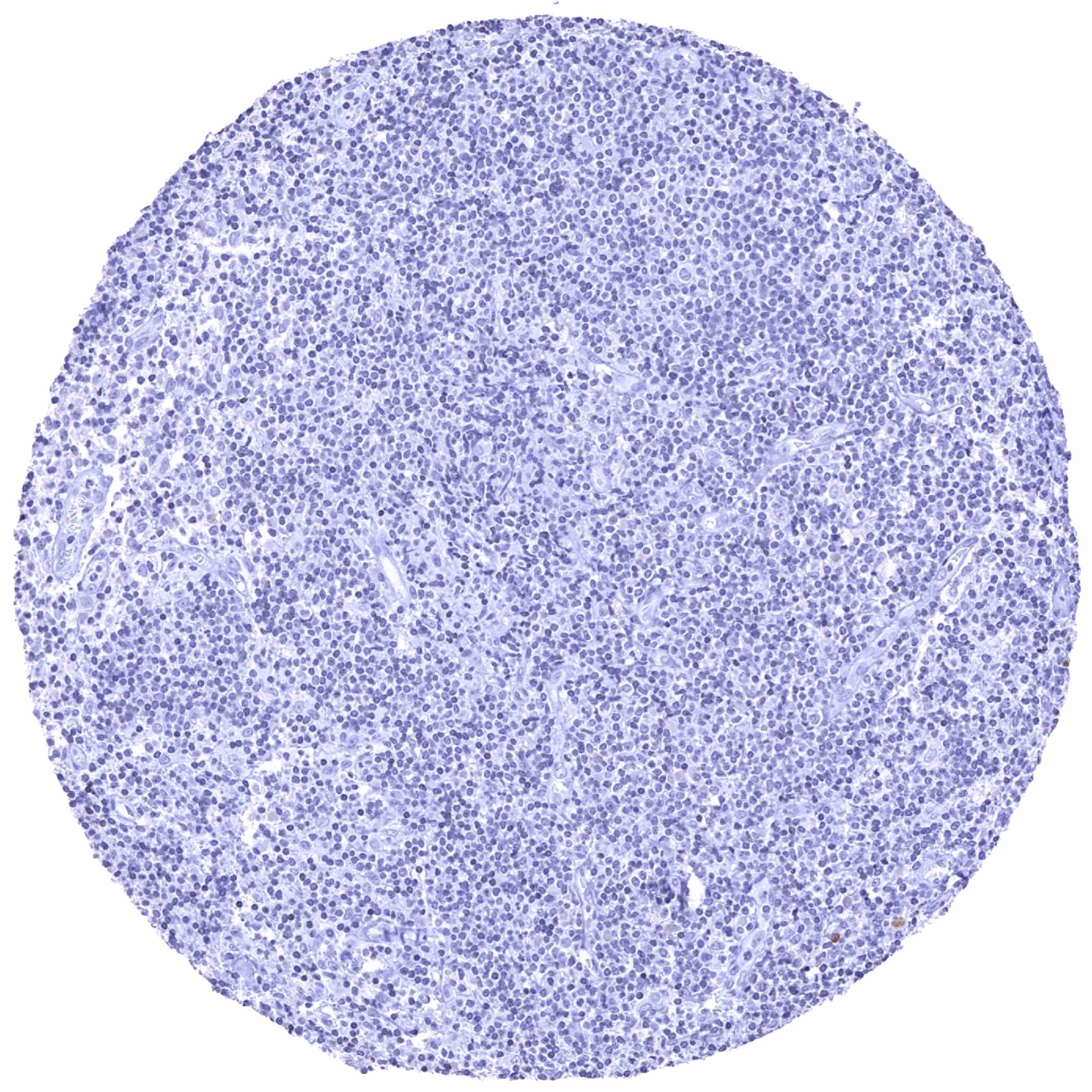
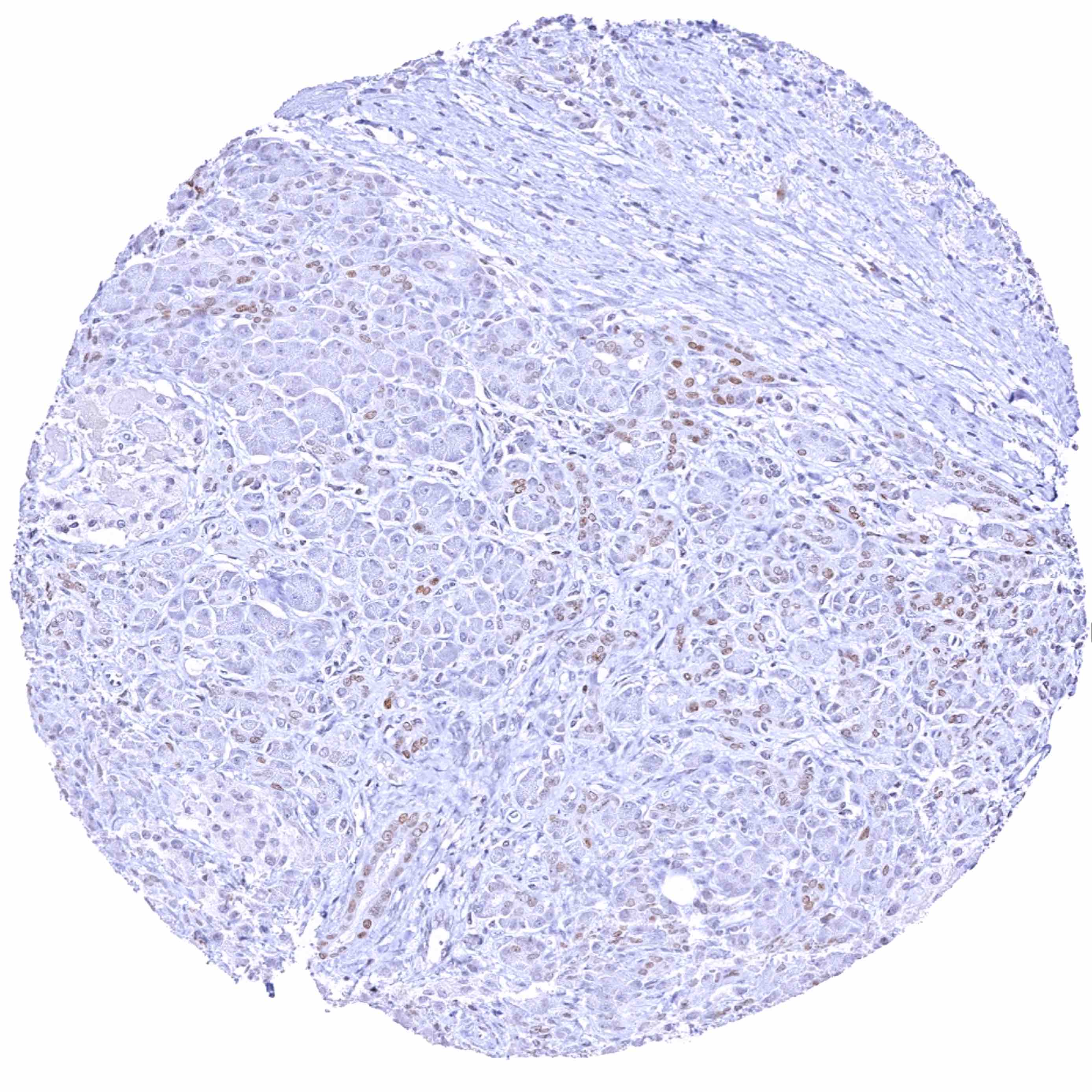
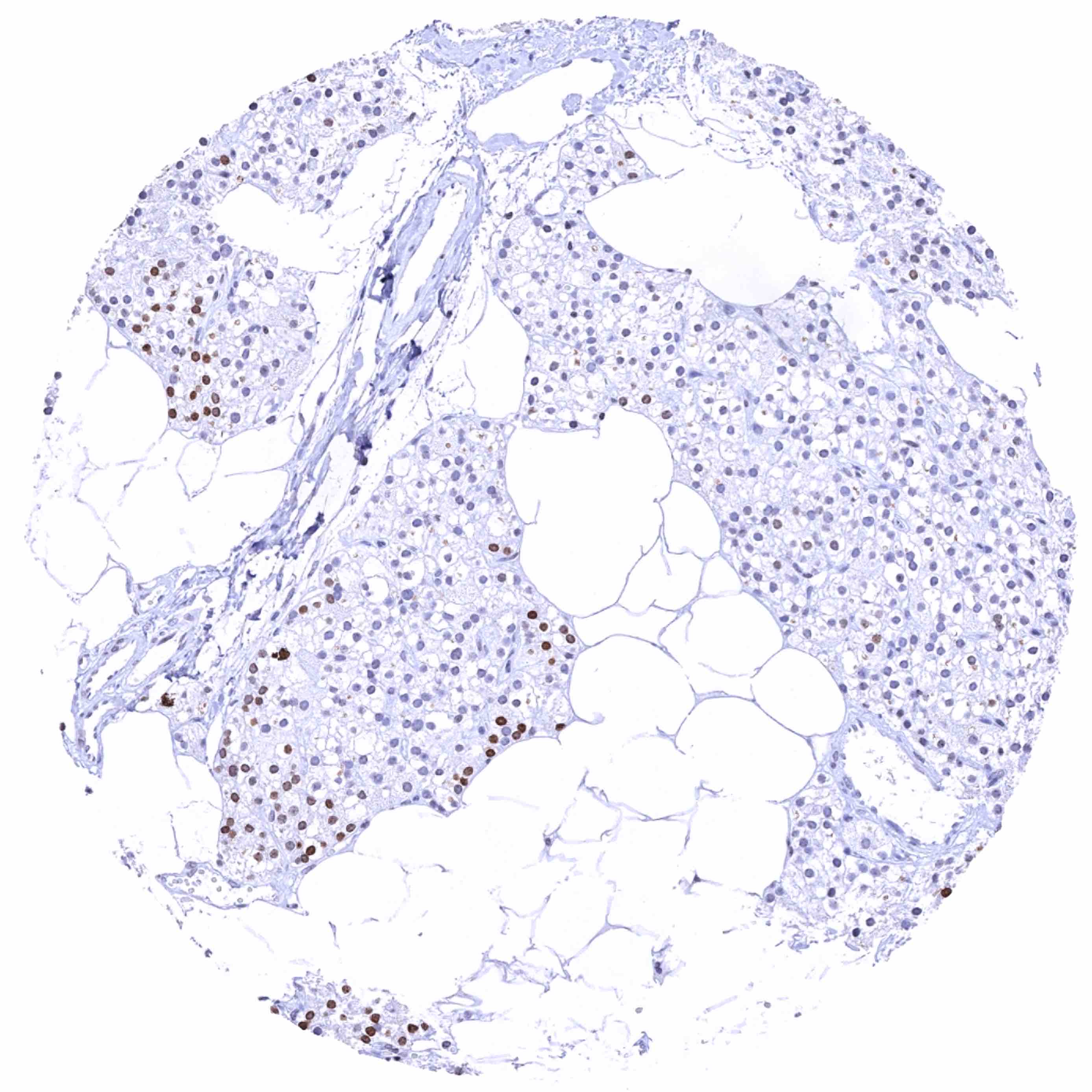
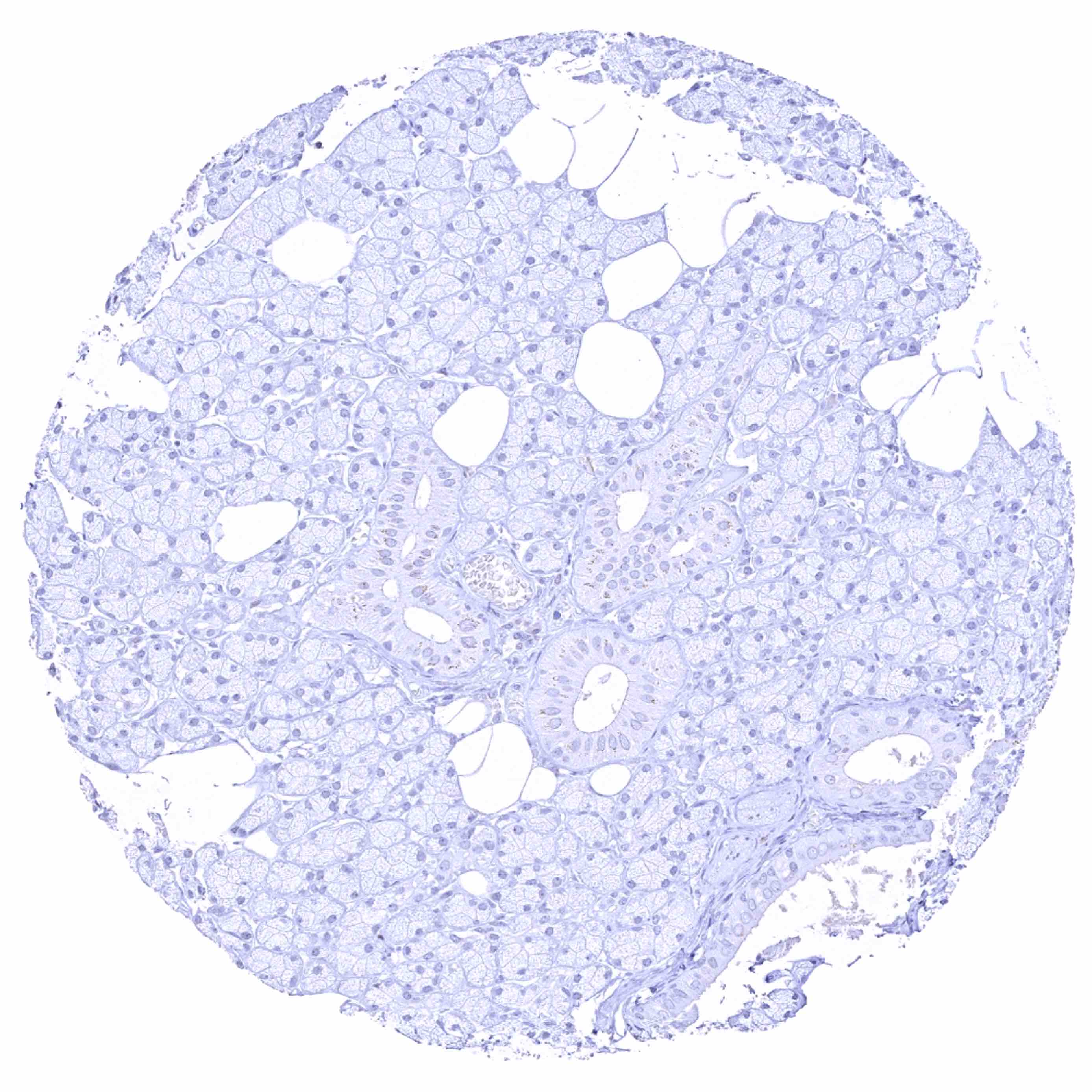
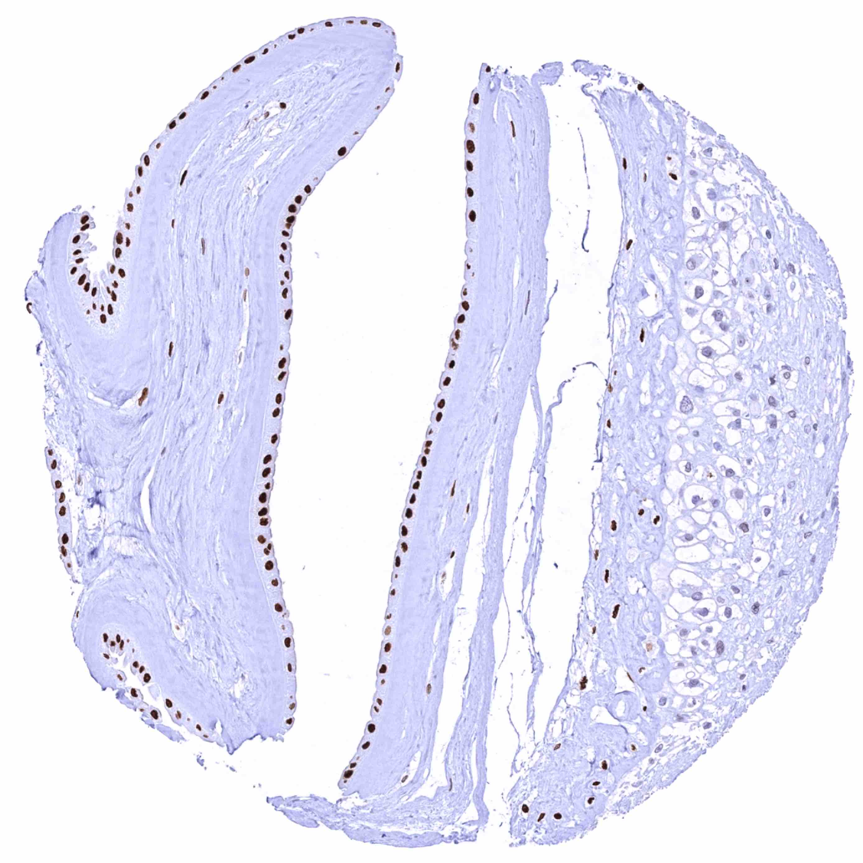
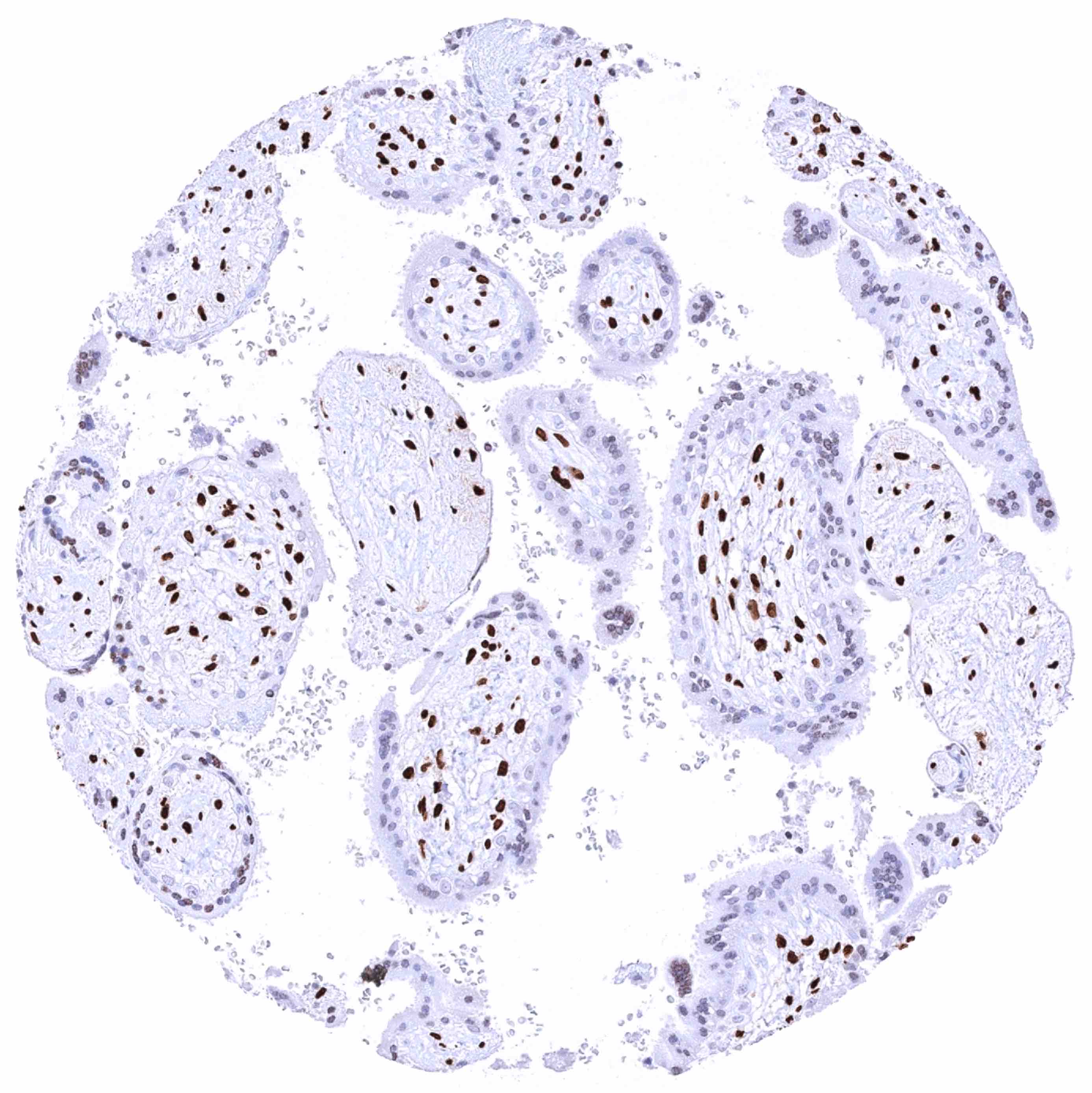
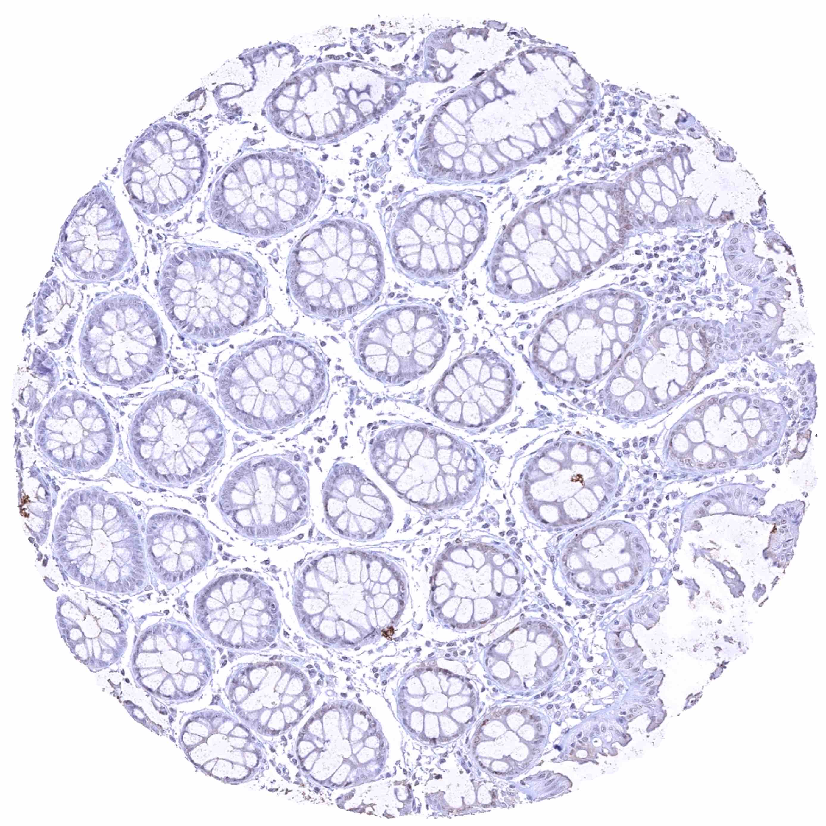
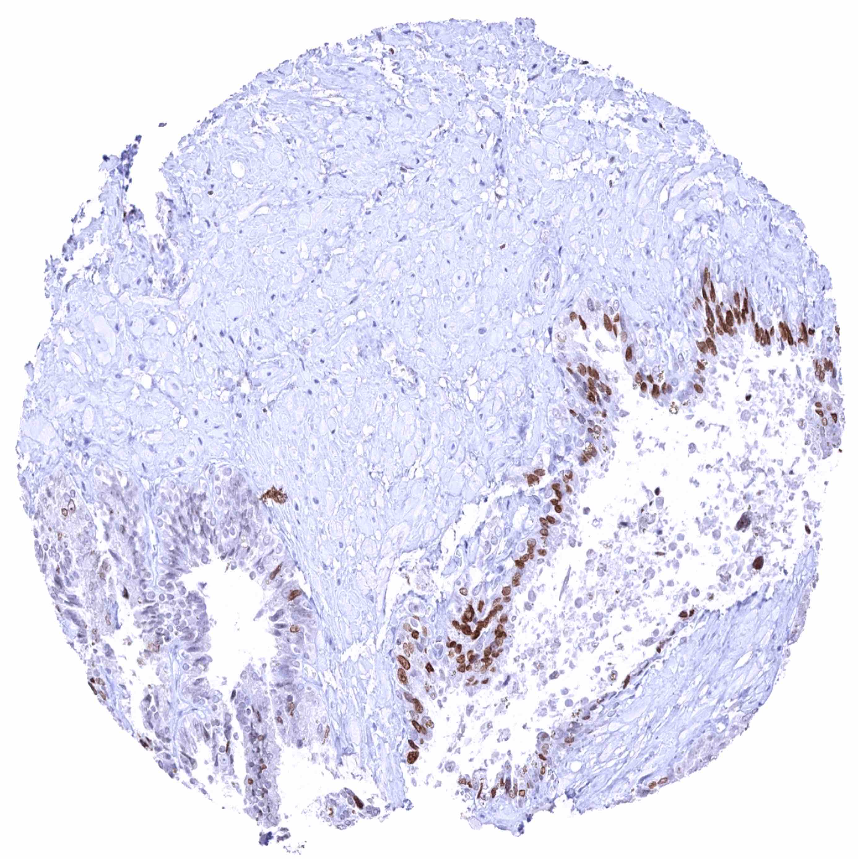
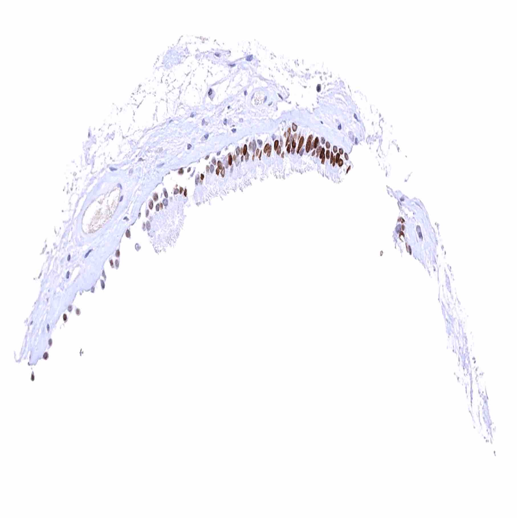
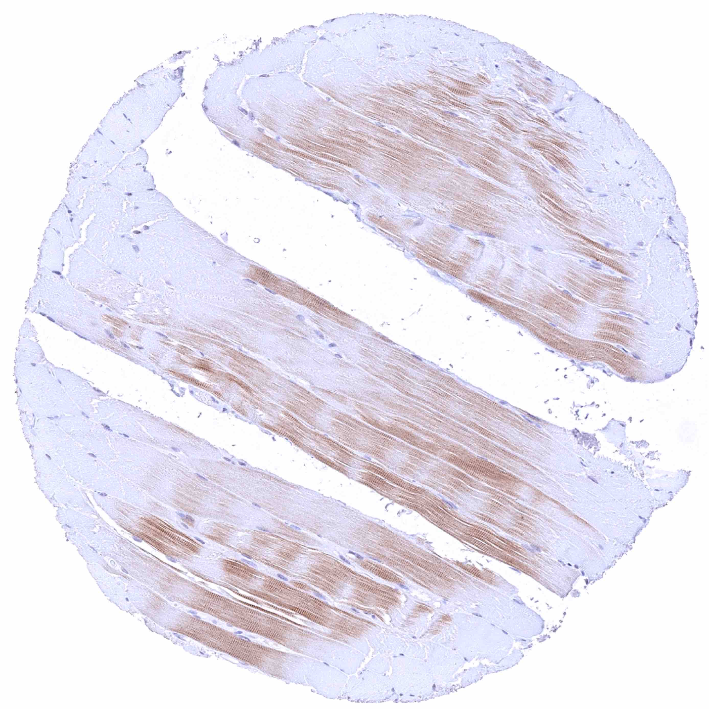
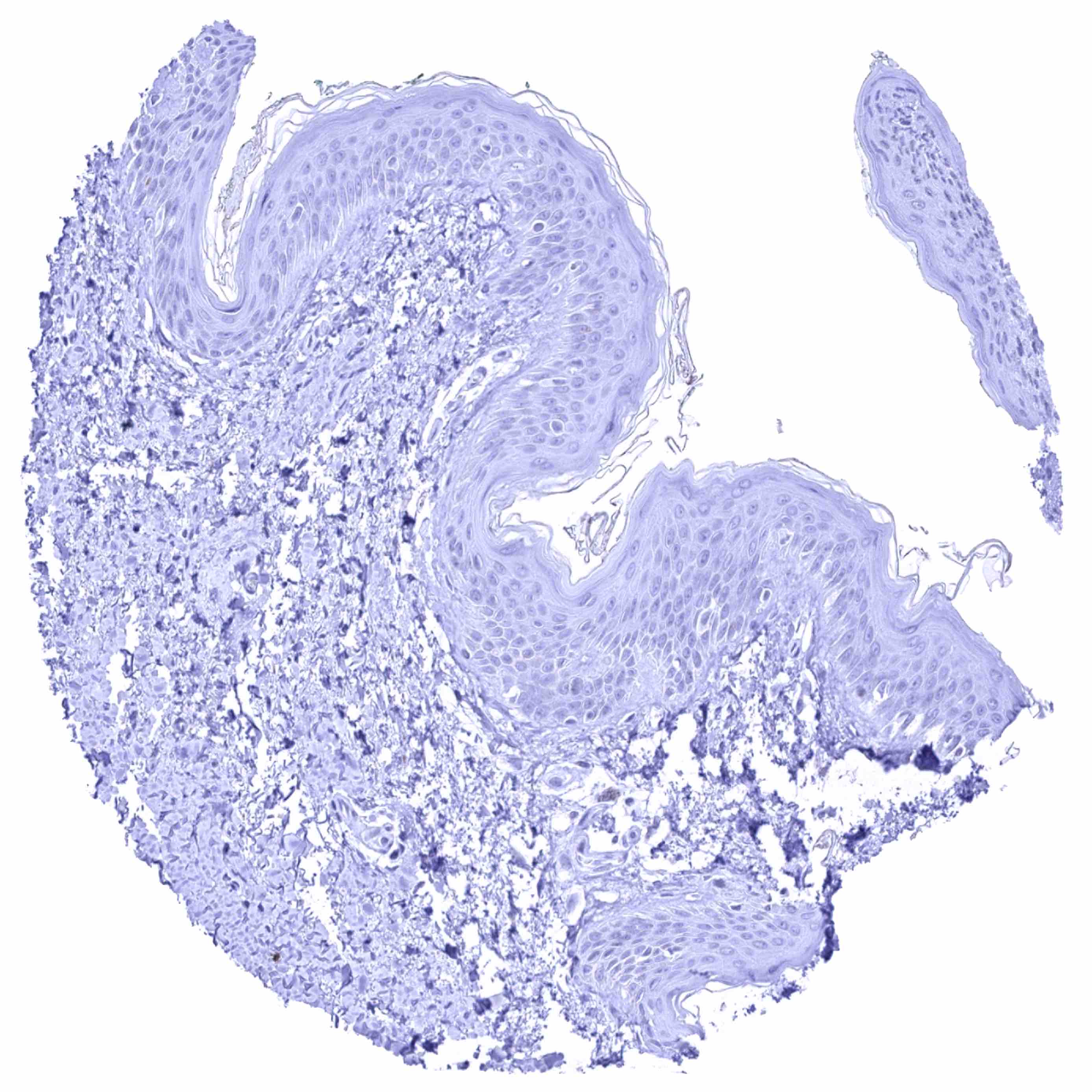
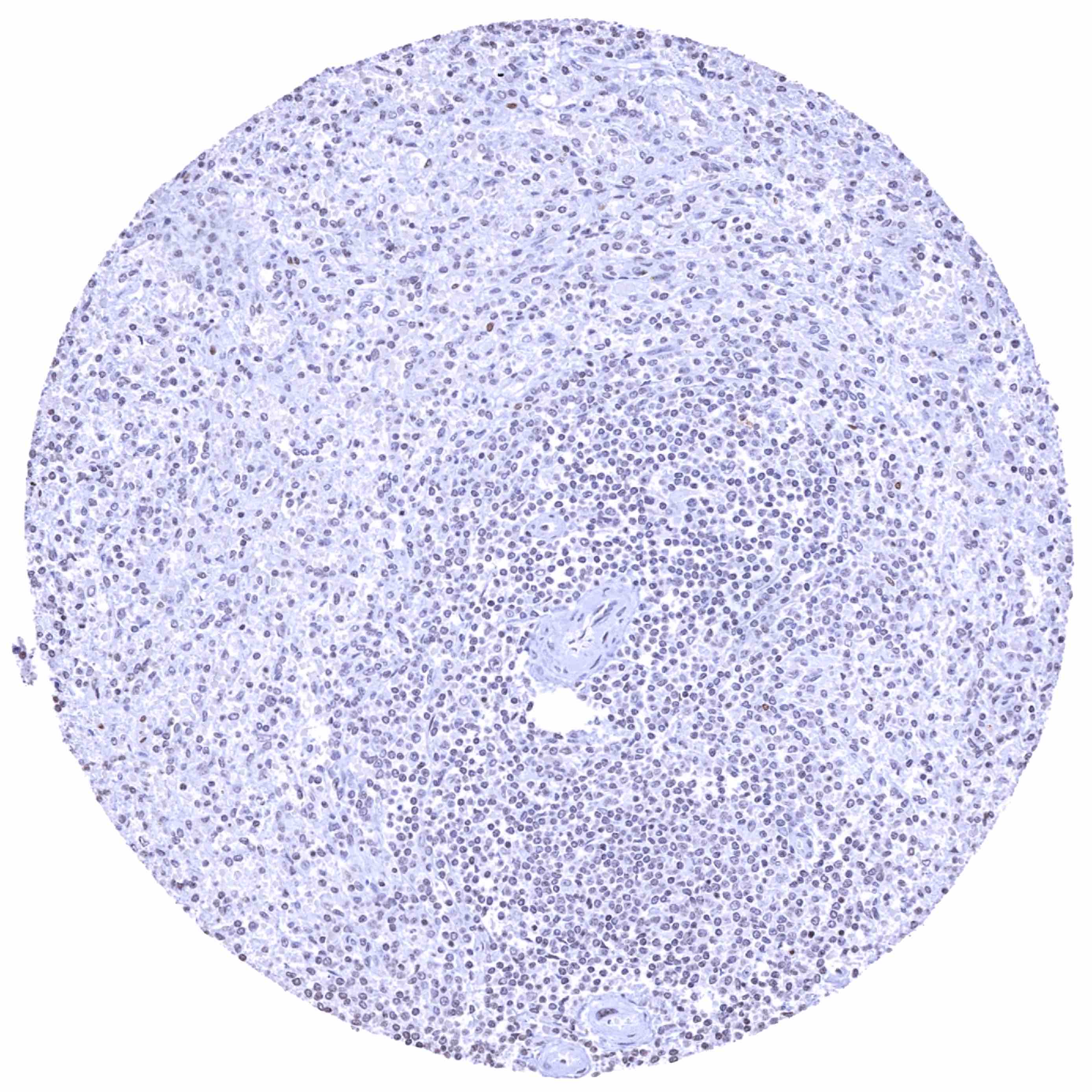
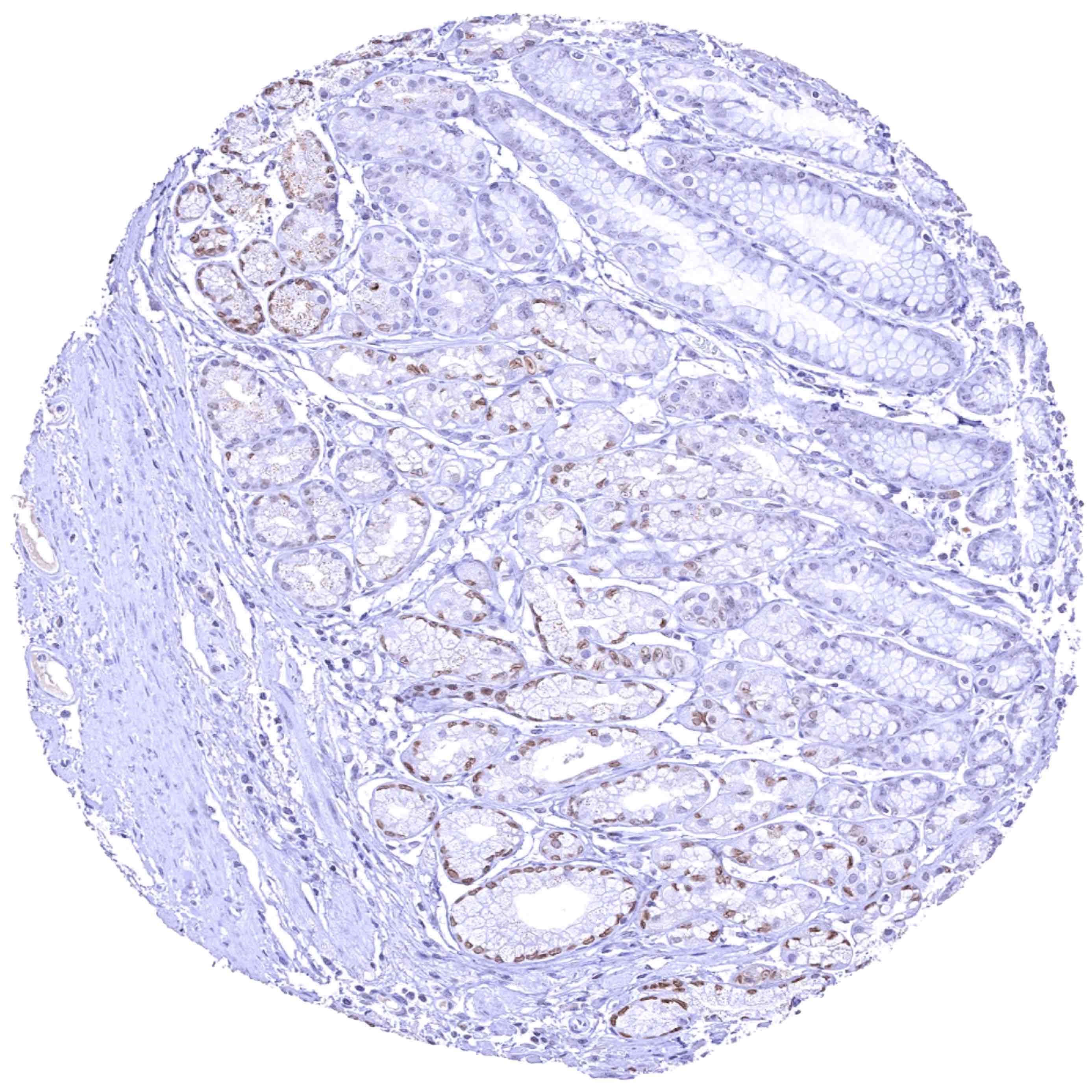
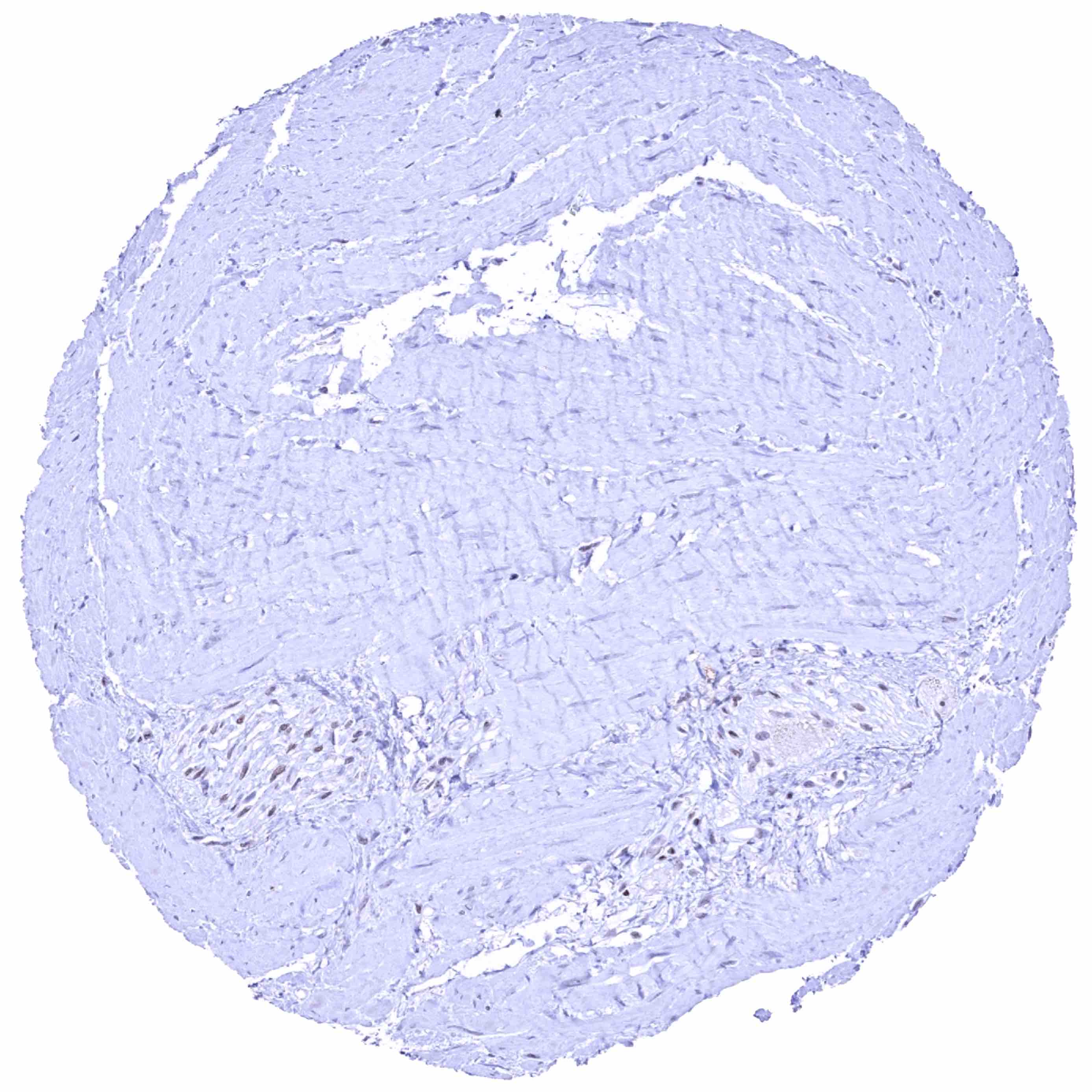
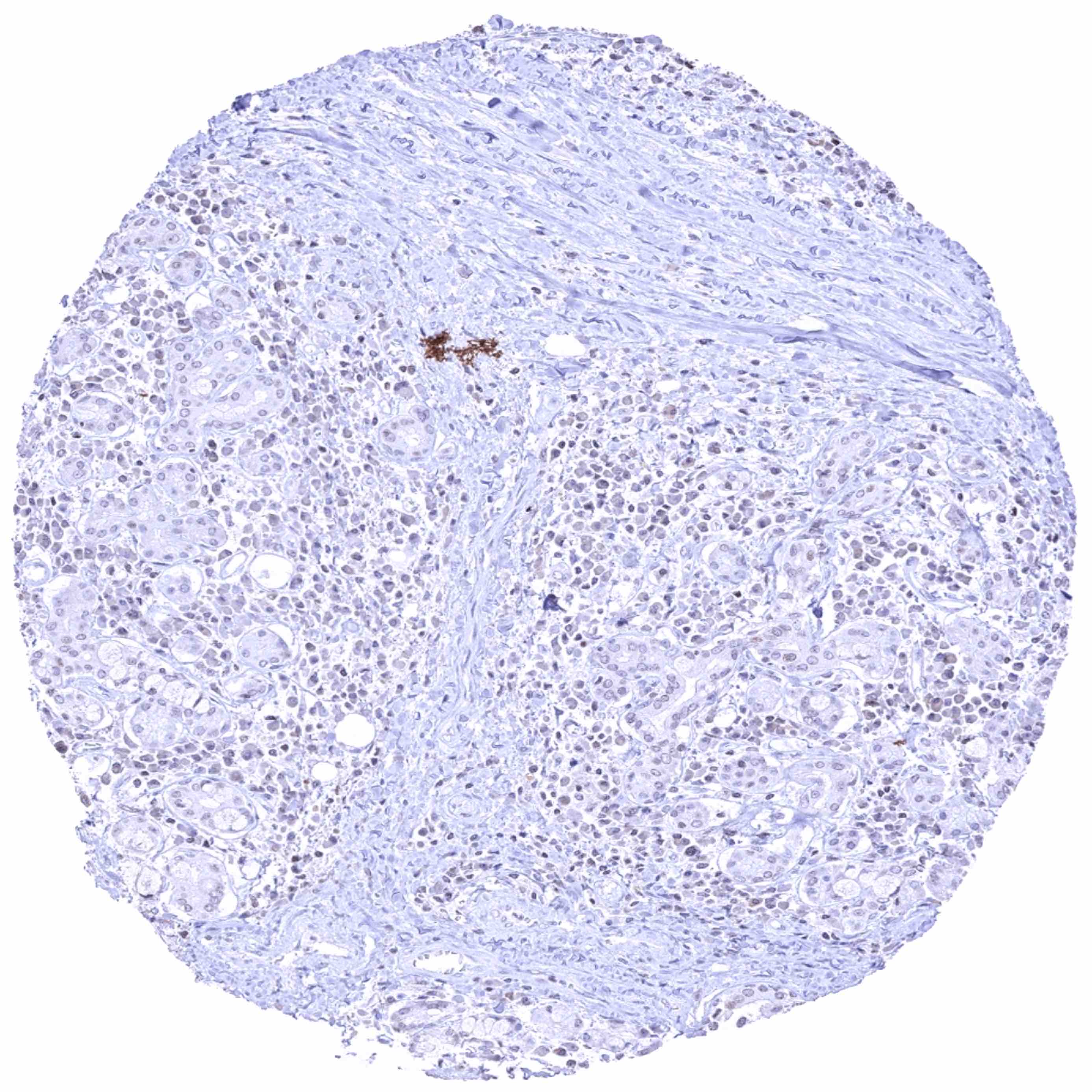
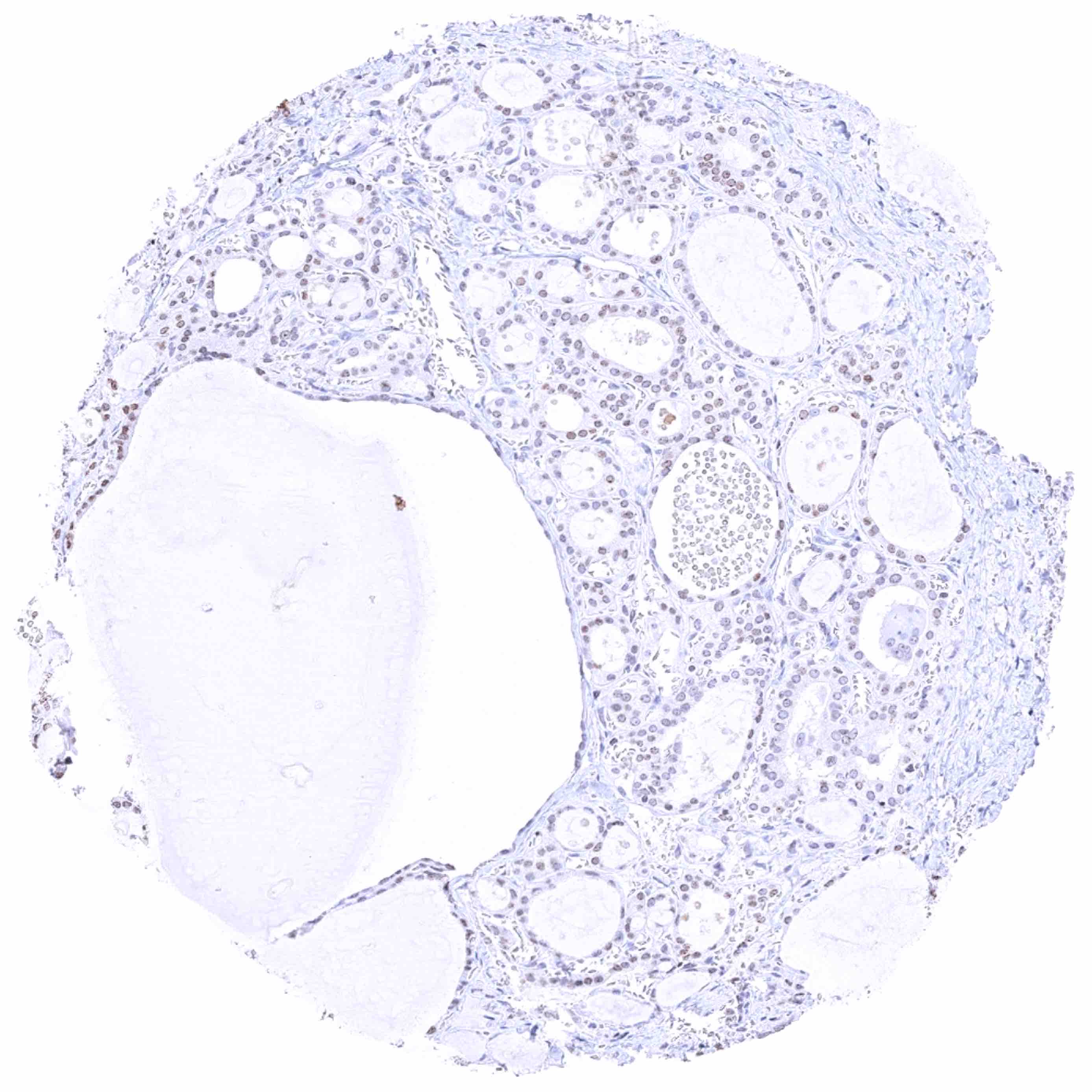
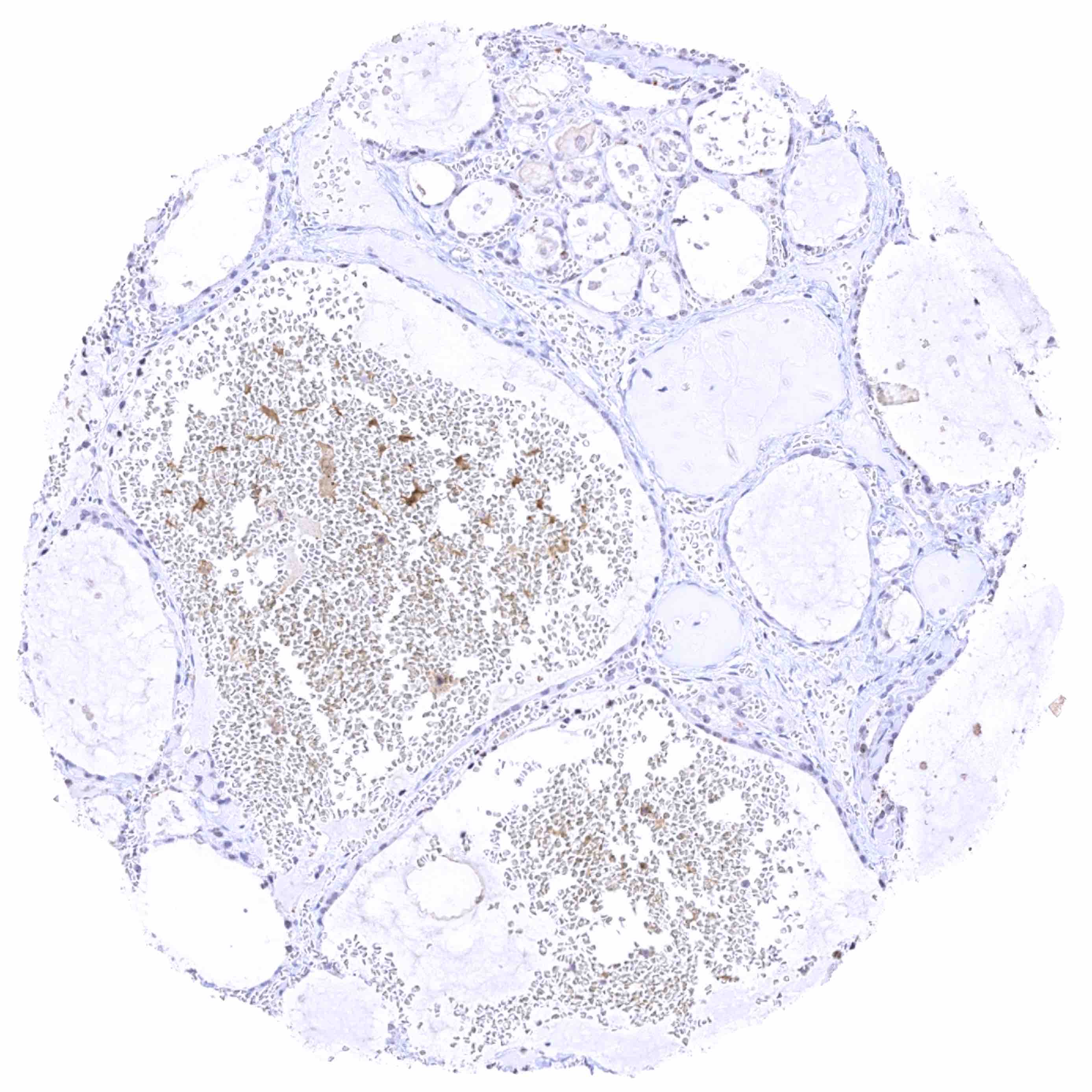
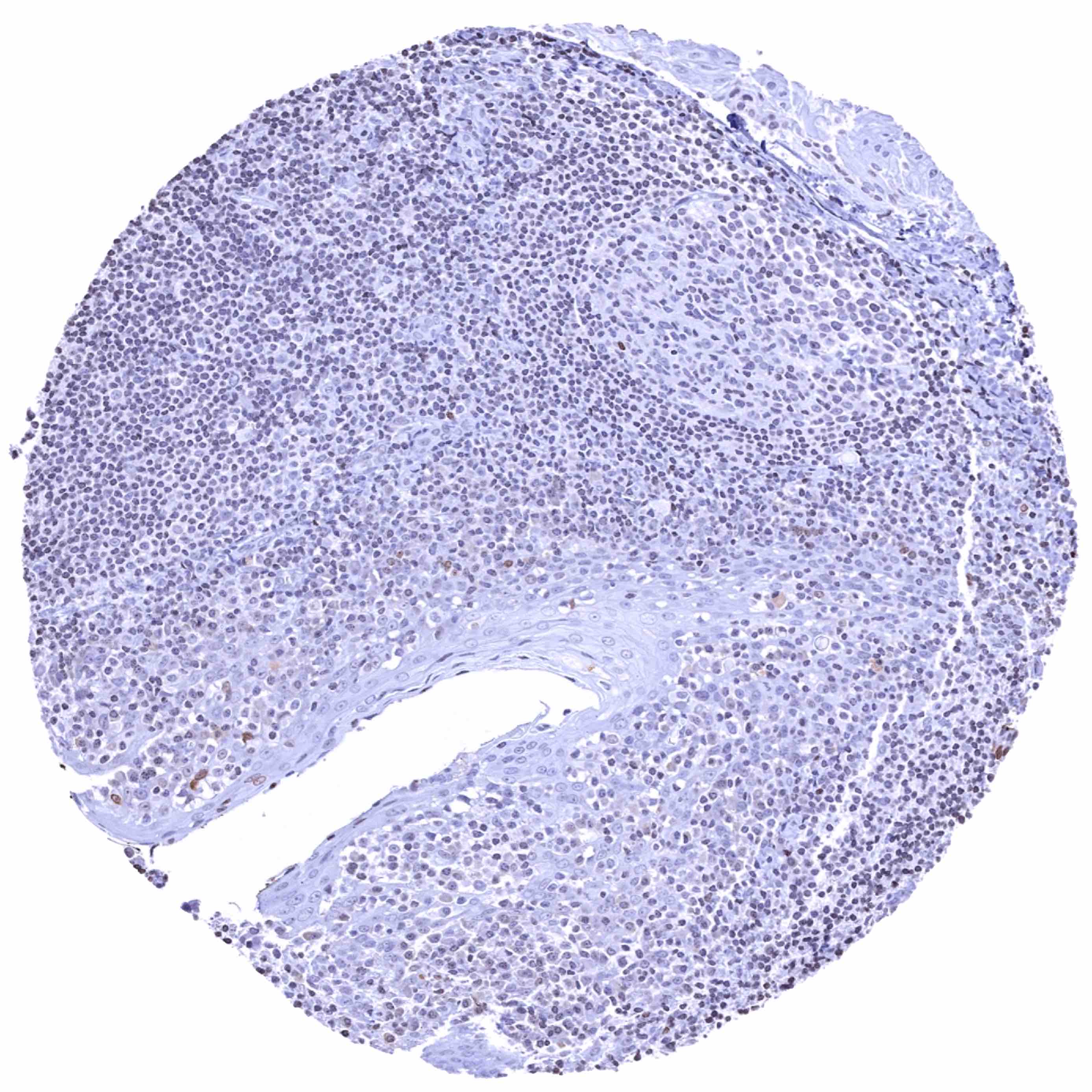
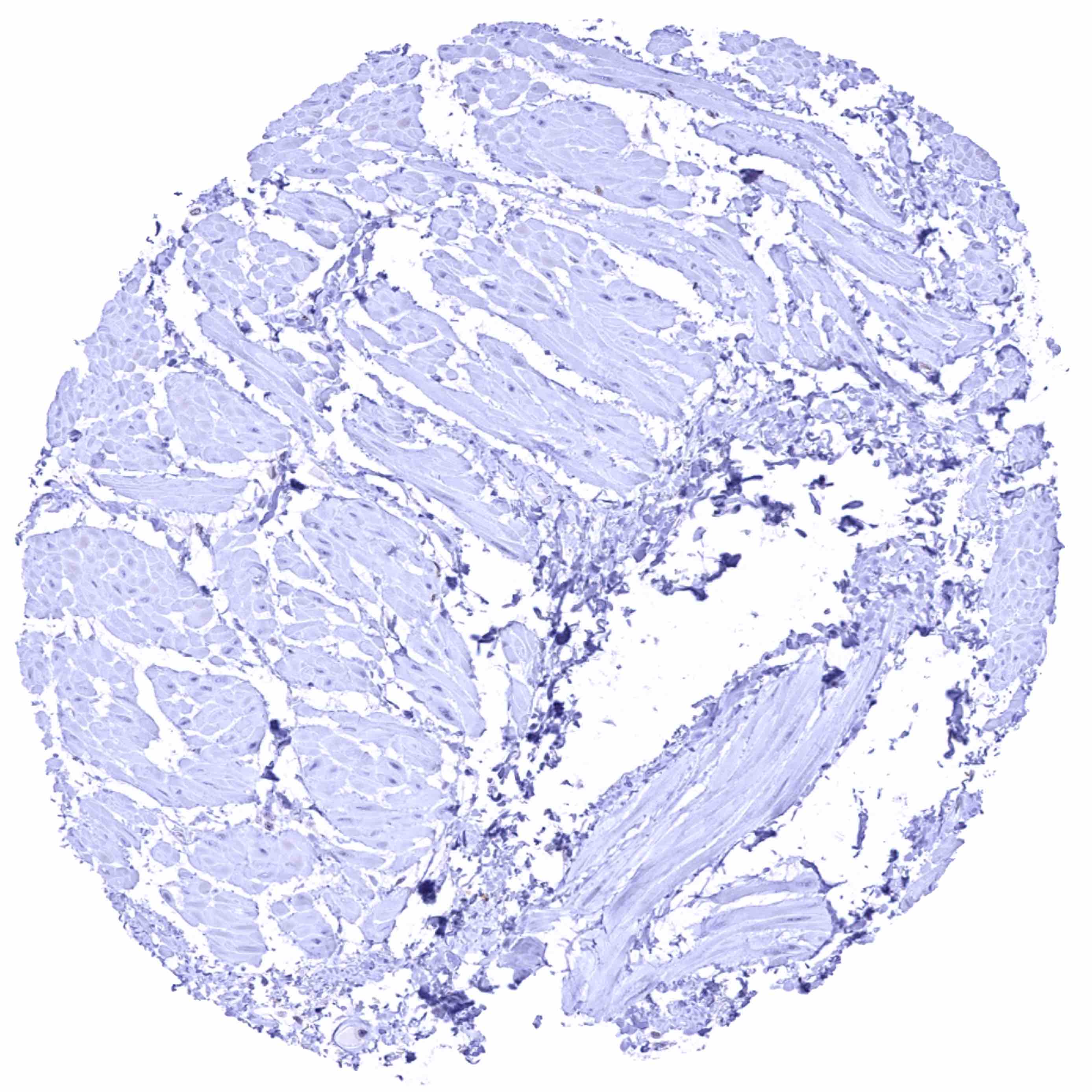
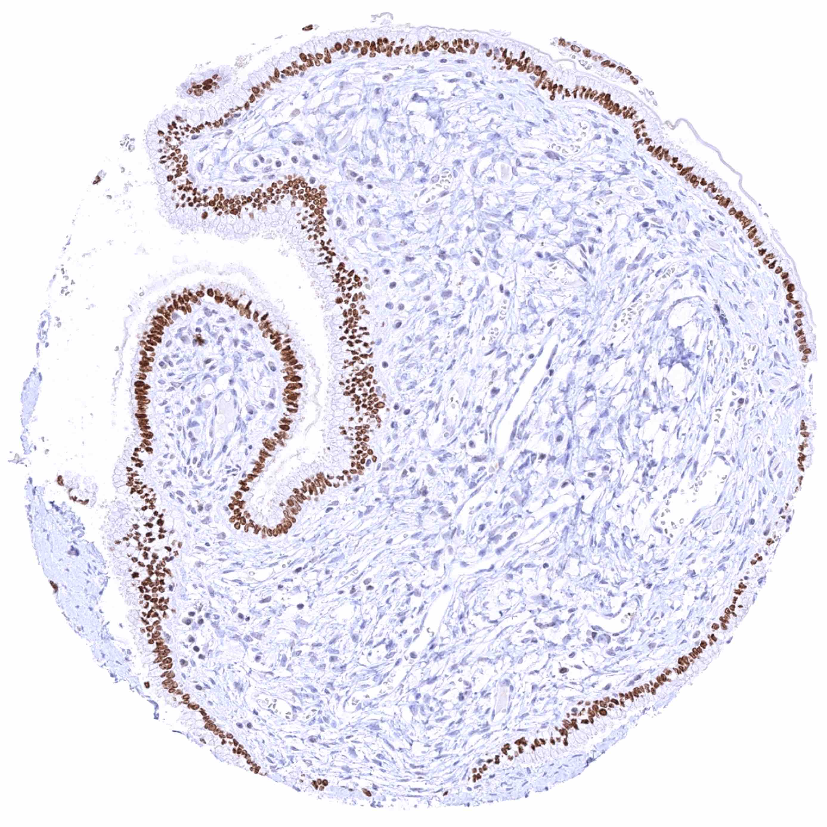
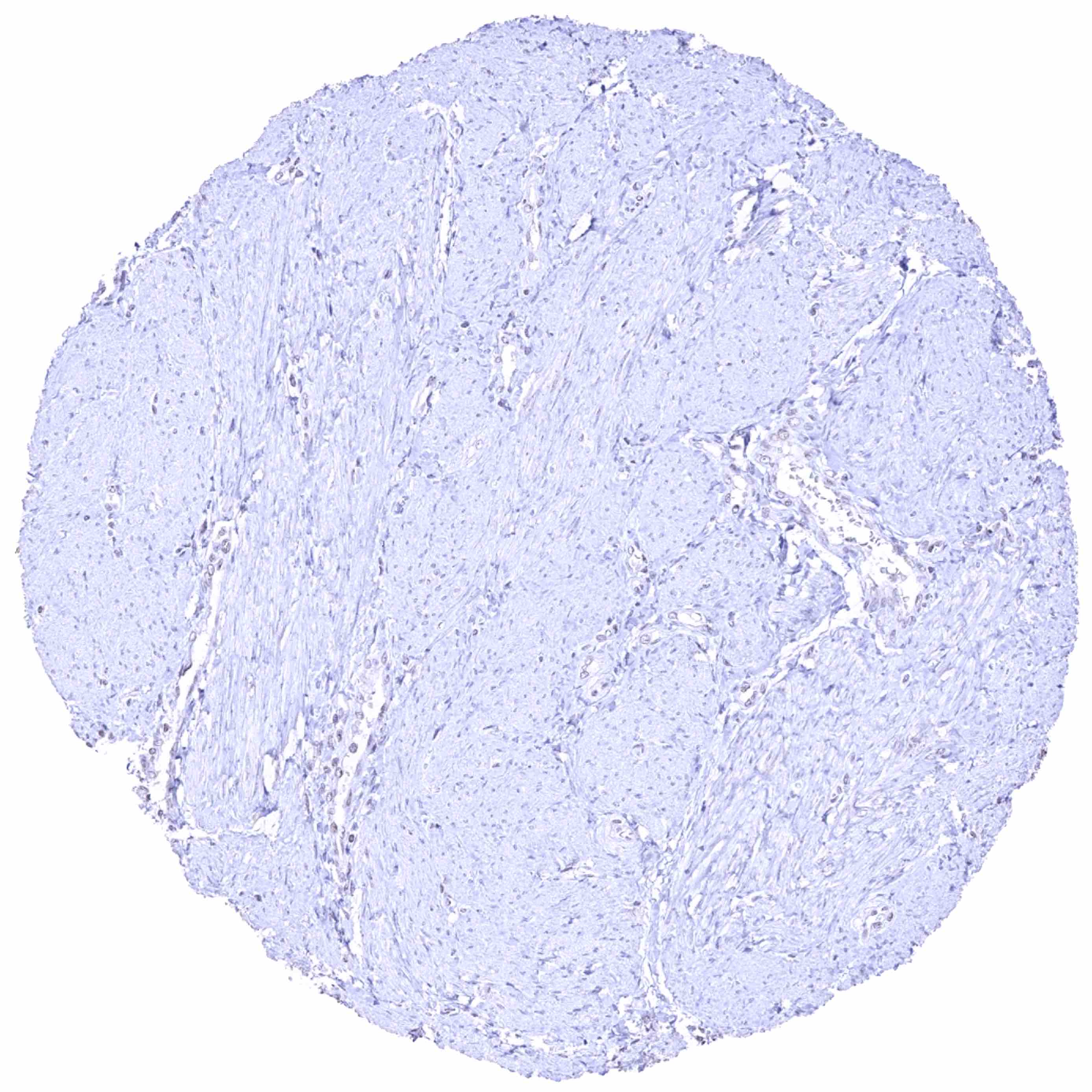
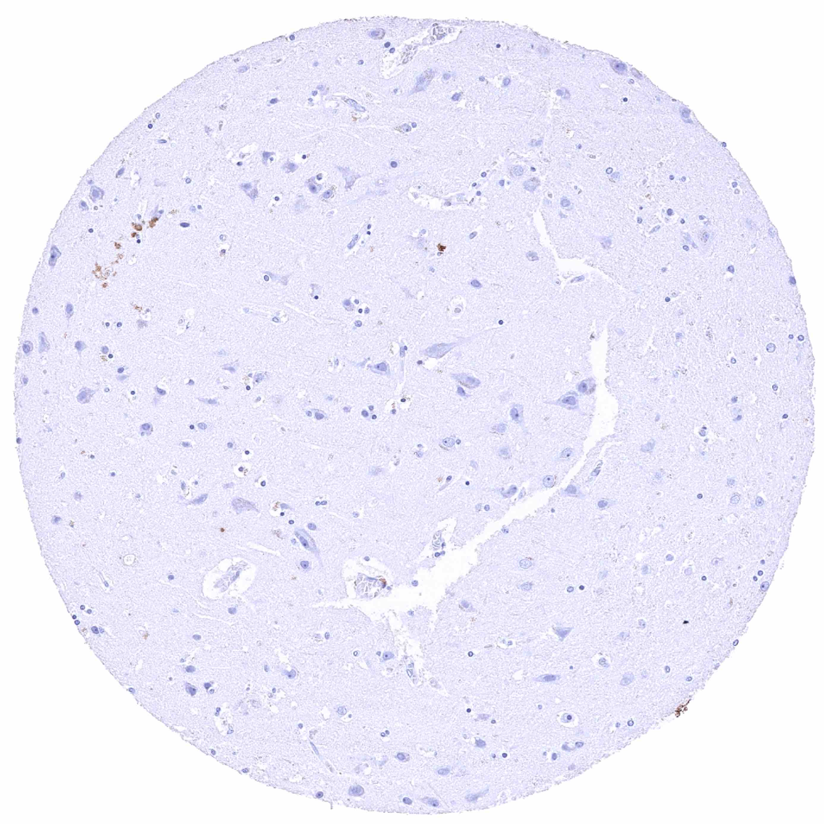
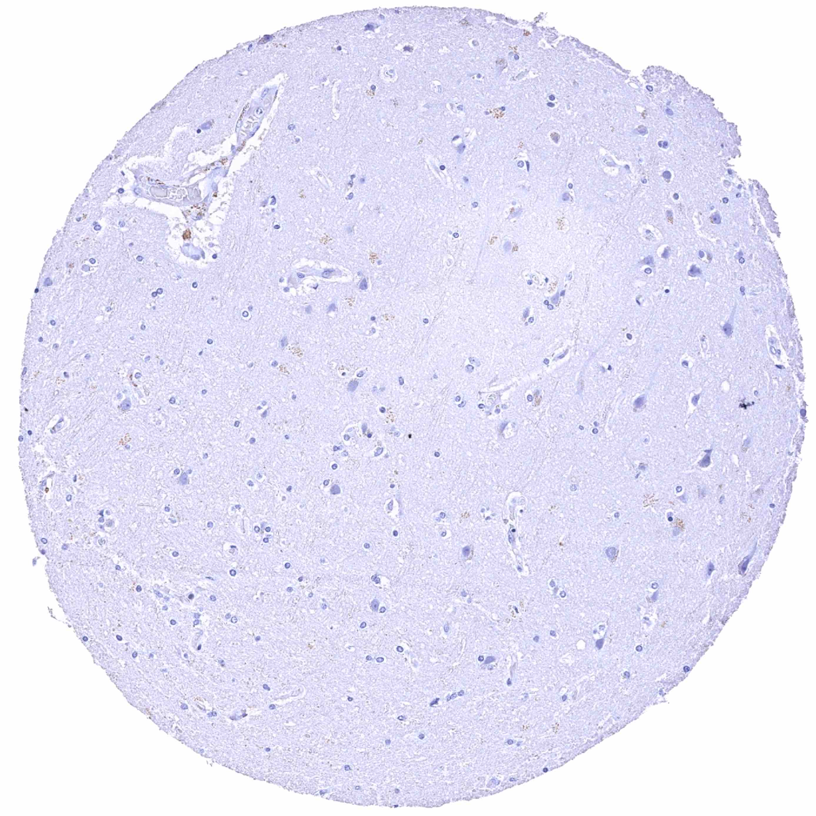
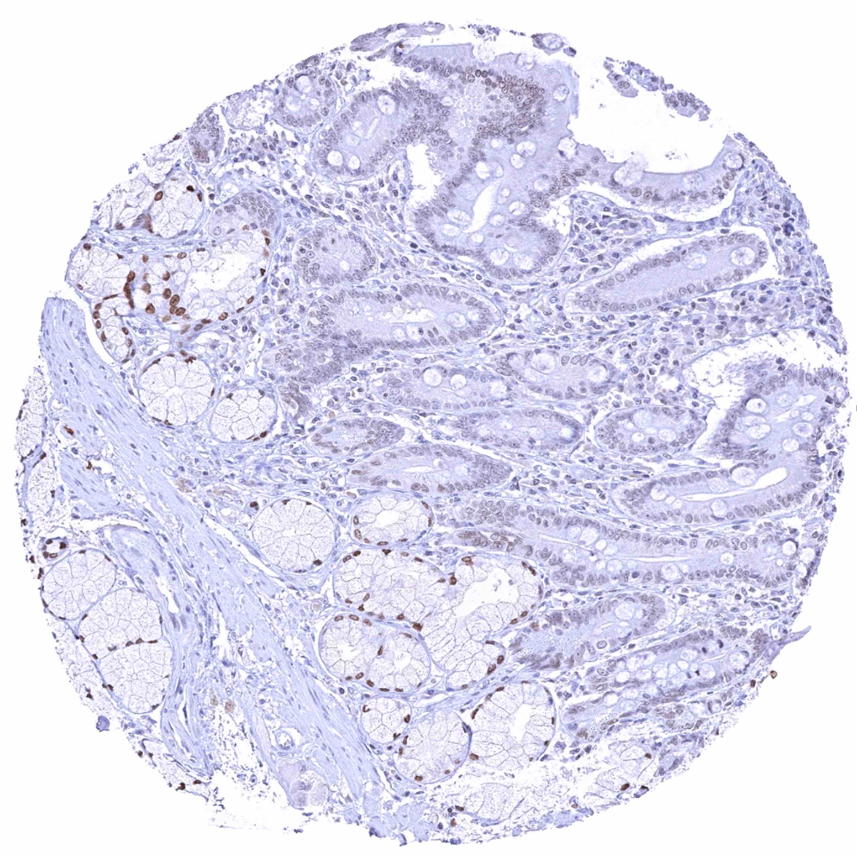
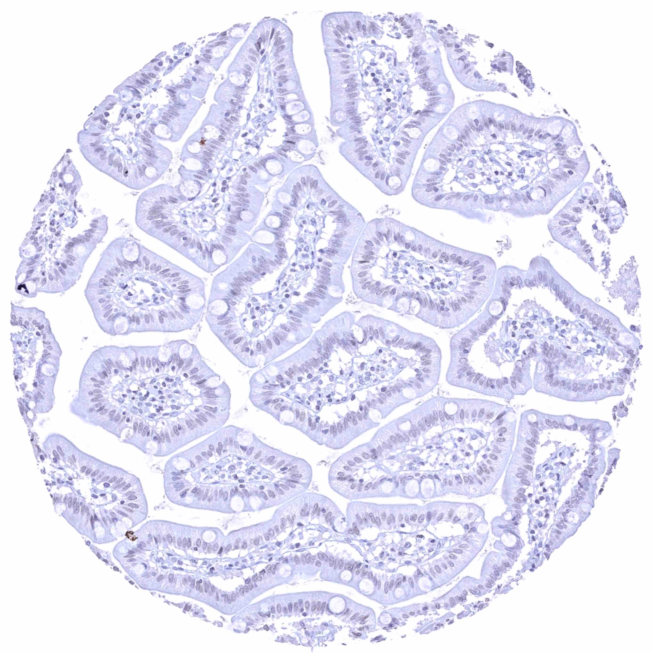
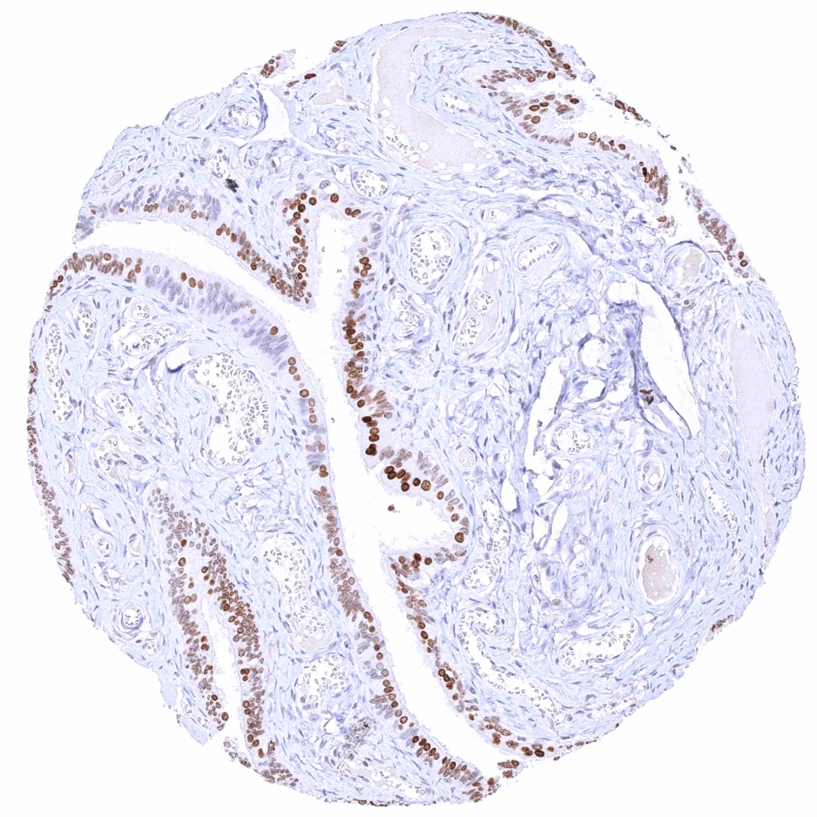
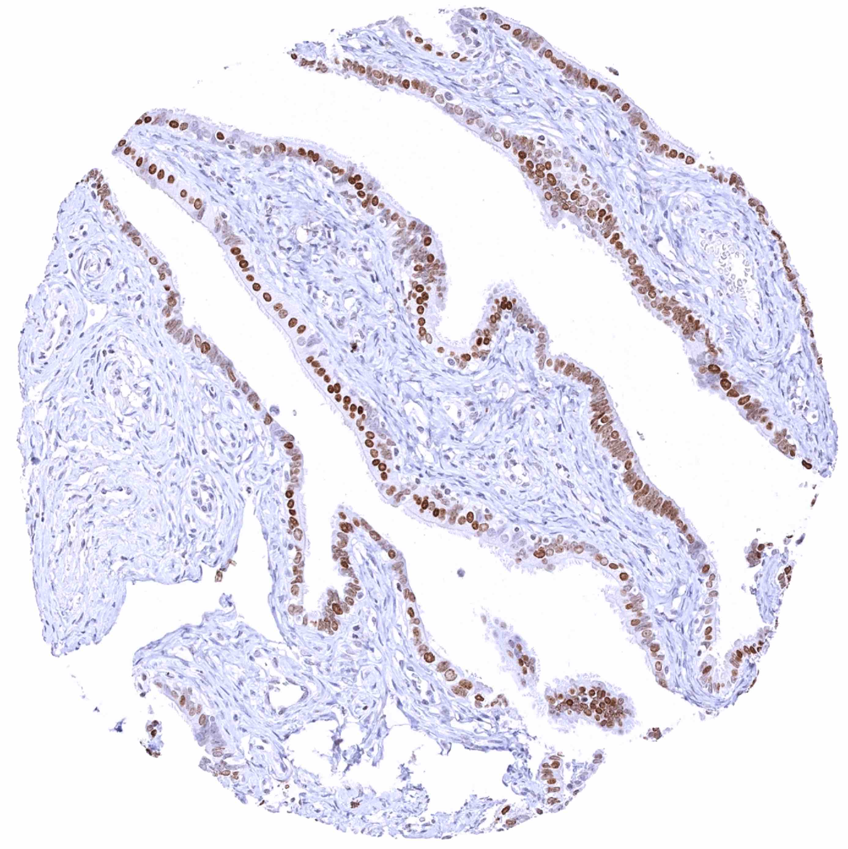
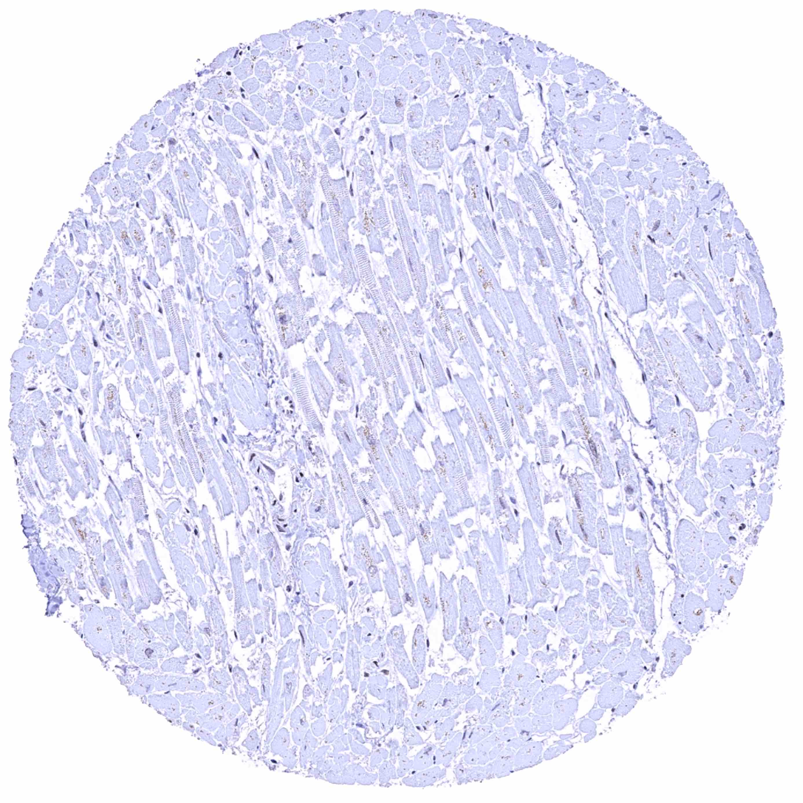
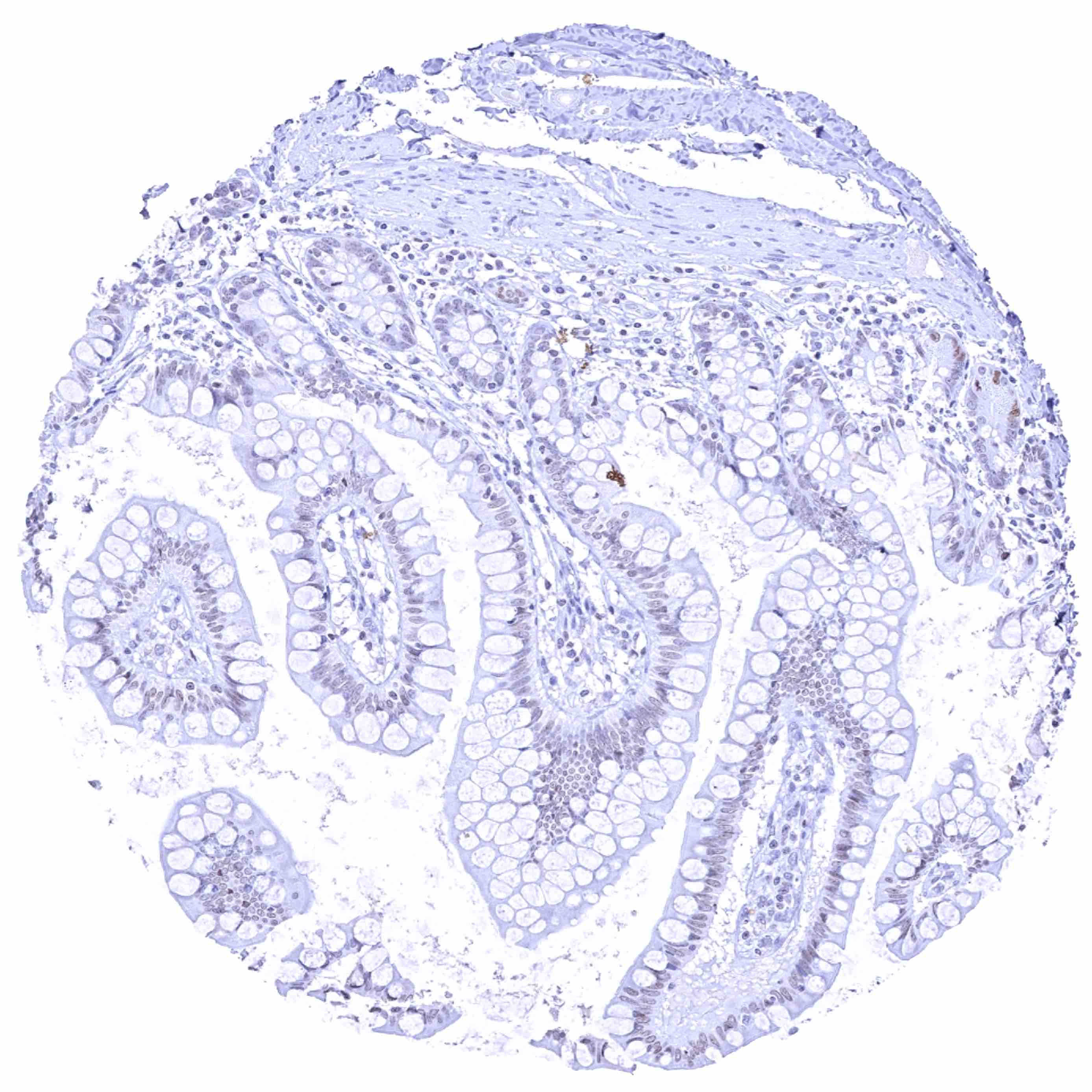
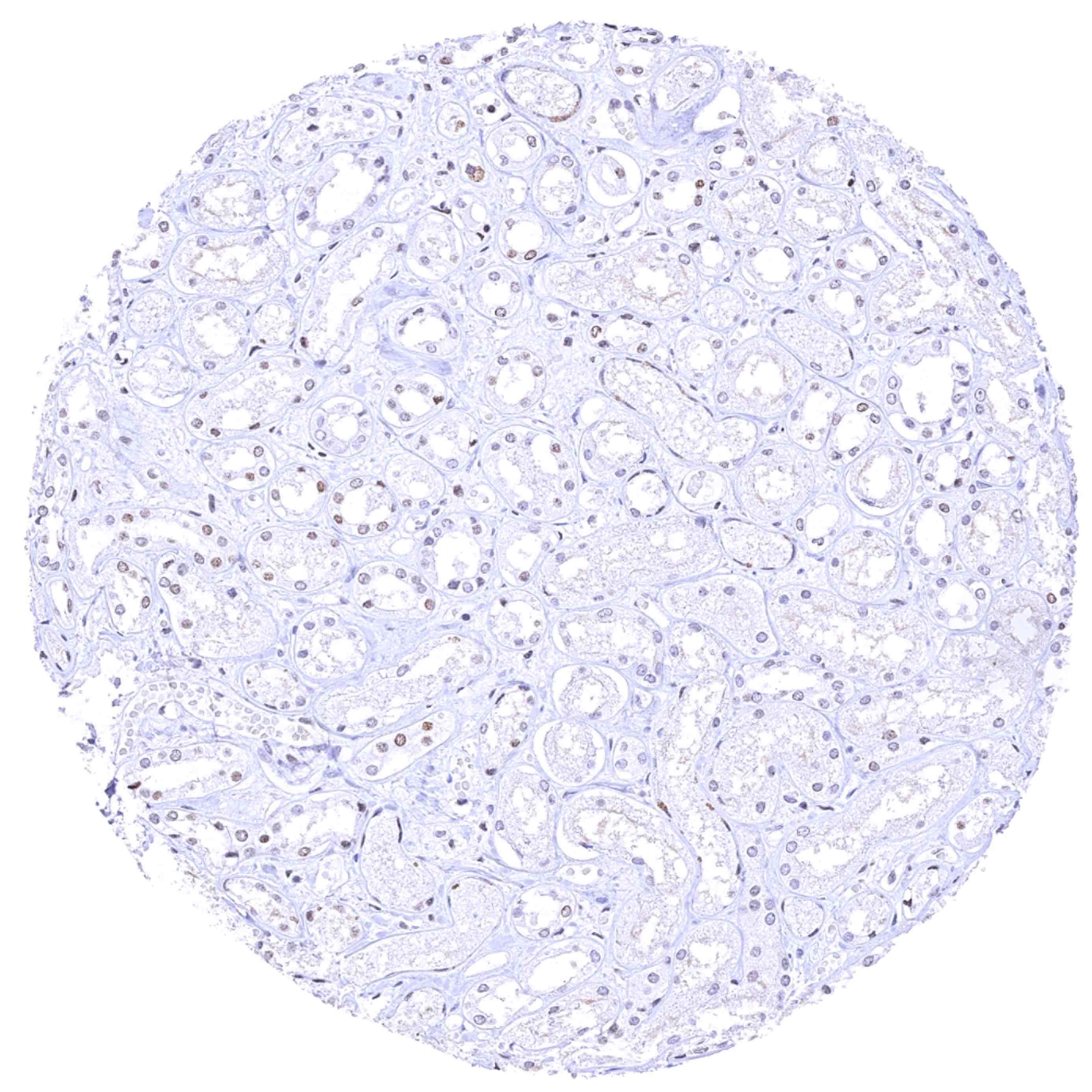
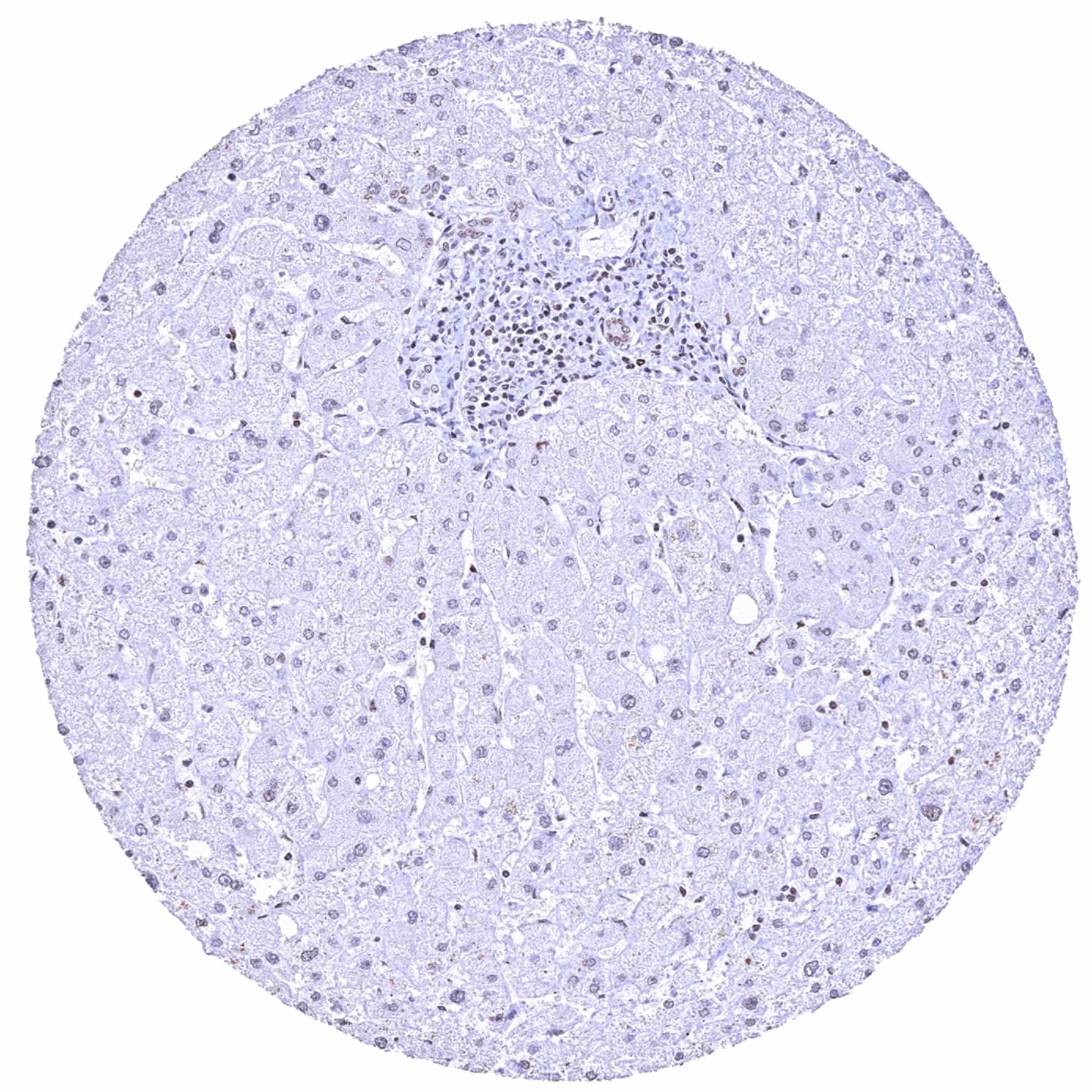
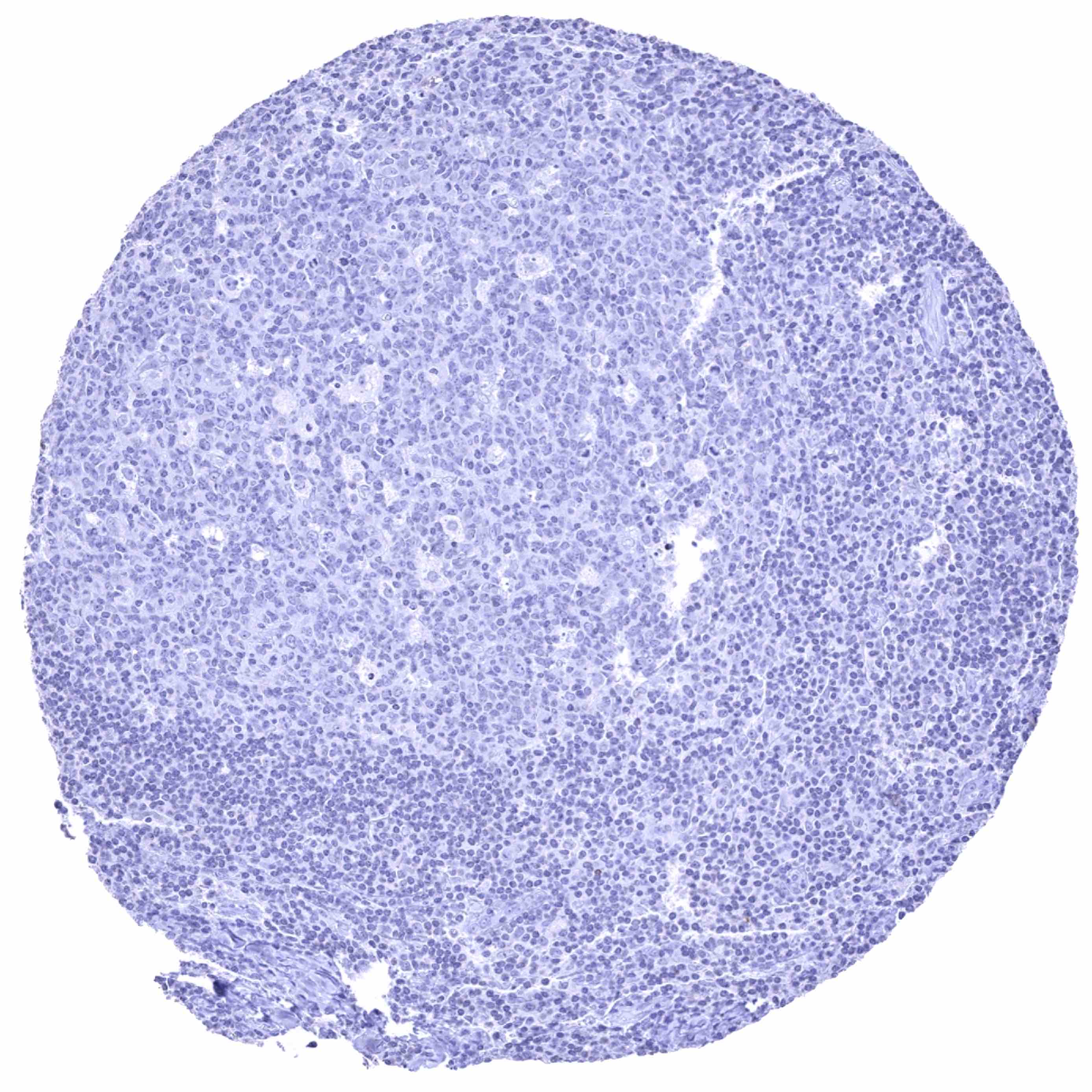
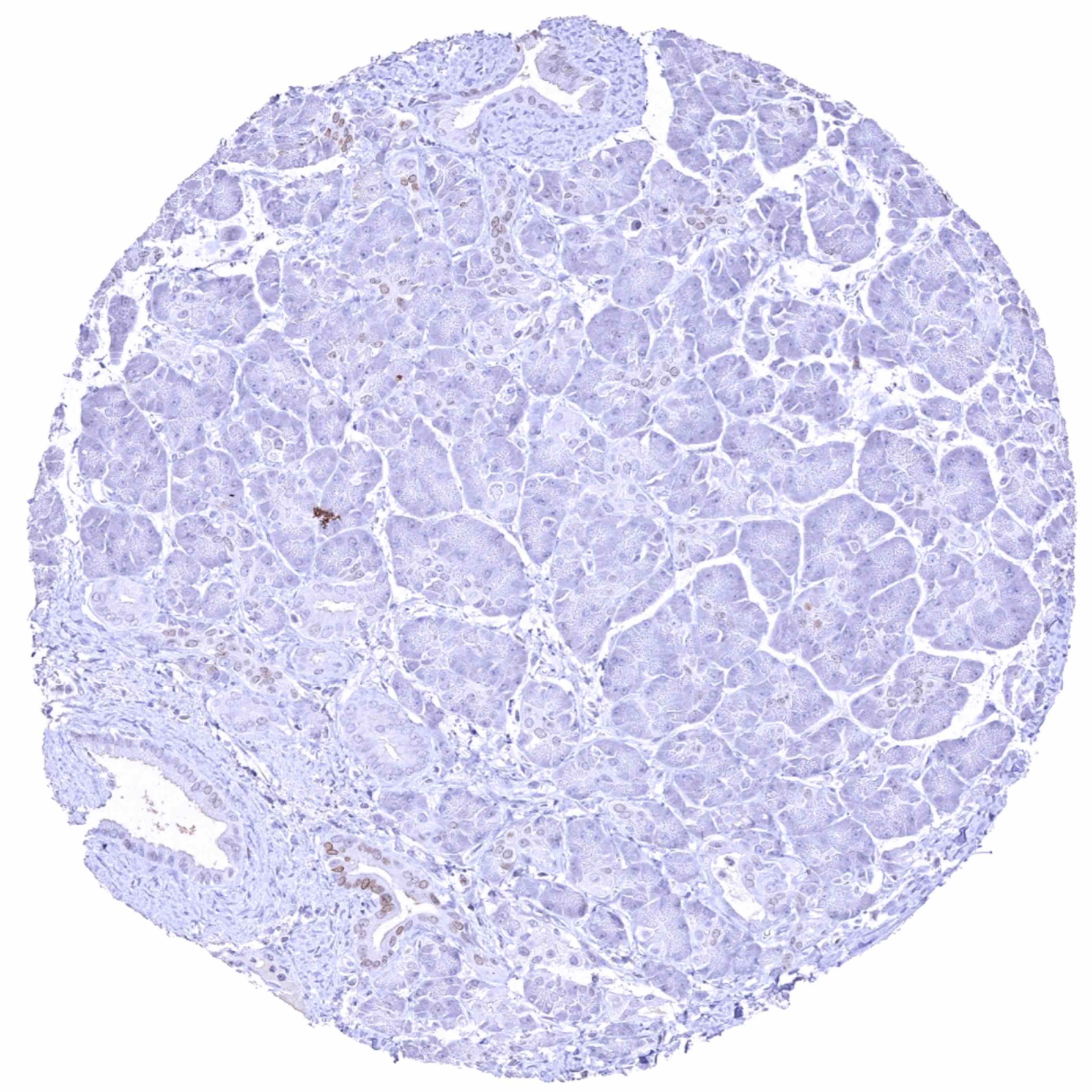
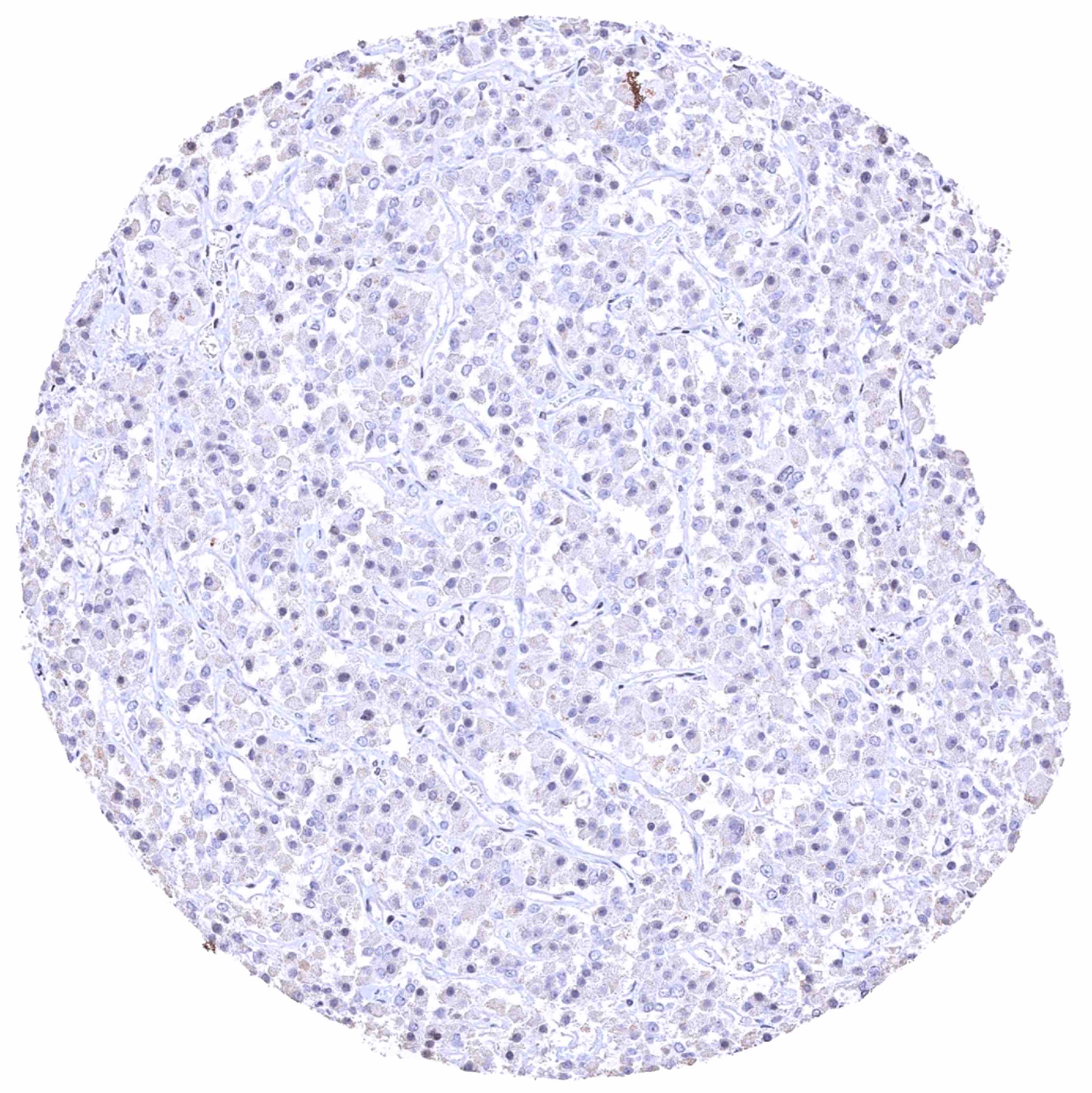
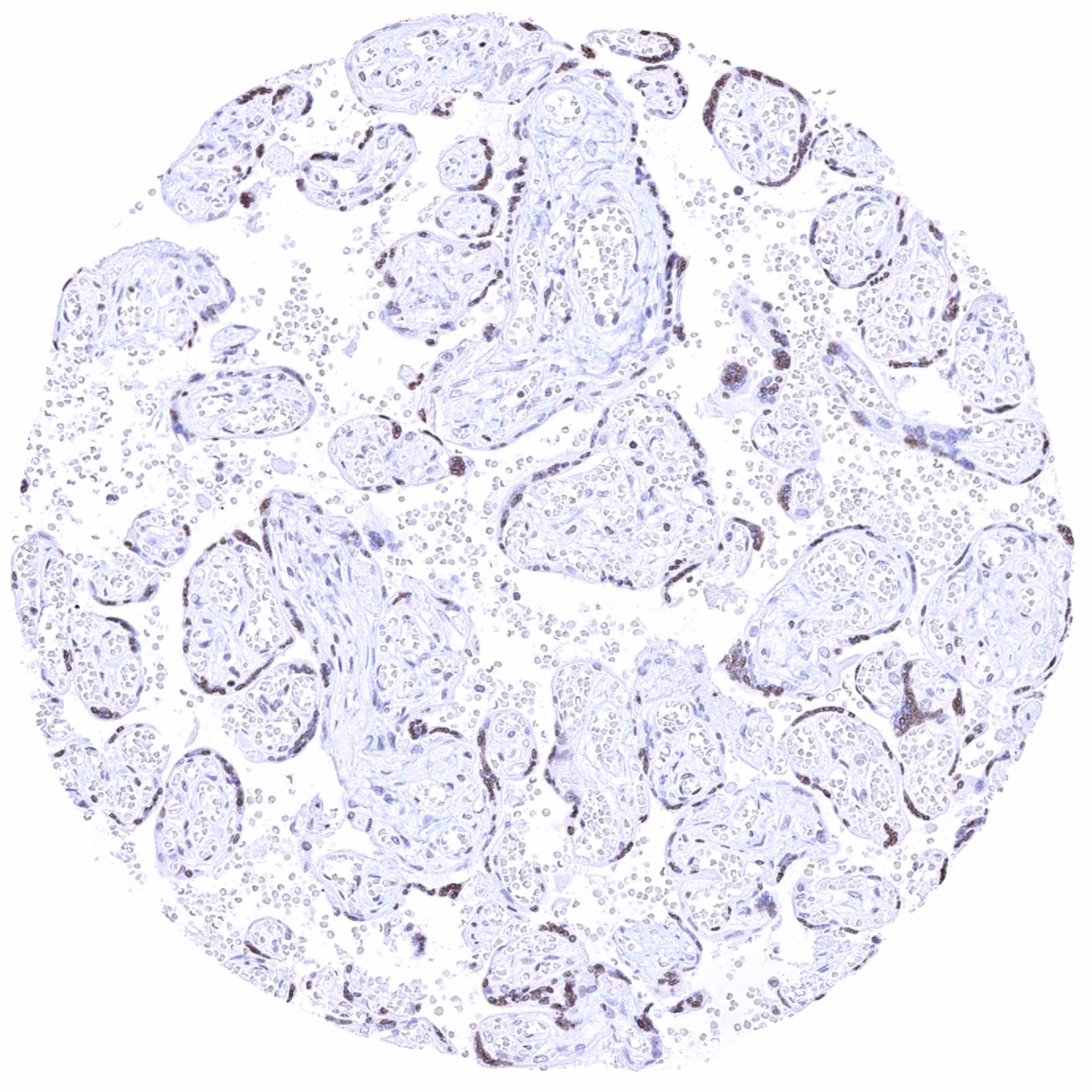
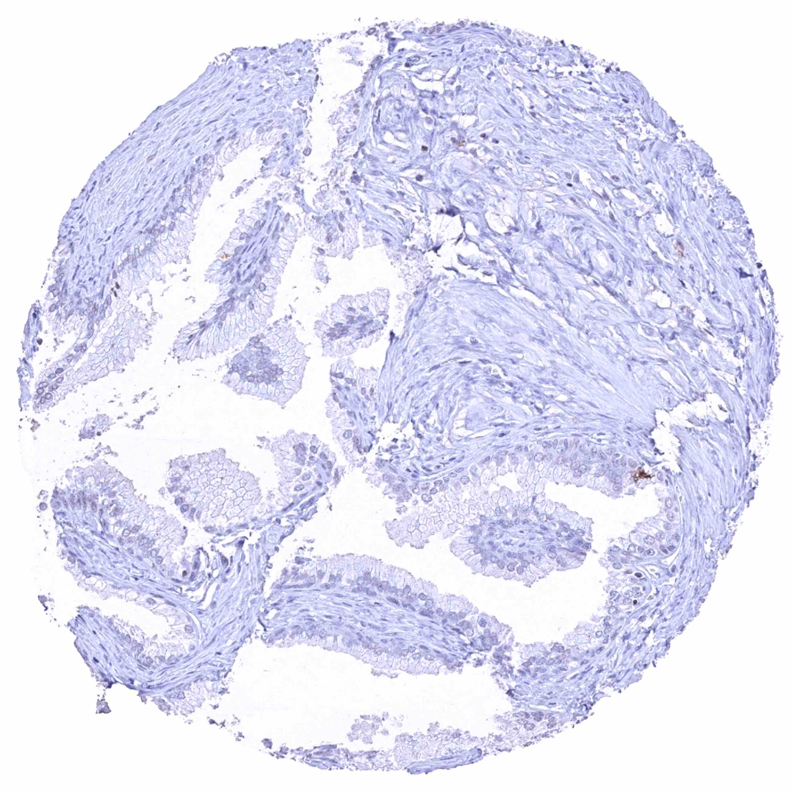
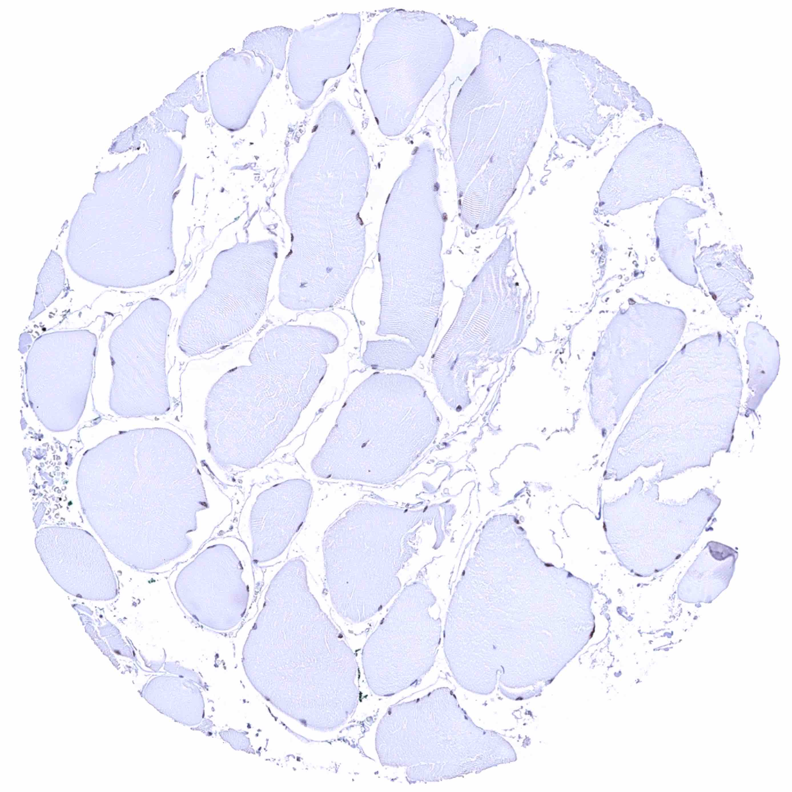
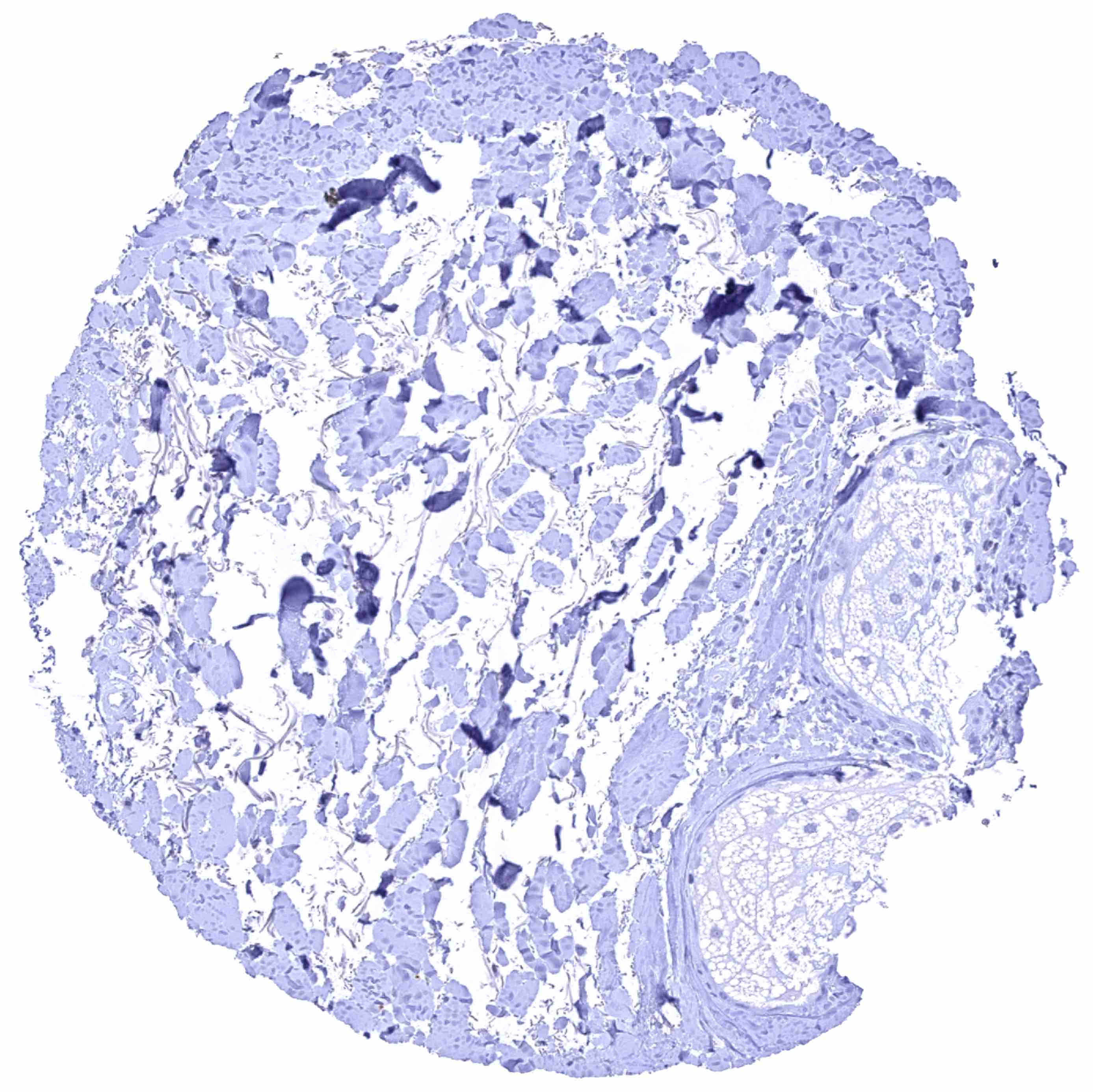
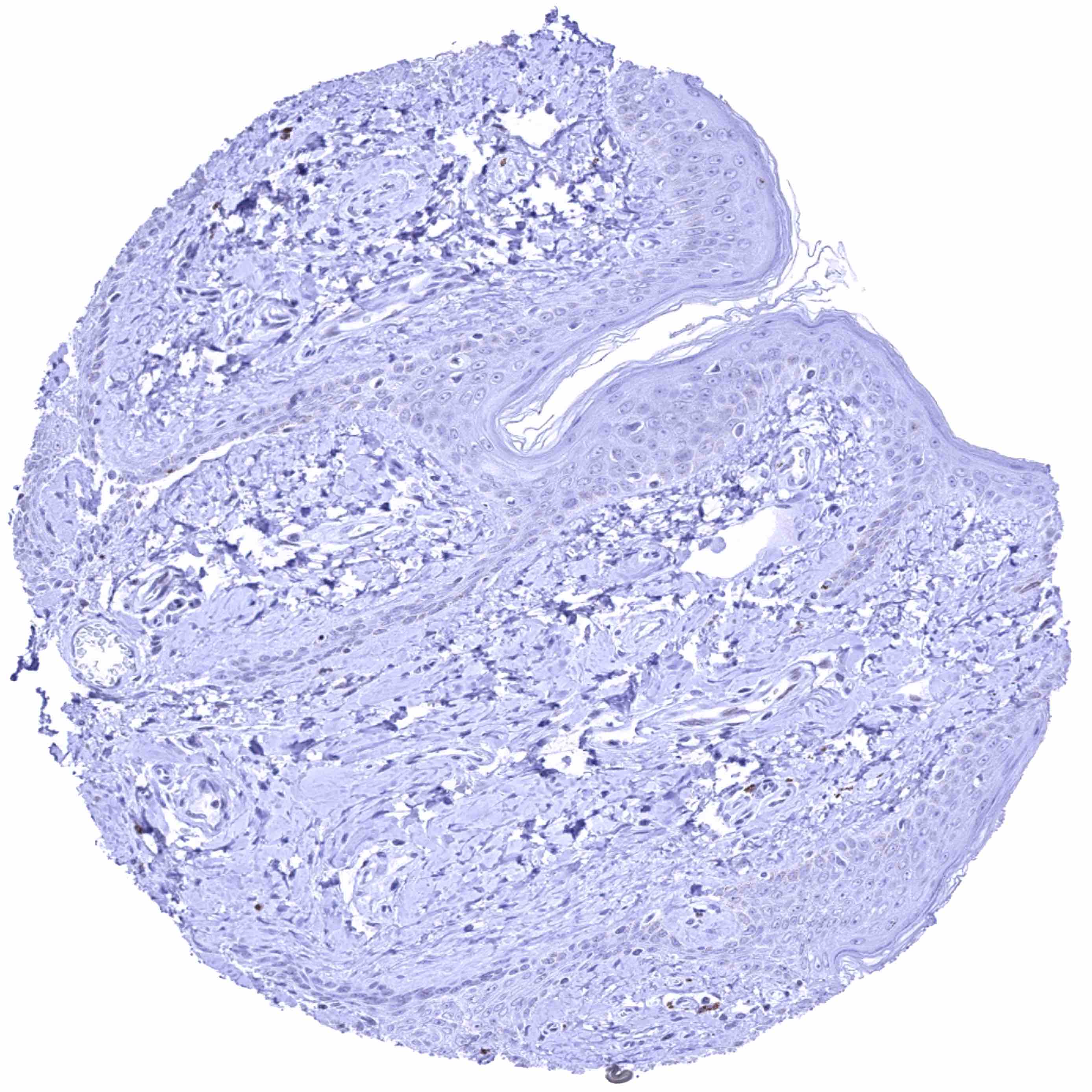
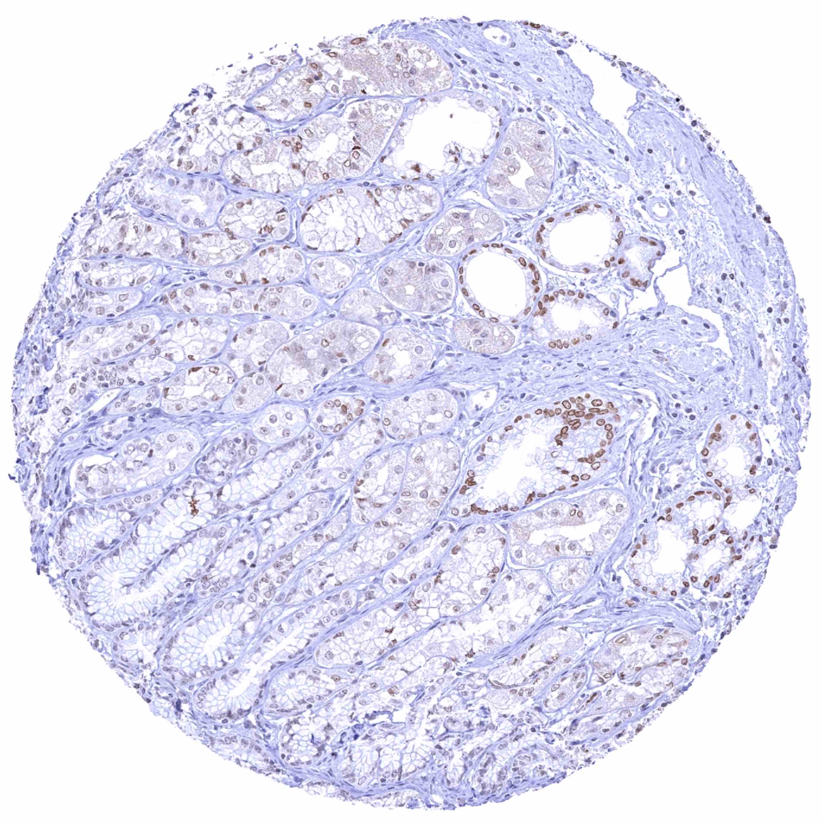
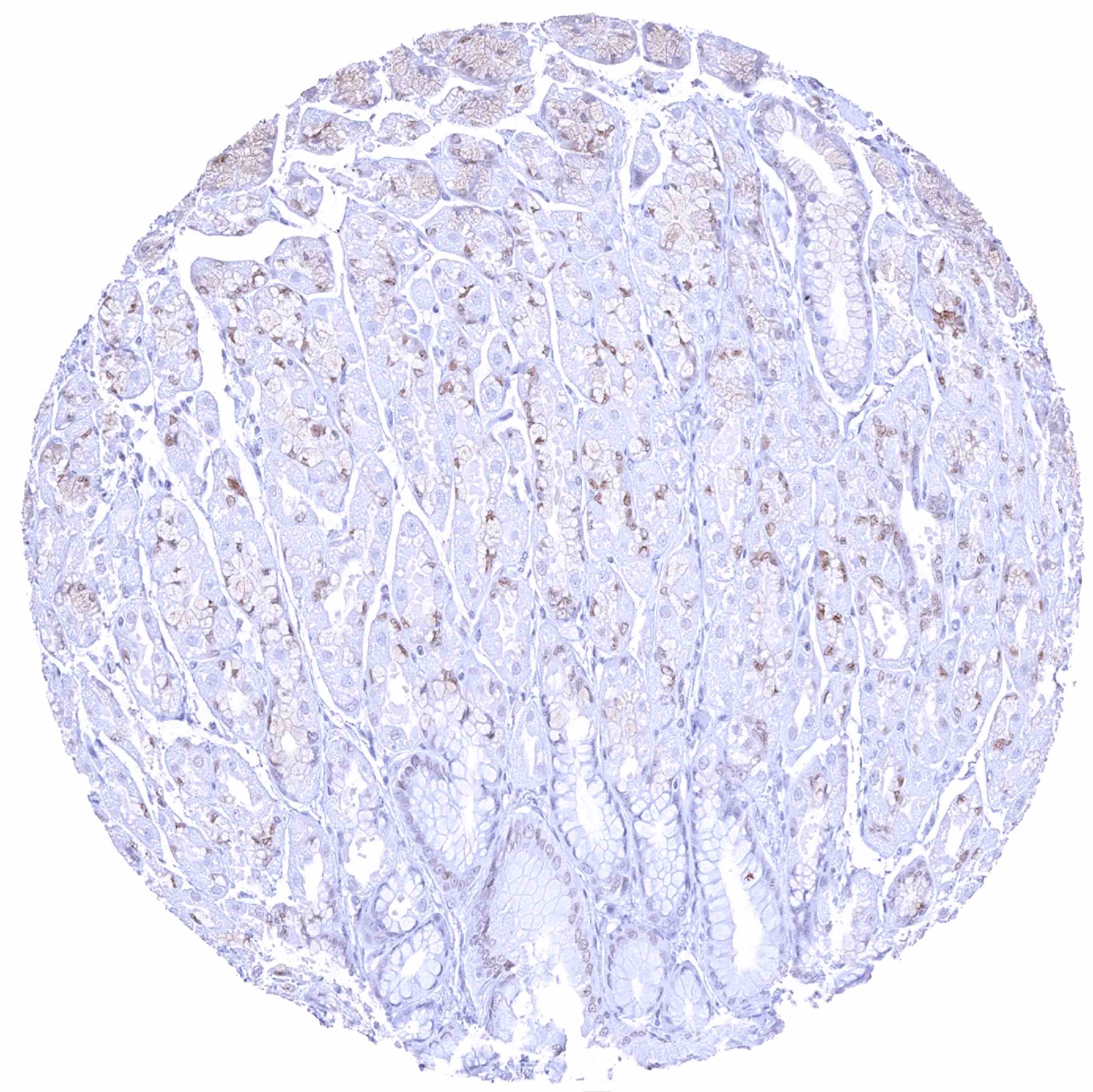
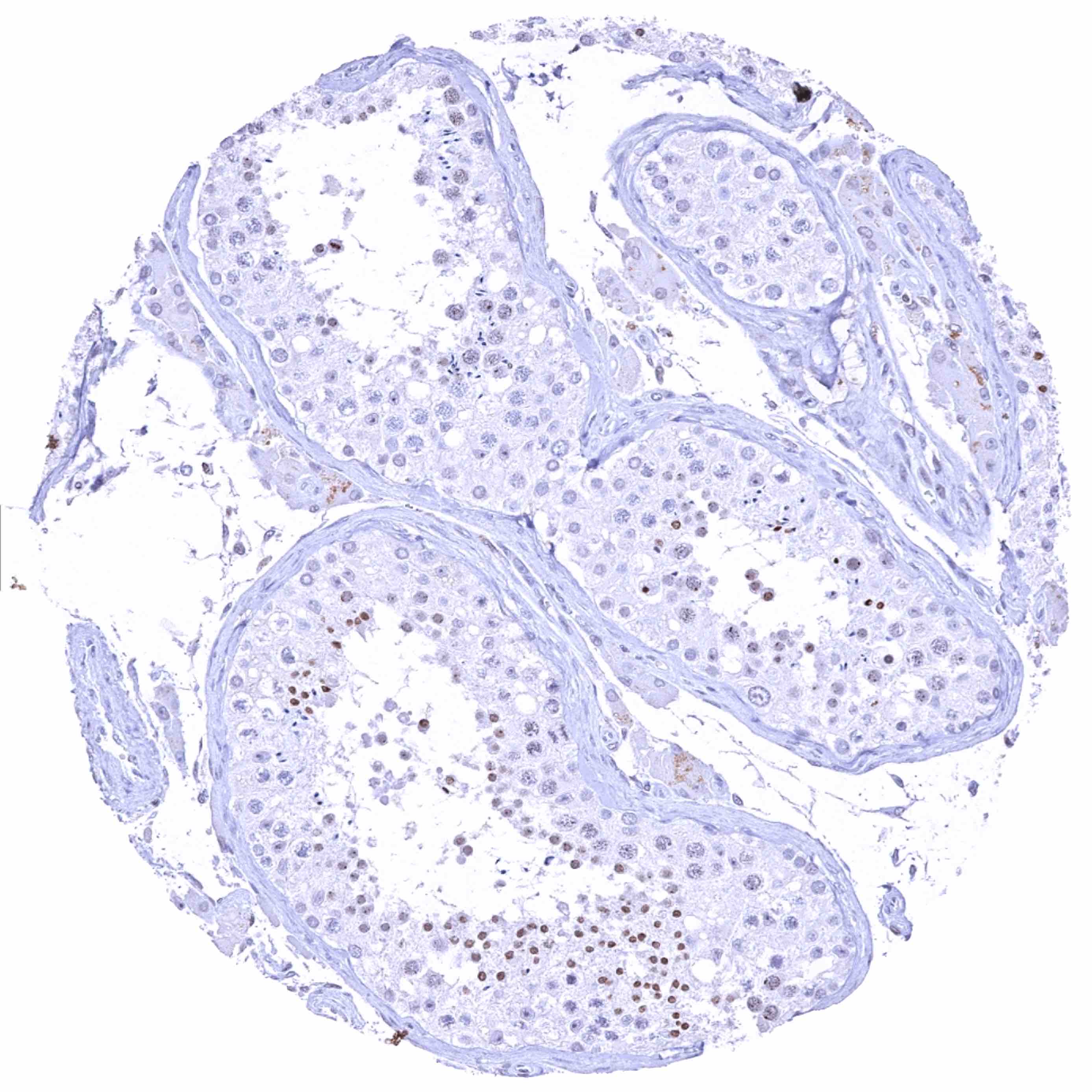
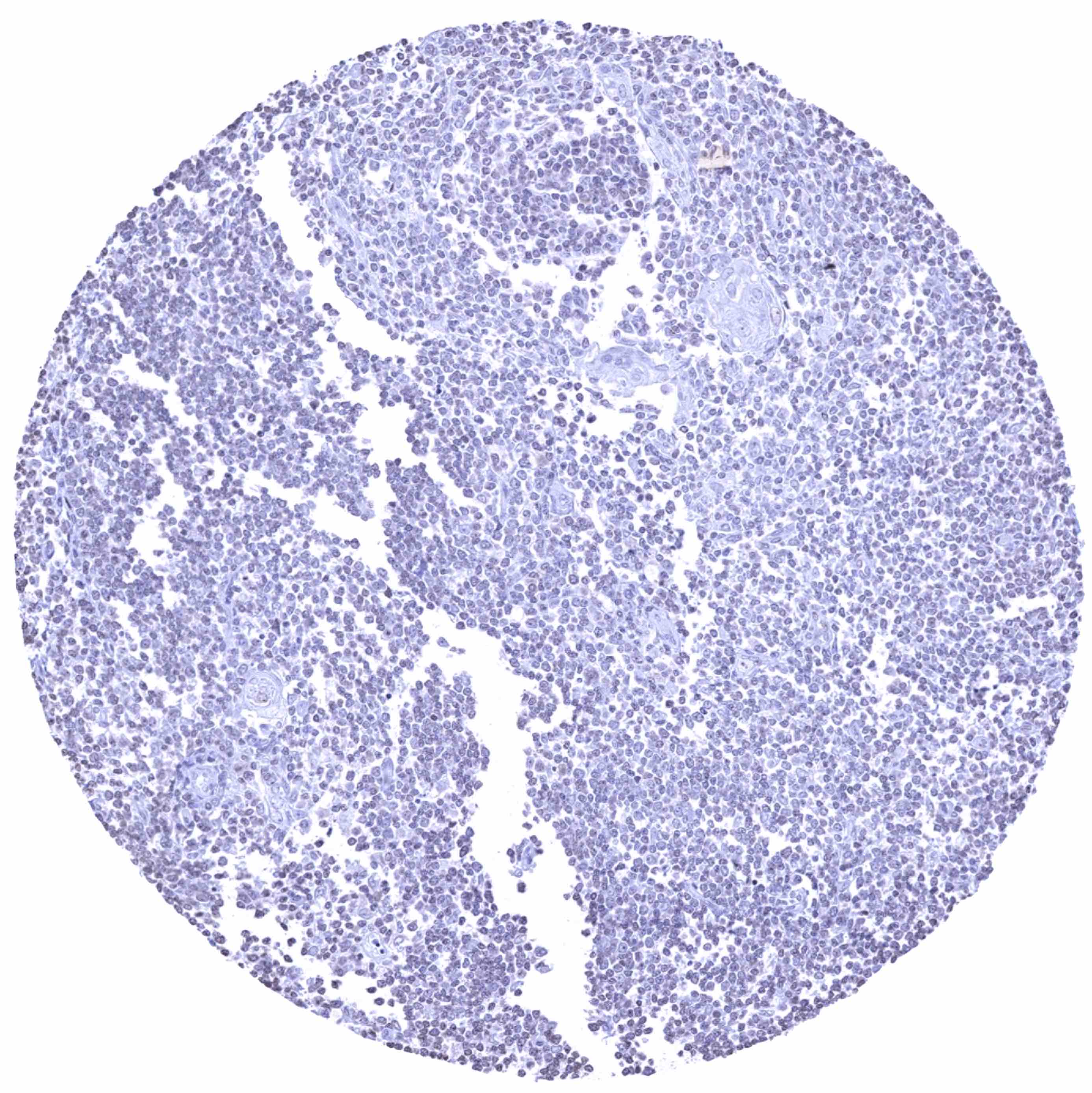
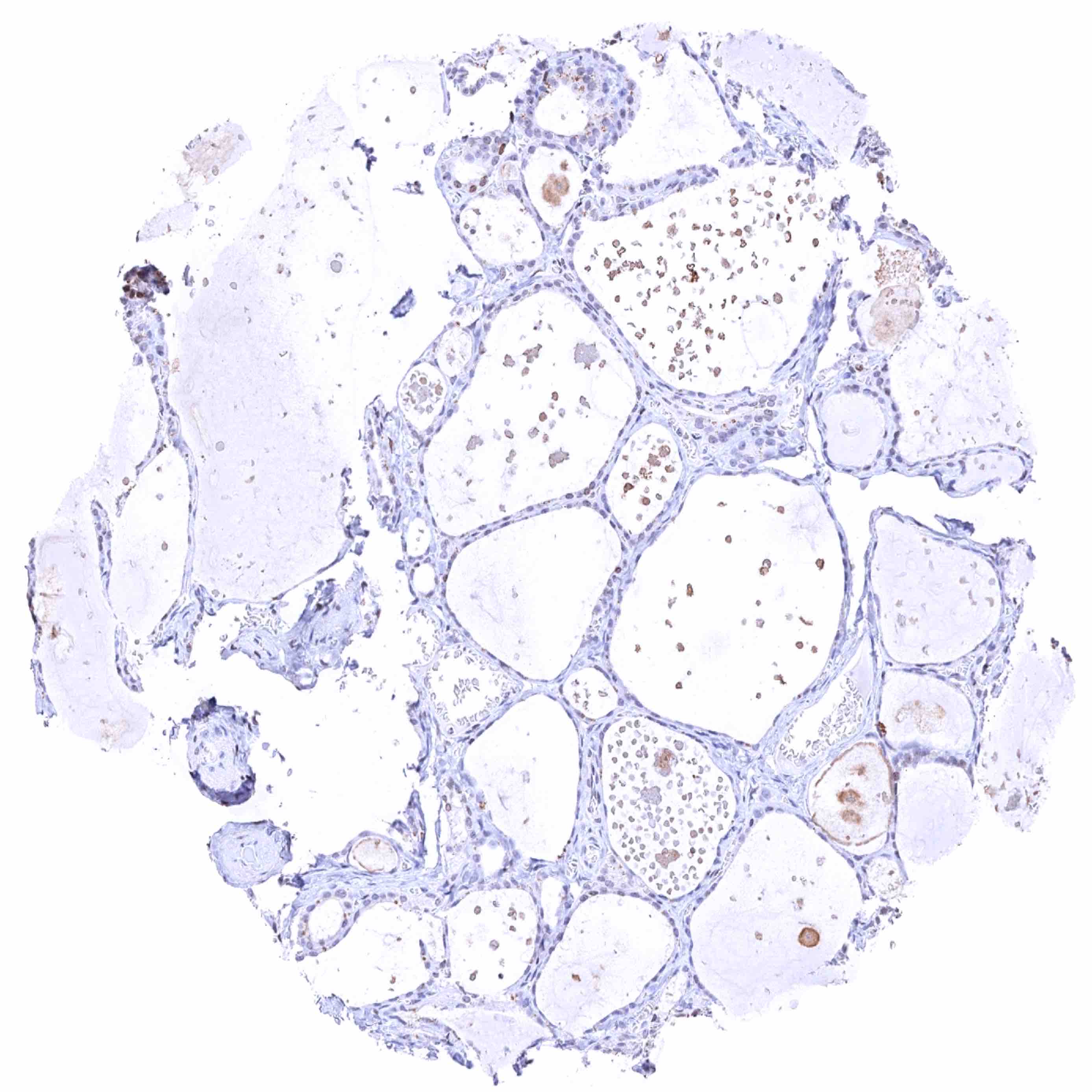
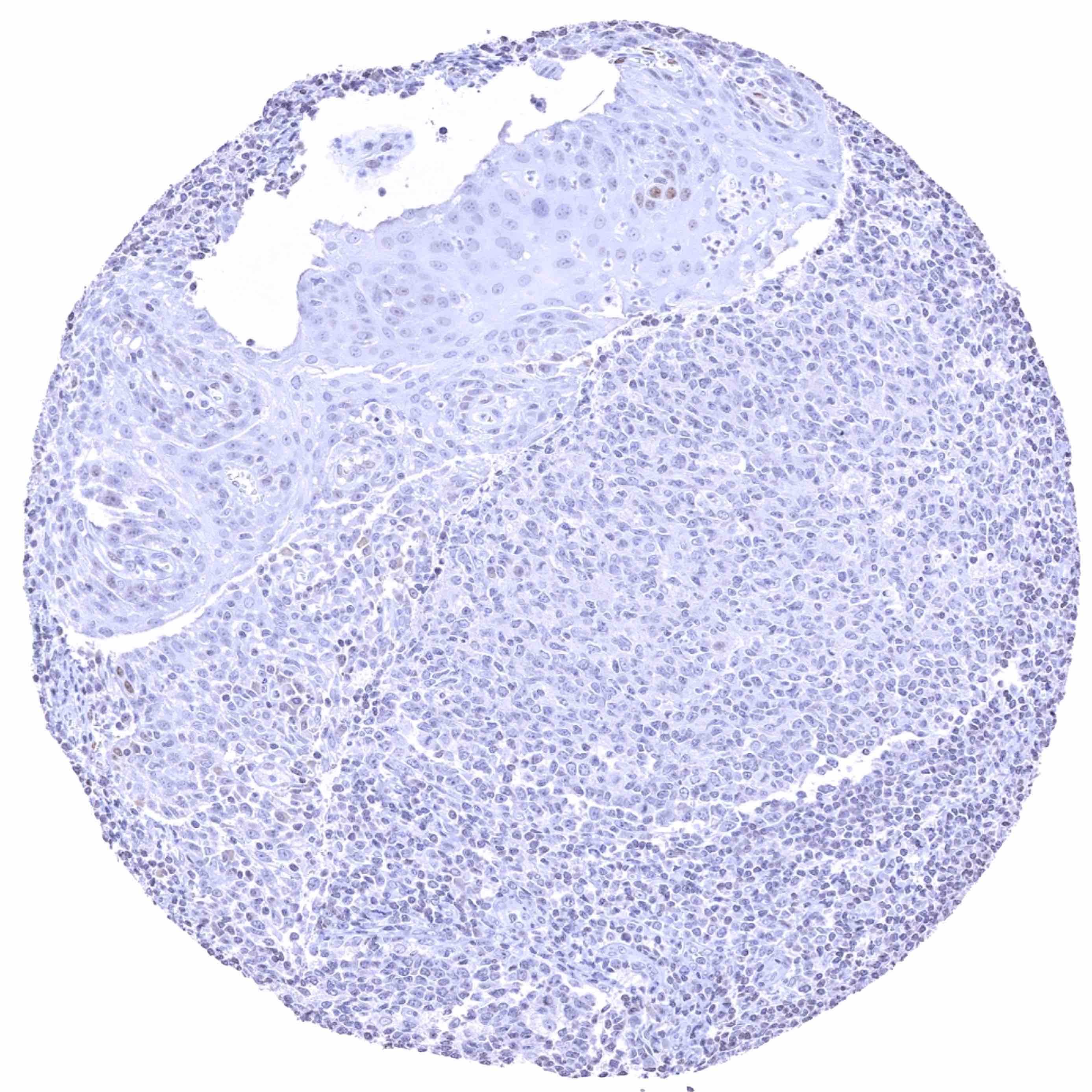
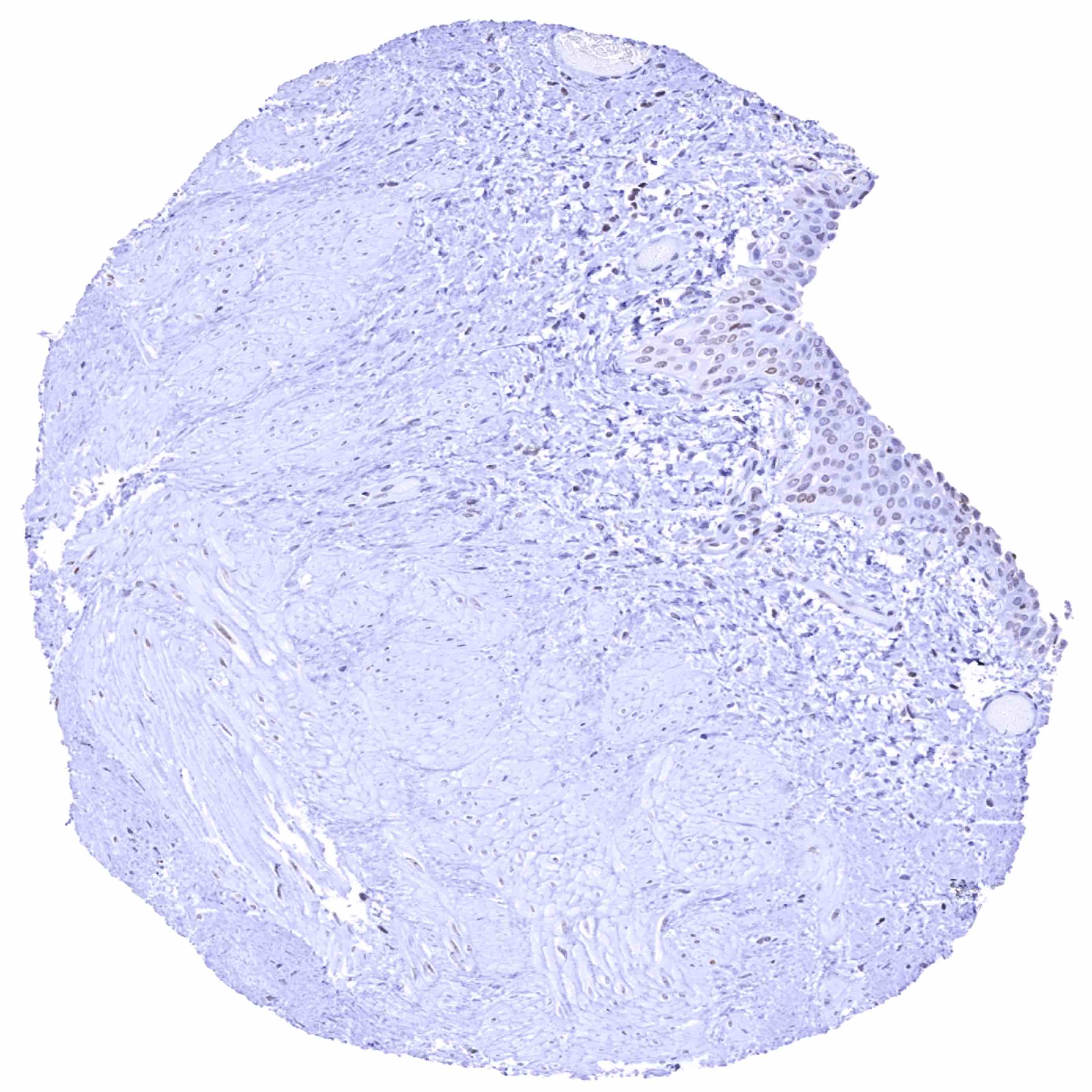
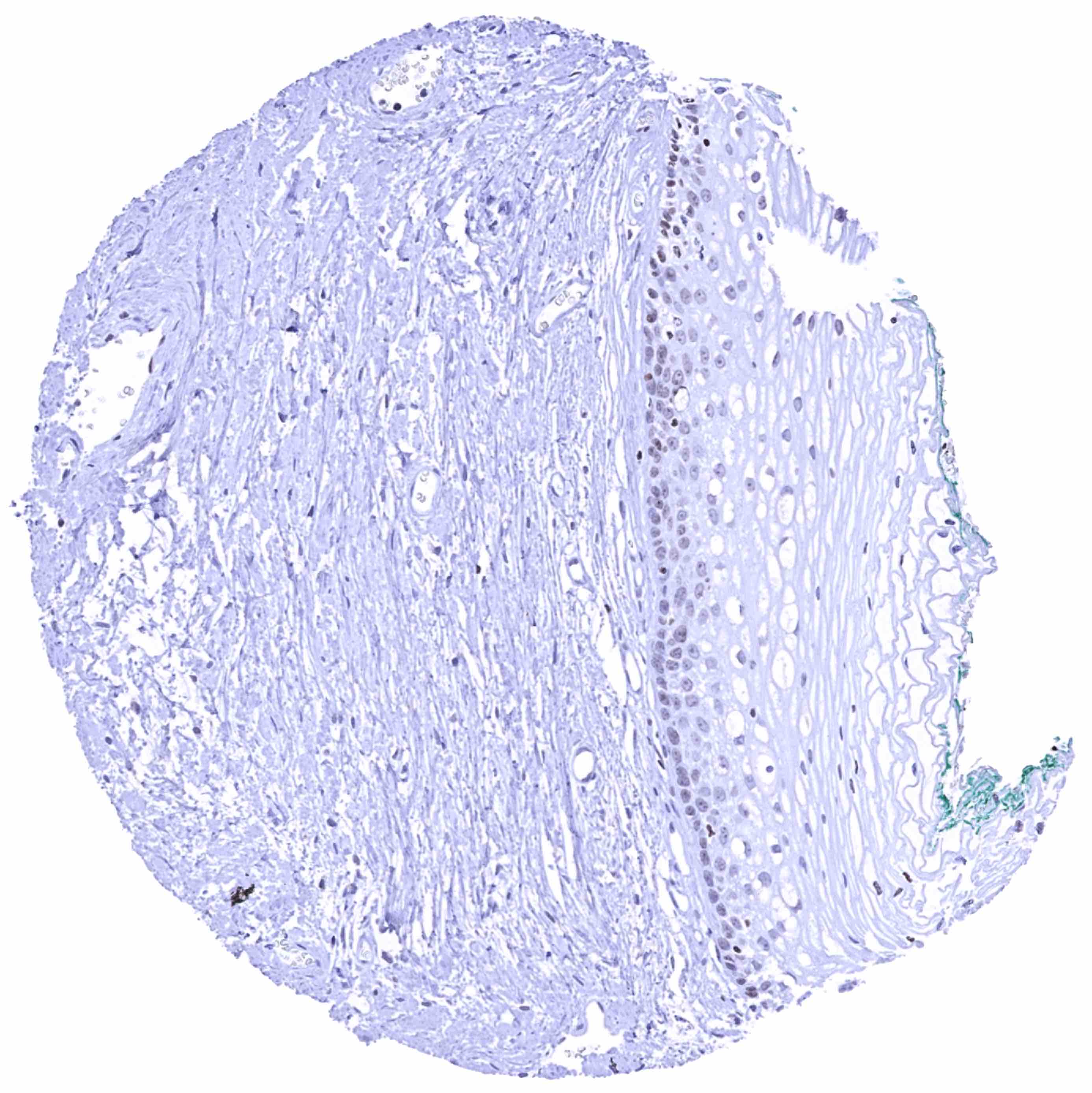
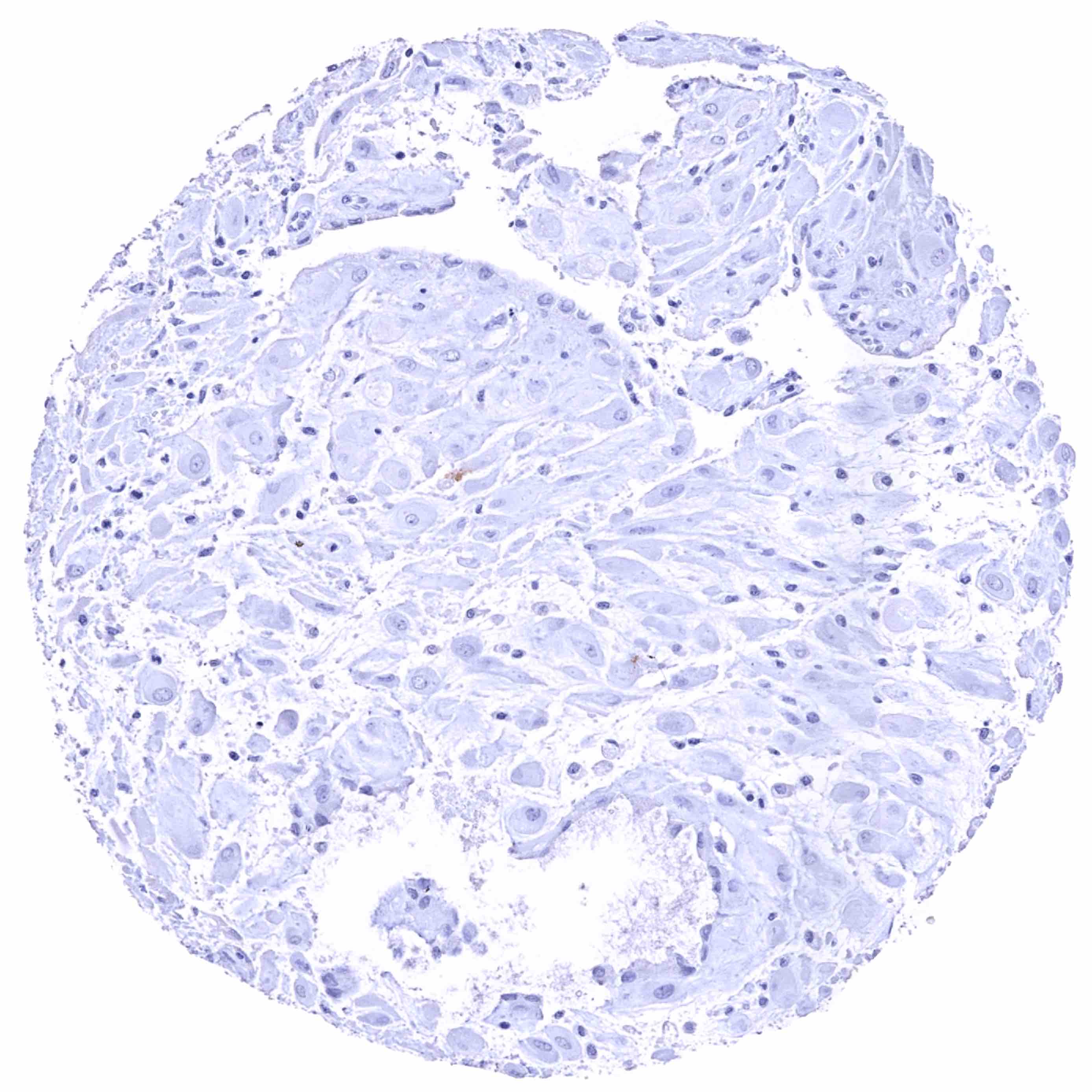
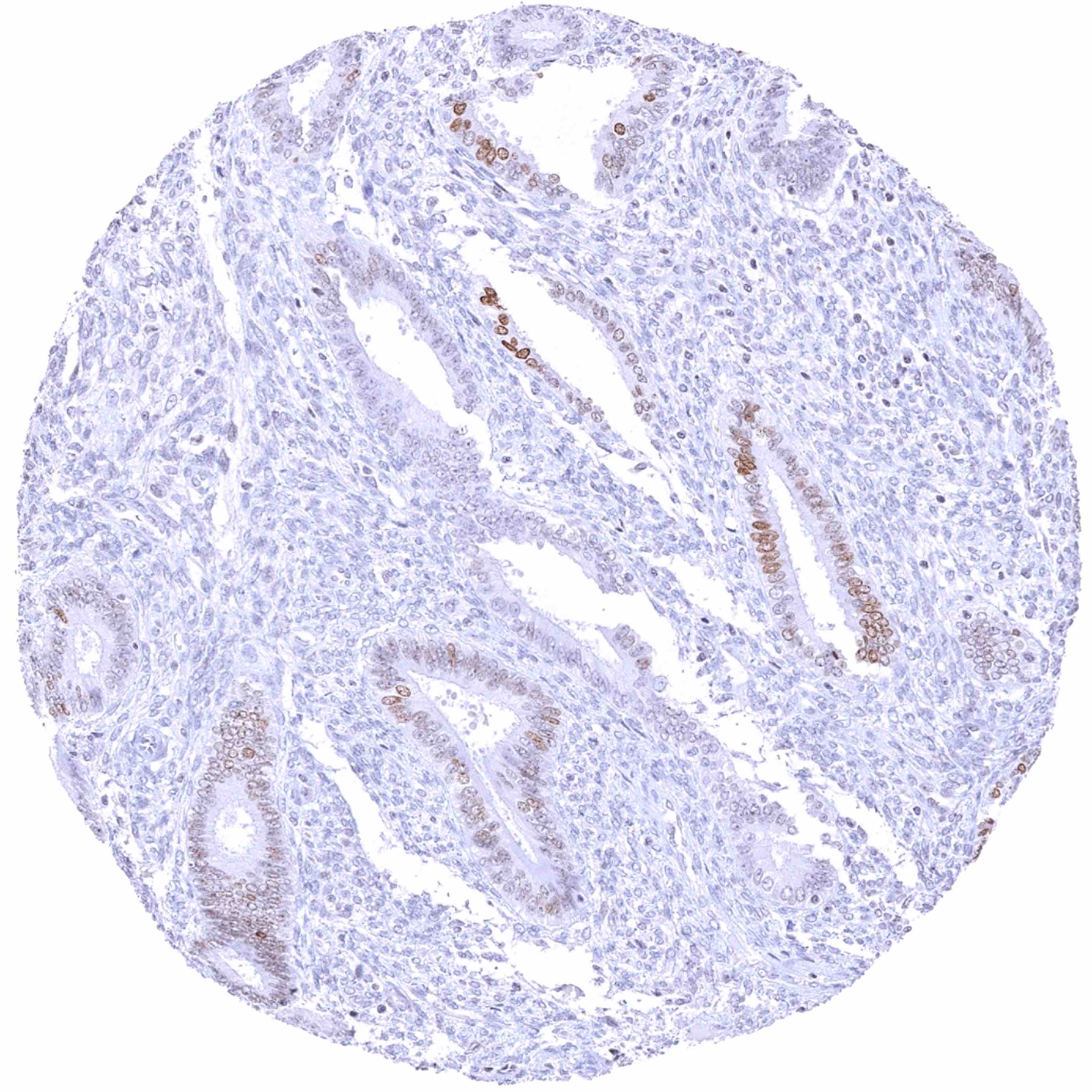
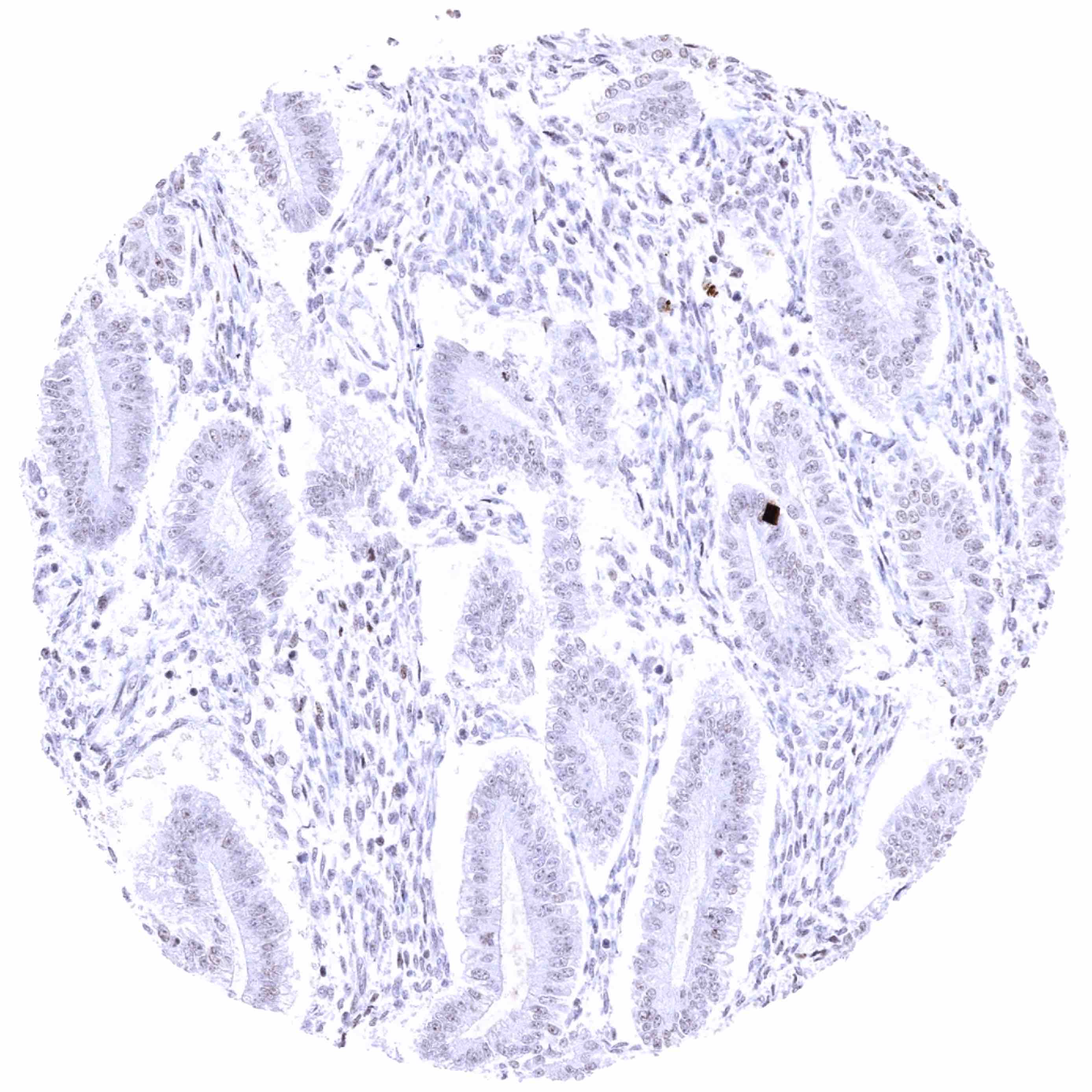
HMGA2 antibody [HMV314] HistoMAXTM


Go to Cancer Tissue Gallery
  
 Go back to product page
  

Go to HistoMAX page


Adrenal gland

Adrenal gland

Aorta, media

Appendix, muscular wall


Bone marrow – Moderate nuclear HMGA2 staining of a fraction of cells

Breast

Bronchus, mucosa – Strong nuclear HMGA2 staining of most respiratory epithelial cells

Cerebellum (white matter)


Cerebellum, cortex (molecular layer, Purkinje cell layer, granule cell layer, white matter)

Cerebellum, cortex (molecular layer, Purkinje cell layer, granule cell layer, white matter)

Cerebrum, grey matter

Cerebrum, white matter


Colon descendens, mucosa – Weak nuclear HMGA2 staining of a fraction of epithelial cells

Colon descendens, muscular wall

Duodenum, brunner gland – Weak nuclear HMGA2 staining of duodenal crypt cells. Moderate HMGA2 positivity of Brunner glands

Duodenum, mucosa


Epididymis – Weak nuclear HMGA2 staining of a fraction of epithelial cells in the cauda

Epididymis

Esophagus, squamous epithelium

Fallopian tube, mucosa – Moderate to strong nuclear HMGA2 staining of a large subset of epithelial cells


Fallopian tube, mucosa – Strong nuclear HMGA2 staining of most epithelial cells

Fat

Gallbladder, epithelium

Heart muscle


Ileum, mucosa – Very faint nuclear HMGA2 positivity of some epithelial cells

Kidney, cortex – Absence of HMGA2 staining in this sample

Kidney, cortex – Moderate nuclear HMGA2 staining of tubular cells in areas of tissue damage

Kidney, medulla – Faint nuclear HMGA2 staining of a fraction of collecting duct cells


Liver – Weak nuclear HMGA2 staining of a fraction of bile duct epithelial cells

Lung

Lymph node

Lymph node


Ovary, corpus luteum

Ovary, follicular cyst

Pancreas – Faint nuclear HMGA2 staining of cells from some small ducts

Pancreas – Weak nuclear HMGA2 staining of a fraction of acinar and ductal (small ducts) cells


Parathyroid gland – Some epithelial cell groups show a moderate to strong nuclear HMGA2 staining

Parotid gland

Pituitary gland, anterior lobe

Pituitary gland, posterior lobe – Weak to moderate nuclear HMGA2 staining of pituicytes


placenta (amnion and chorion) – Strong nuclear HMGA2 positivity of amnion and stroma cells while chorion cells are negative

Placenta, early – Strong nuclear HMGA2 staining of stroma cells. Trophoblast cells remain HMGA2 negative

Placenta, mature – Some nuclear HMGA2 staining of trophoblast cells in some samples. Stroma cells are HMGA2 negative

Prostate


Rectum, mucosa – Faint nuclear HMGA2 staining of a fraction of epithelial cells

Rectum, mucosa – Faint nuclear HMGA2 staining of a fraction of epithelial cells

Sinus paranasales – Moderate nuclear HMGA2 staining of most respiratory epithelial cells

Skeletal muscle – Weak to moderate HMGA2 staining of some myocytes


Skeletal muscle

Skin

Skin, hairfollicel and sebaceous glands

Skin


Spleen

Stomach, antrum – Weak to moderate nuclear HMGA2 positivity of a fraction of glandular cells.

Stomach, antrum – Weak to moderate nuclear HMGA2 positivity of a fraction of glandular cells

Stomach, corpus – Weak to moderate nuclear HMGA2 positivity of a fraction of glandular cells


Stomach, muscular wall

Sublingual gland

Testis – Weak to moderate nuclear HMGA2 staining of a spermatocytes and of spermatozoa

Thymus


Thyroid gland – Most follicular cells show a faint to weak nuclear HMGA2 staining

Thyroid gland

Thyroid gland

Tonsil


Tonsil, surface epithelium

Urinary bladder, muscular wall

Urinary bladder, urothelium – Few urothelial cells show a weak nuclear HMGA2 positivity

Uterus, ectocervix


Uterus, endocervix – Strong nuclear HMGA2 staining of epithelial cells

Uterus, endometrium (pregnancy)

Uterus, endometrium (proliferation) – Weak to moderate nuclear HMGA2 staining of a subset of epithelial cells

Uterus, endometrium (secretion)


Uterus, myometrium


| TOP |