Normal Tissue Gallery- GTX639935

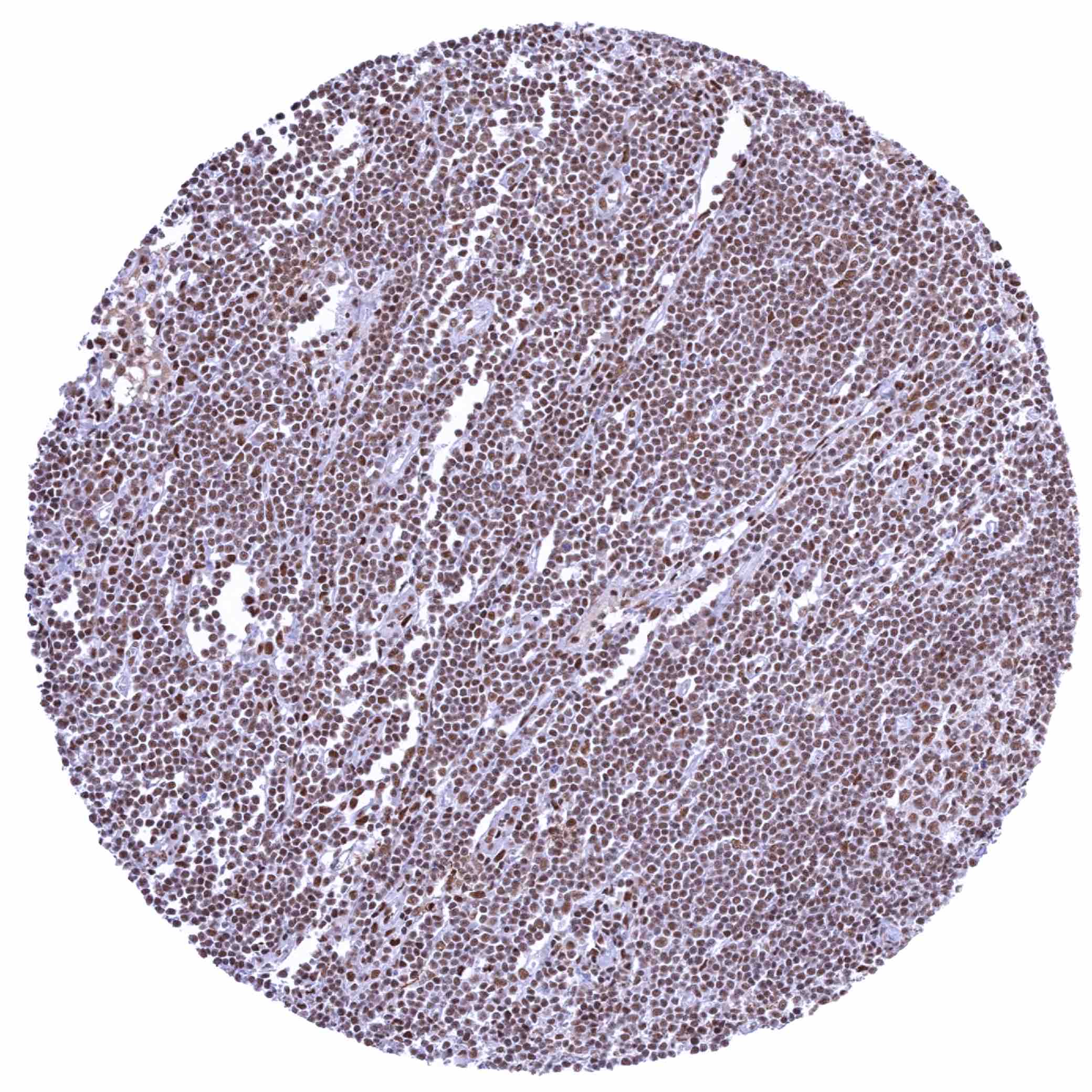
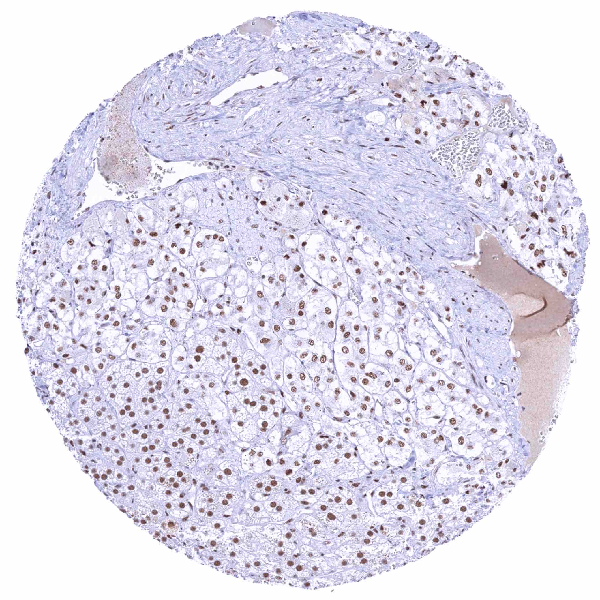
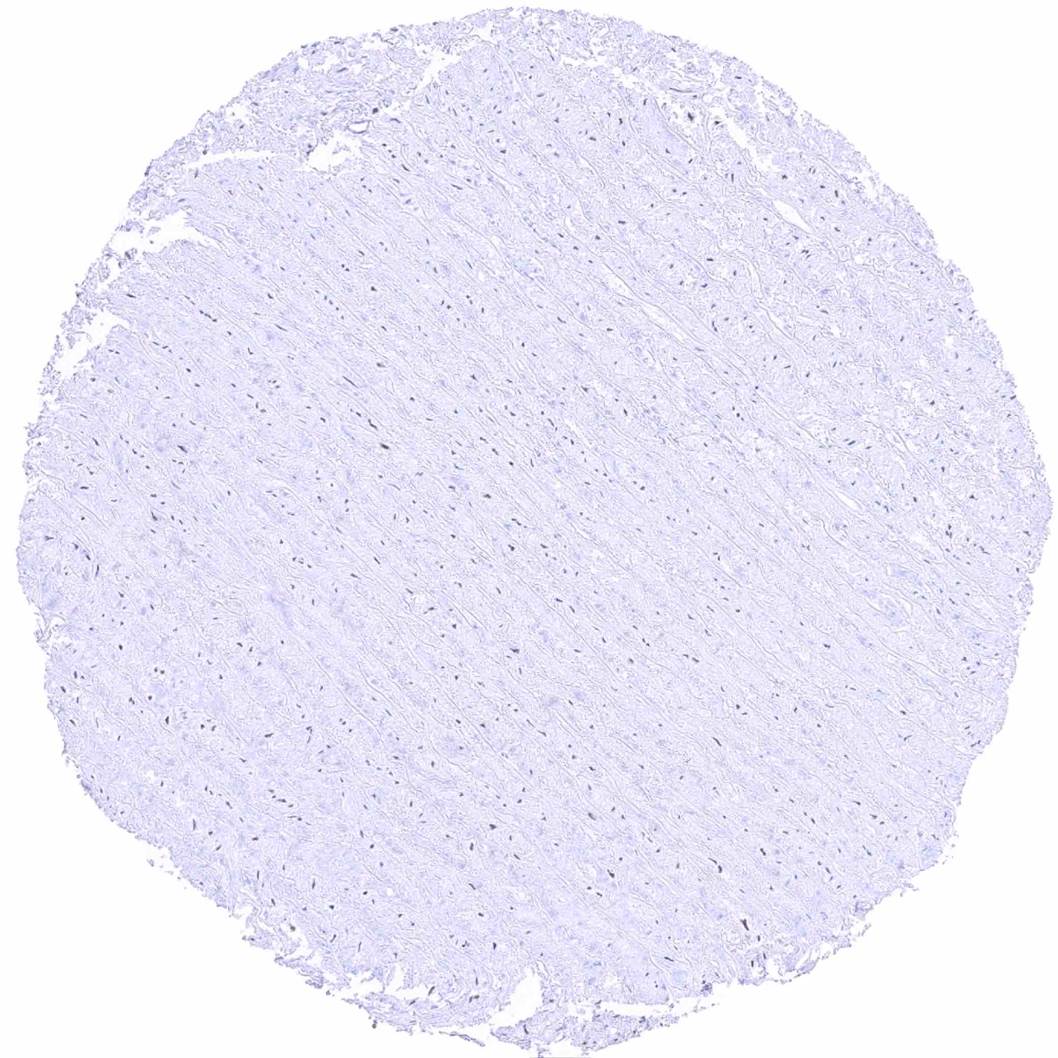
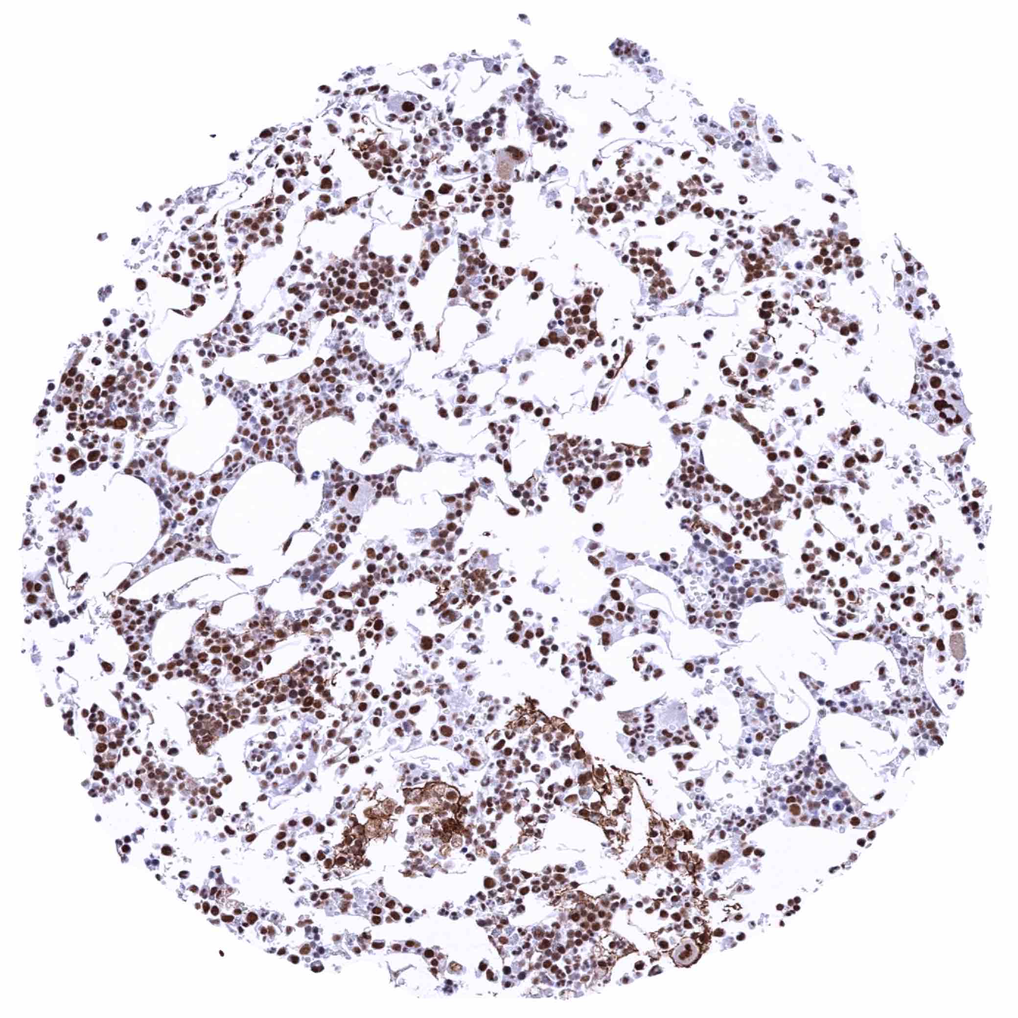
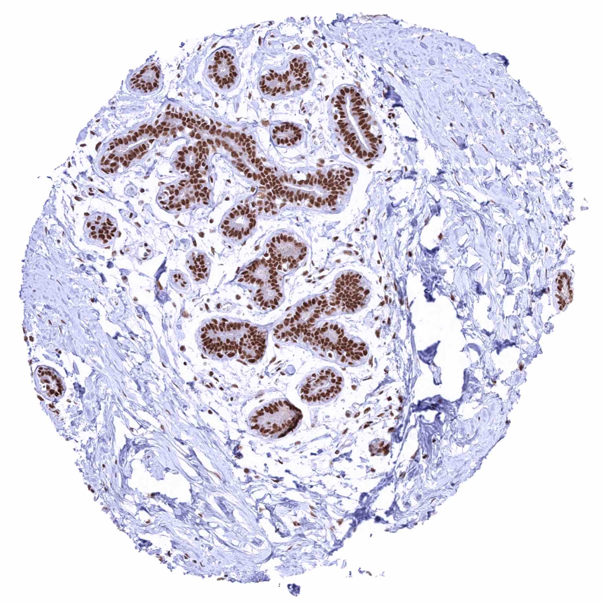
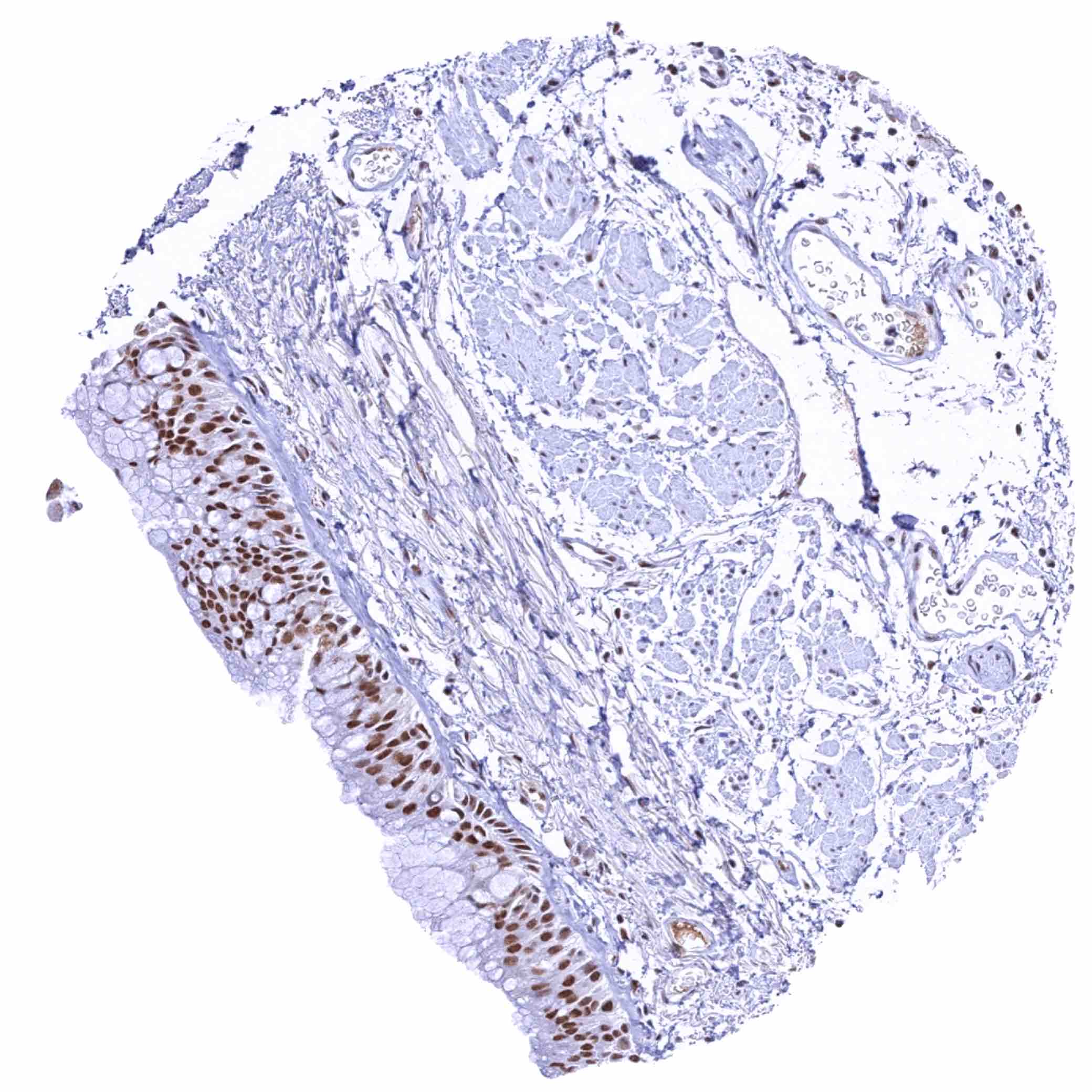
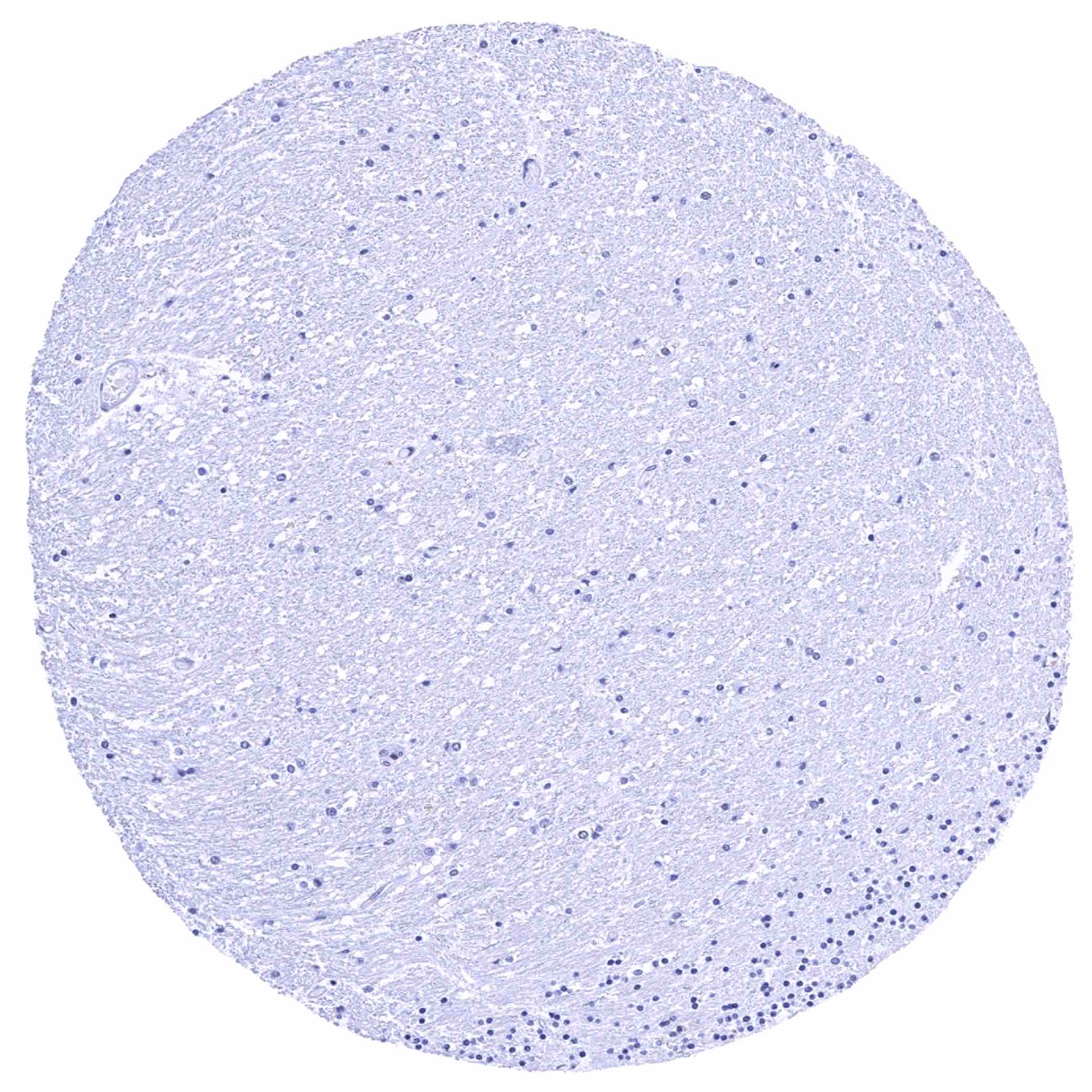
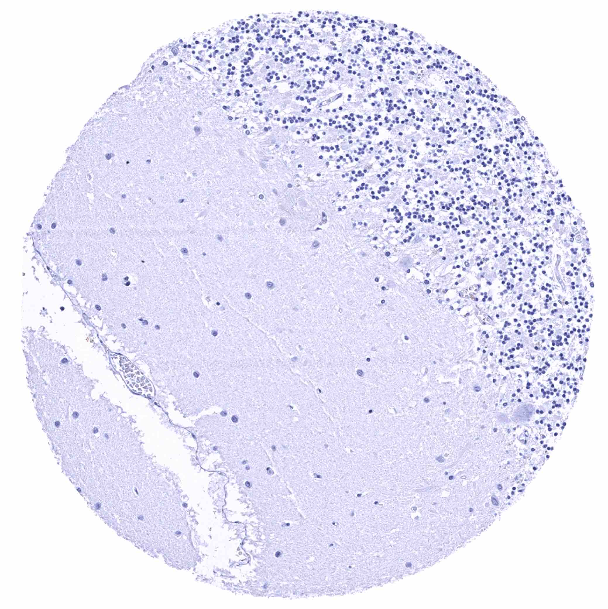
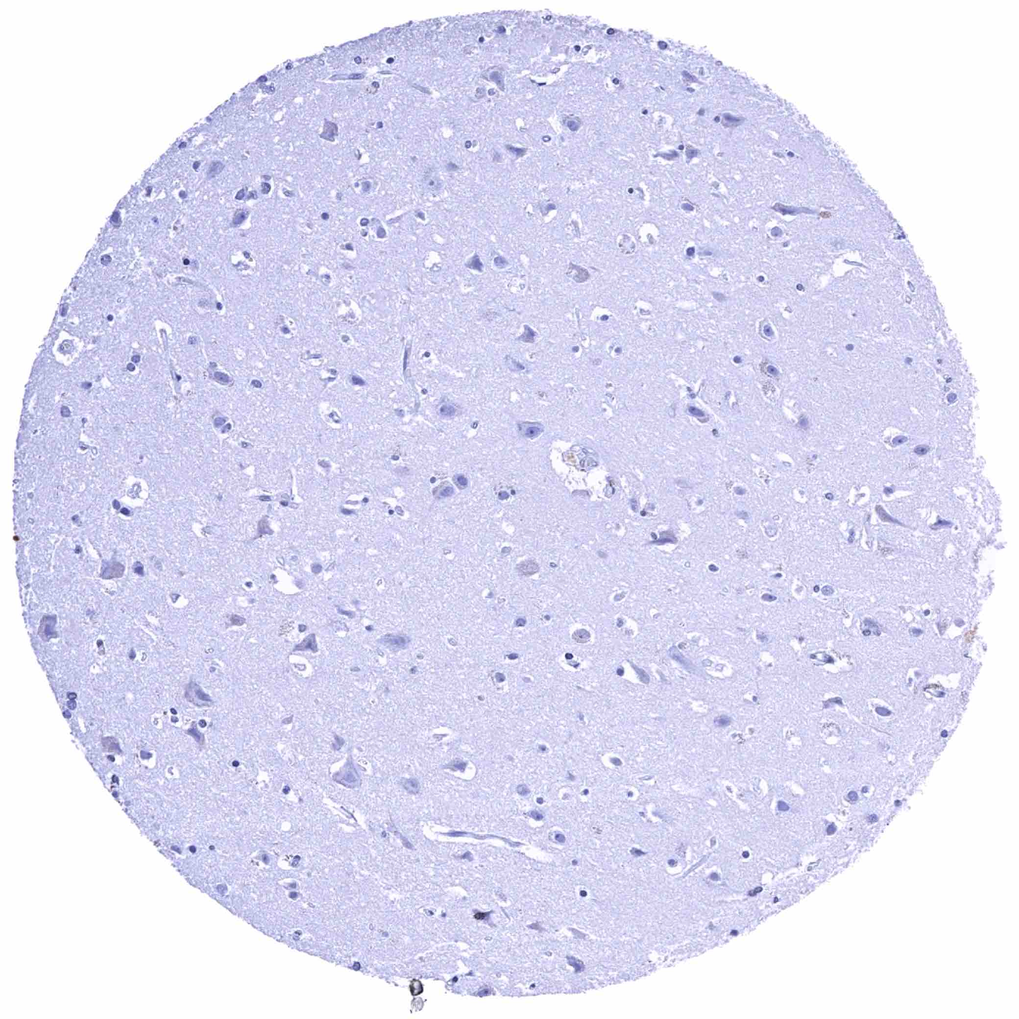
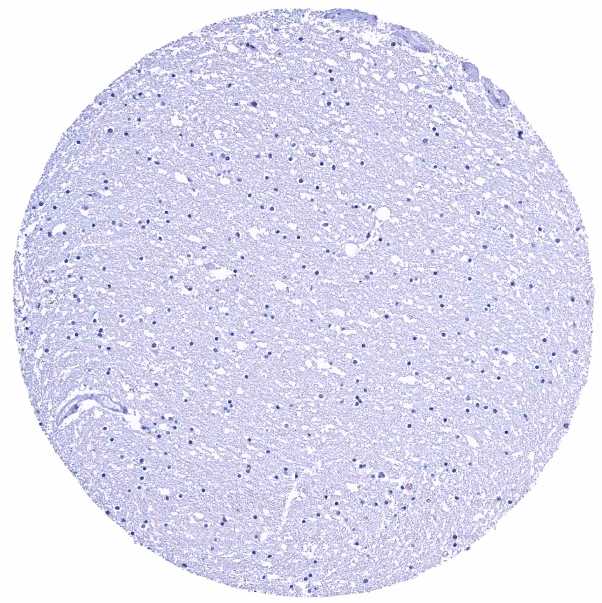
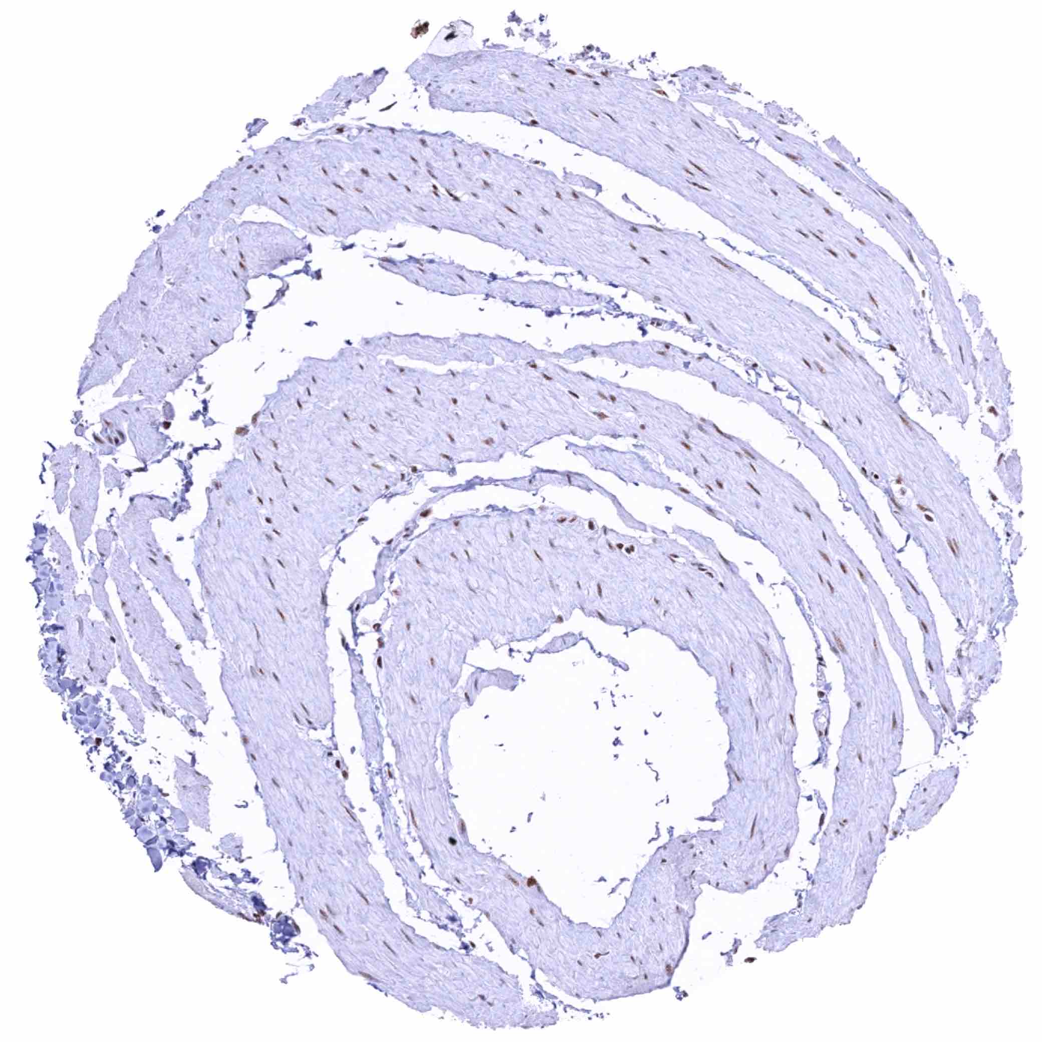
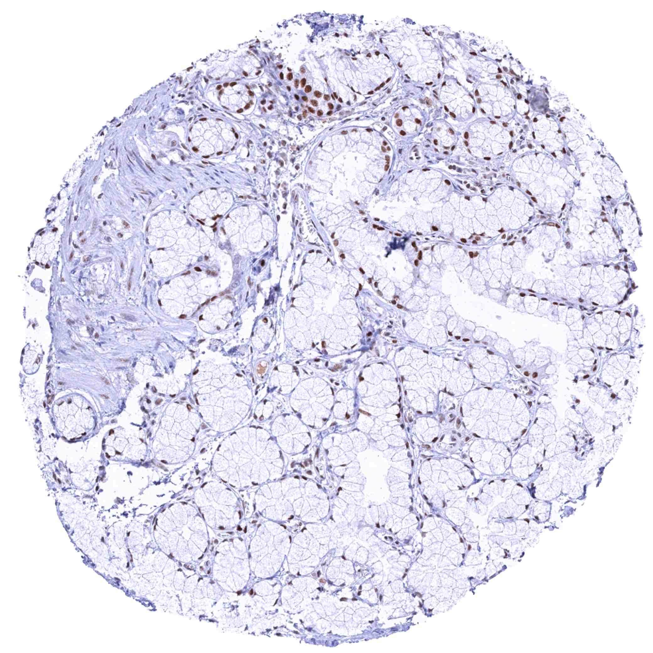
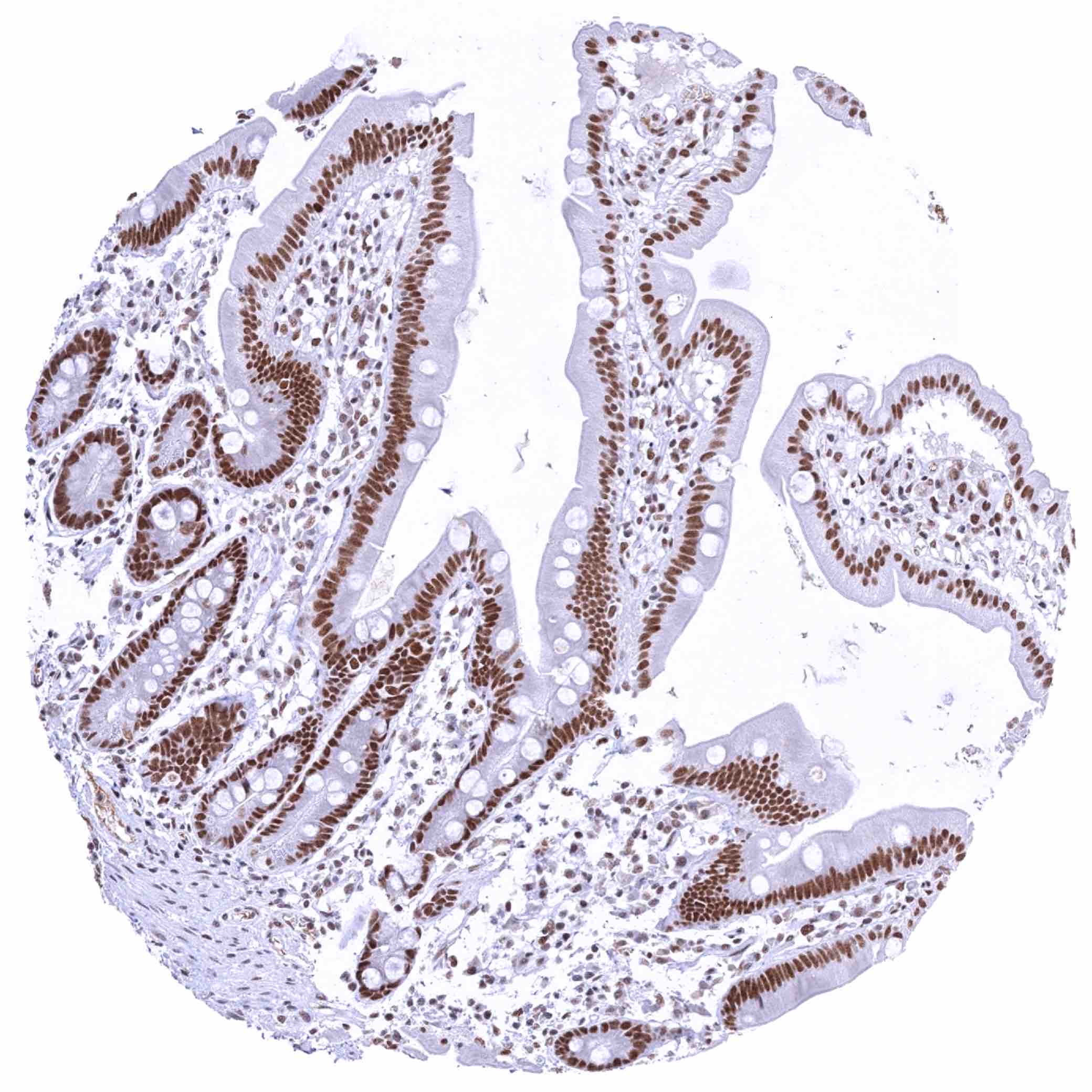
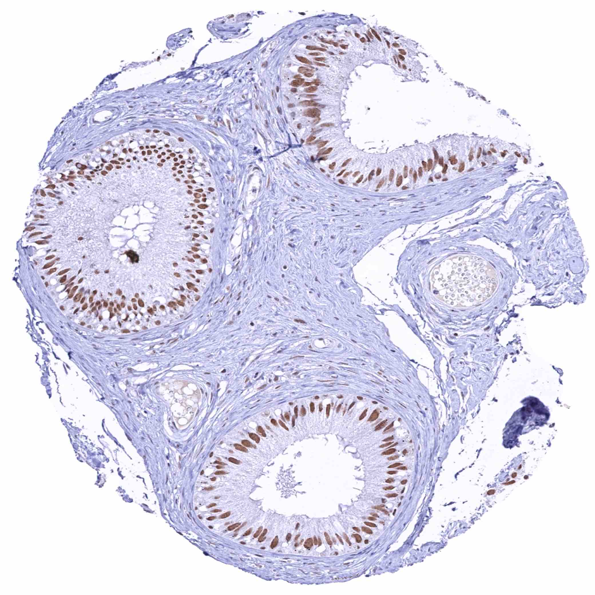
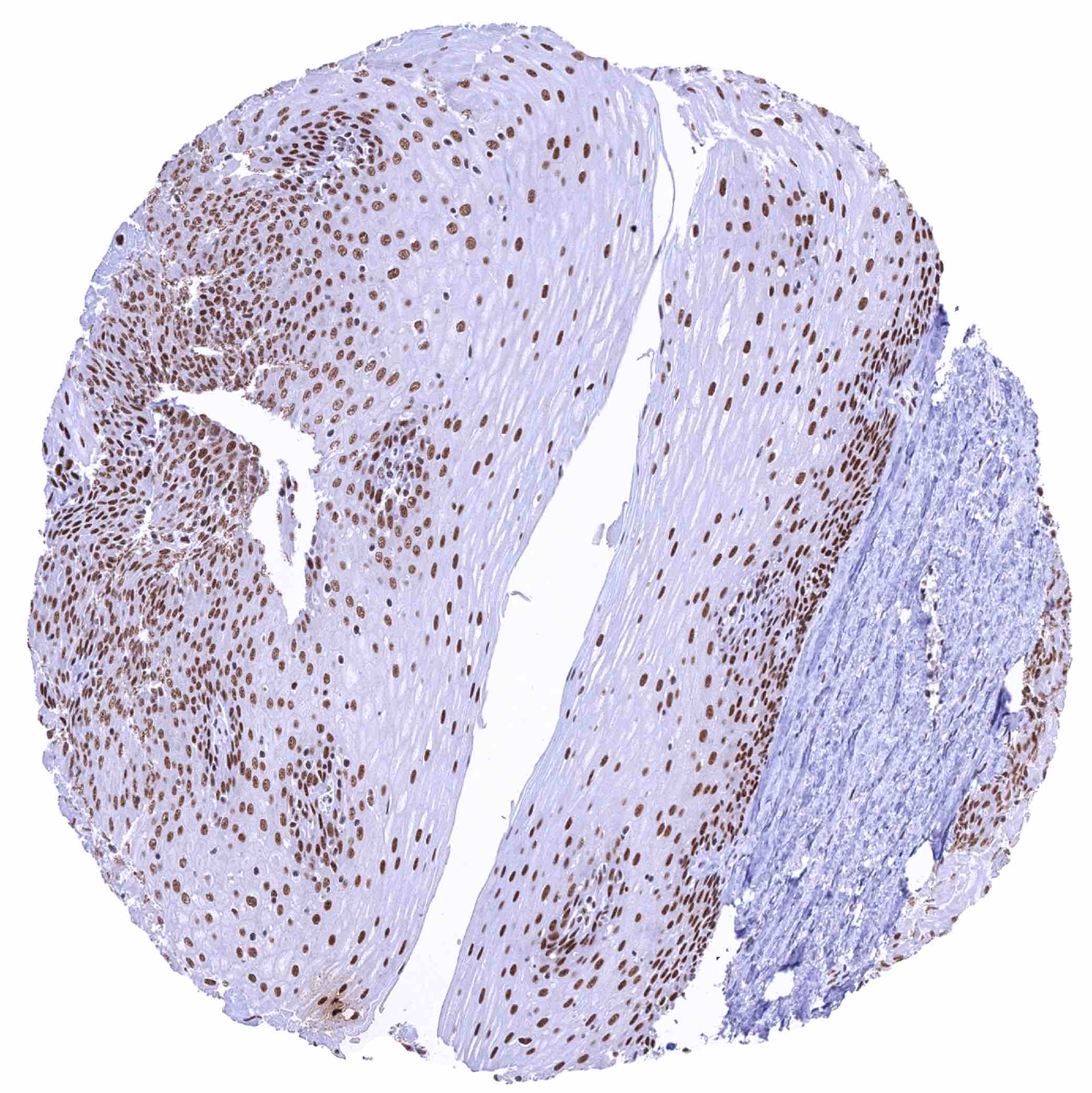
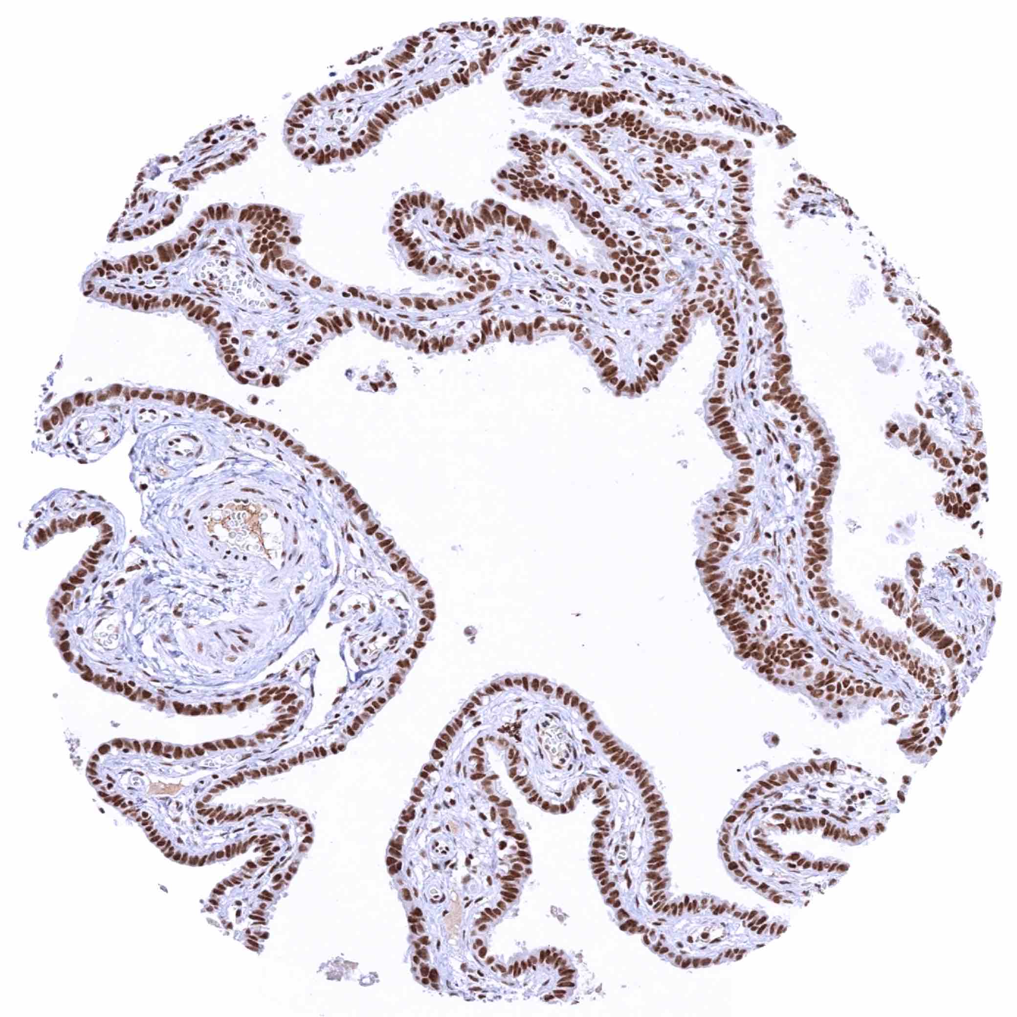
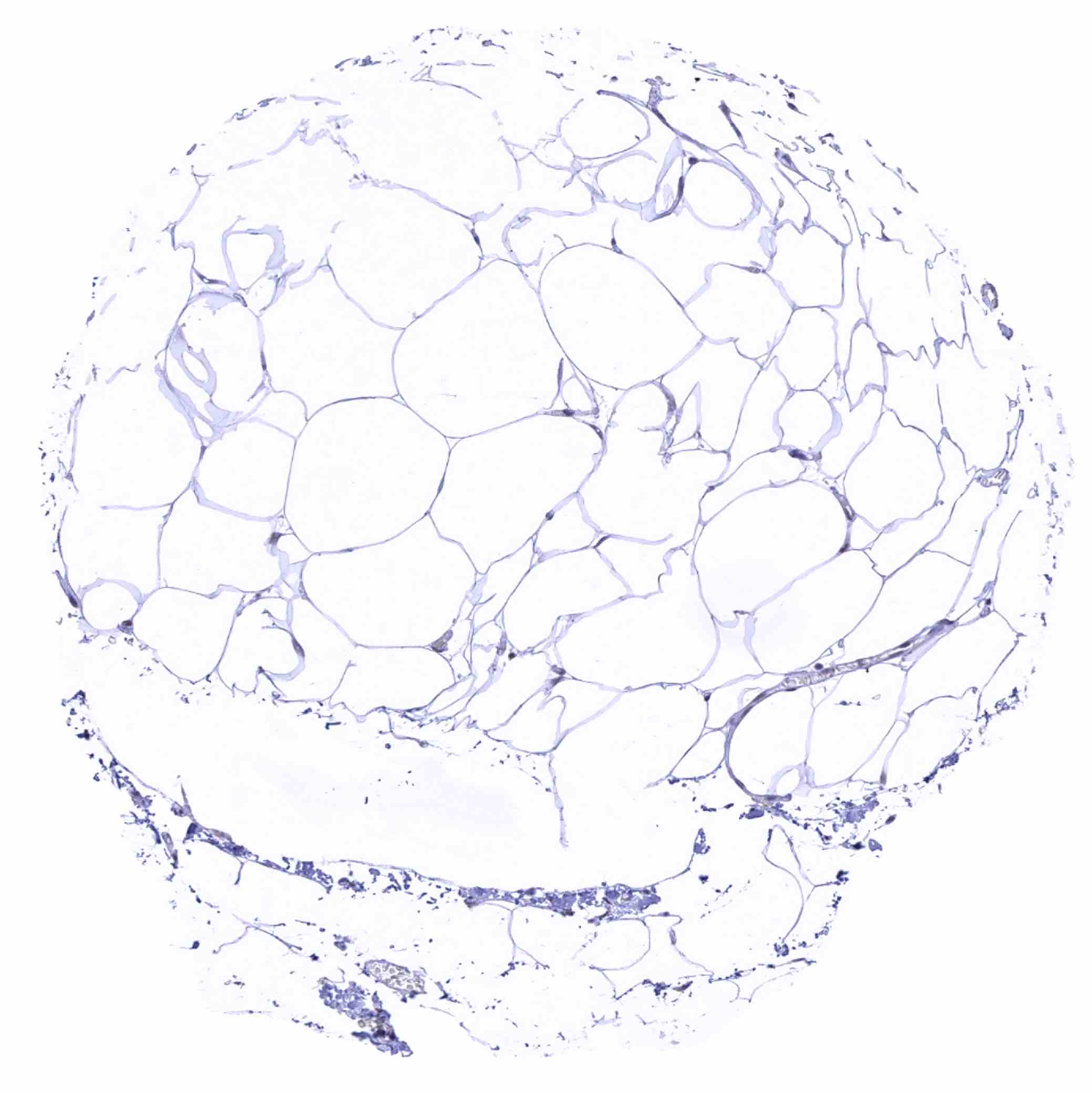
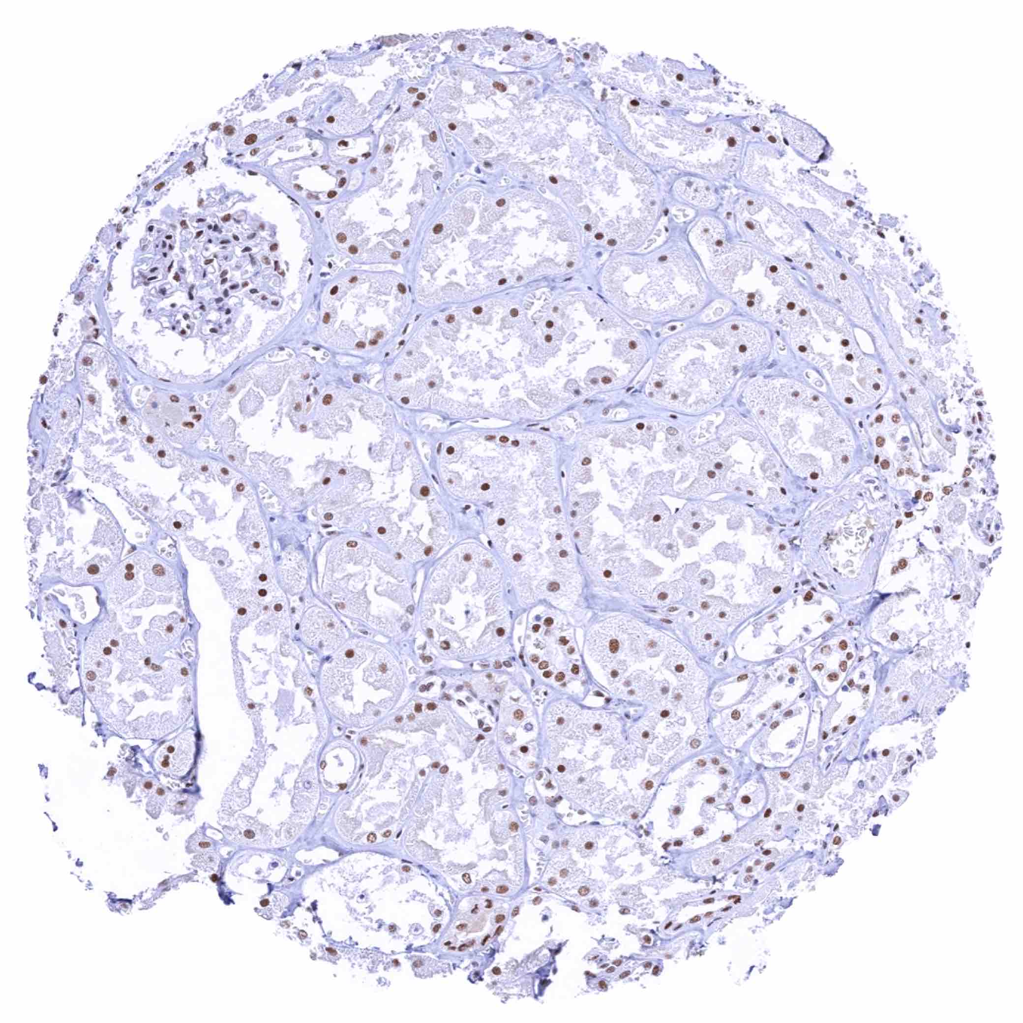
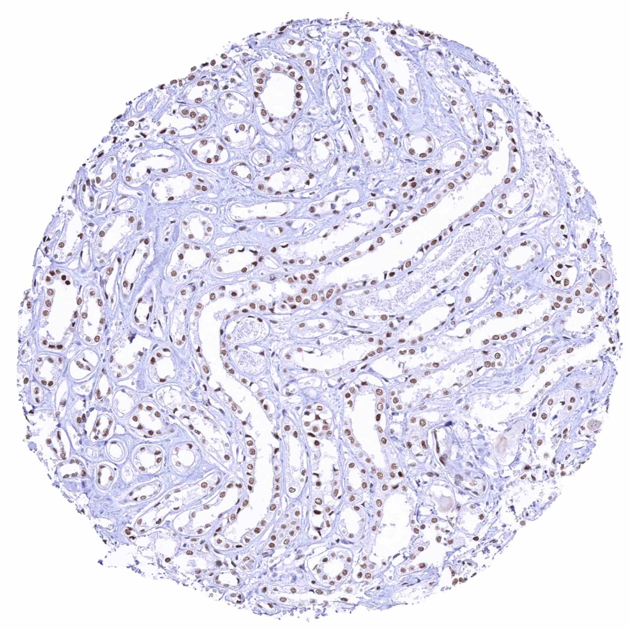
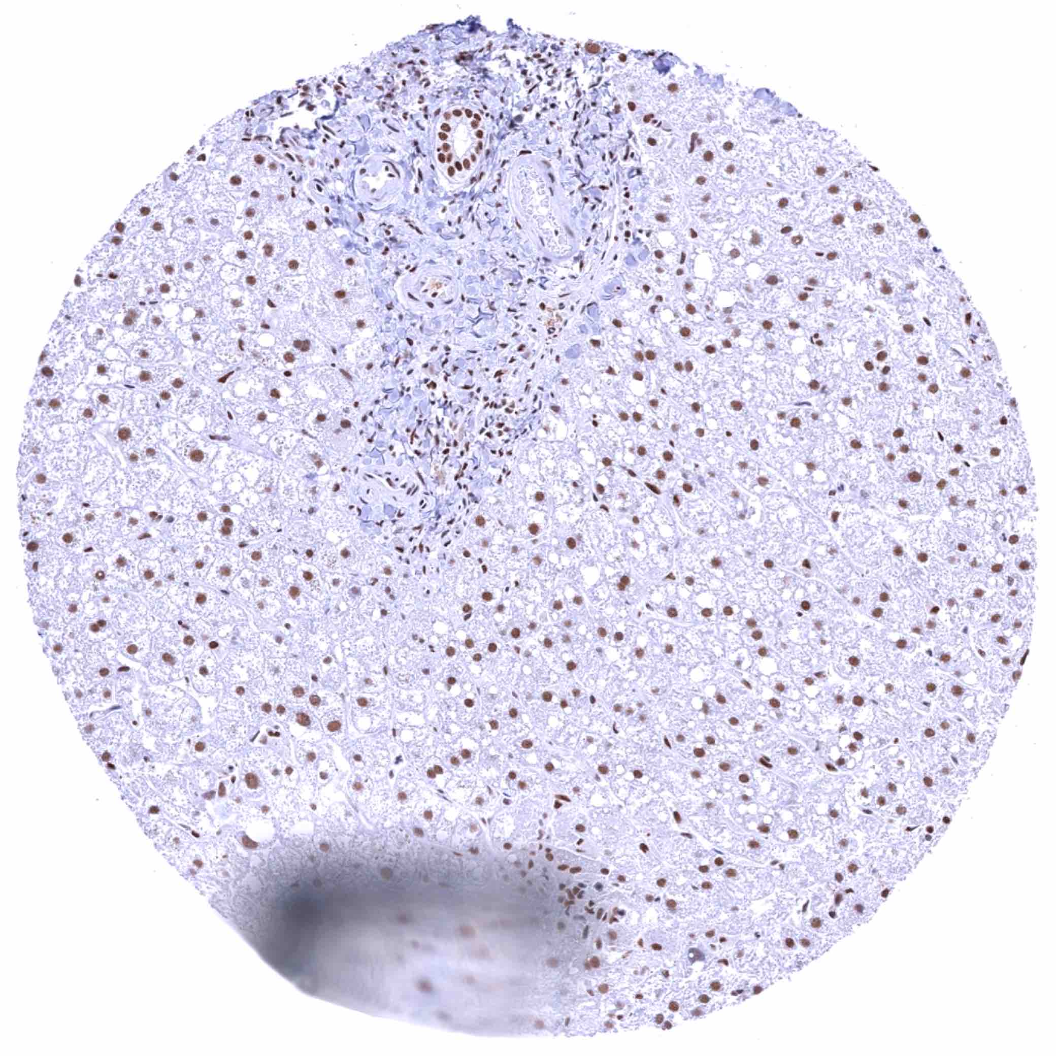
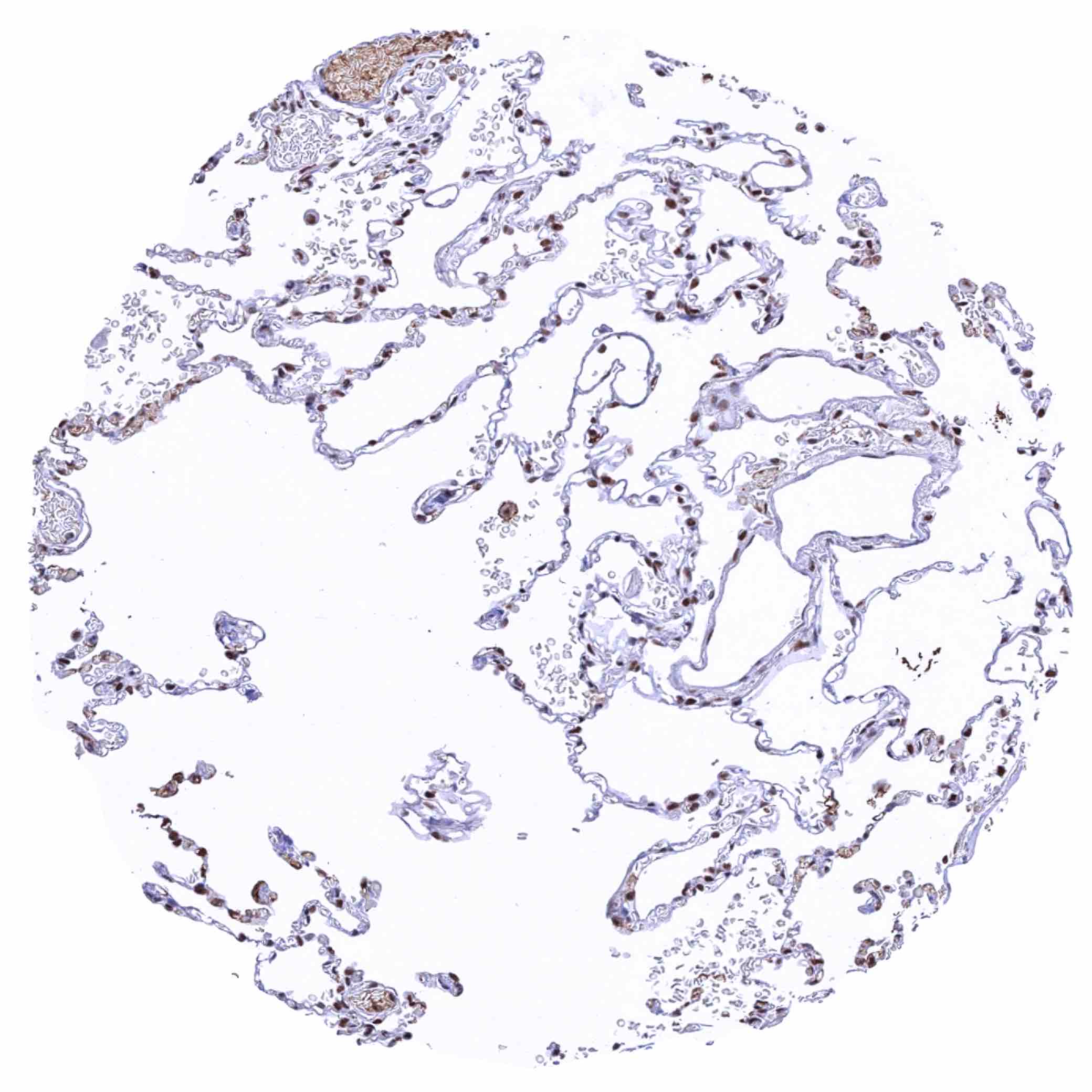
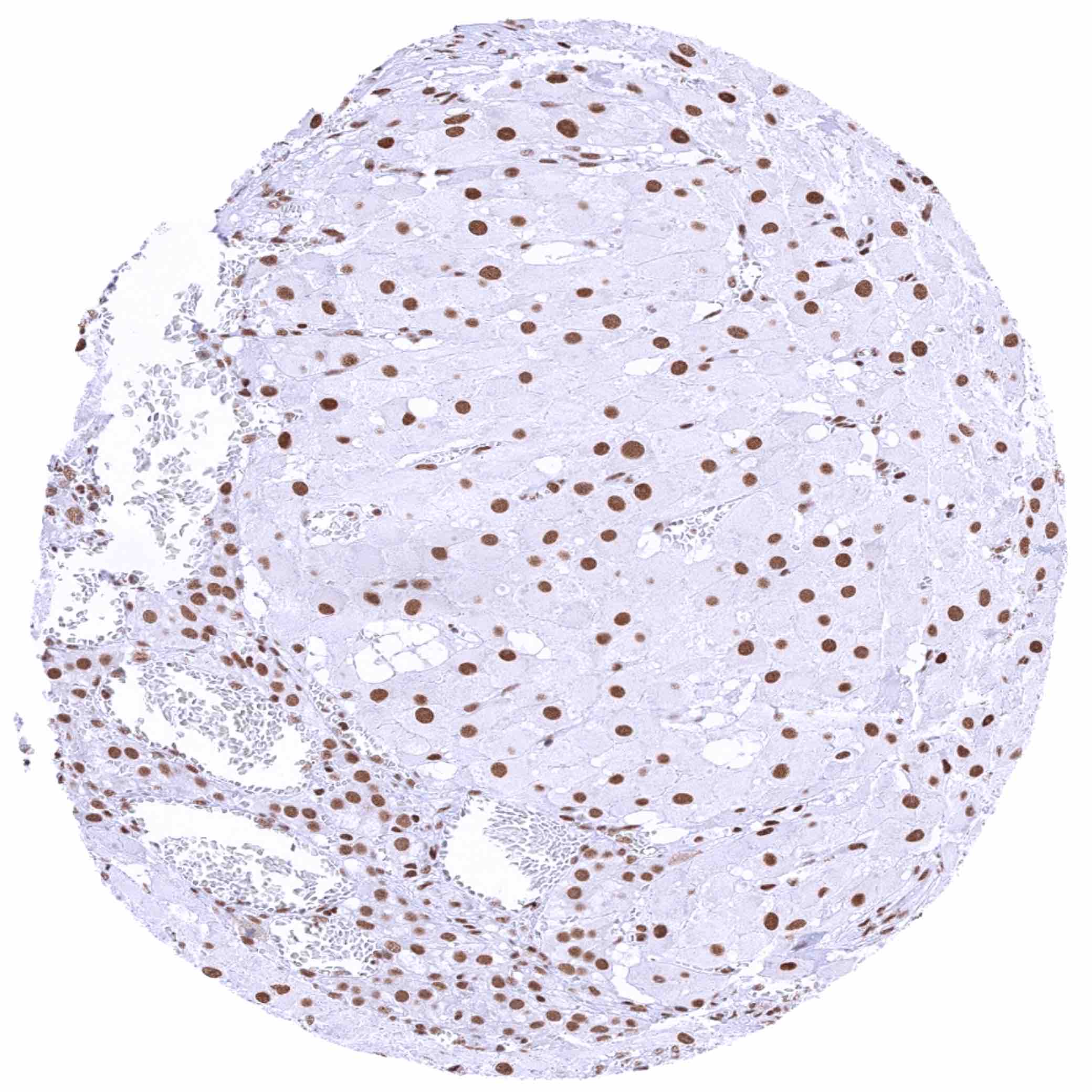
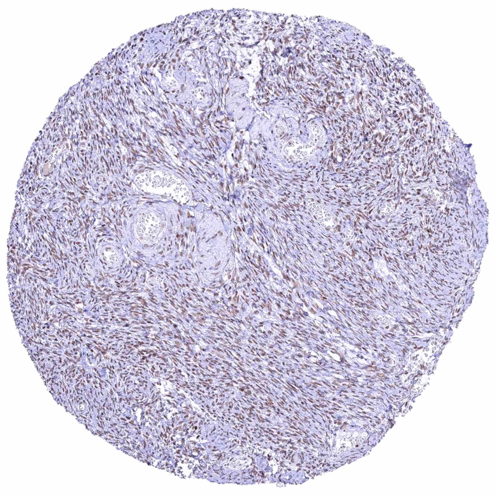
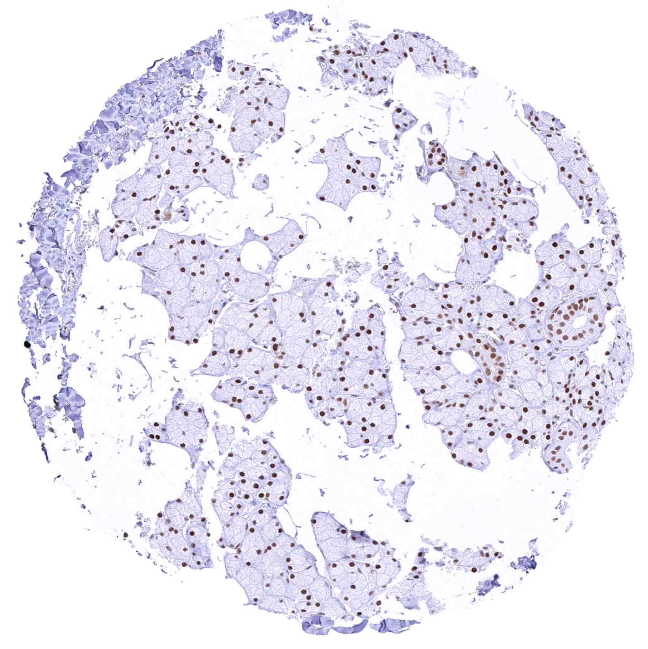
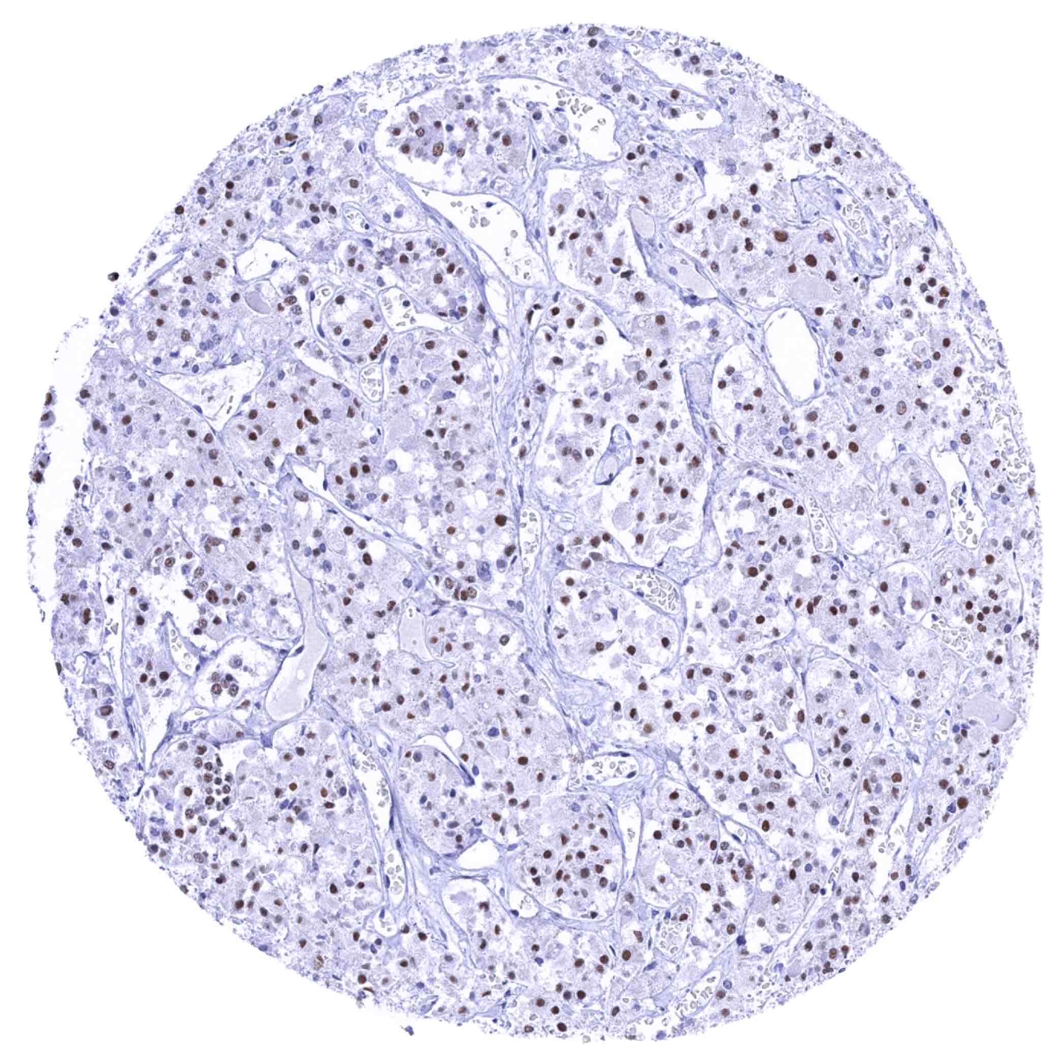
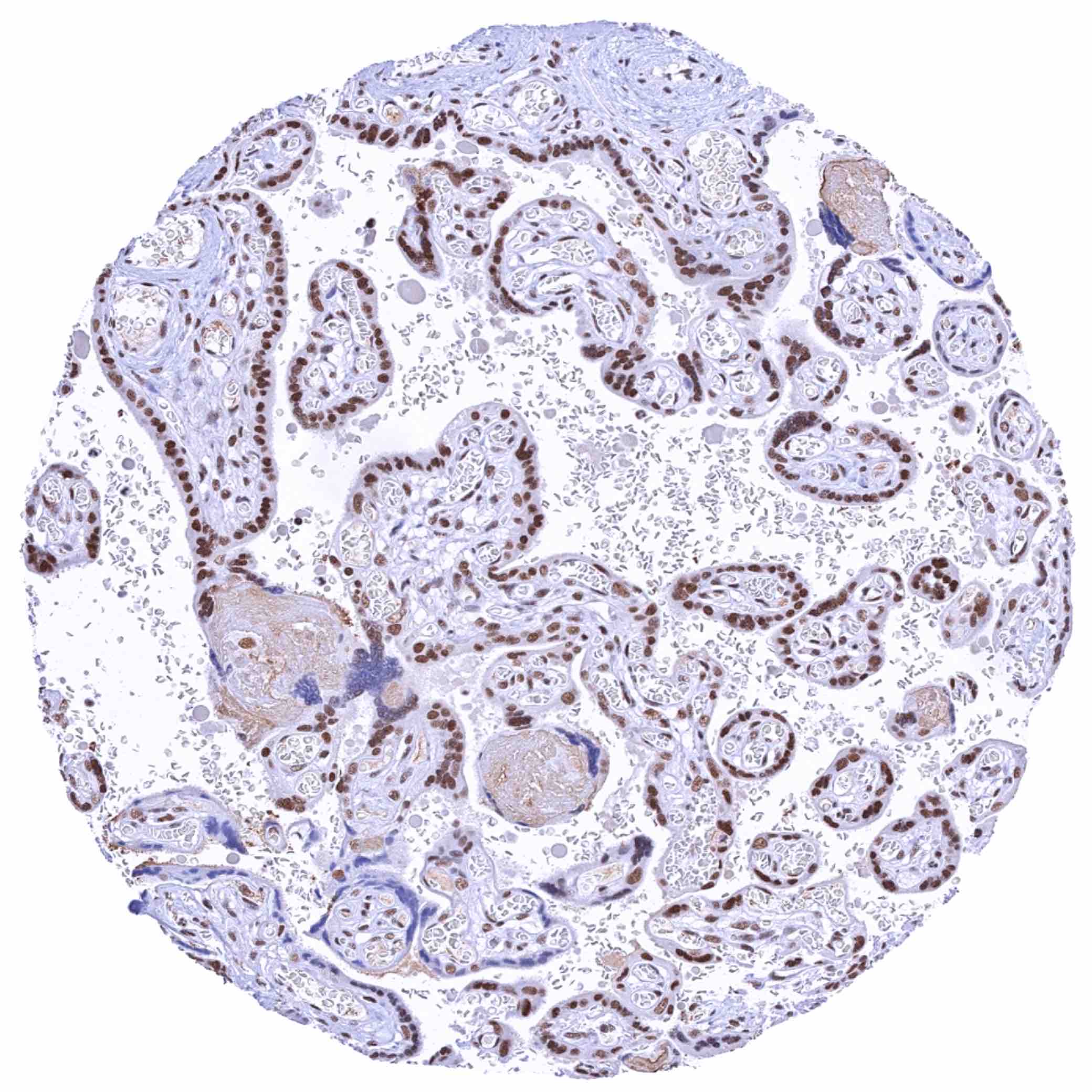
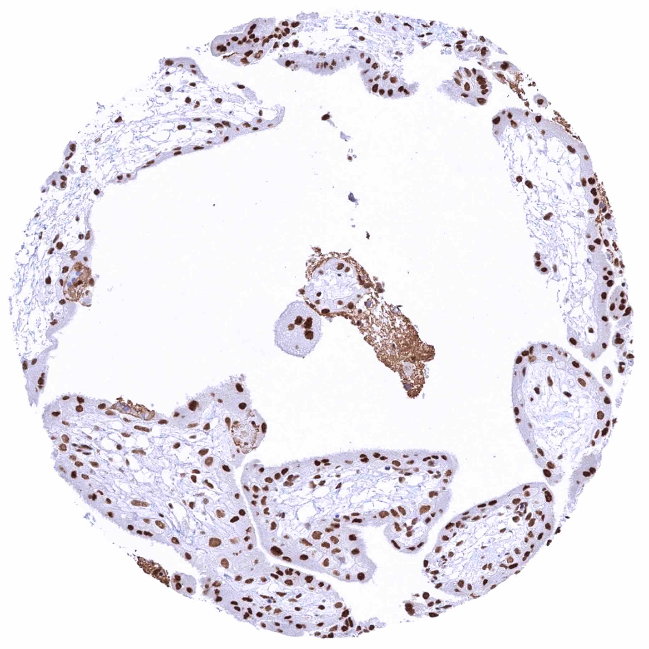
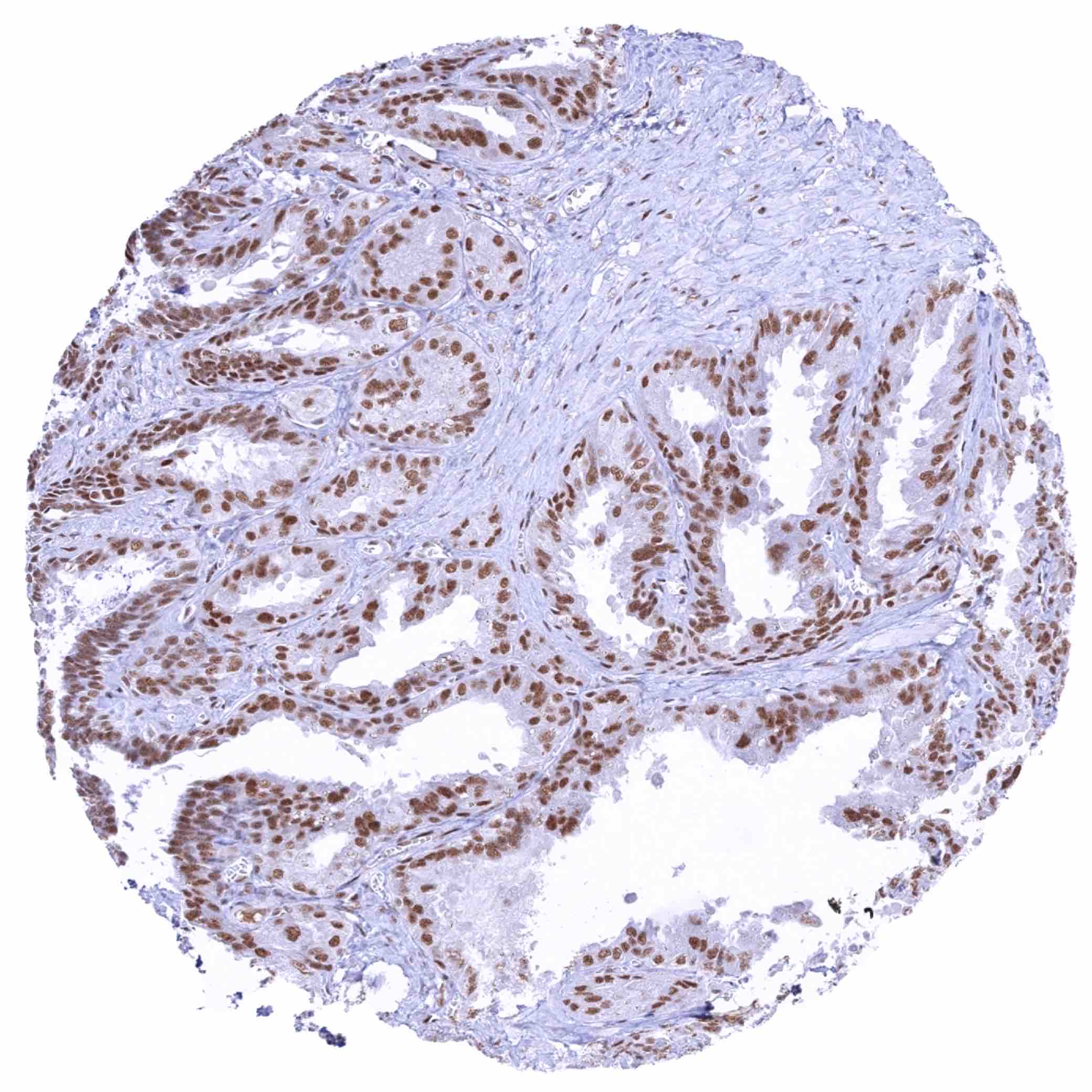
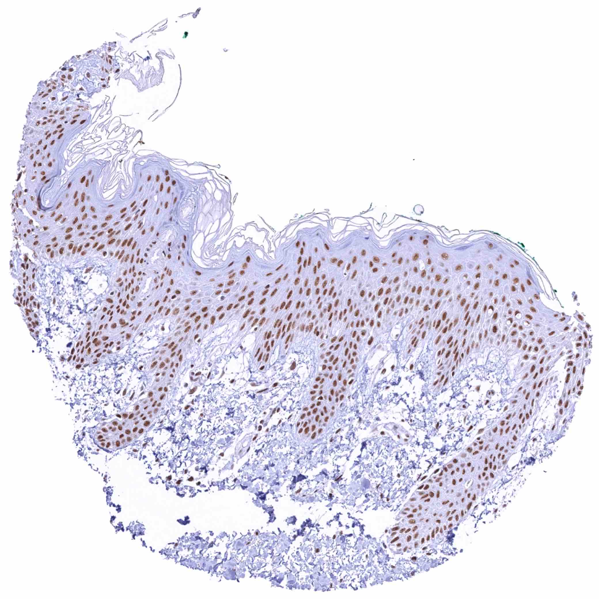
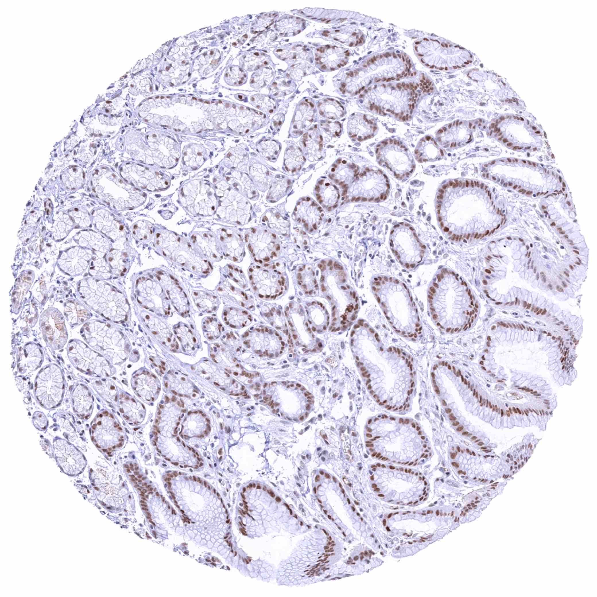
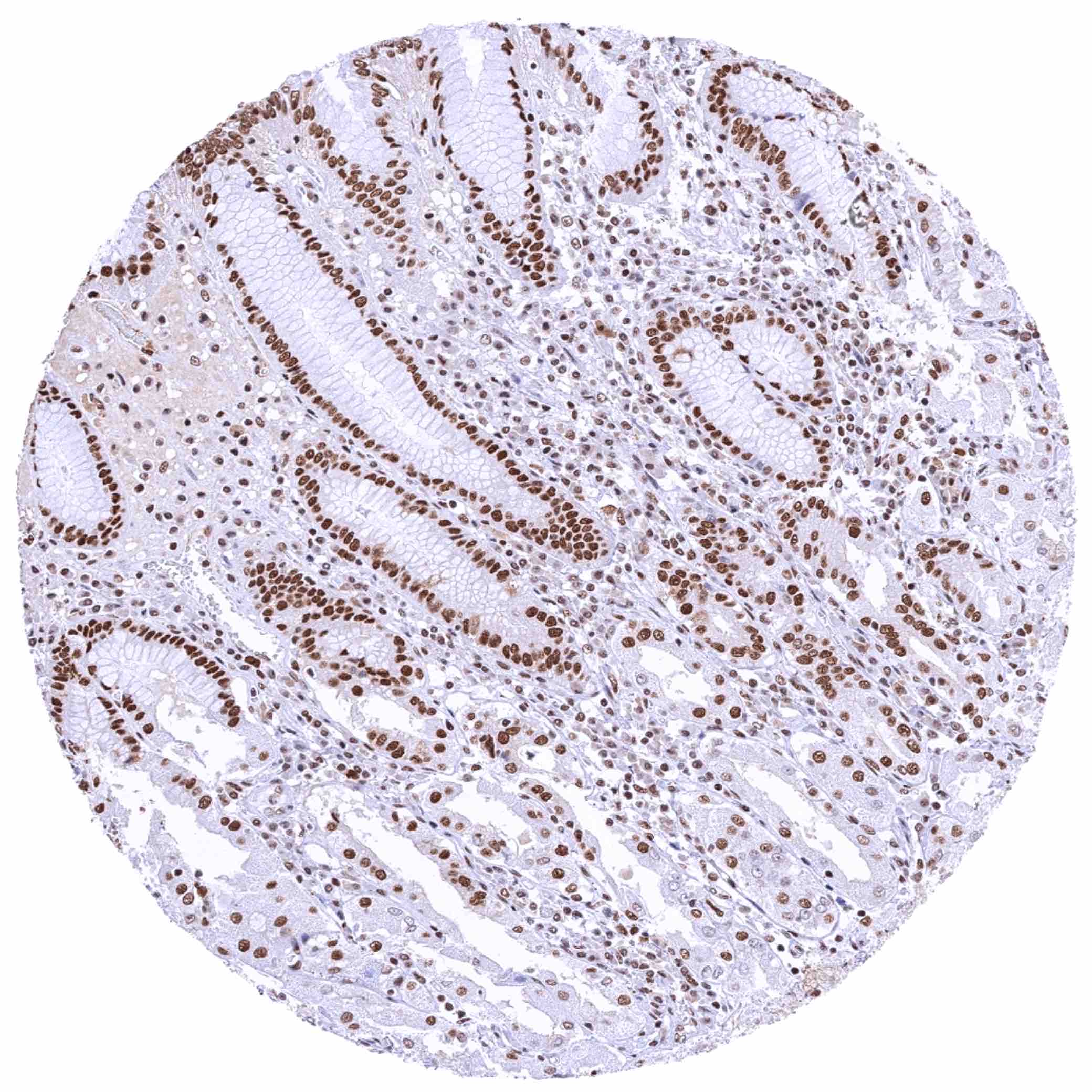
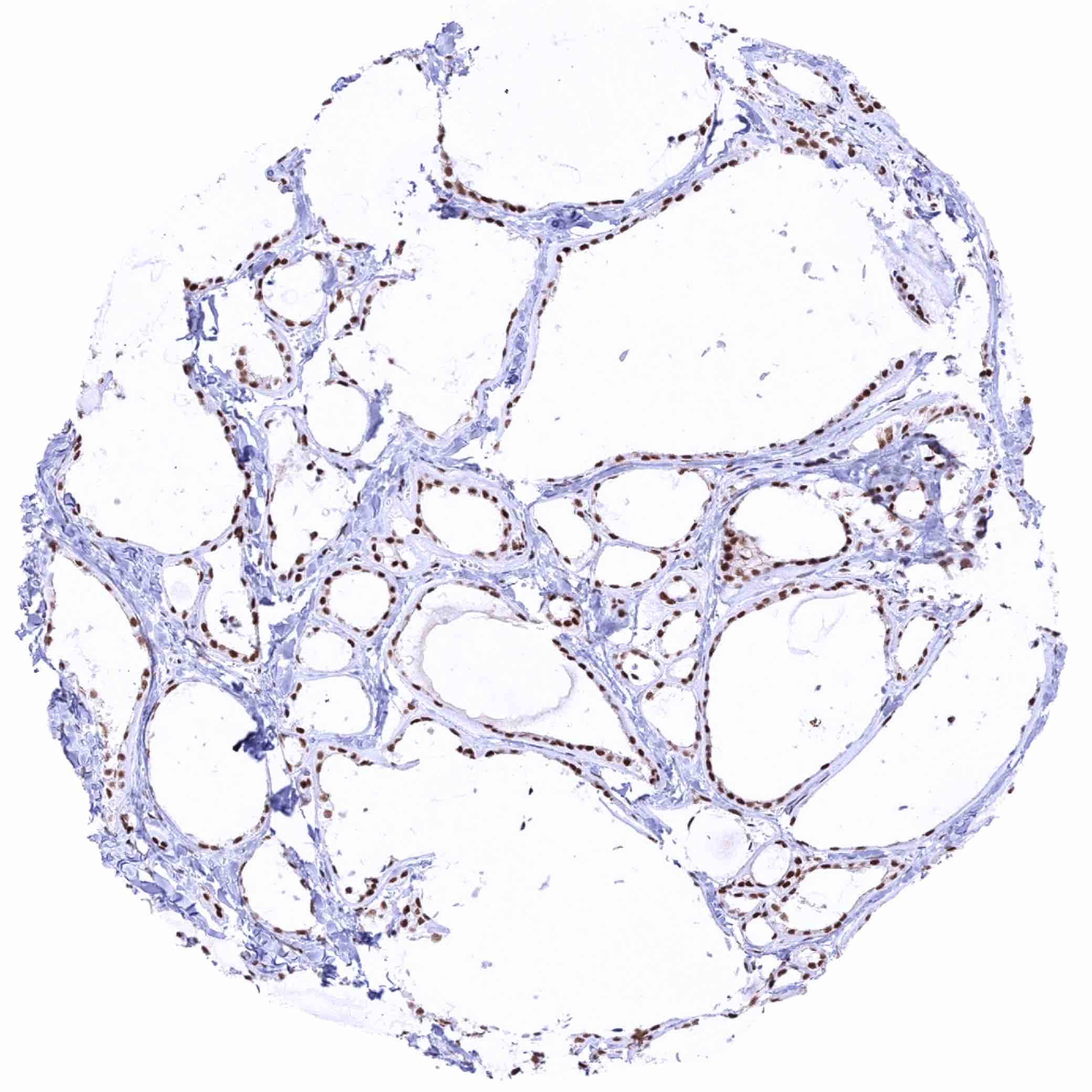
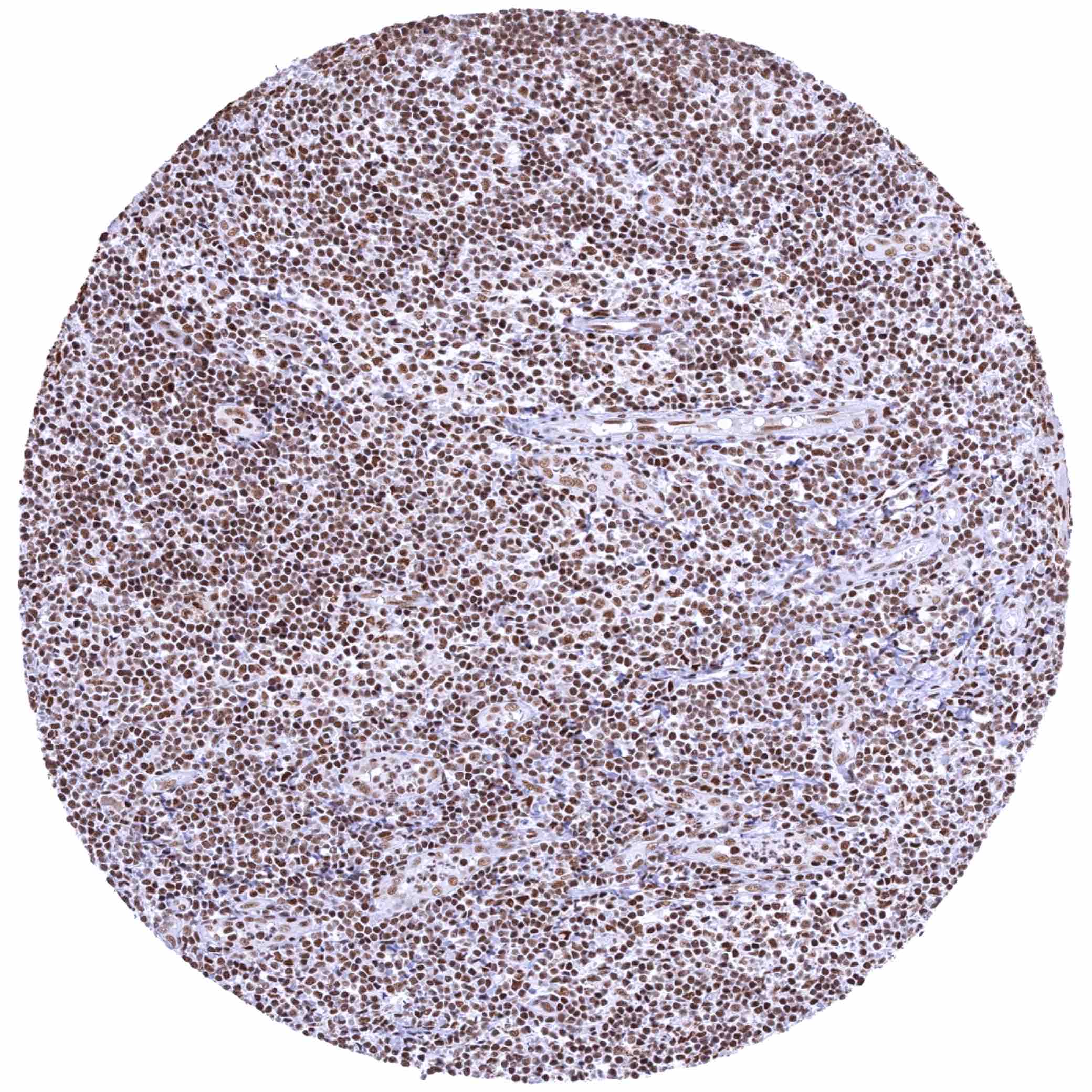
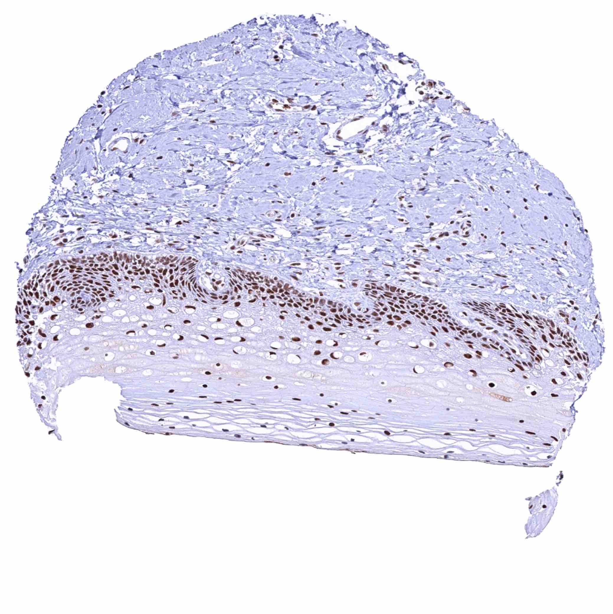
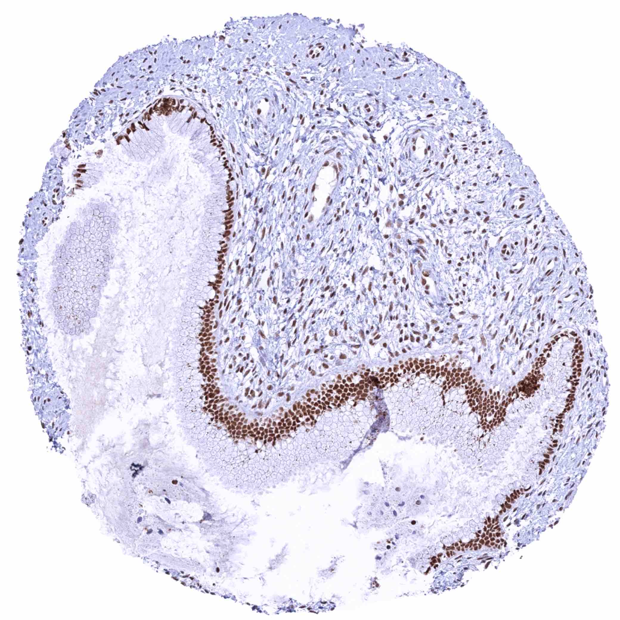
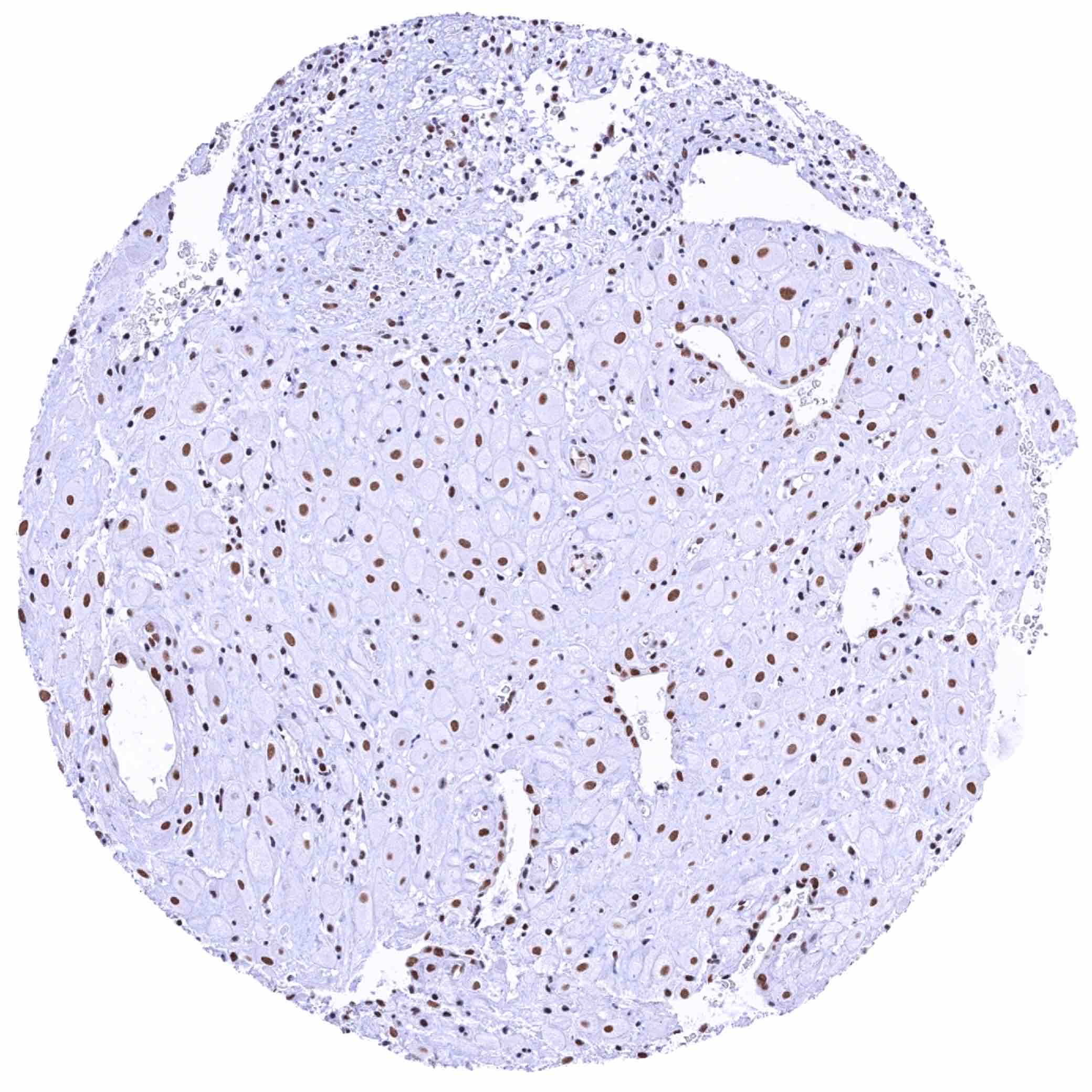
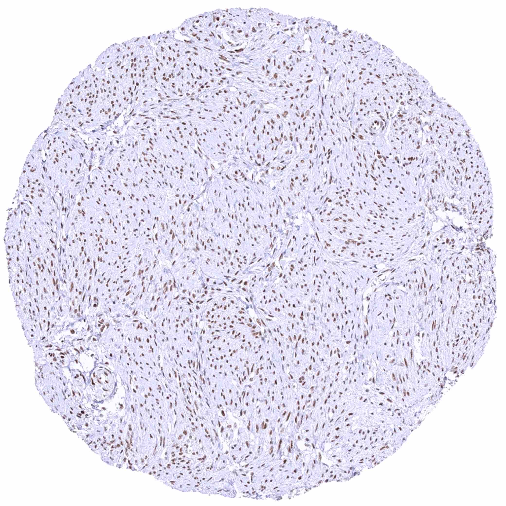
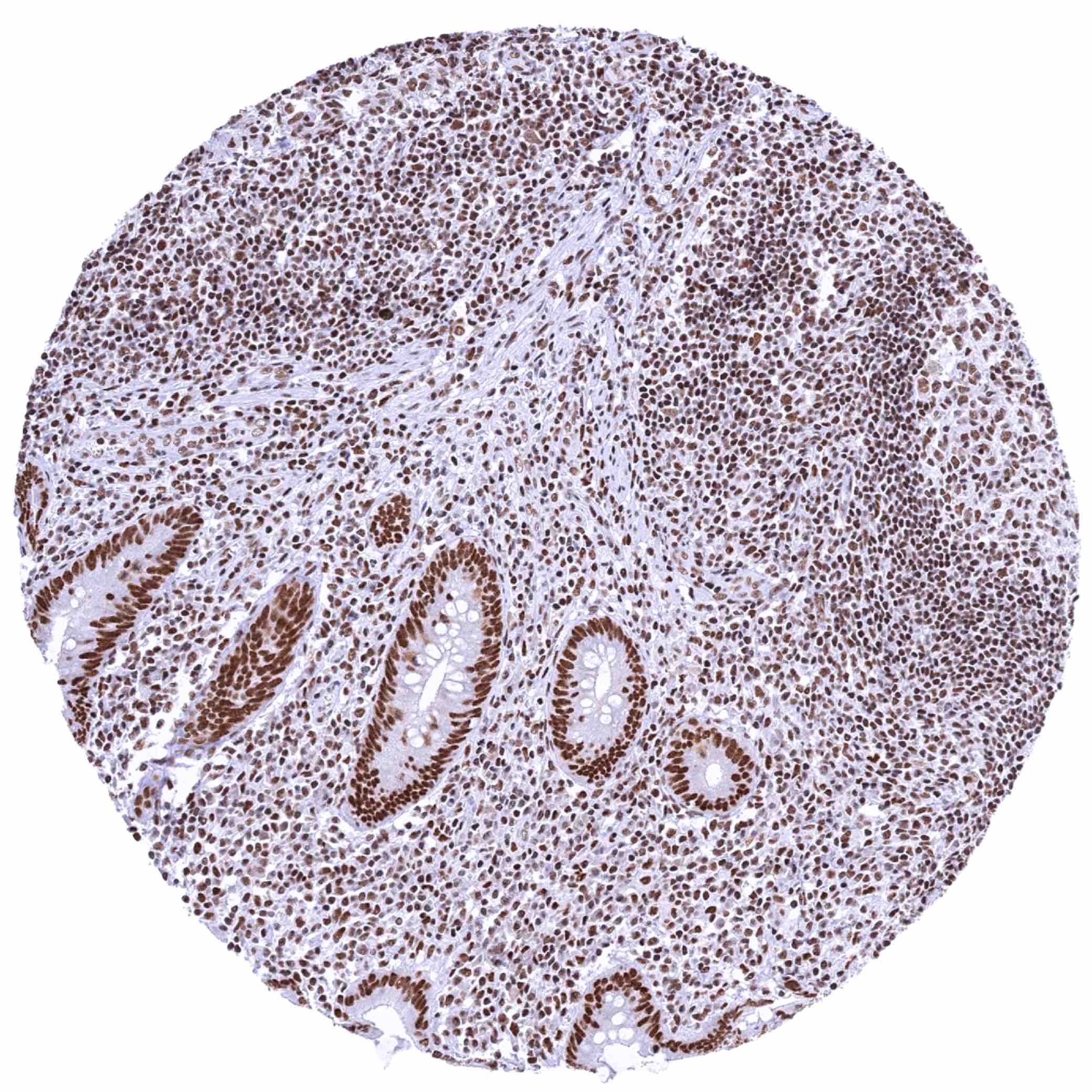
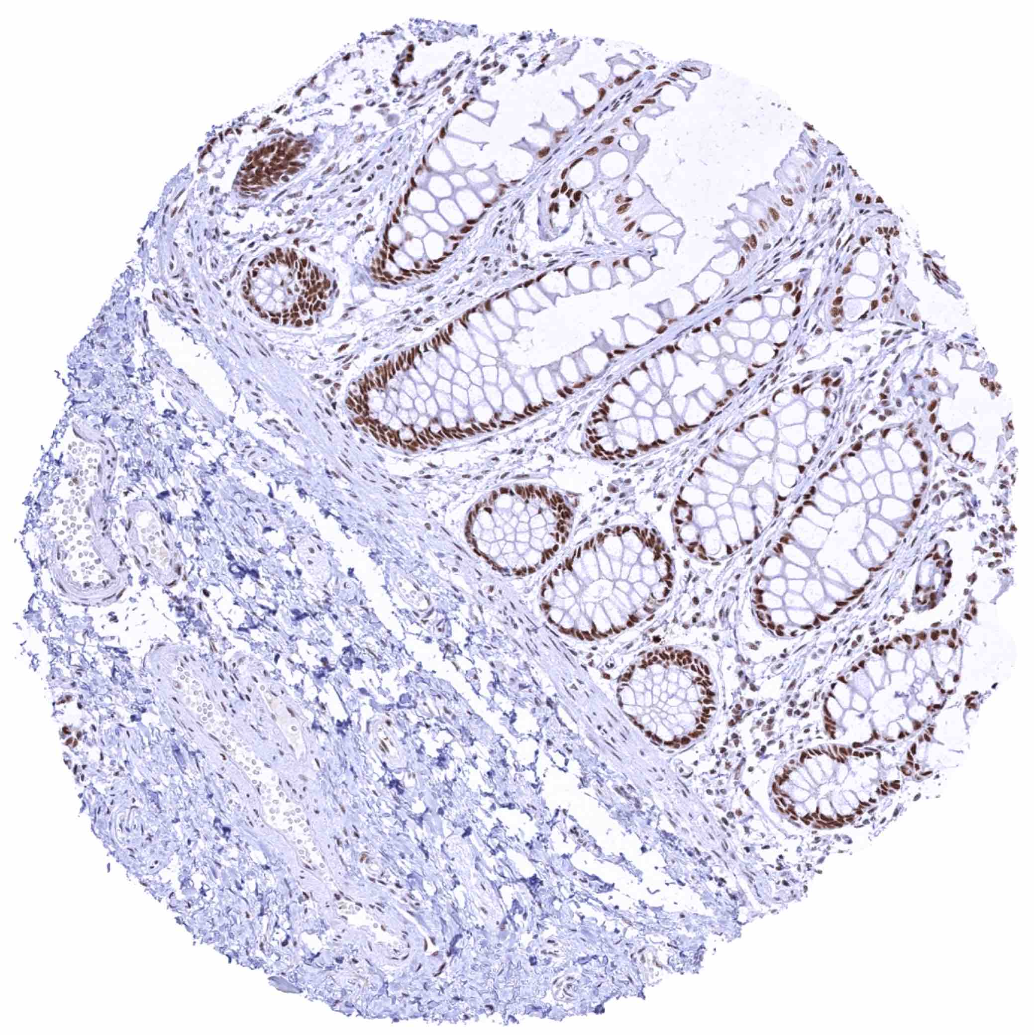
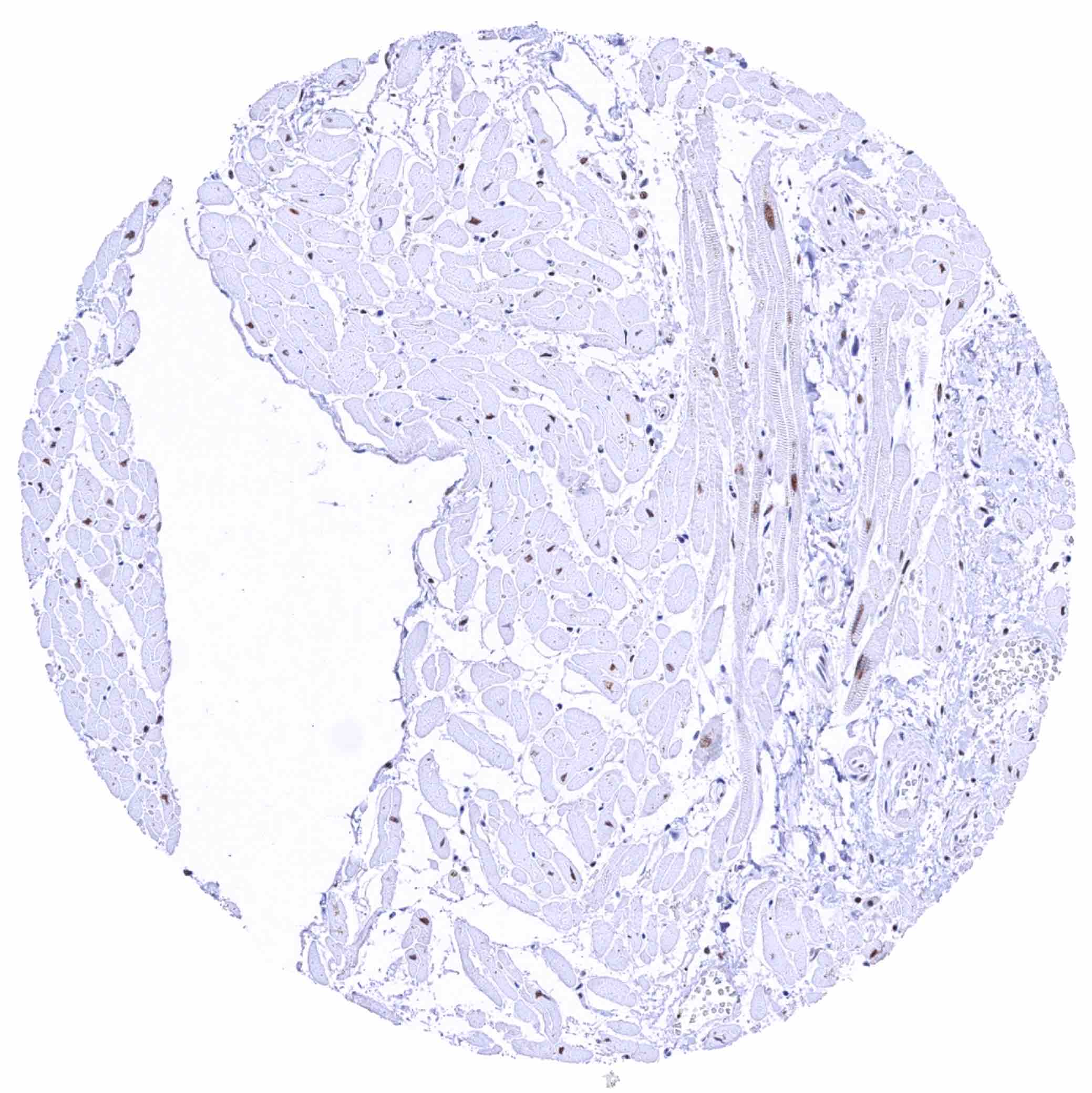
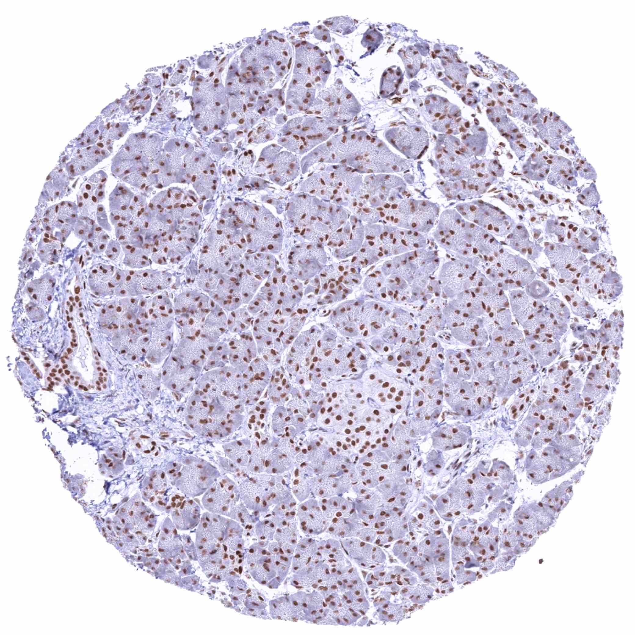
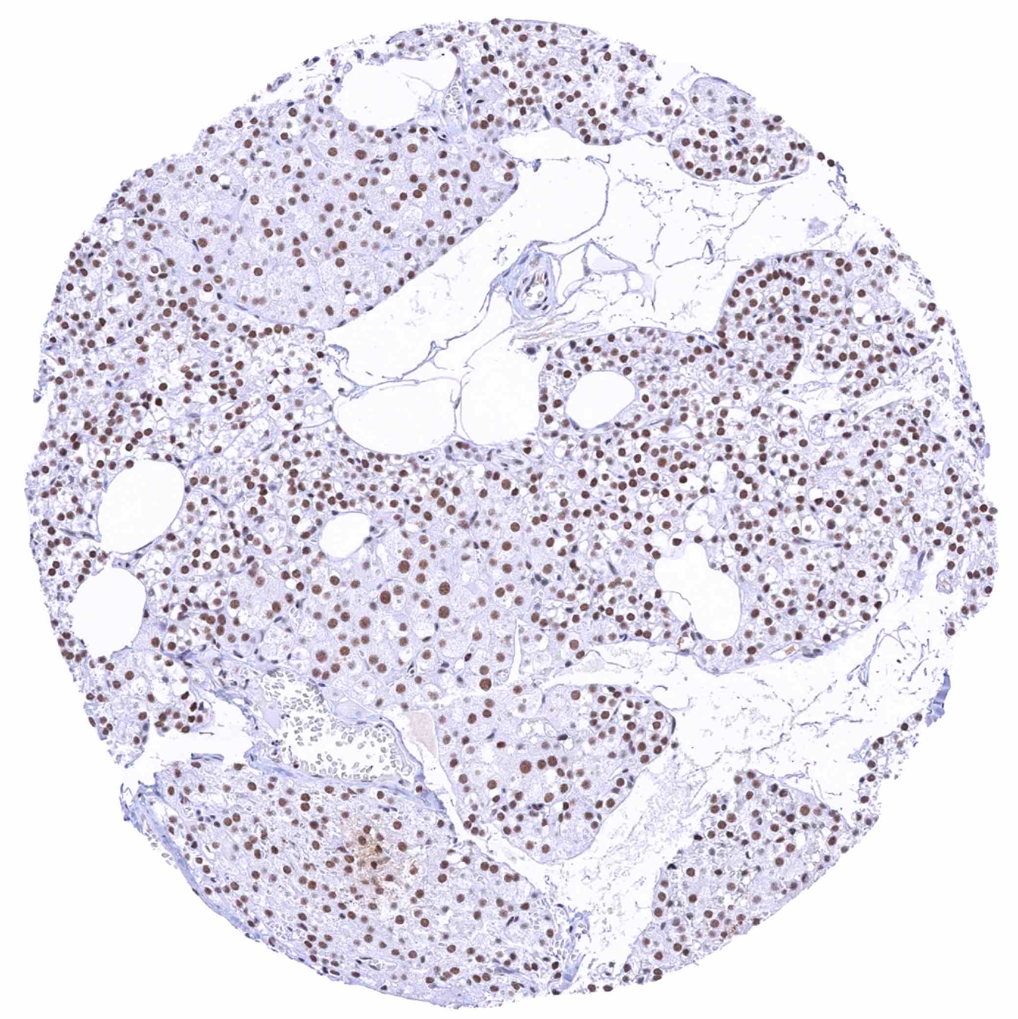
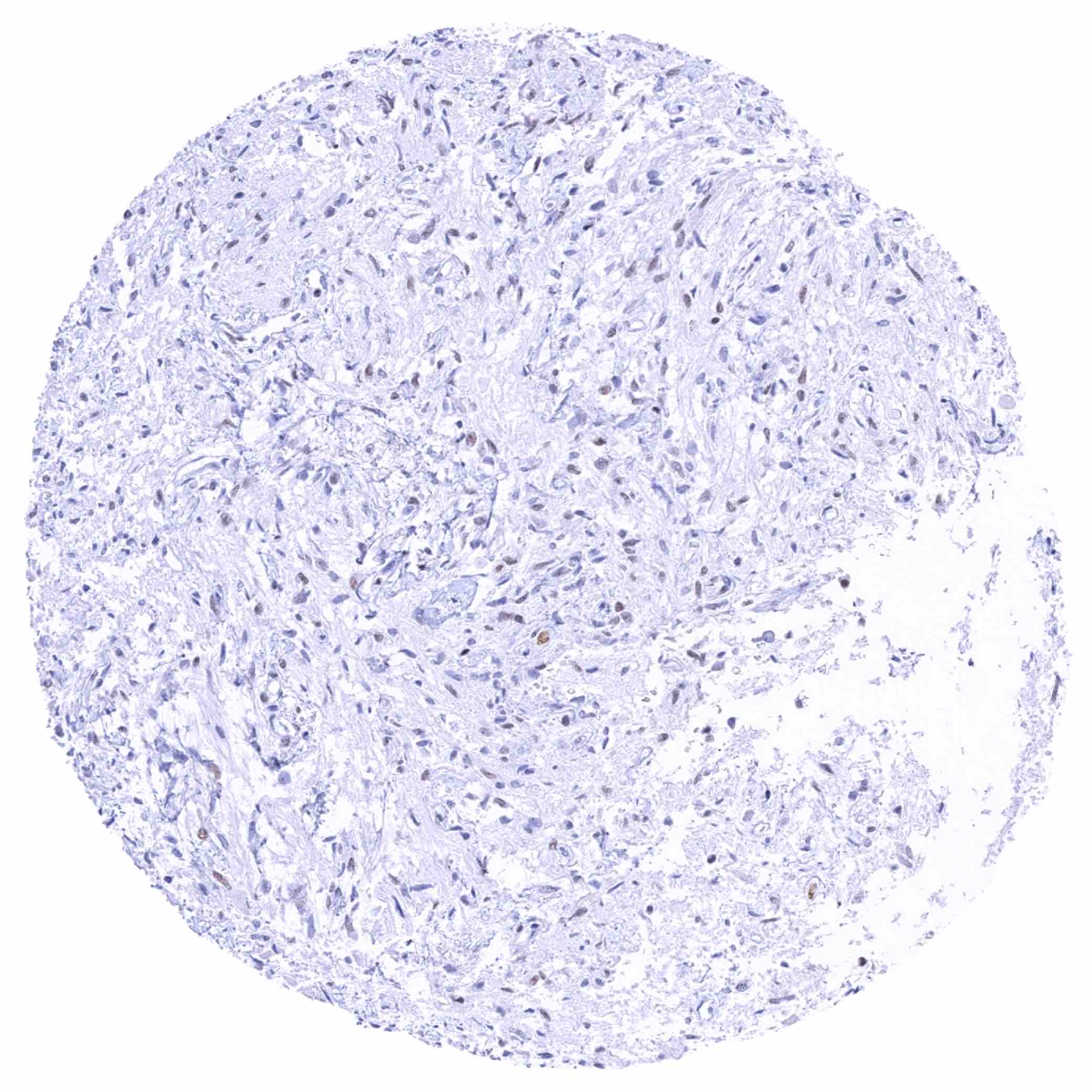
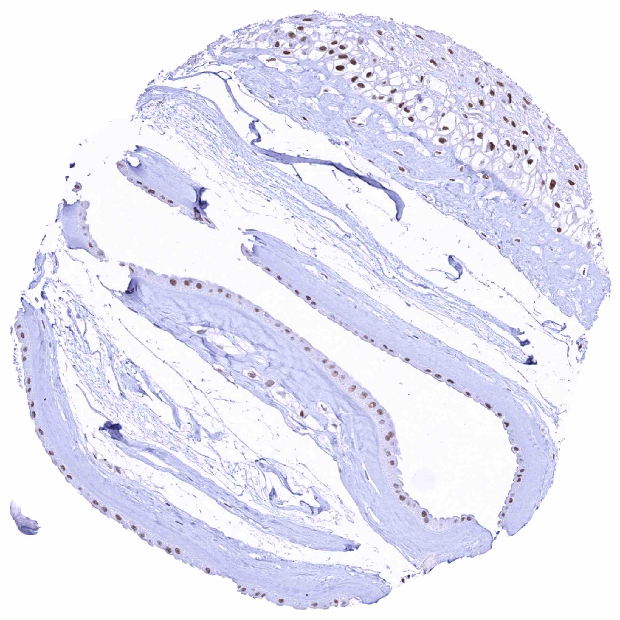
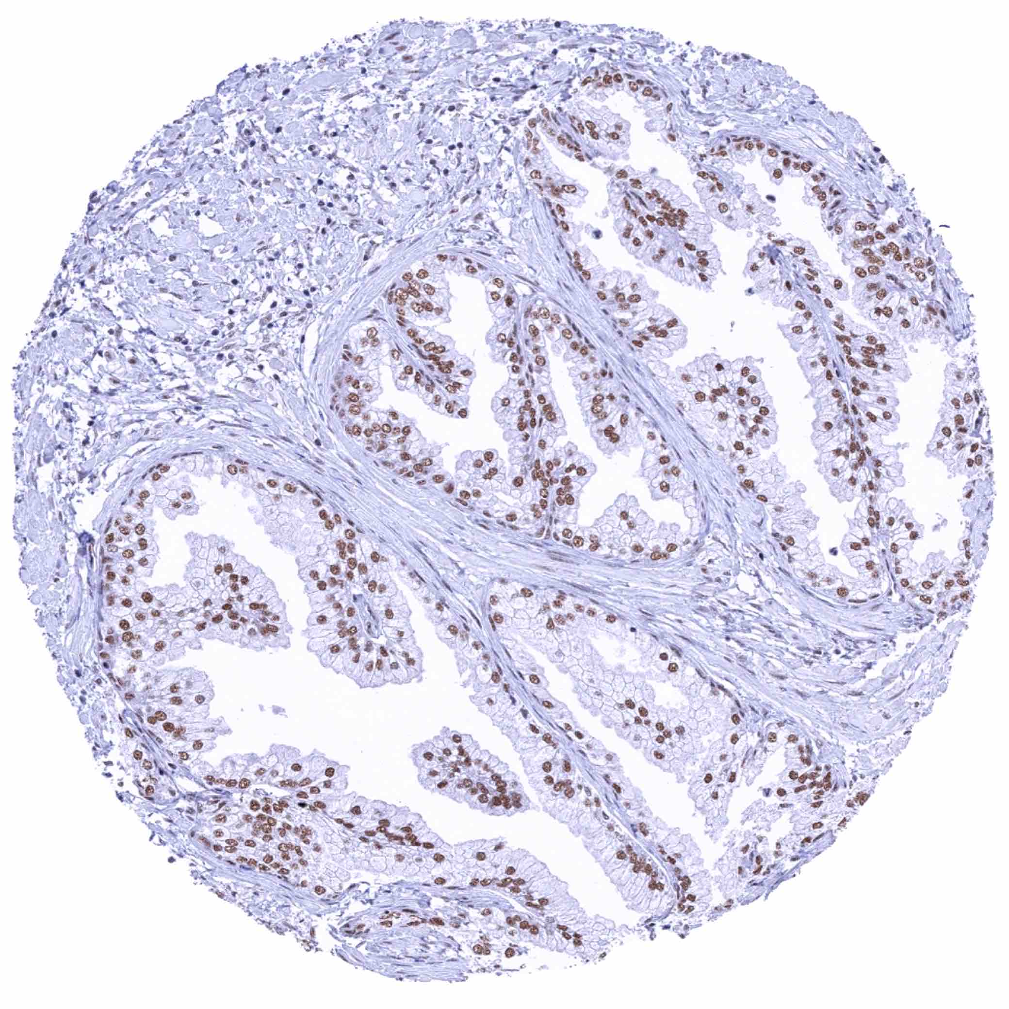
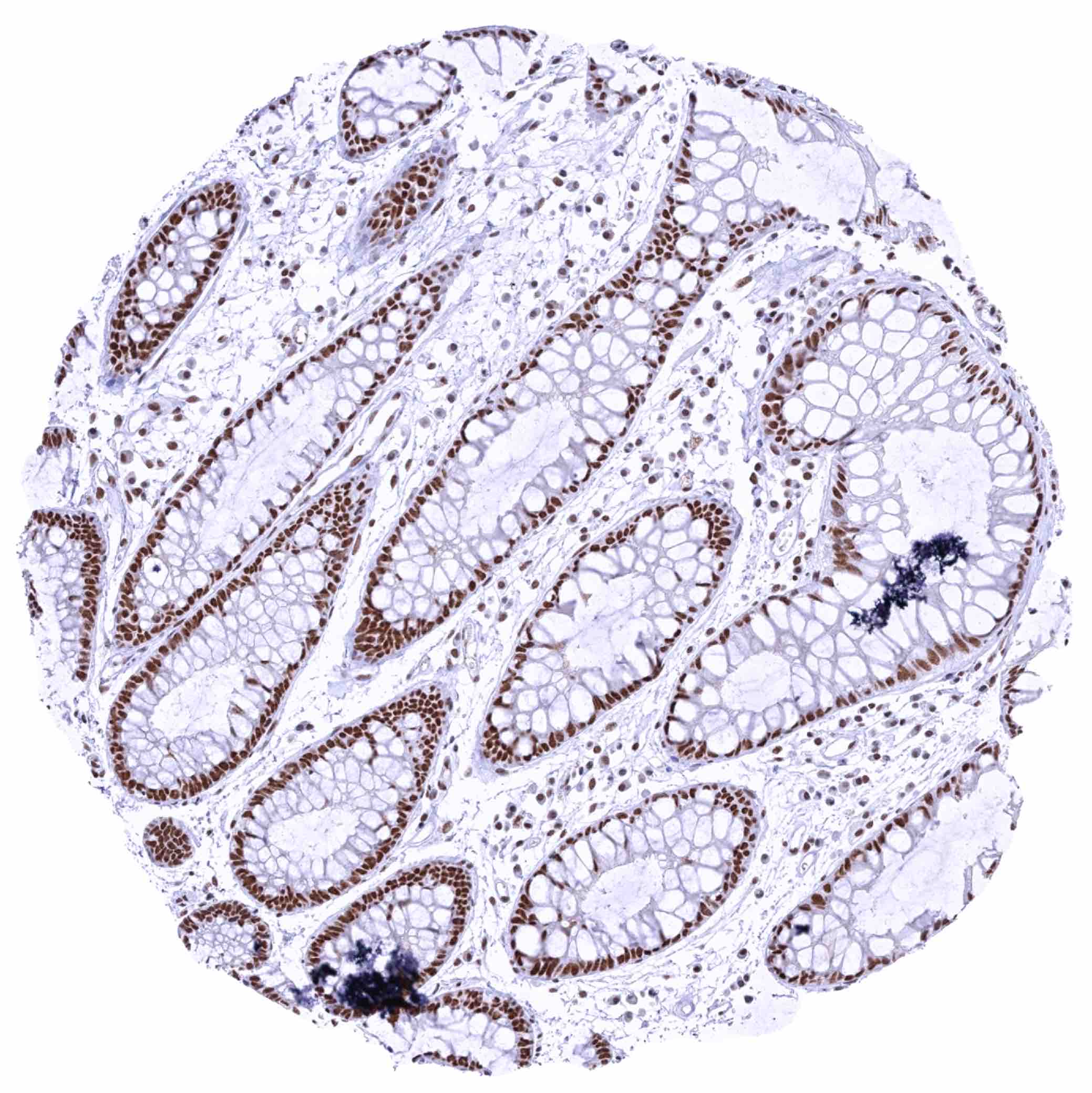
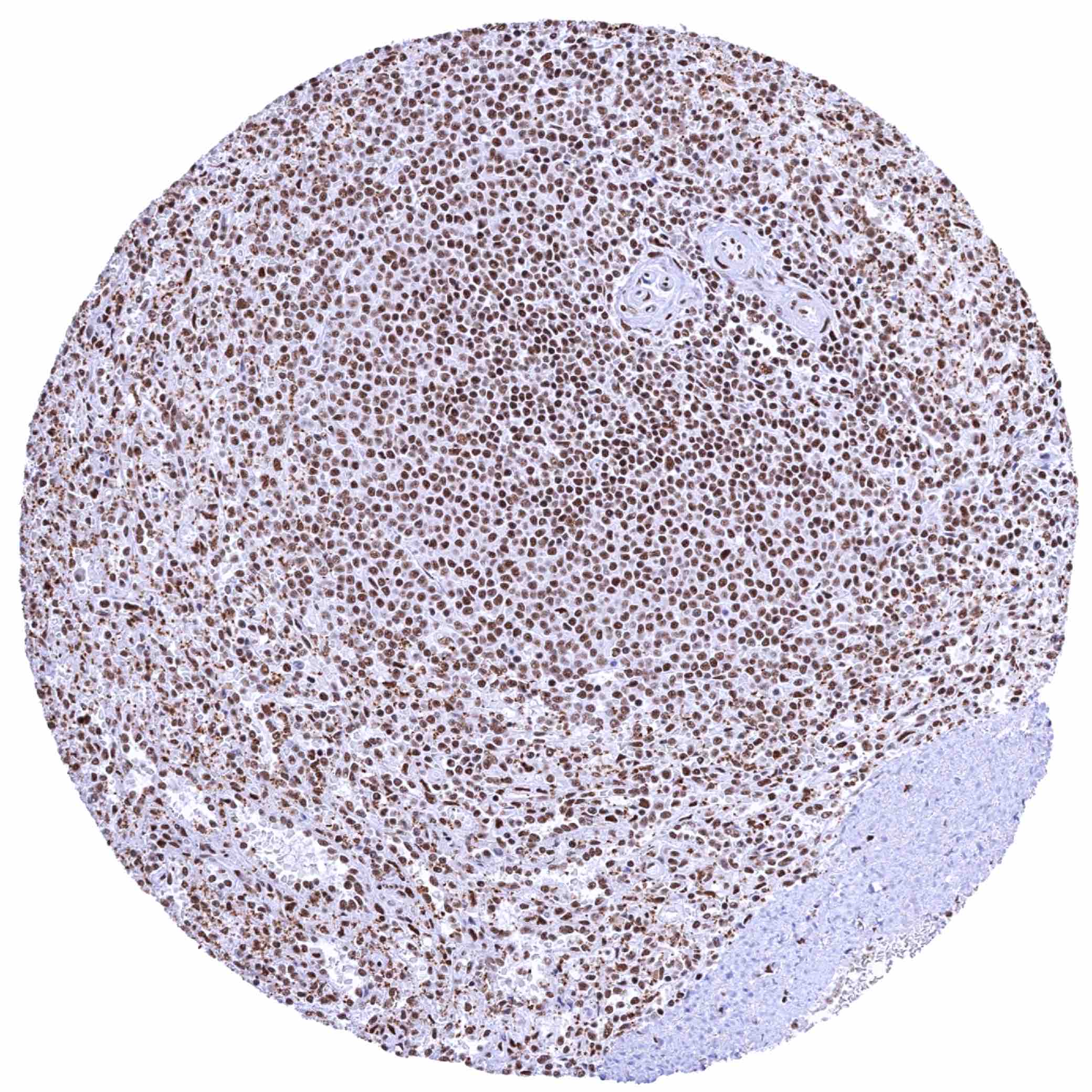
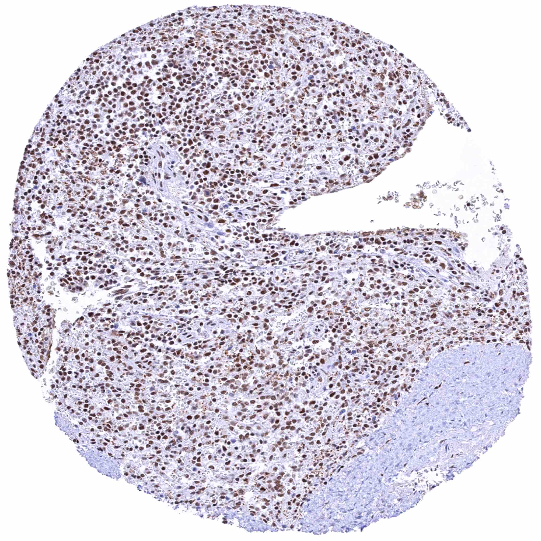
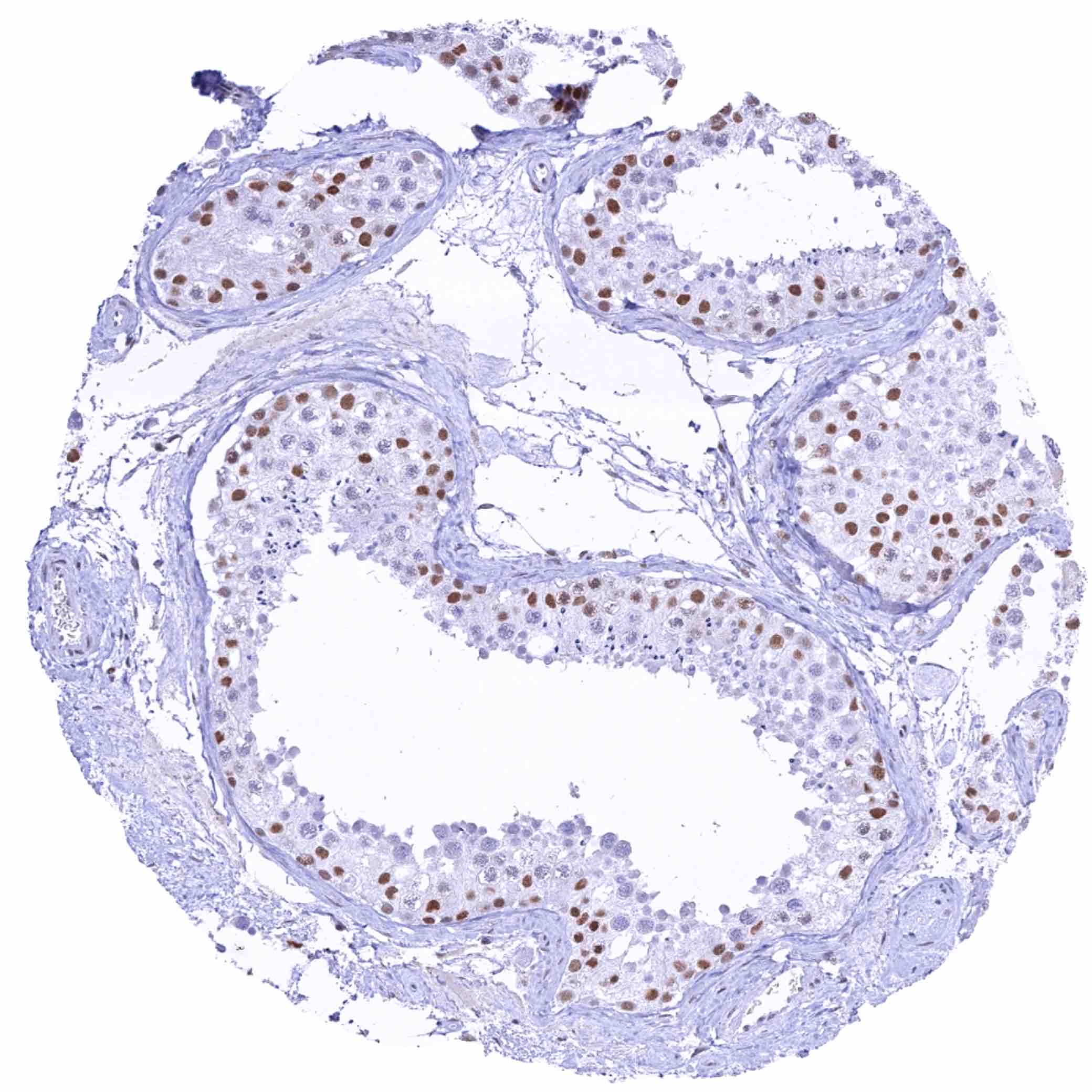
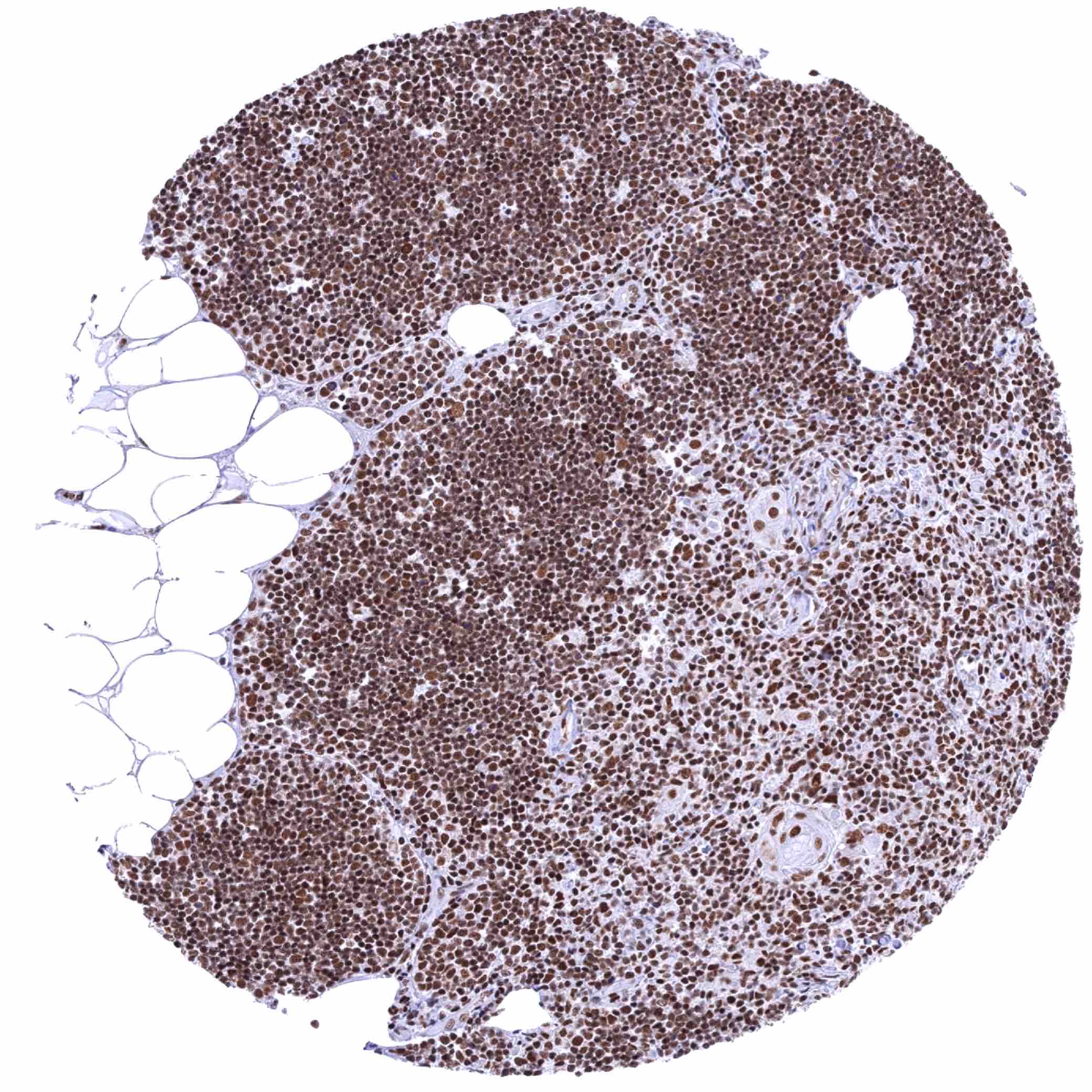
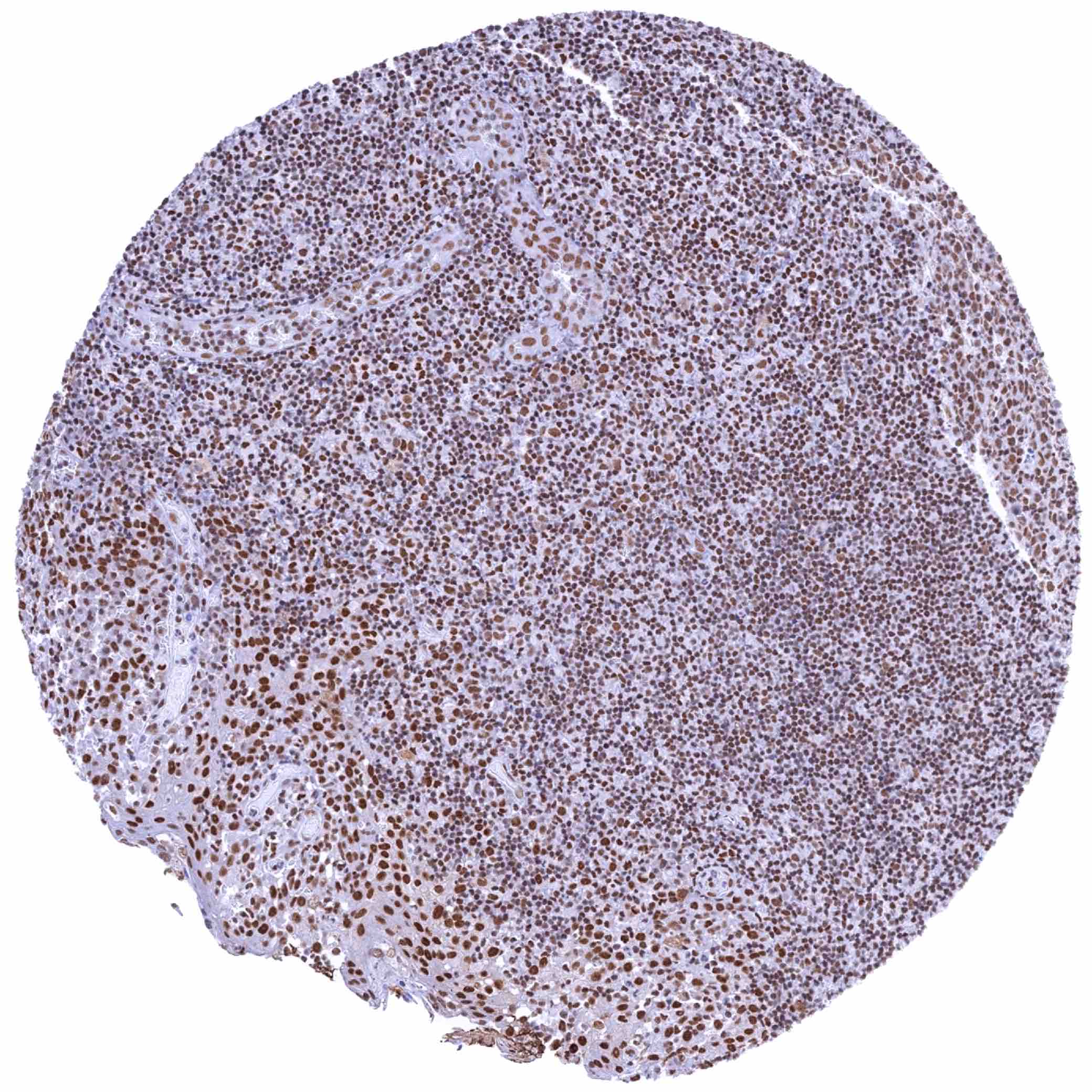
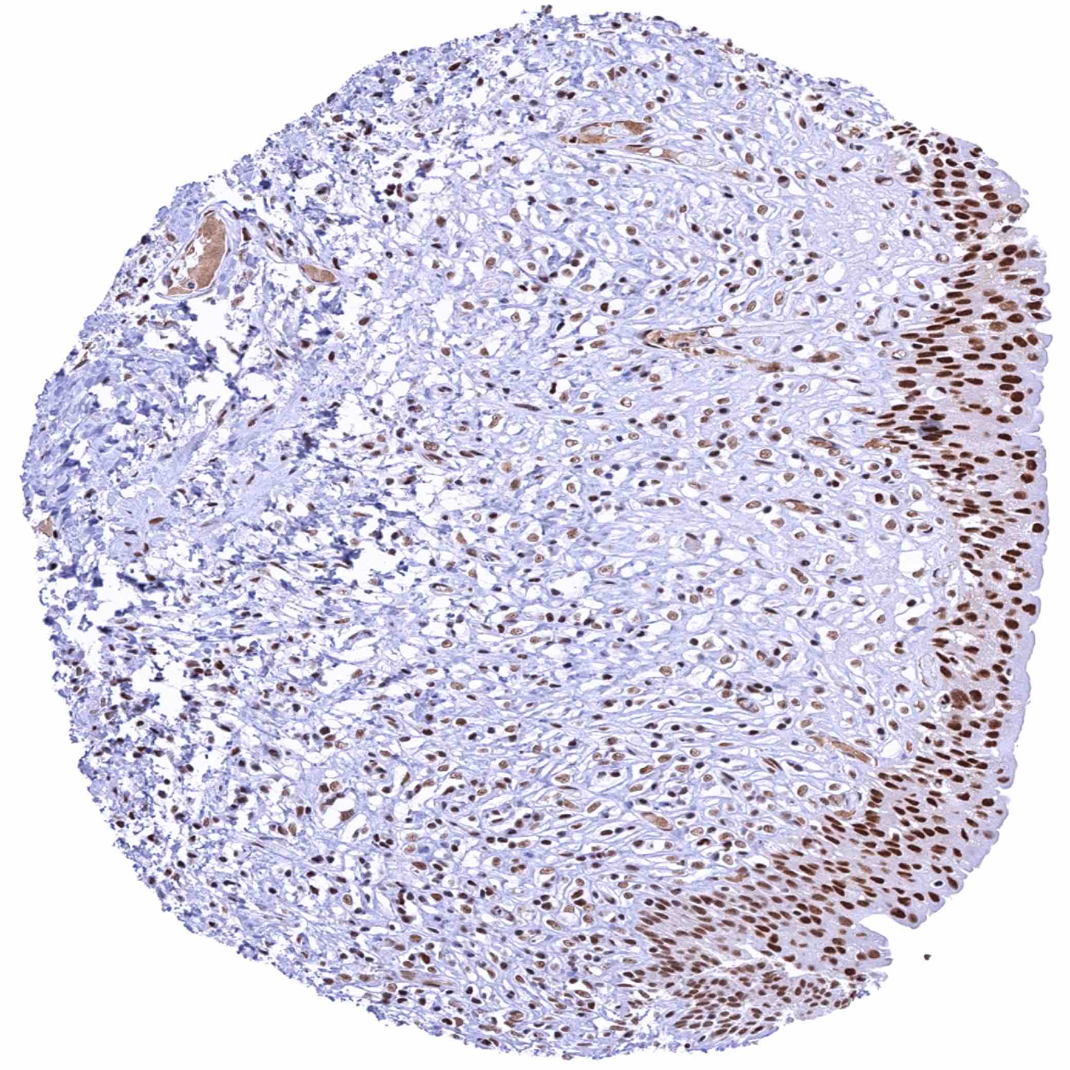
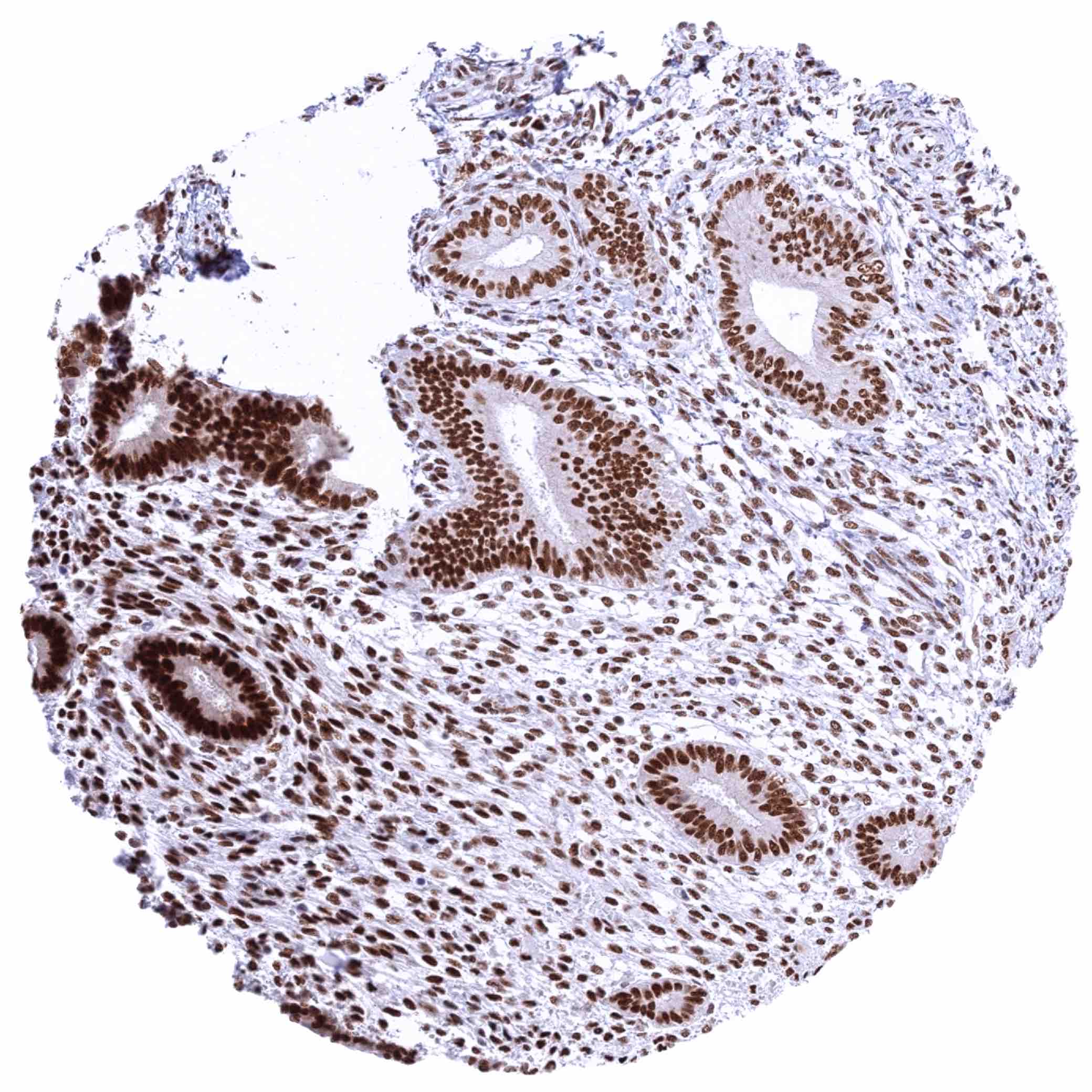
KDM6A antibody [HMV311] HistoMAXTM


Go to Cancer Tissue Gallery
  
 Go back to product page
  

Go to HistoMAX page


Lymph node – Moderate to strong staining of lymphocytes and other cell types

Adrenal gland – Moderate to strong nuclear KDM6A staining of adrenocortical cells

Aorta, media – Weak nuclear KDM6A staining of cells

Appendix, mucosa – Strong nuclear KDM6A staining of epithelial and inflammatory cells


Appendix, muscular wall – Weak to moderate nuclear KDM6A staining of muscular and ganglion cells

Bone marrow – Strong nuclear KDM6A staining of virtually all cells

Breast – Strong nuclear KDM6A staining of basal and luminal cells

Bronchus, glands – Moderate to strong nuclear KDM6A staining of respiratory epithelial cells


Cerebellum (white matter)

Cerebellum, cortex (molecular layer, Purkinje cell layer, granule layer)

Cerebrum, grey matter

Cerebrum, white matter


Colon descendens, mucosa – Moderate to strong nuclear KDM6A staining of epithelial cells. Staining is more intense in the crypt base than in the superficial epithelial cell layers

Colon descendens, muscular wall – Weak to moderate nuclear KDM6A staining of muscular cells

Duodenum, Brunner gland – Moderate to strong nuclear KDM6A staining of Brunner gland cells

Duodenum, mucosa – Moderate to strong nuclear KDM6A staining of epithelial and inflammatory cells


Epididymis – Moderate nuclear KDM6A staining of epithelial cells of the cauda. Staining is less intense in basal cells

Epididymis, Caput – Moderate to strong nuclear KDM6A staining of epithelial cells of the caput. Staining is less intense in basal cells

Esophagus, squamous epithelium – Moderate to strong nuclear KDM6A staining of squamous epithelial cells

Fallopian tube, mucosa – Strong nuclear KDM6A staining of all epithelial cells


Fat – Faint nuclear KDM6A staining of some fat cells

Gallbladder, epithelium – Strong nuclear KDM6A staining of epithelial cells

Heart muscle – Weak to moderate nuclear KDM6A staining of heart muscle cells

Ileum, mucosa – Moderate to strong nuclear KDM6A staining of epithelial and inflammatory cells


Kidney, cortex – Moderate nuclear KDM6A staining of epithelial cells of all types

Kidney, medulla – Moderate nuclear KDM6A staining of collecting ducts cells

Liver – Moderate nuclear KDM6A staining of hepatocytes. Stronger staining of other cell types

Lung – Moderate to strong nuclear KDM6A staining of all cell types


Ovary, corpus luteum – Strong nuclear KDM6A staining of corpus luteum cells

Ovary, stroma – Moderate nuclear KDM6A staining of stroma cells

Pancreas – Moderate to strong nuclear KDM6A staining of acinar cells. Stronger staining of ductal and islet cells

Parathyroid gland – Moderate to strong nuclear KDM6A staining of epithelial cells


Parotid gland – Moderate to strong nuclear KDM6A staining of all epithelial cells

Pituitary gland, anterior lobe – Moderate to strong nuclear KDM6A staining of epithelial cells

Pituitary gland, posterior lobe – Faint nuclear KDM6A staining of pituicytes

Placenta (amnion and chorion) – Weak to moderate nuclear KDM6A staining of amnion cells. Moderate to strong staining of chorion cells


Placenta, mature – Strong nuclear KDM6A staining of trophoblastic cells, stroma cells and endothelial cells

Placenty, early – Strong nuclear KDM6A staining of trophoblastic cells, stroma cells and endothelial cells

Prostate – Moderate nuclear KDM6A staining of all epithelial cells

Rectum, mucosa – Moderate to strong nuclear KDM6A staining of epithelial cells


Seminal vesicle – Strong nuclear KDM6A staining of epithelial cells

Skeletal muscle – Moderate nuclear KDM6A staining of muscular cells

Skin – Moderate to strong nuclear KDM6A staining of squamous epithelial cells

Spleen – Moderate to strong staining of all cell types


Spleen

Stomach, antrum – Moderate to strong nuclear KDM6A staining of epithelial cells. The staining is weaker in glandular cells than in superficial epithelium

Stomach, corpus – Moderate to strong nuclear KDM6A staining of epithelial cells. The staining is weaker in glandular cells than in superficial epithelium

Testis – Moderate to strong nuclear KDM6A staining of Sertoli cells. KDM6A staining is weak in spermatogonia and virtually absent in more mature cells of spermatogenesis


Thymus – Intense nuclear KDM6A staining of all cell types

Thyroid gland – Moderate to strong nuclear KDM6A staining of follicular epithelial cells

Tonsil – Intense nuclear KDM6A staining of all cell types

Tonsil, surface epithelium – Strong nuclear KDM6A staining of squamous epithelial cells and of lymphocytes


Urinary bladder, muscular wall – Weak to moderate nuclear KDM6A staining of muscular cells

Urinary bladder, urothelium – Strong nuclear KDM6A staining of urothelial cells

Uterus, ectocervix – Moderate to strong nuclear KDM6A staining of squamous epithelial cells

Uterus, endocervix – Strong nuclear KDM6A staining of epithelial cells


Uterus, endometrium (pregnancy) – Moderate to strong nuclear KDM6A staining of all cells

Uterus, endometrium (proliferation) – Strong nuclear KDM6A staining of epithelial and stromal cells

Uterus, endometrium (secretion) – Strong nuclear KDM6A staining most glandular cells

Uterus, myometrium – Moderate to strong nuclear KDM6A staining of muscular cells


| TOP |