Normal Tissue Gallery- GTX04475

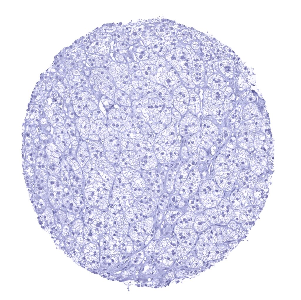
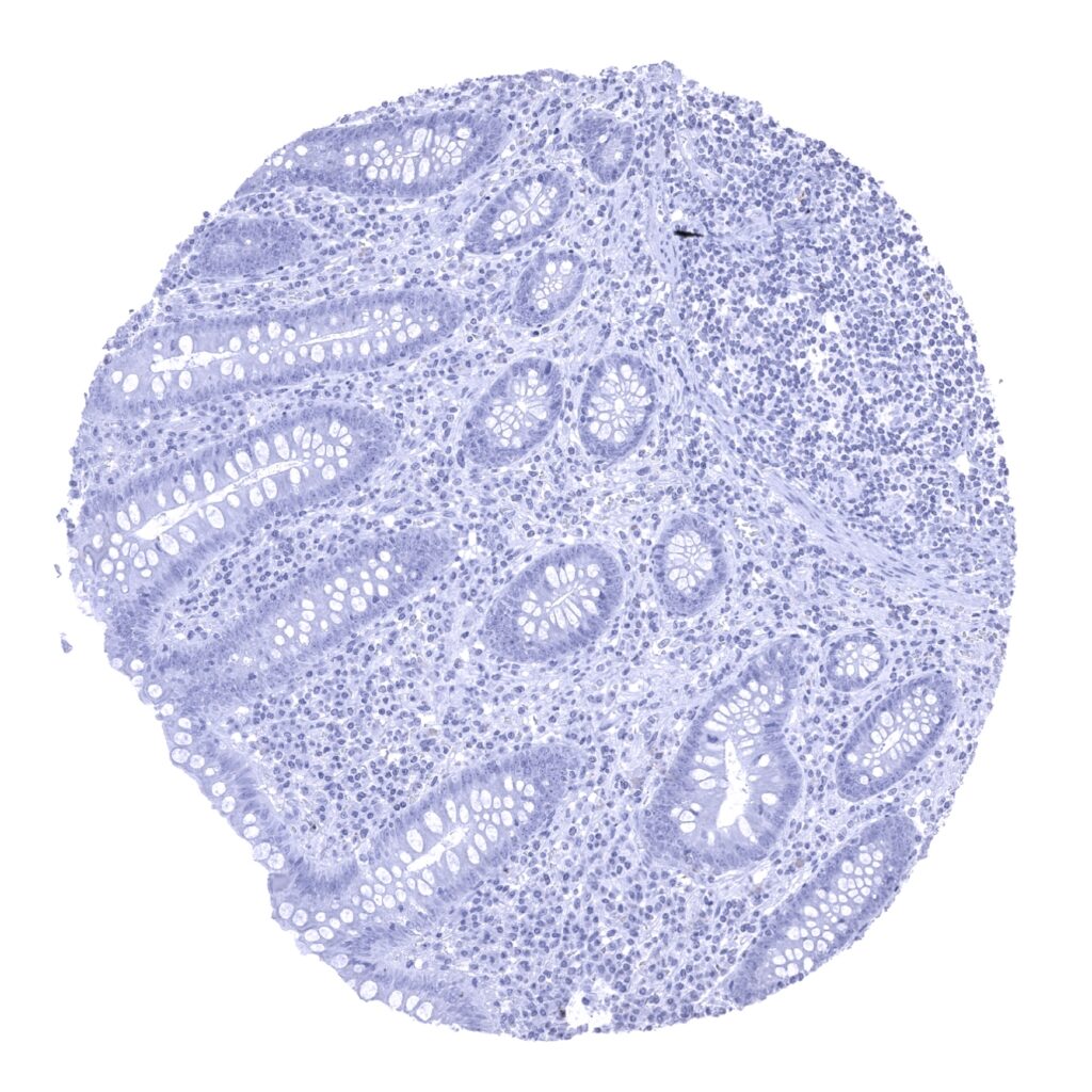
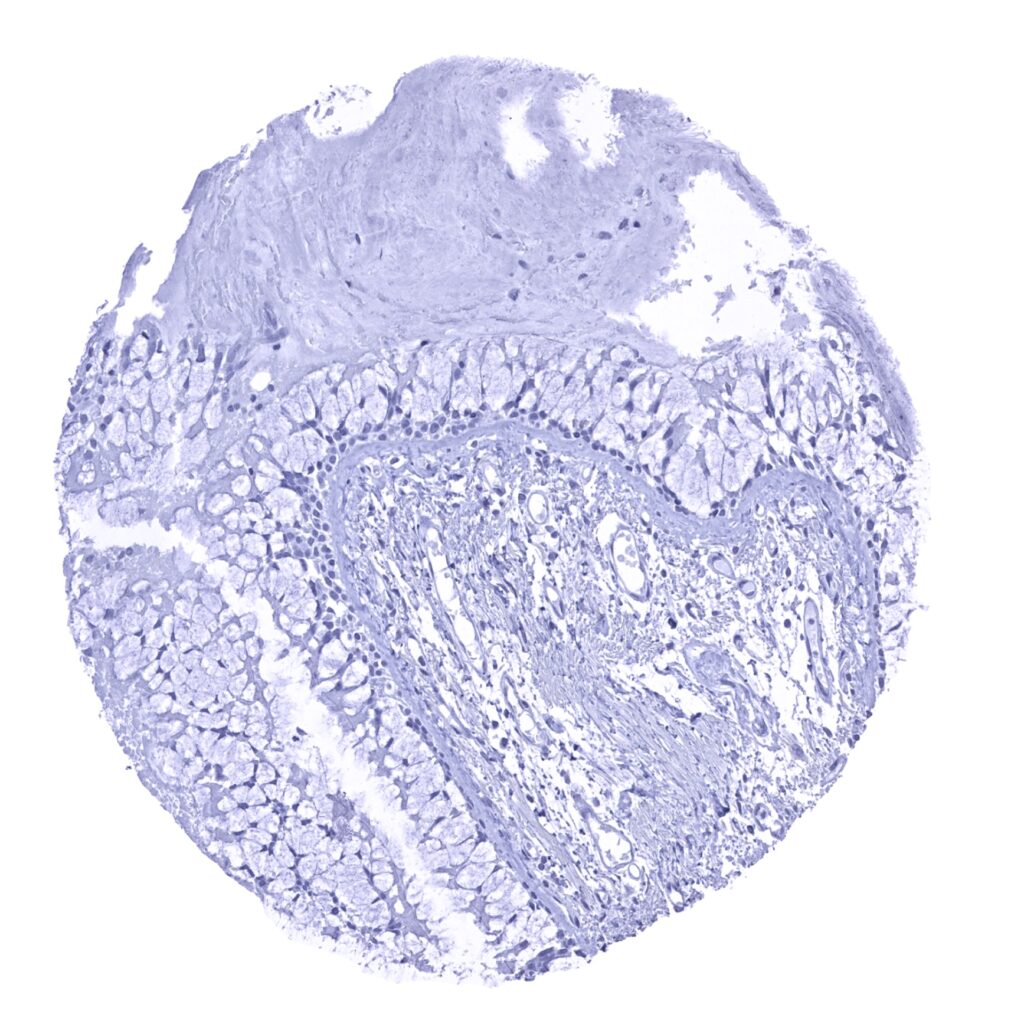
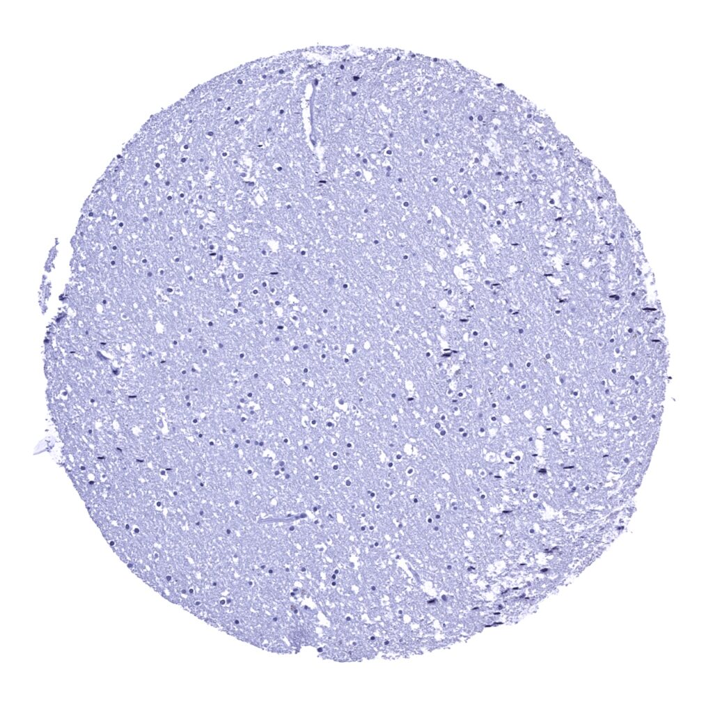
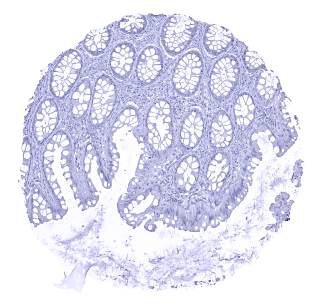
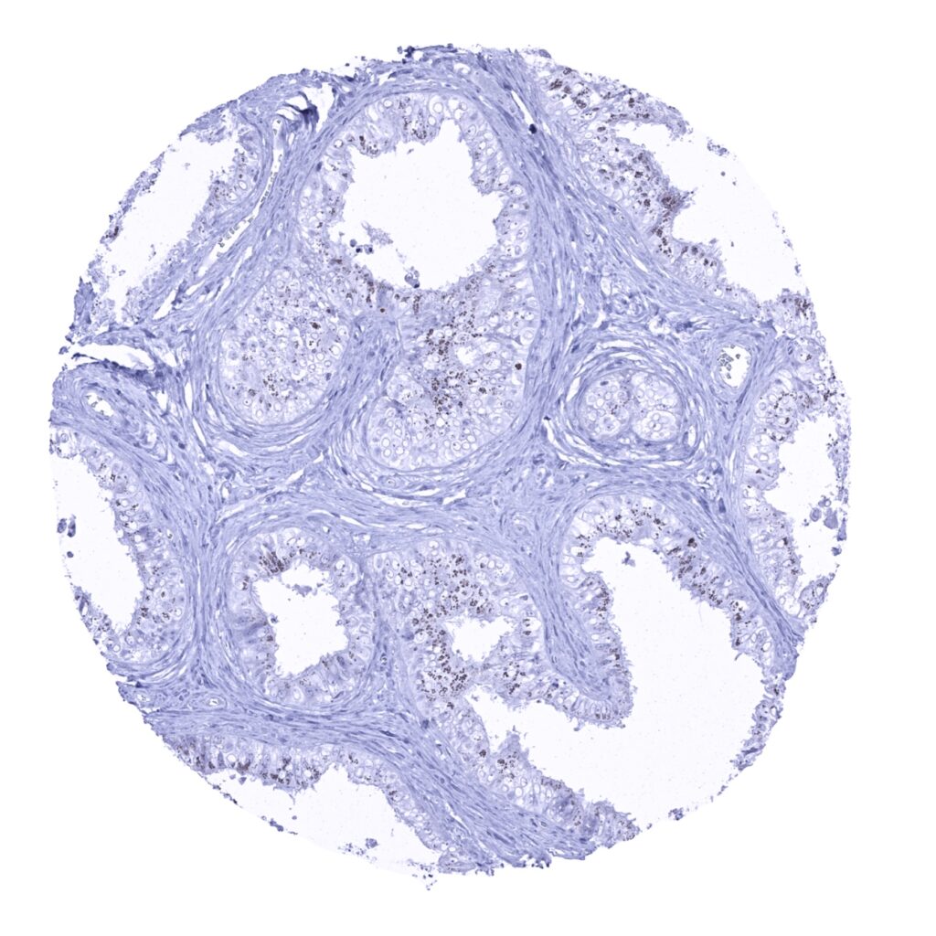
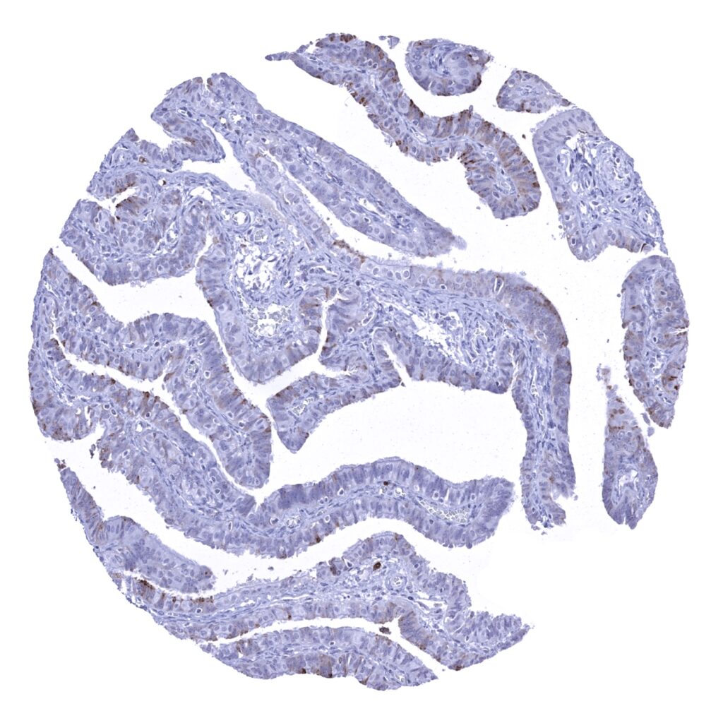
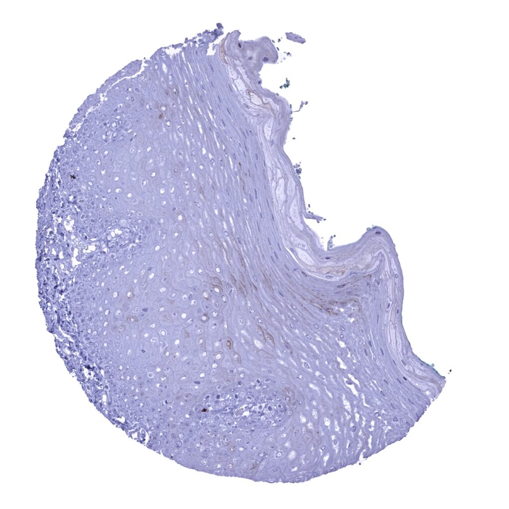
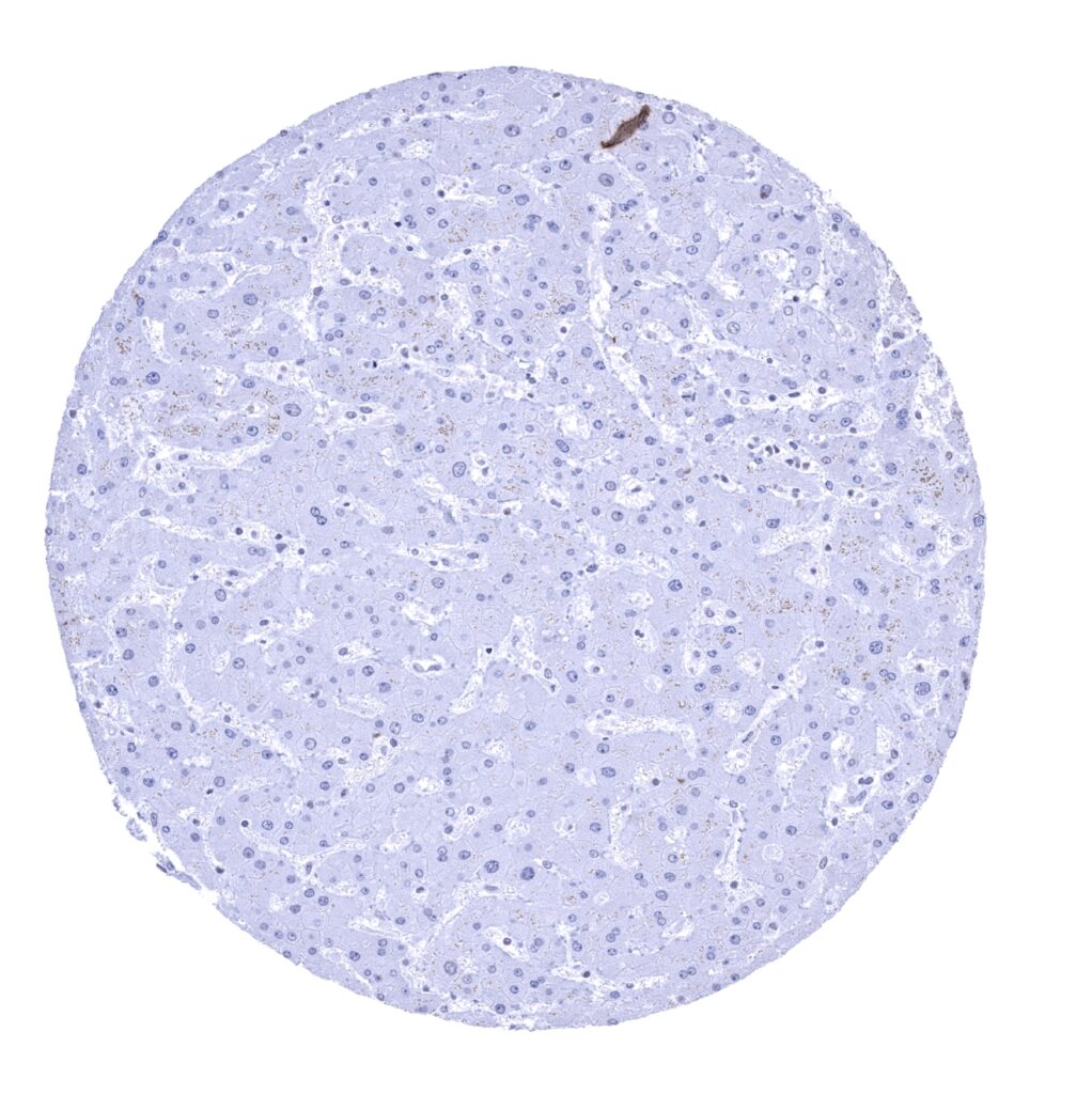
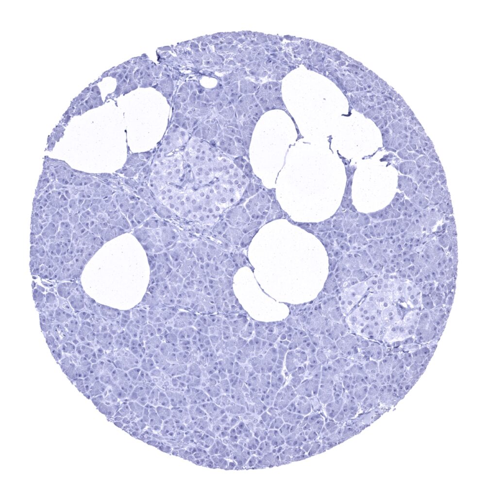
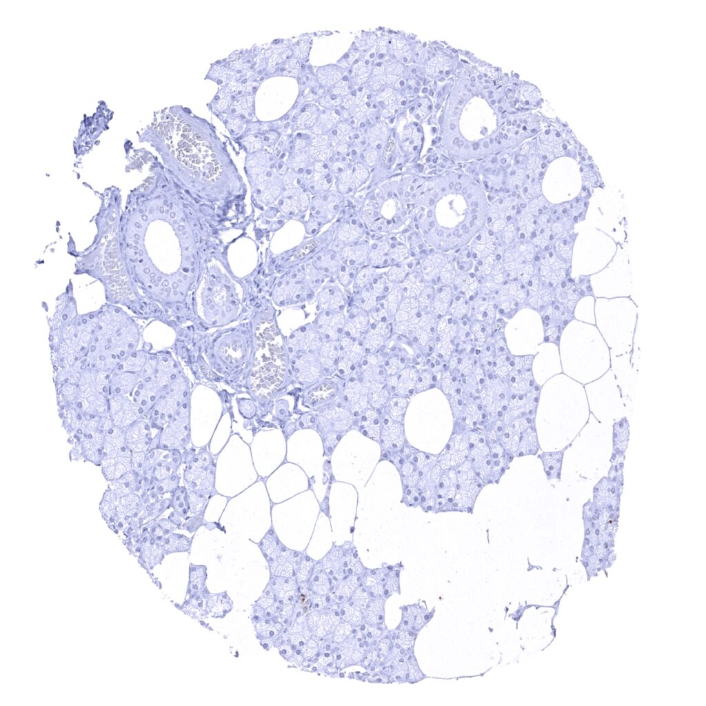
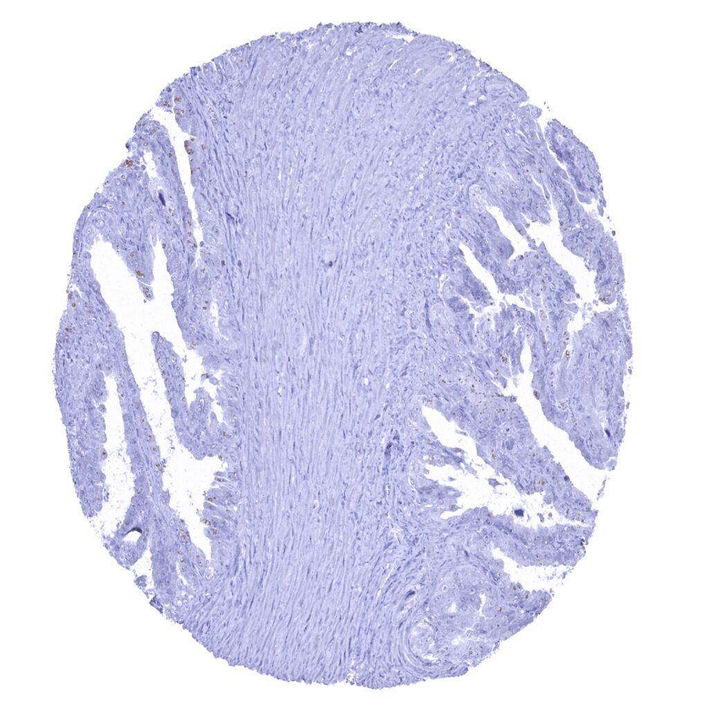
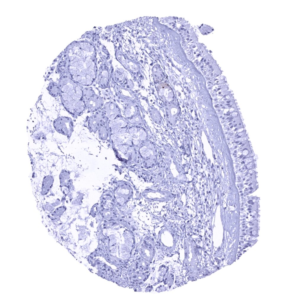
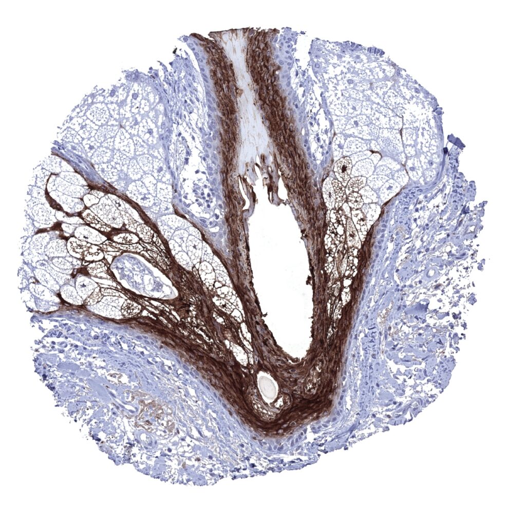
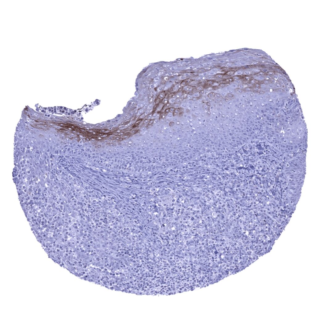
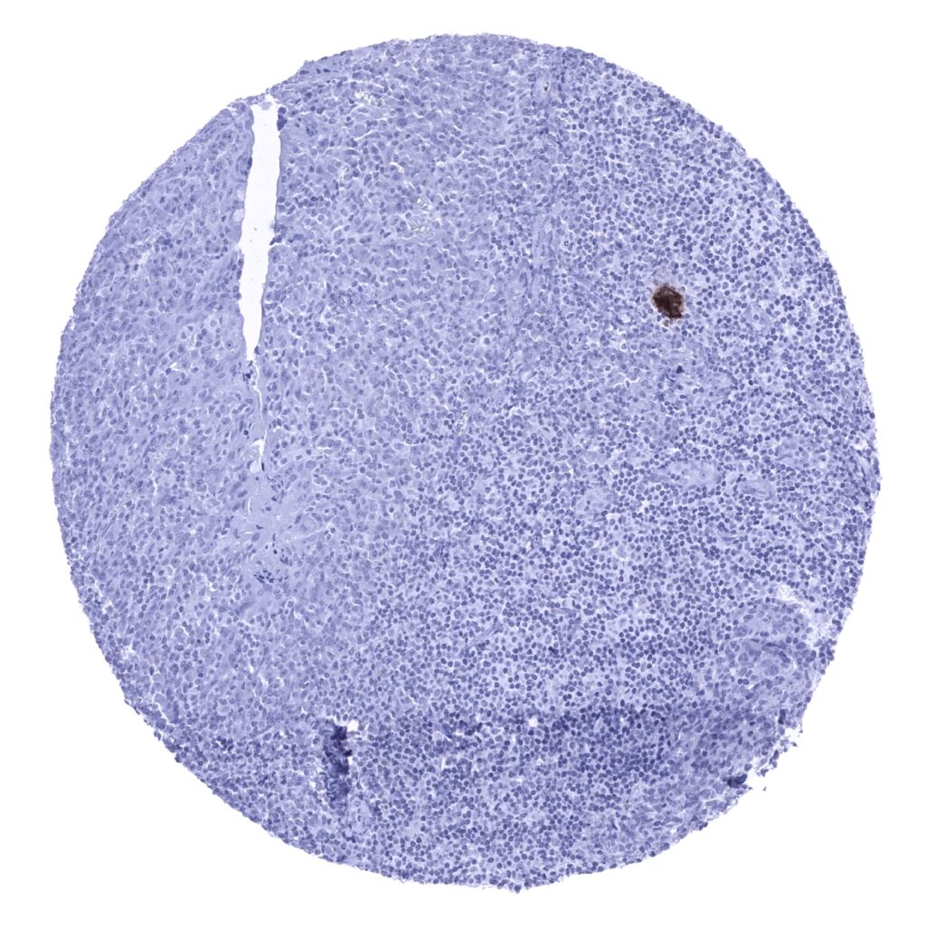
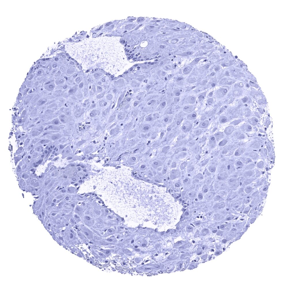
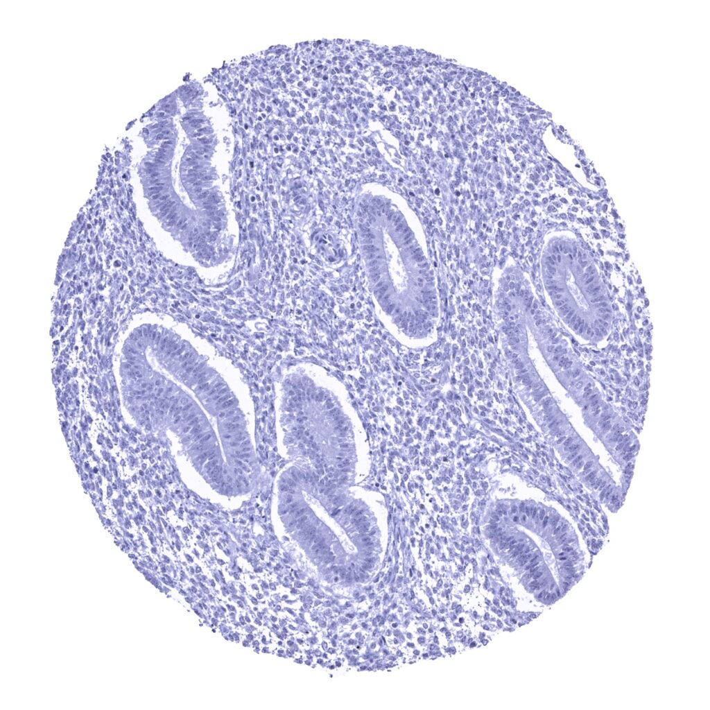
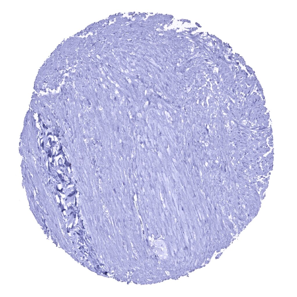
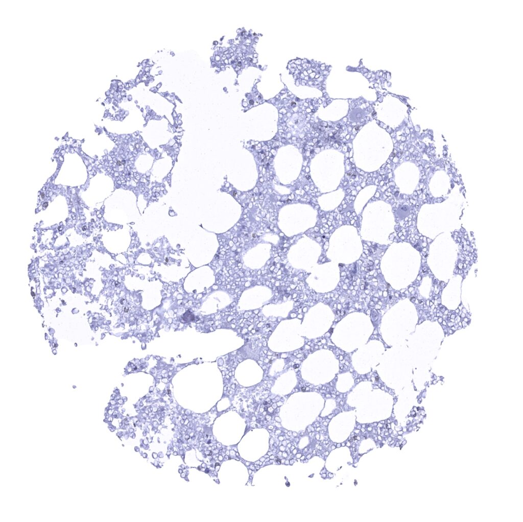
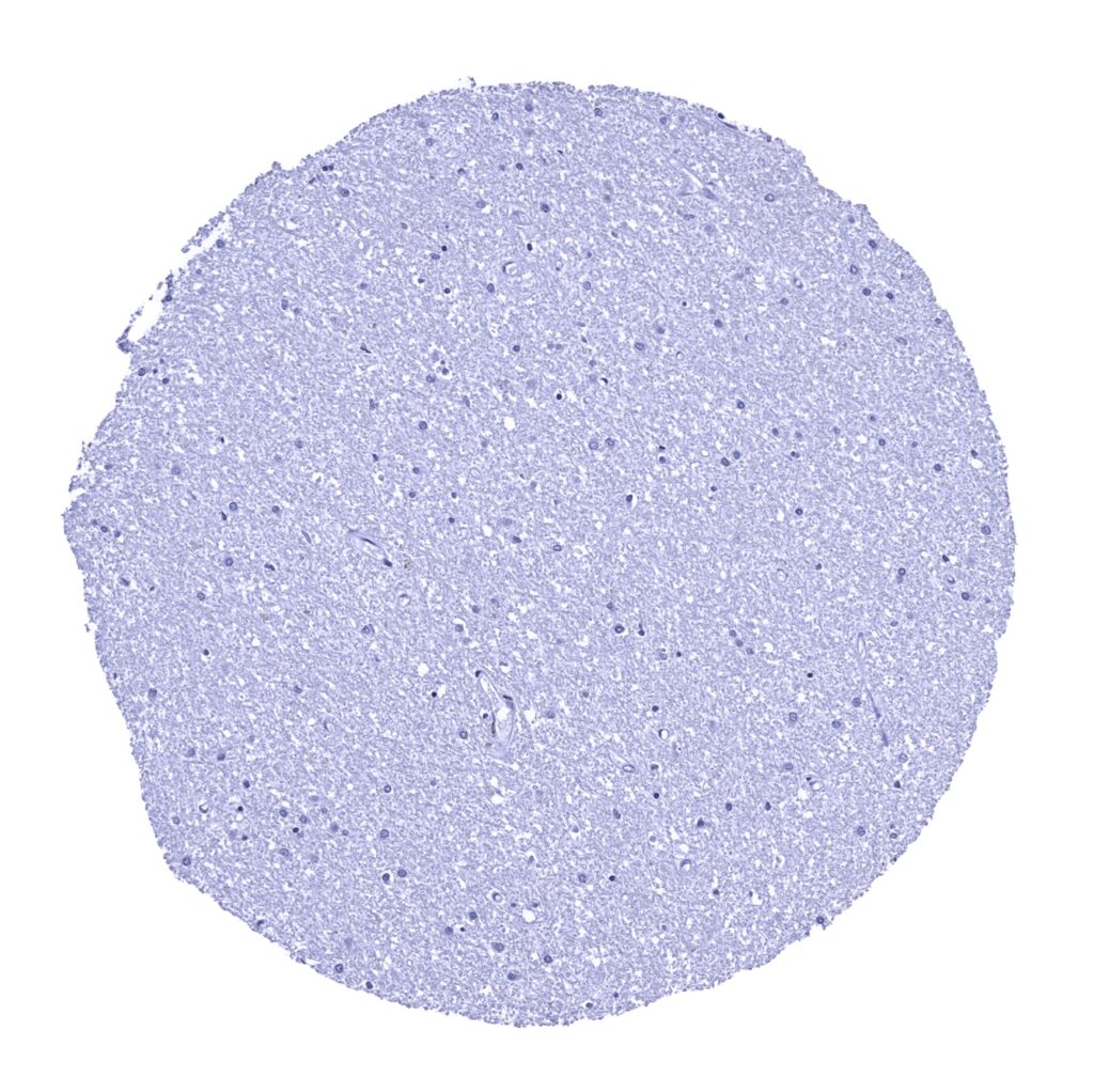
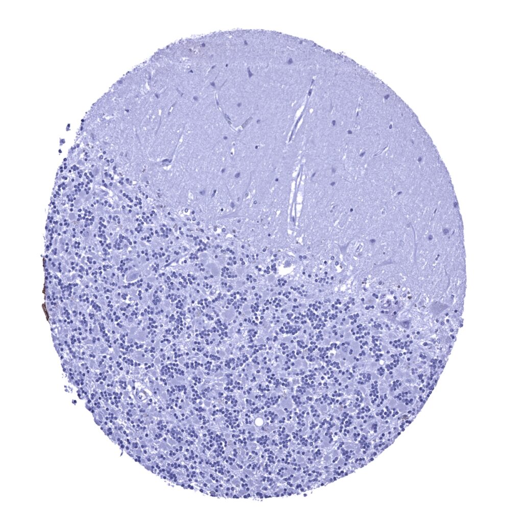
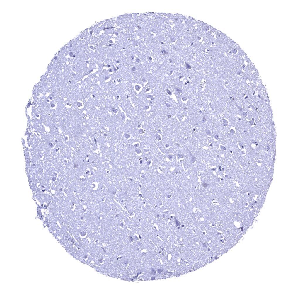
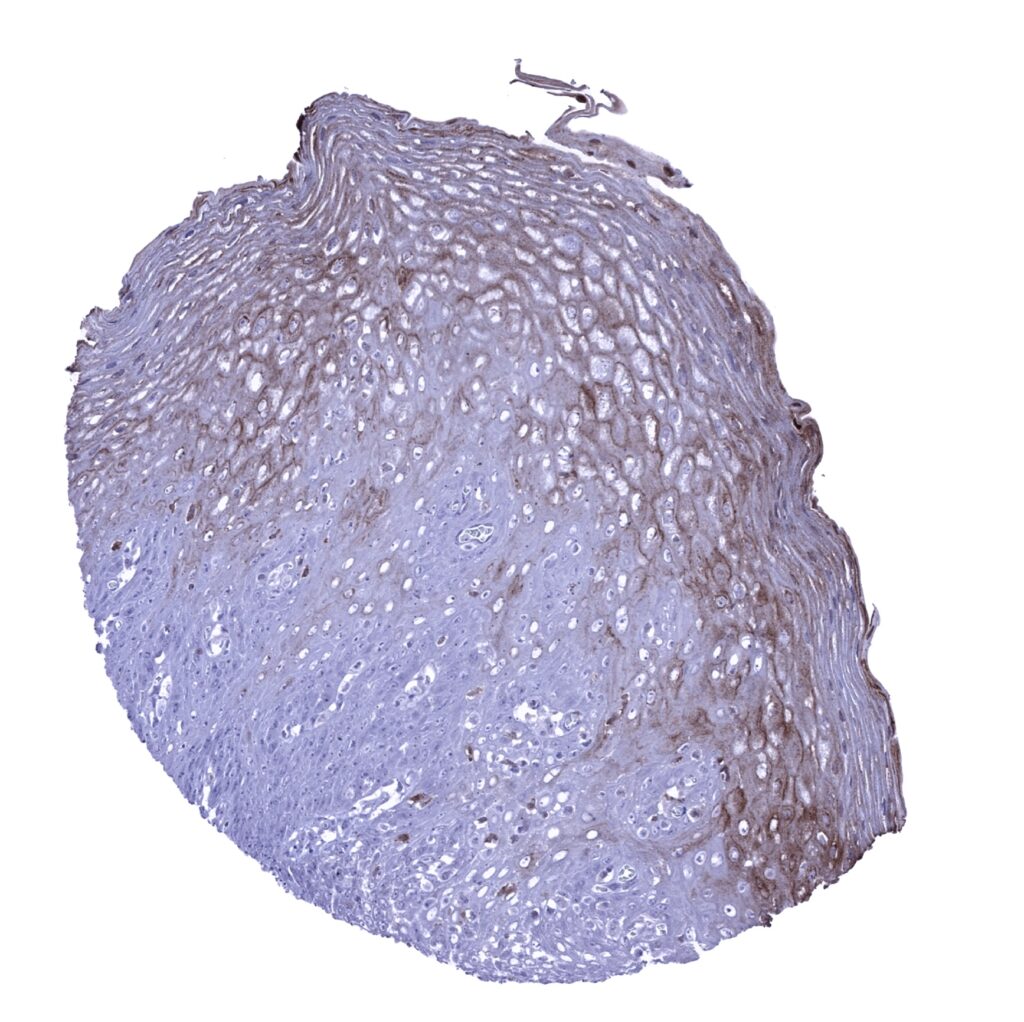
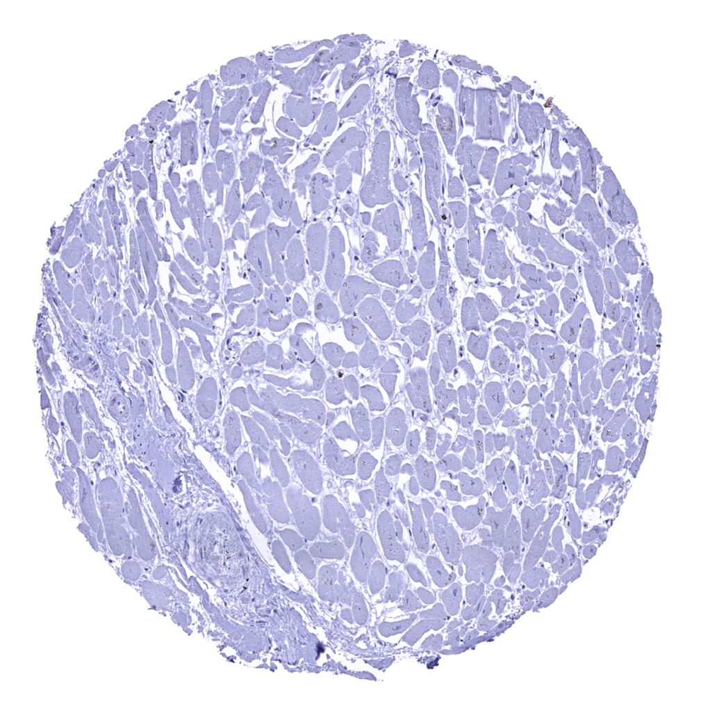
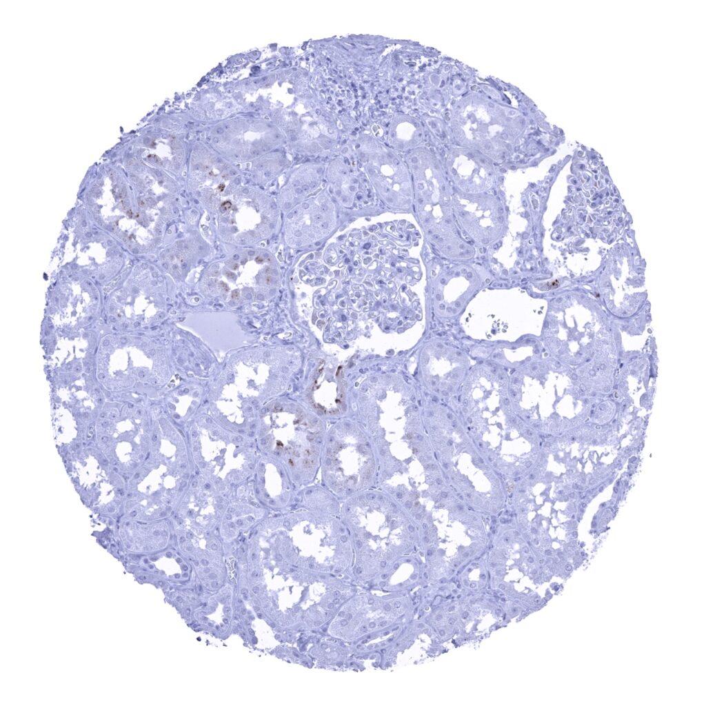
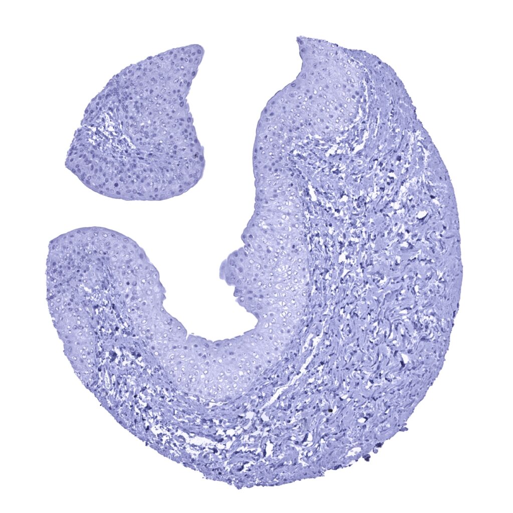
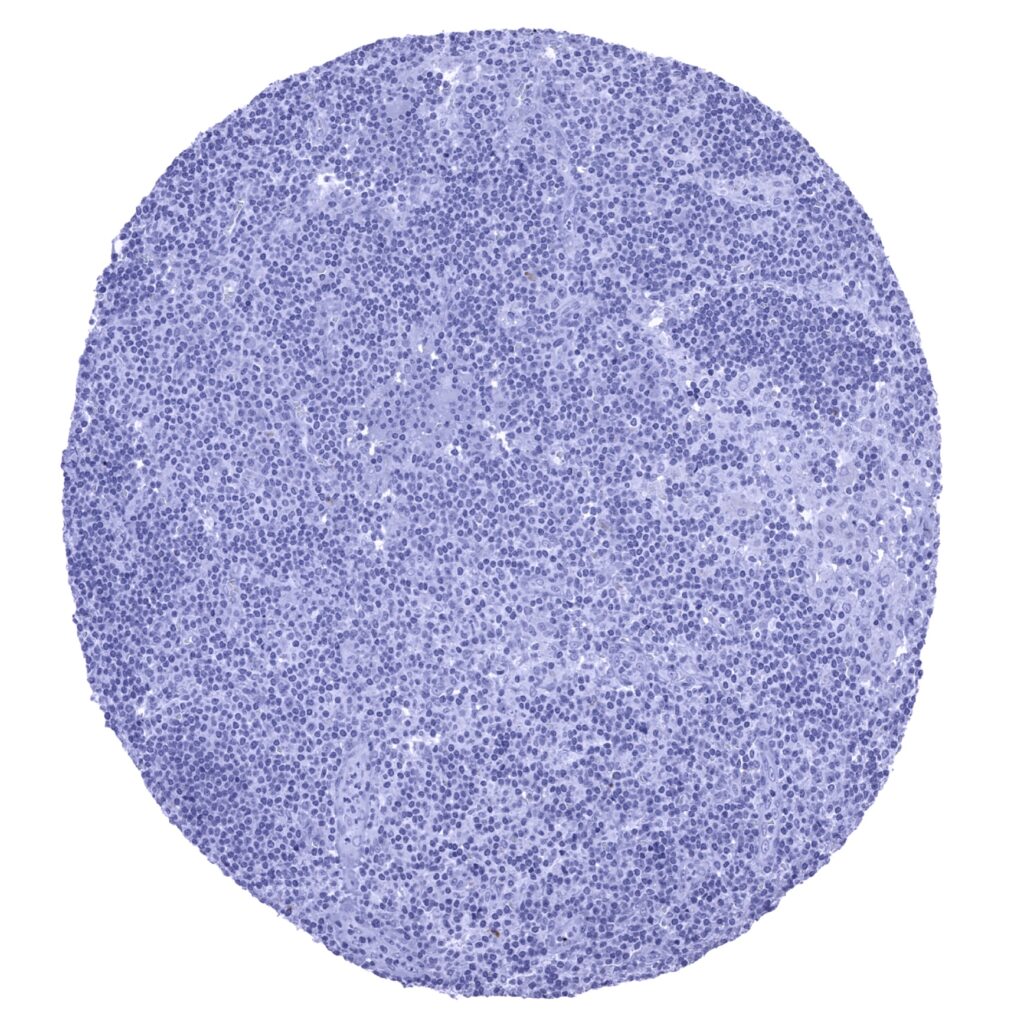
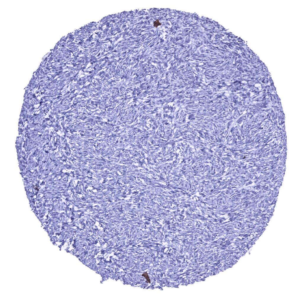
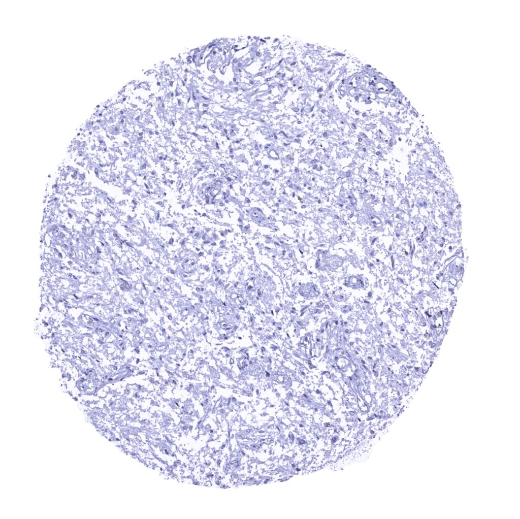
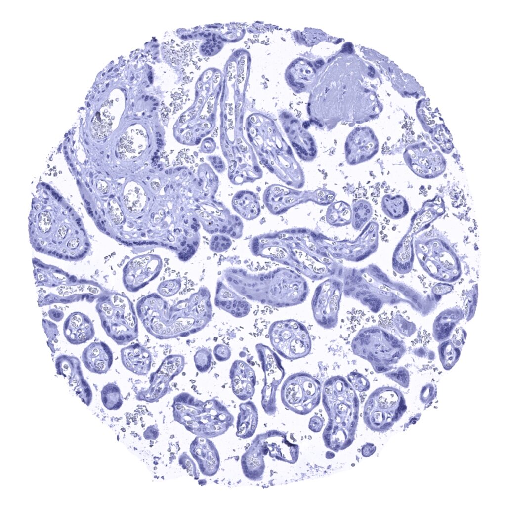
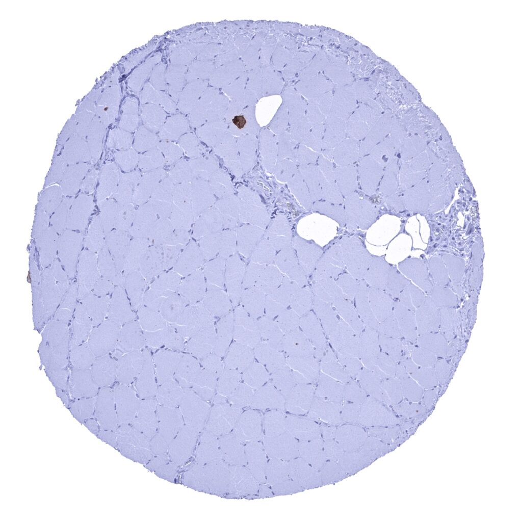
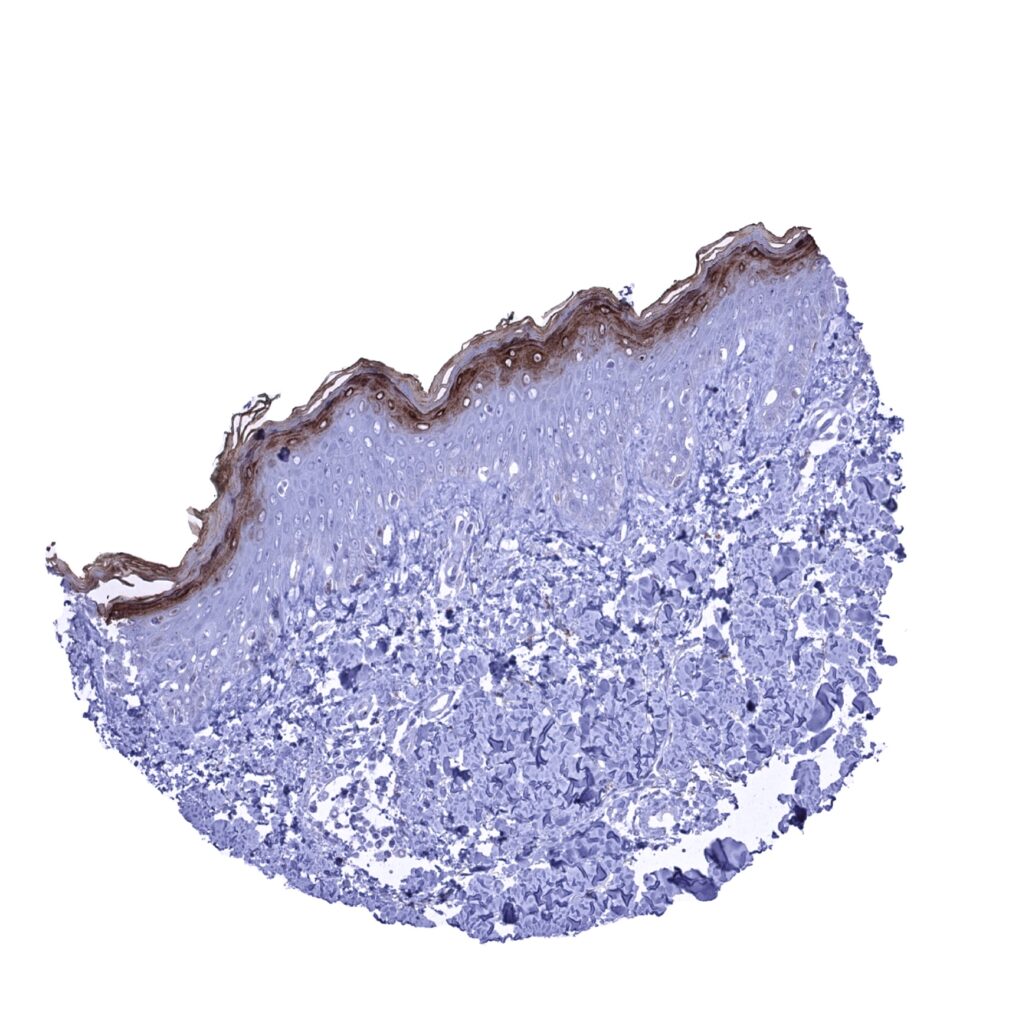
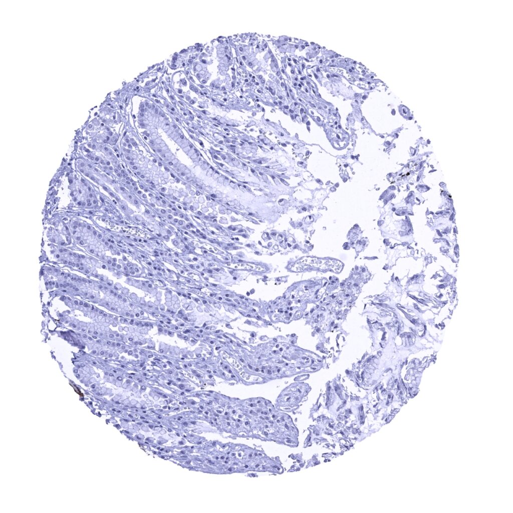
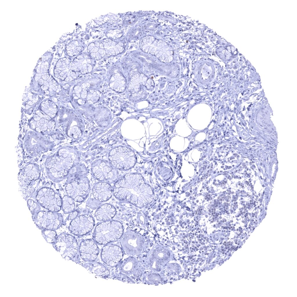
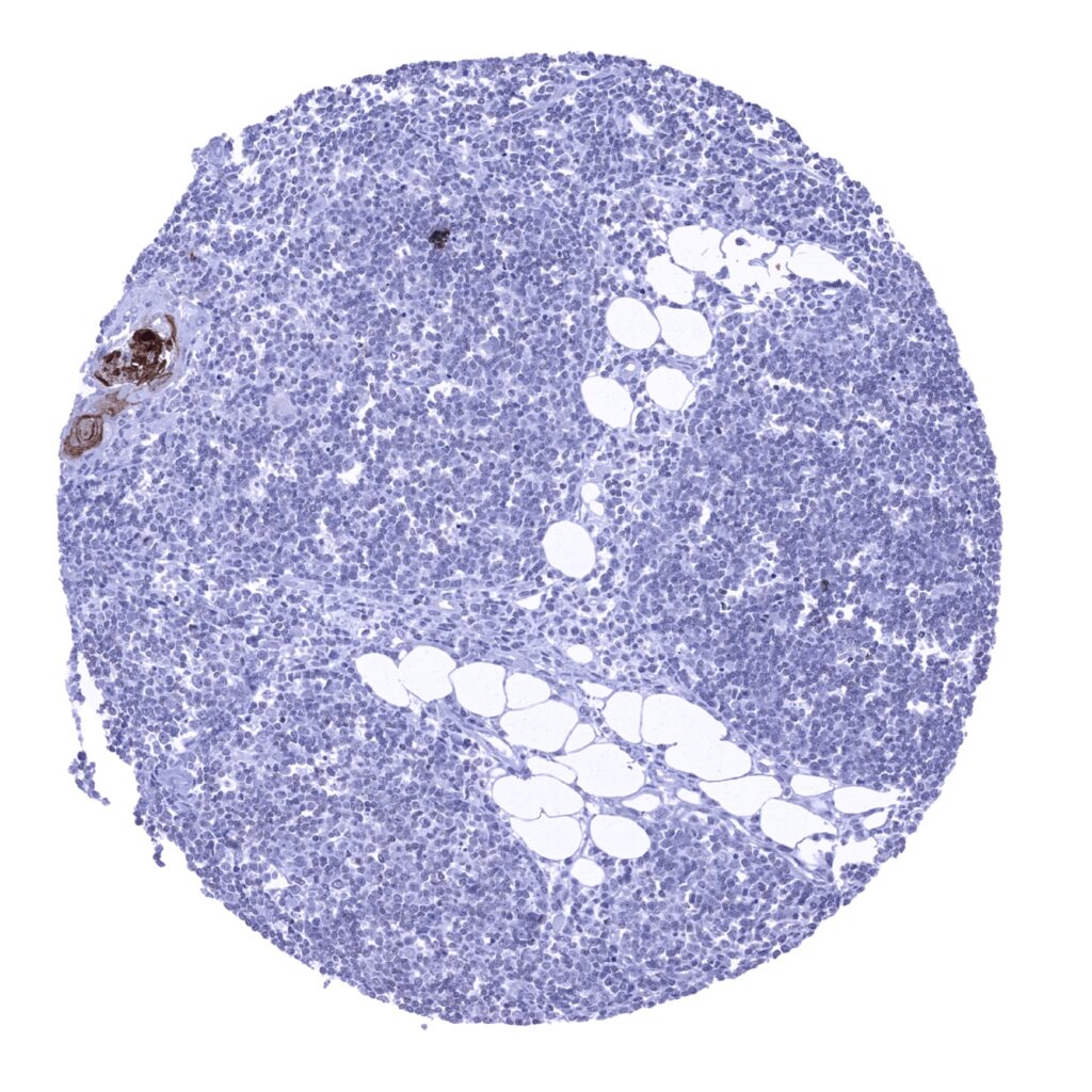
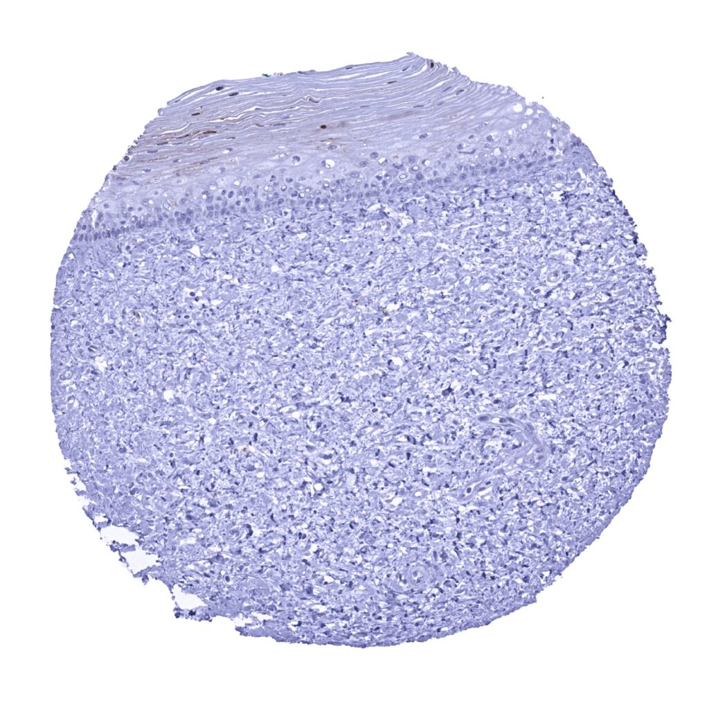
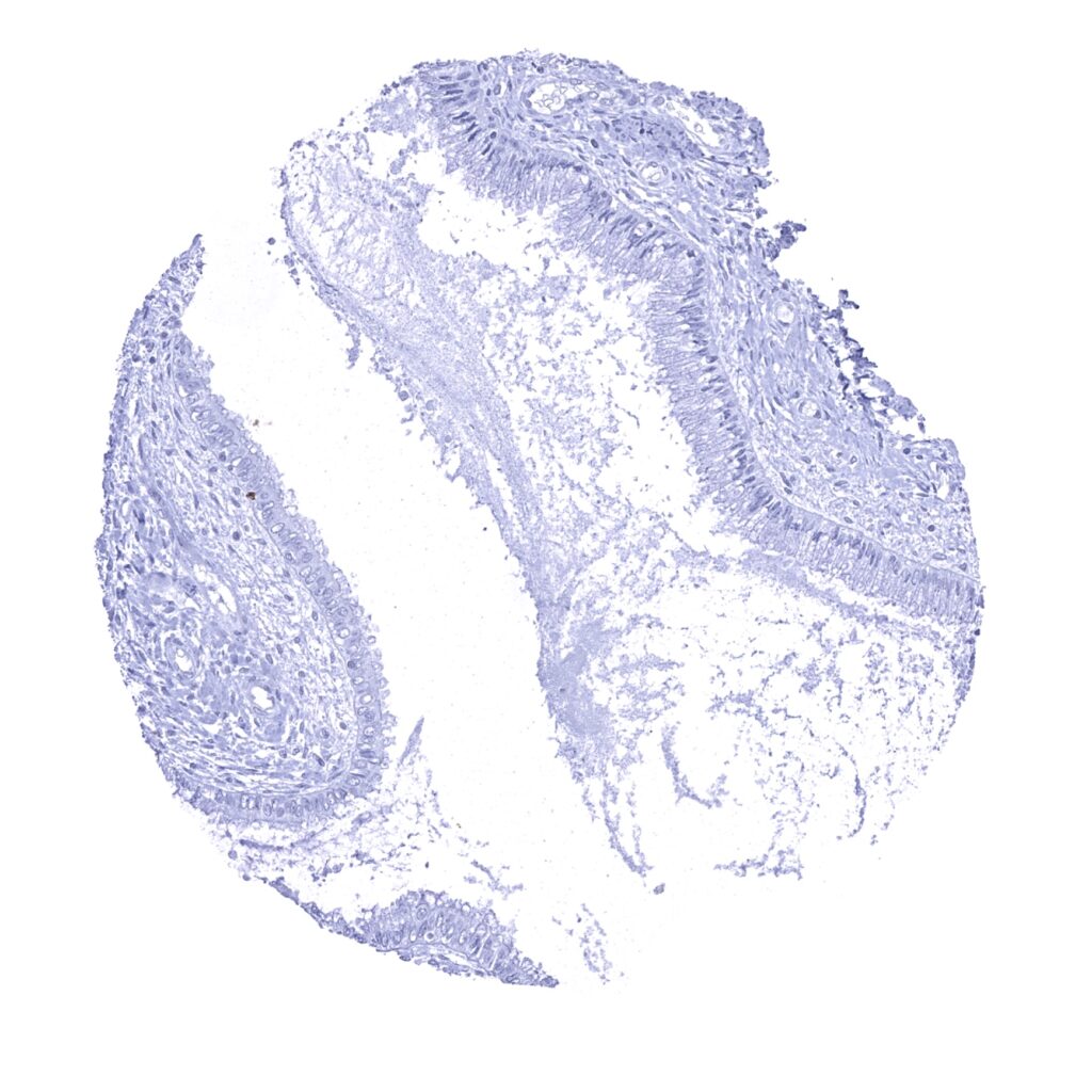
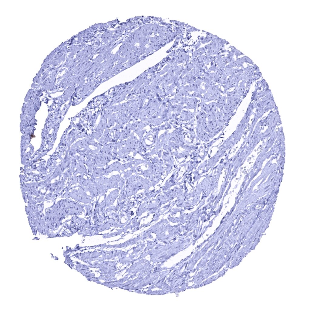
Kallikrein 7 antibody [MSVA-707M] HistoMAX


Go to Cancer Tissue Gallery
  
 Go back to product page
  

Go to HistoMAX page


Adrenal gland

Aorta, media

Appendix, mucosa

Appendix, muscular wall


Bone marrow

Breast

Bronchus, mucosa

Cerebellum (white matter)


Cerebellum, cortex (molecular, Purkinje cell, and granule cell layers)

Cerebrum, grey matter

Cerebrum, white matter

Colon descendens, mucosa


Colon descendens, muscular wall

Duodenum, mucosa

Epididymis – Granular cytoplasmic staining in this sample reflects pigment and is unrelated to Kallikrein 7.

Epididymis


Esophagus, squamous epithelium – Weak to moderate Kallikrein 7 immunostaining in the superficial half of the squamous epithelium

Fallopian tube, mucosa – A predominantly apical cytoplasmic Kallikrein 7 staining in a small fraction of epithelial cells.

Fallopian tube, mucosa – Kallikrein 7 staining of epithelial cells is not seen in this sample.

Fat


Gallbladder, epithelium

Heart muscle

Ileum, mucosa

Kidney, cortex – Focal Kallikrein 7 staining in few tubuli of this kidney sample.


Kidney, cortex – Kallikrein 7 staining is not seen in this kidney sample.

Kidney, medulla – Kallikrein 7 staining is not seen in this kidney sample.

Kidney, pelvis, urothelium

Lip, oral mucosa – Kallikrein 7 immunostaining is almost absent in this squamous epithelium sample.


Liver

Lung

Lymph node

Ovary, stroma


Pancreas

Parotid gland

Pituitary gland, anterior lobe

Pituitary gland, posterior lobe


Placenta (amnion and chorion)

Placenta, early

Placenta, mature

Prostate


Rectum, mucosa

Seminal vesicle

Sinus paranasales

Skeletal muscle


Skin – A strong Kallikrein 7 immunostaining is seen in the stratum granulosum and the keratinizing superficial layer of the skin

Skin (hair follicles and sebaceous glands) – A strong Kallikrein 7 immunostaining is seen in the cortex of hair follicles and the luminal area of sebaceous glands

Spleen

Stomach, antrum


Stomach, corpus

Sublingual gland

Testis

Thymus – Strong Kallikrein 7 immunostaining in keratinizing squamous epithelial cells of a corpuscle of Hassall's


Thyroid gland

Tonsil, surface epithelium – Moderate Kallikrein 7 staining in the top third of the squamous epithelium, a layer that corresponds to the stratum granulosum

Tonsil

Urinary bladder, muscular wall


Urinary bladder, urothelium

Uterus, ectocervix – Kallikrein 7 immunostaining is almost absent in this squamous epithelium sample

Uterus, ectocervix – Kallikrein 7 staining is undetectable in this squamous epithelium

Uterus, endocervix


Uterus, endometrium (pregnancy)

Uterus, endometrium (proliferation)

Uterus, endometrium (secretion)

Uterus, myometrium


| TOP |