Normal Tissue Gallery- GTX04448

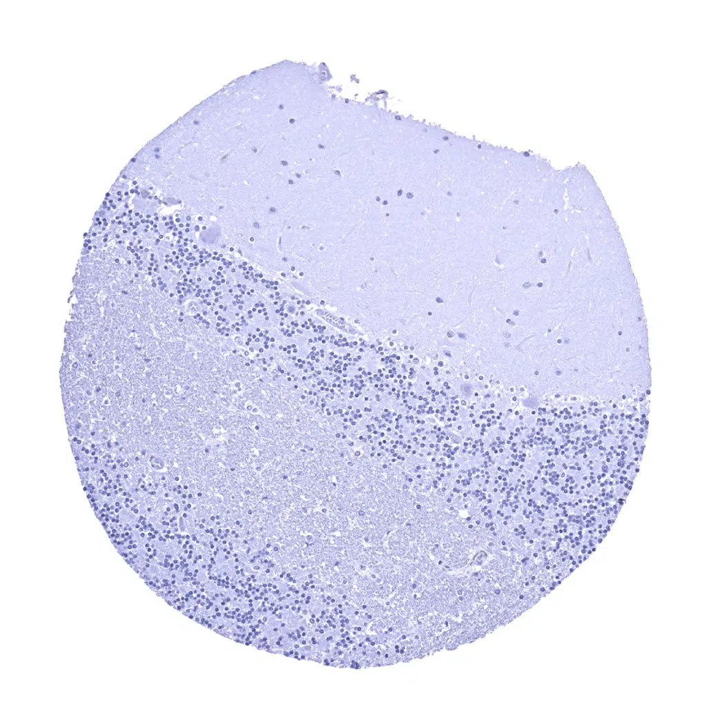
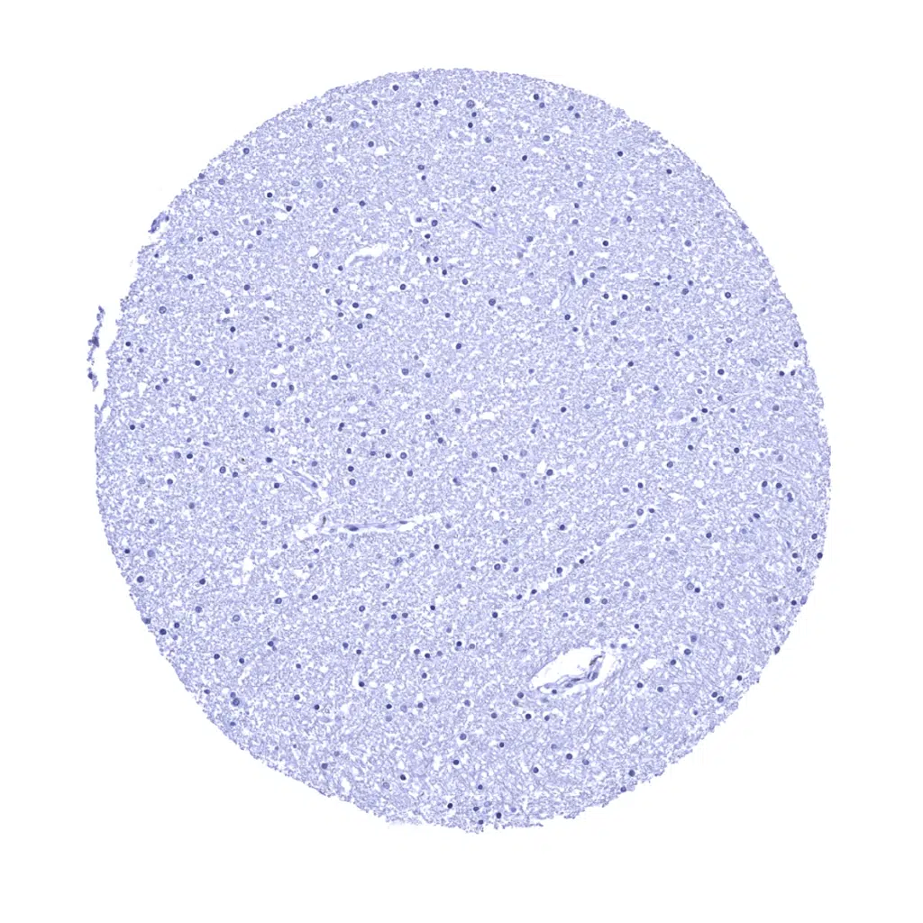
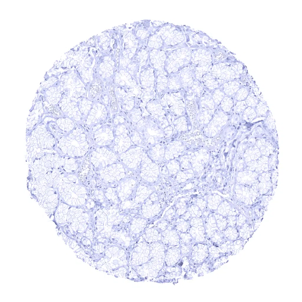
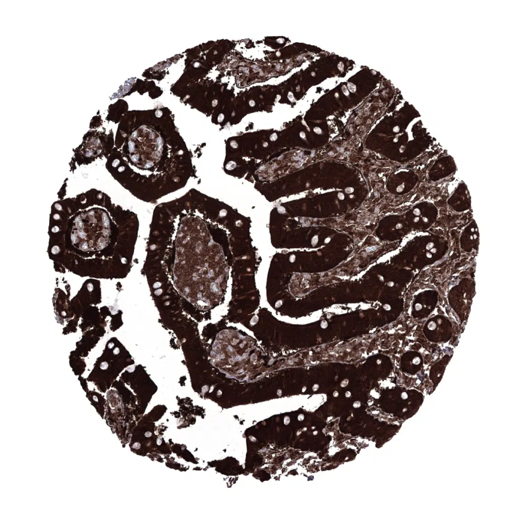
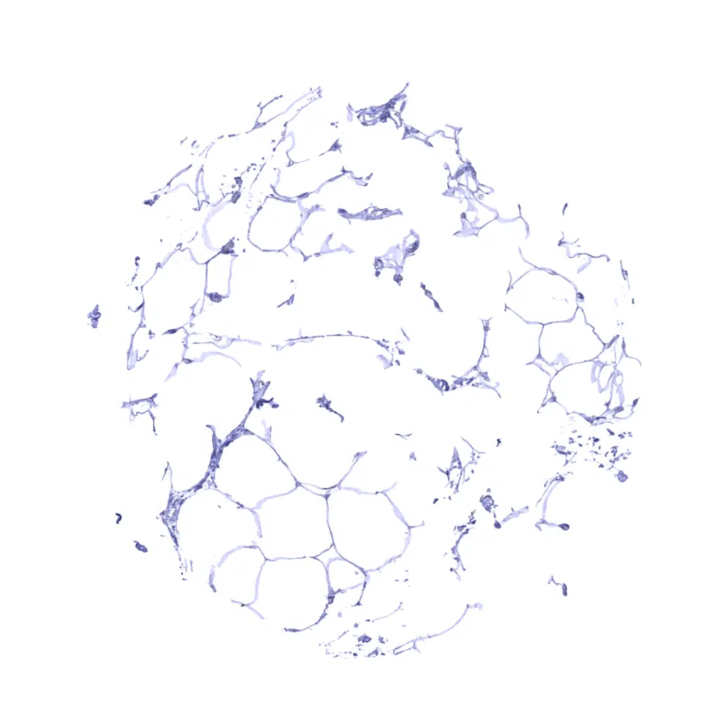
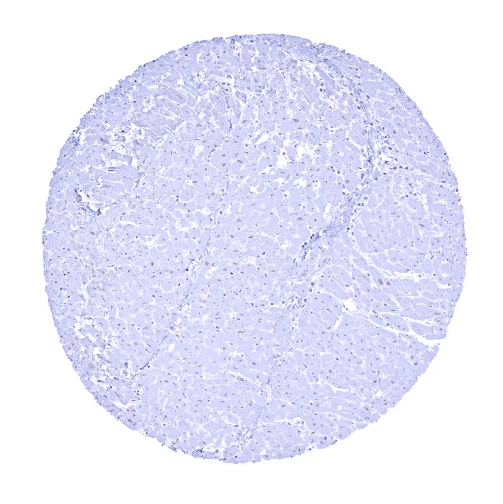
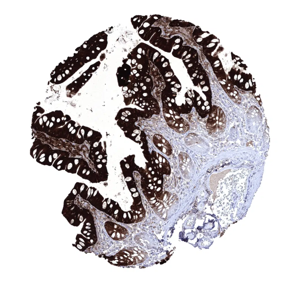
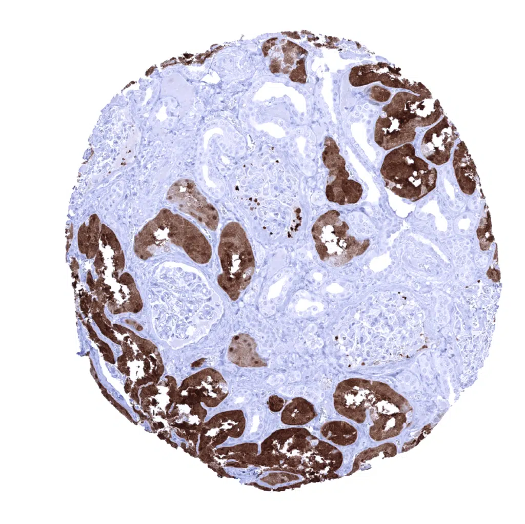
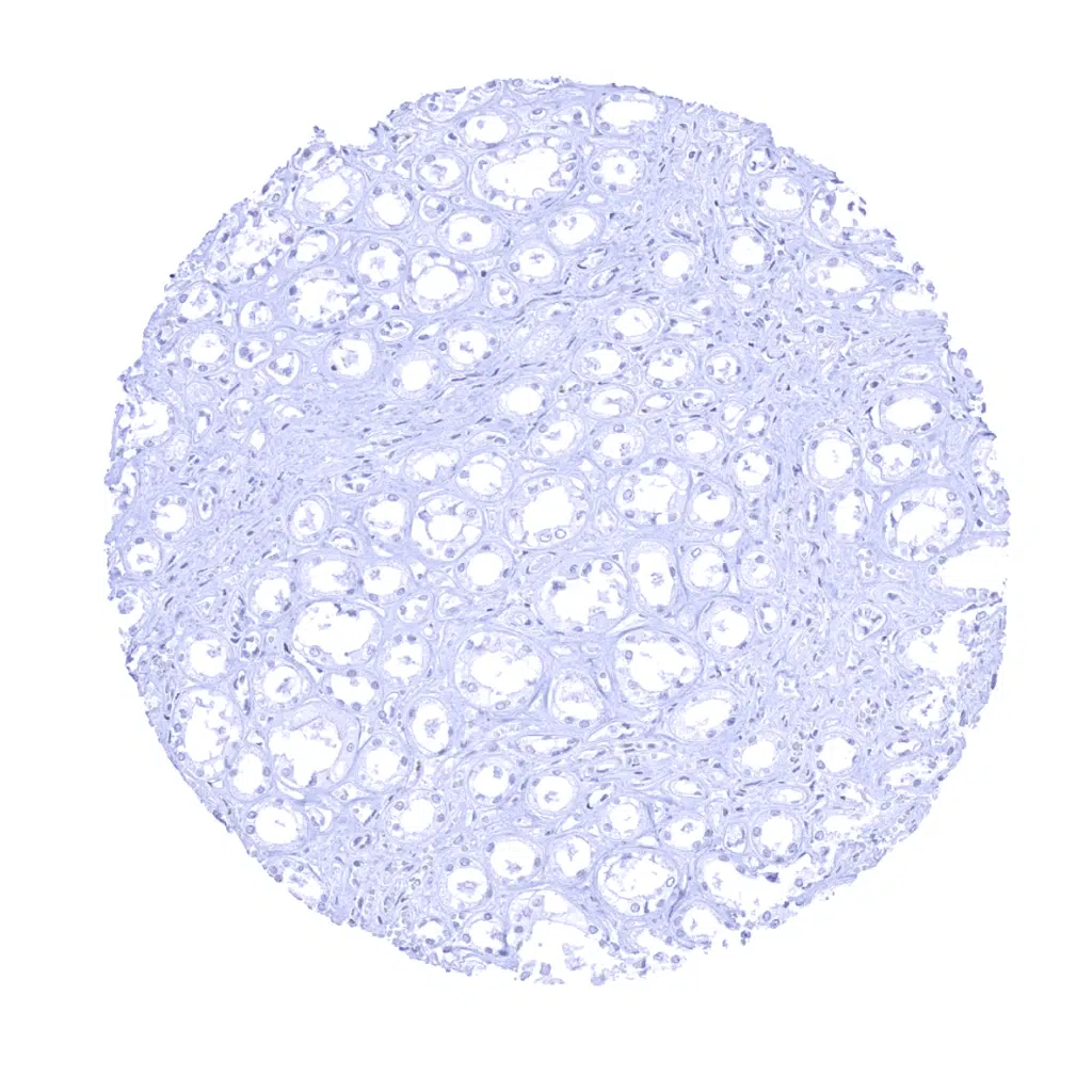
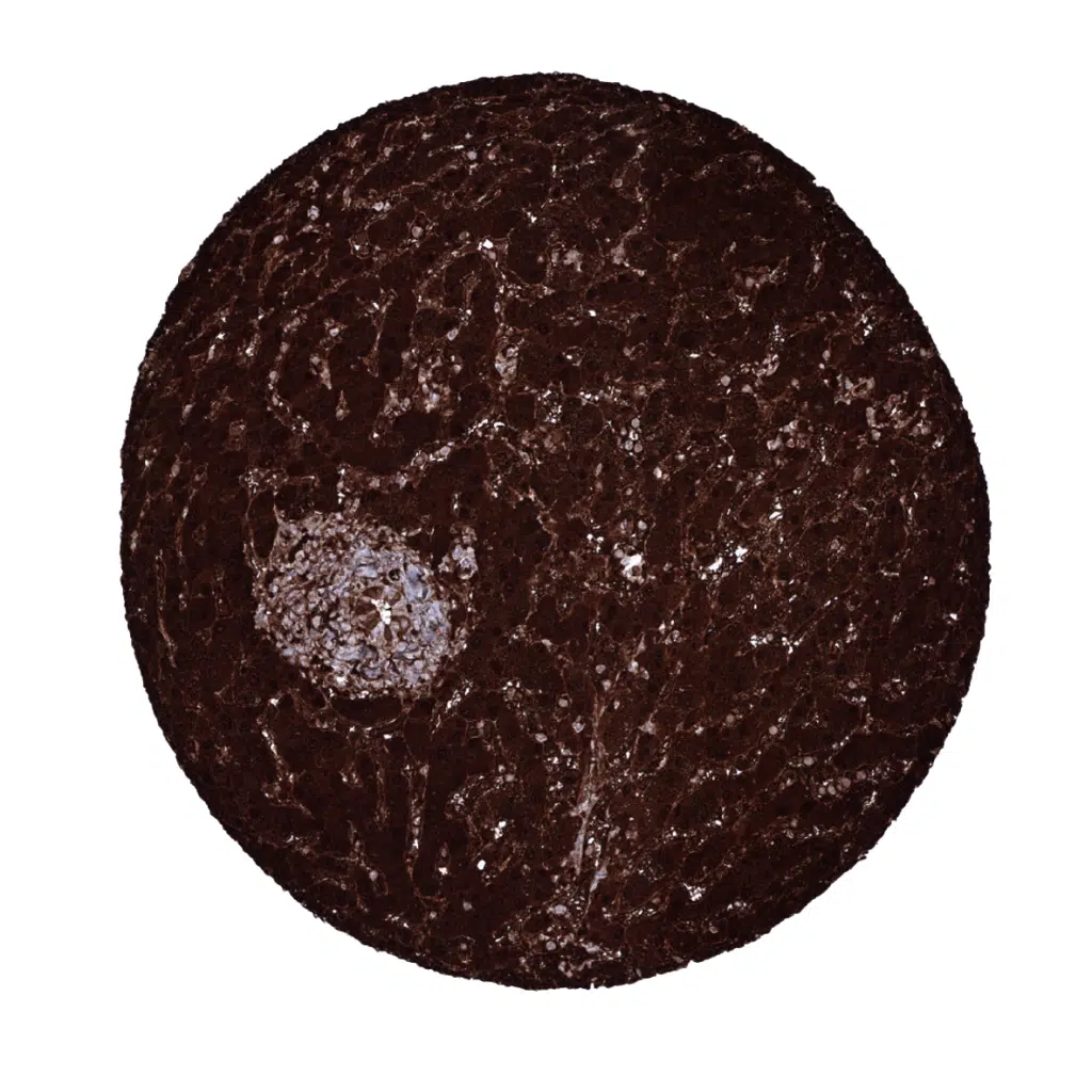
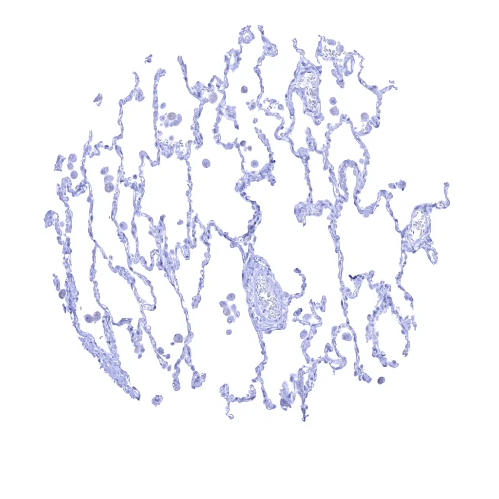
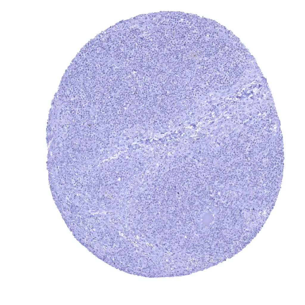
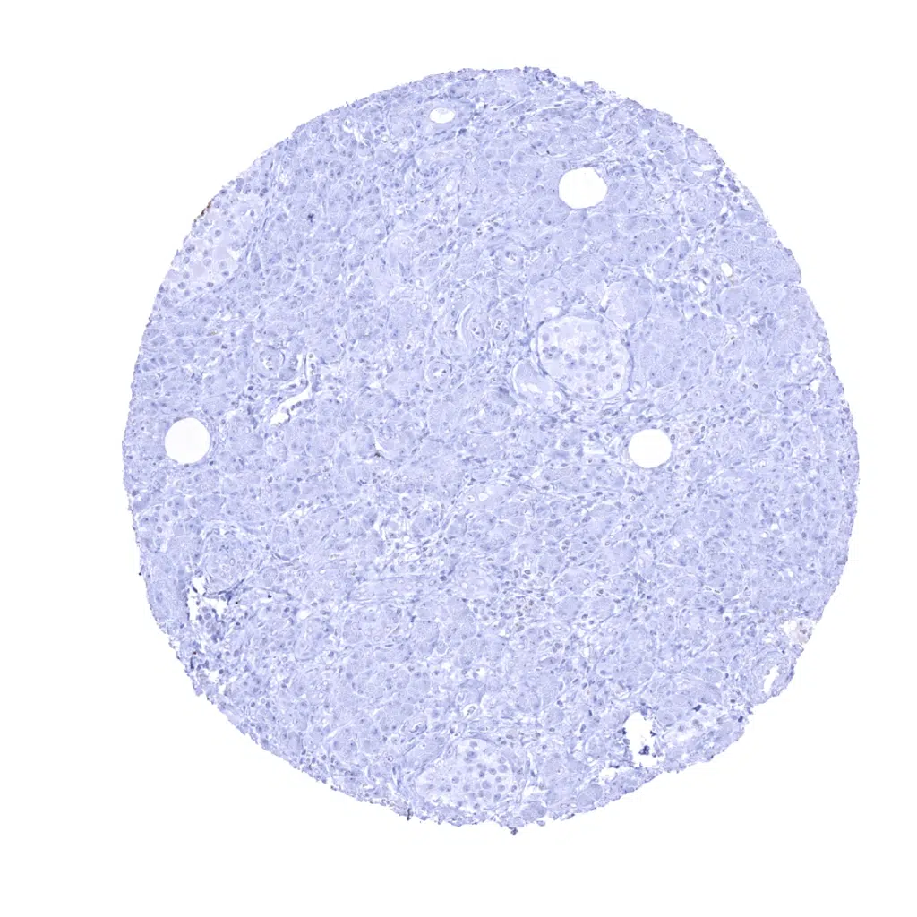
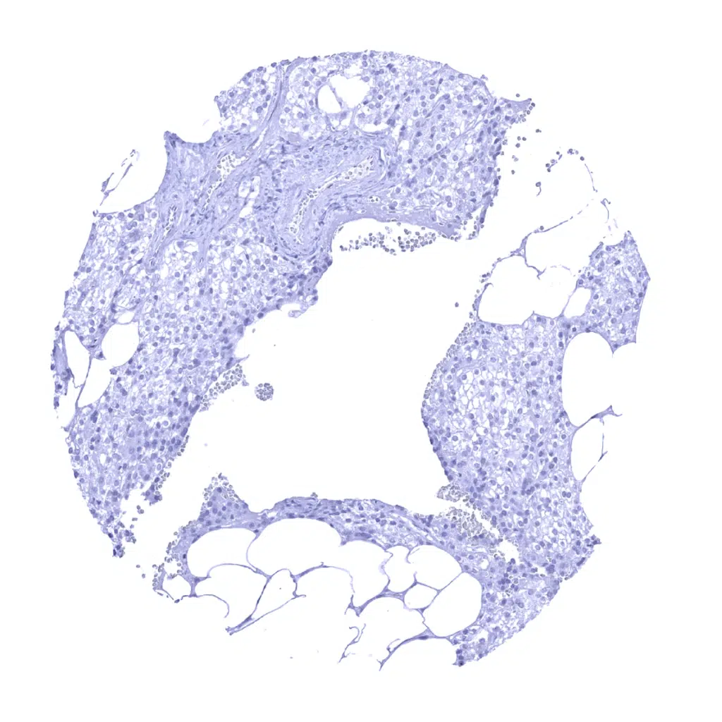
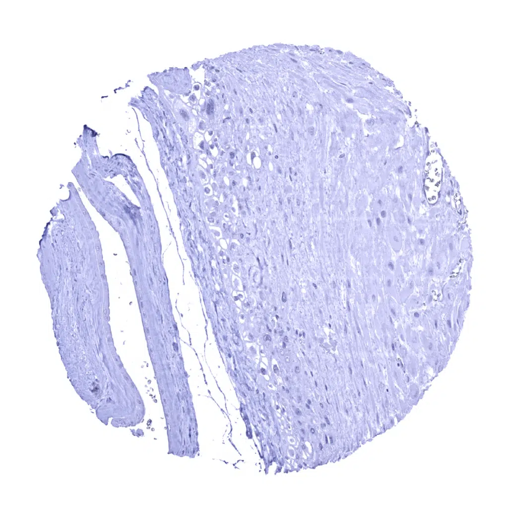
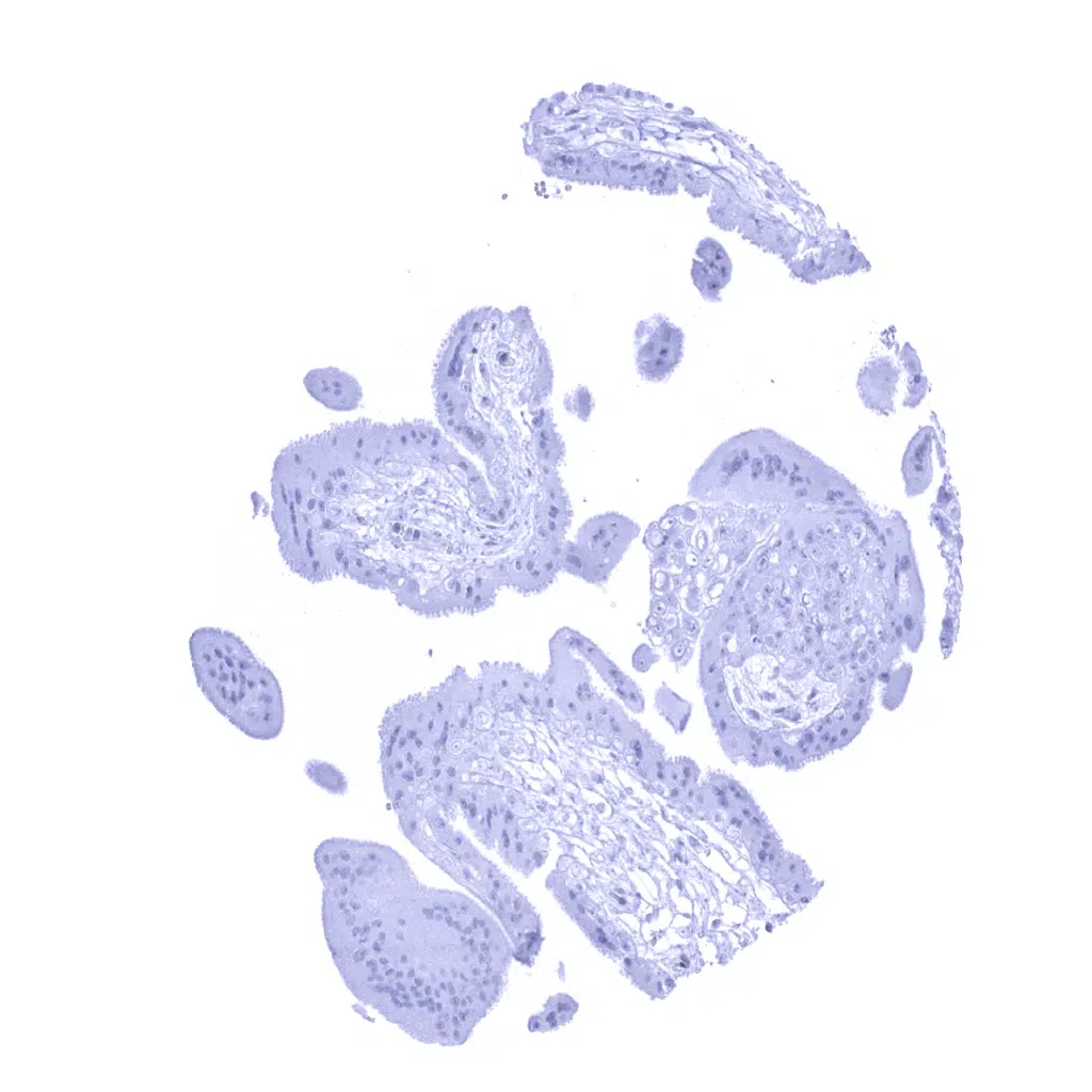
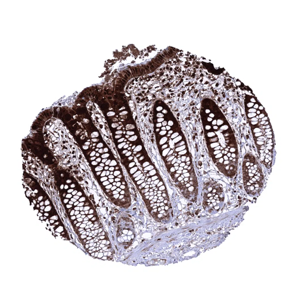
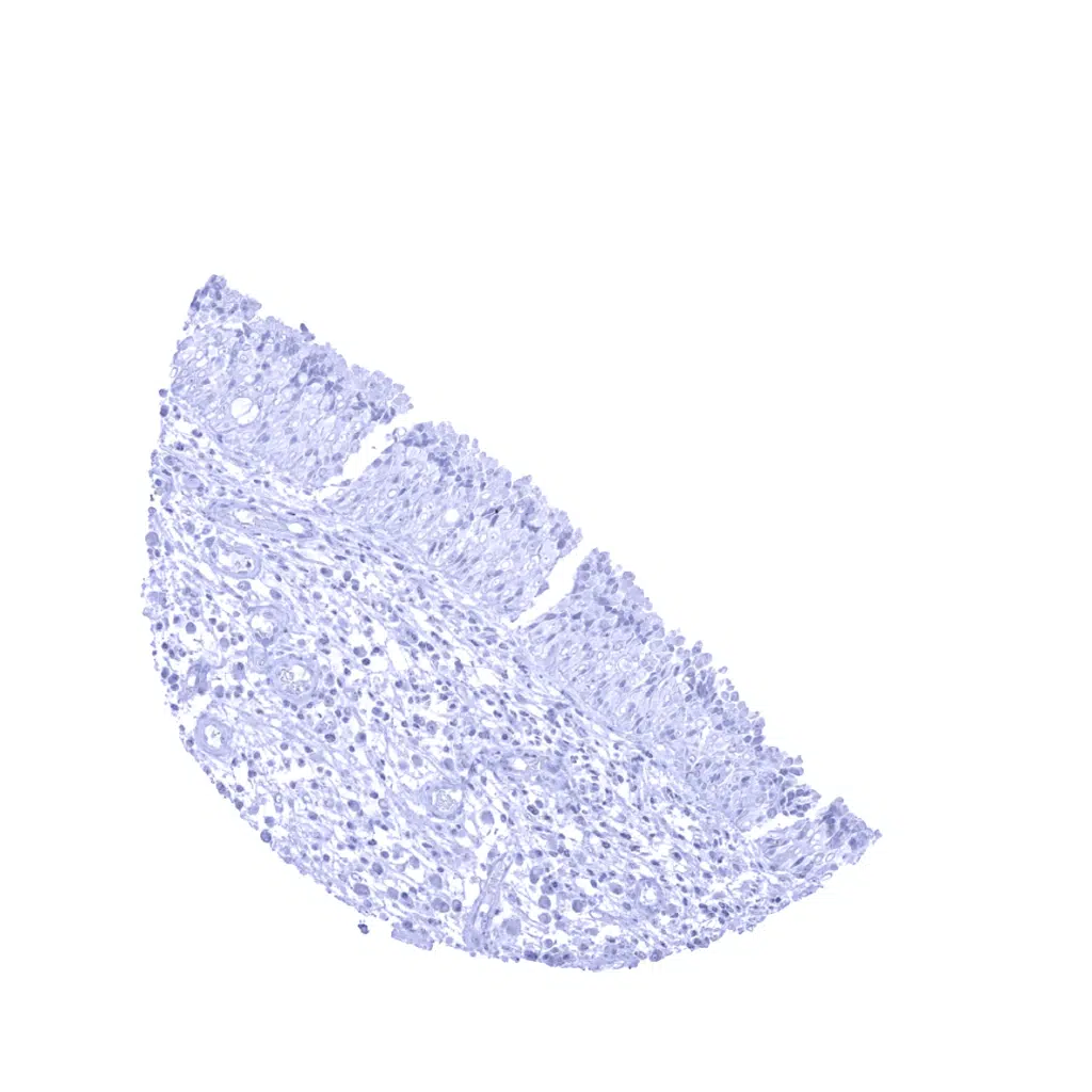
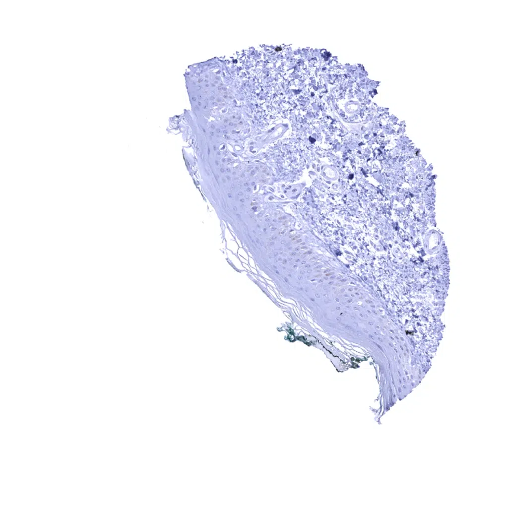
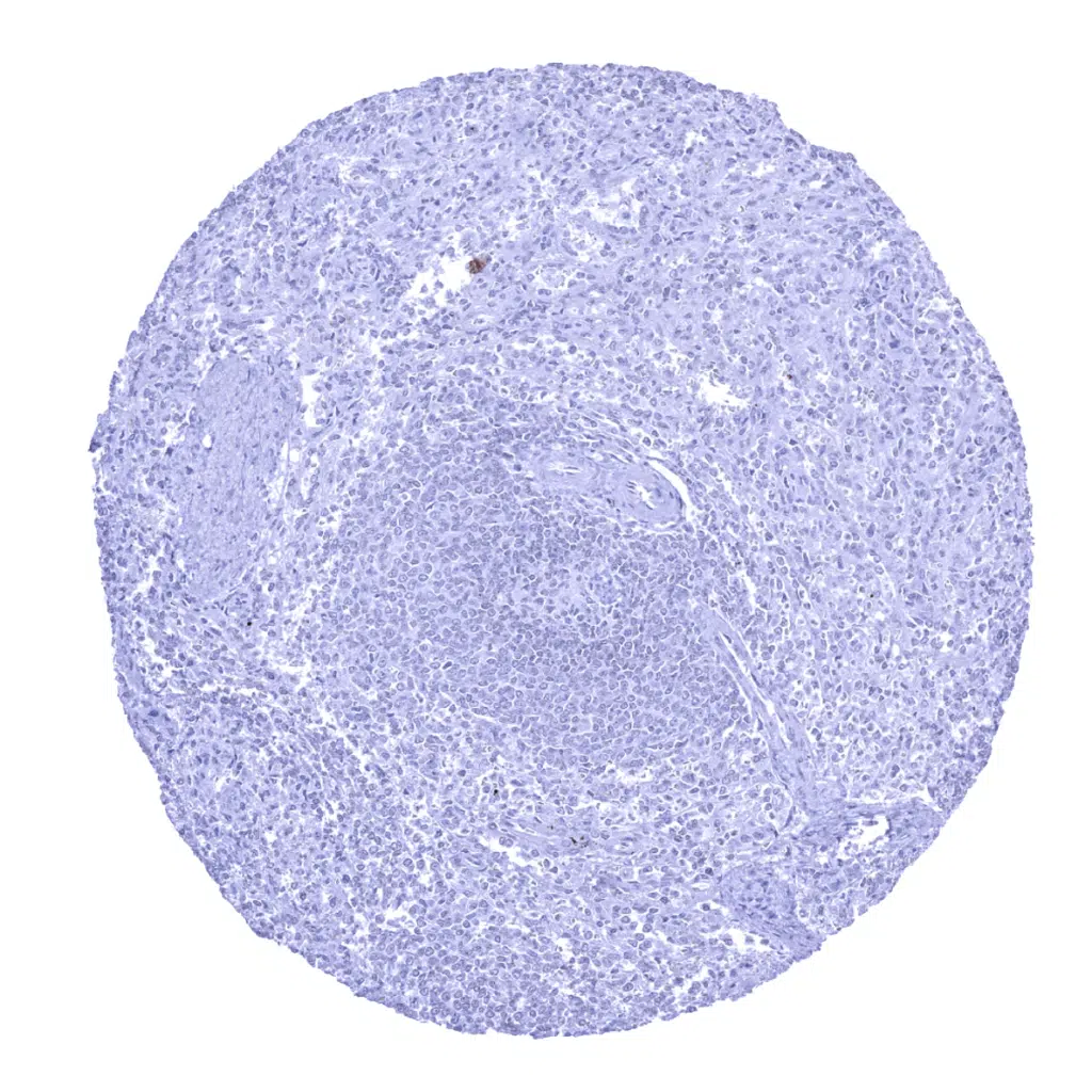
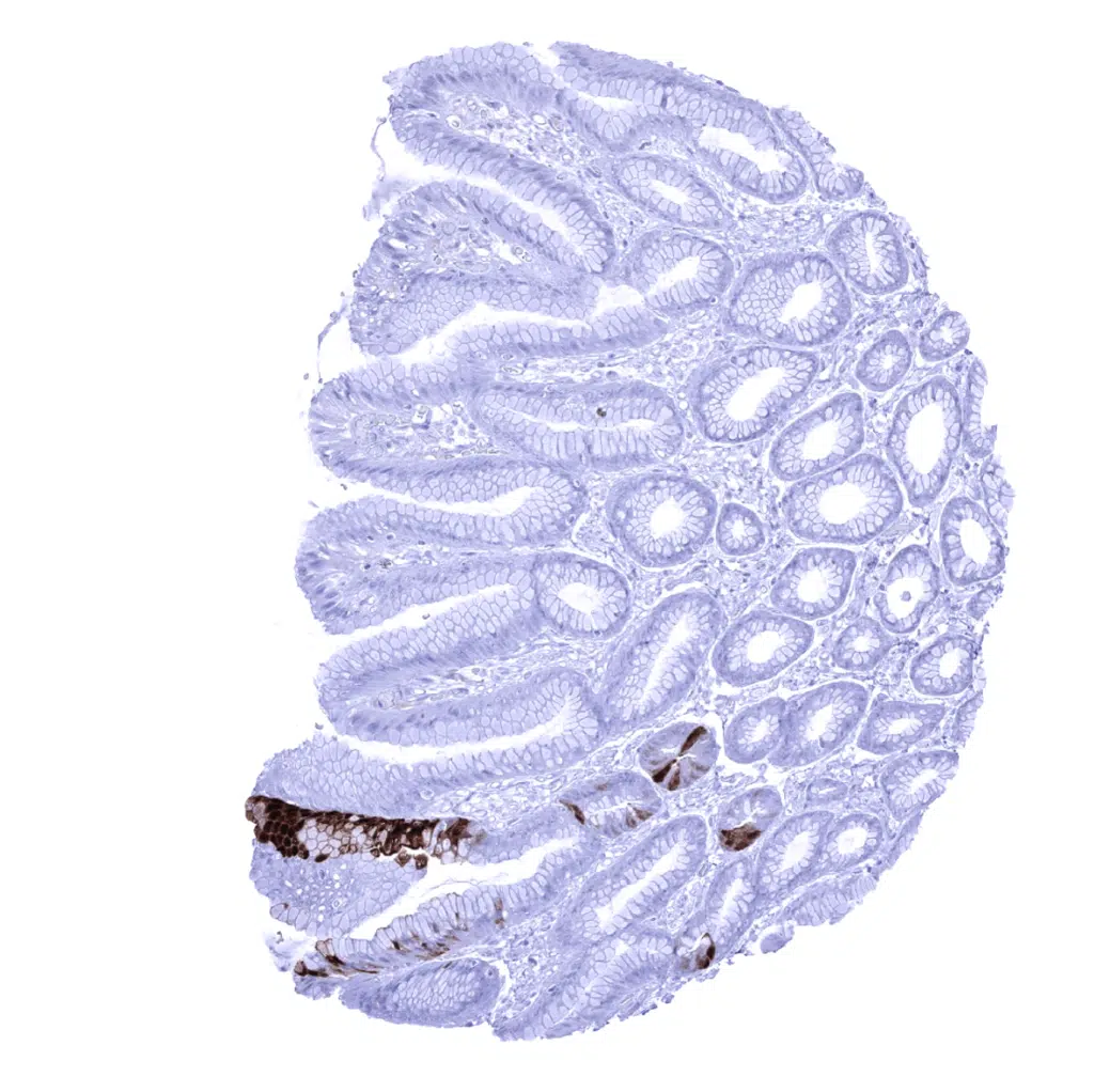
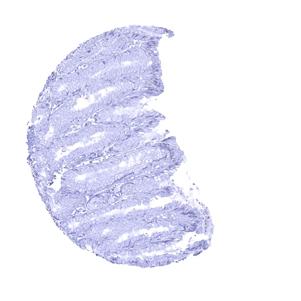
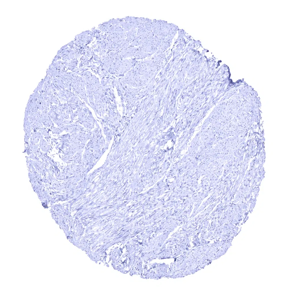
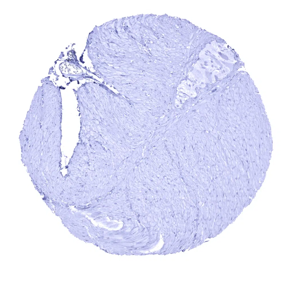
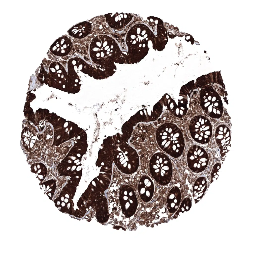
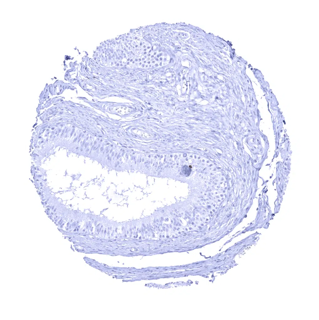
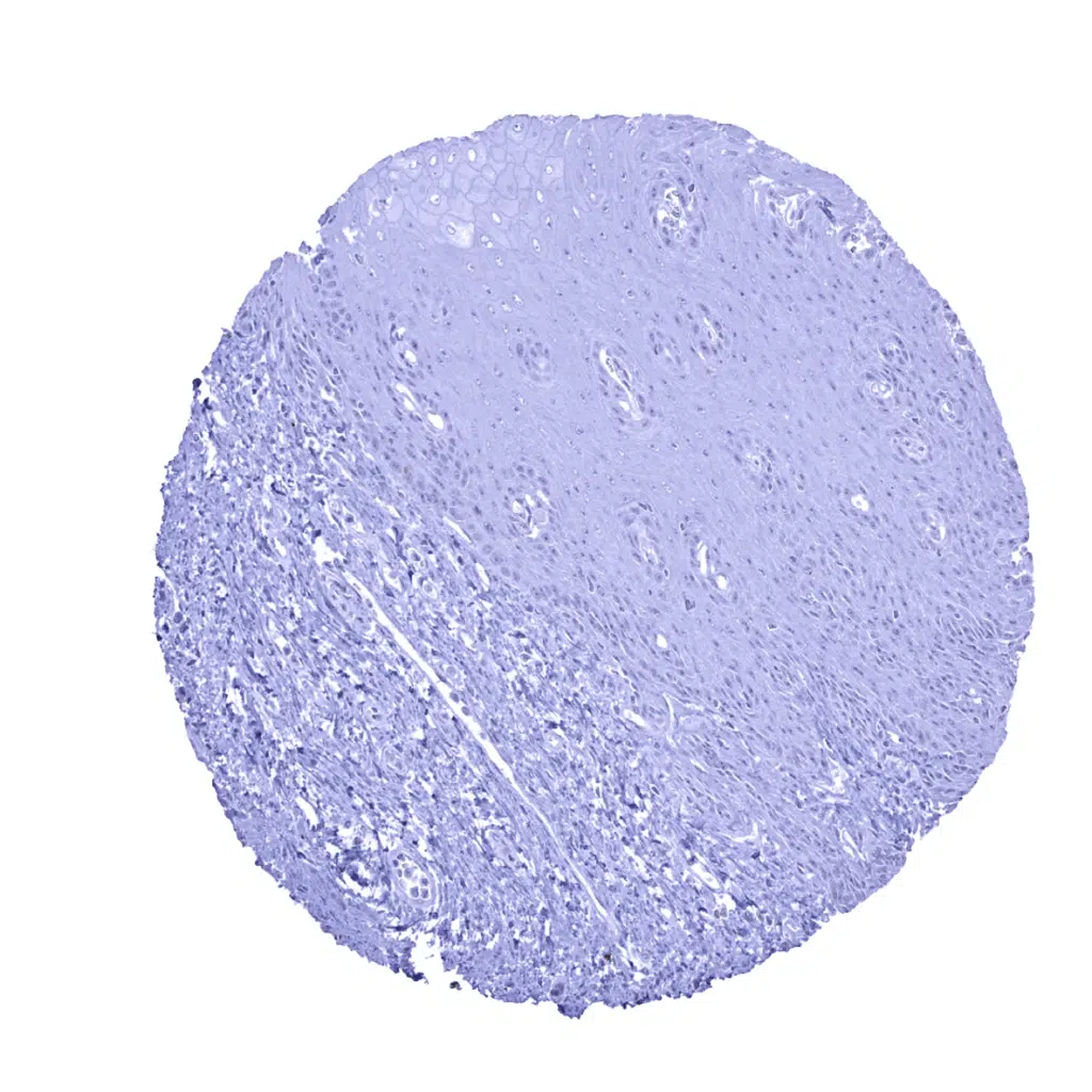
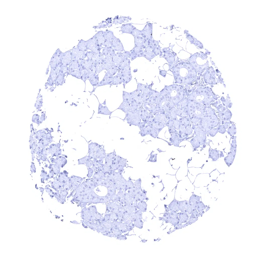
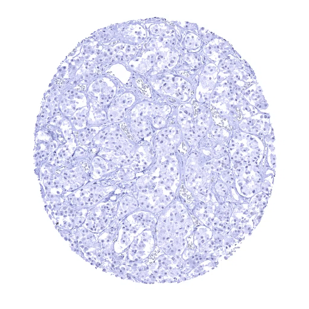
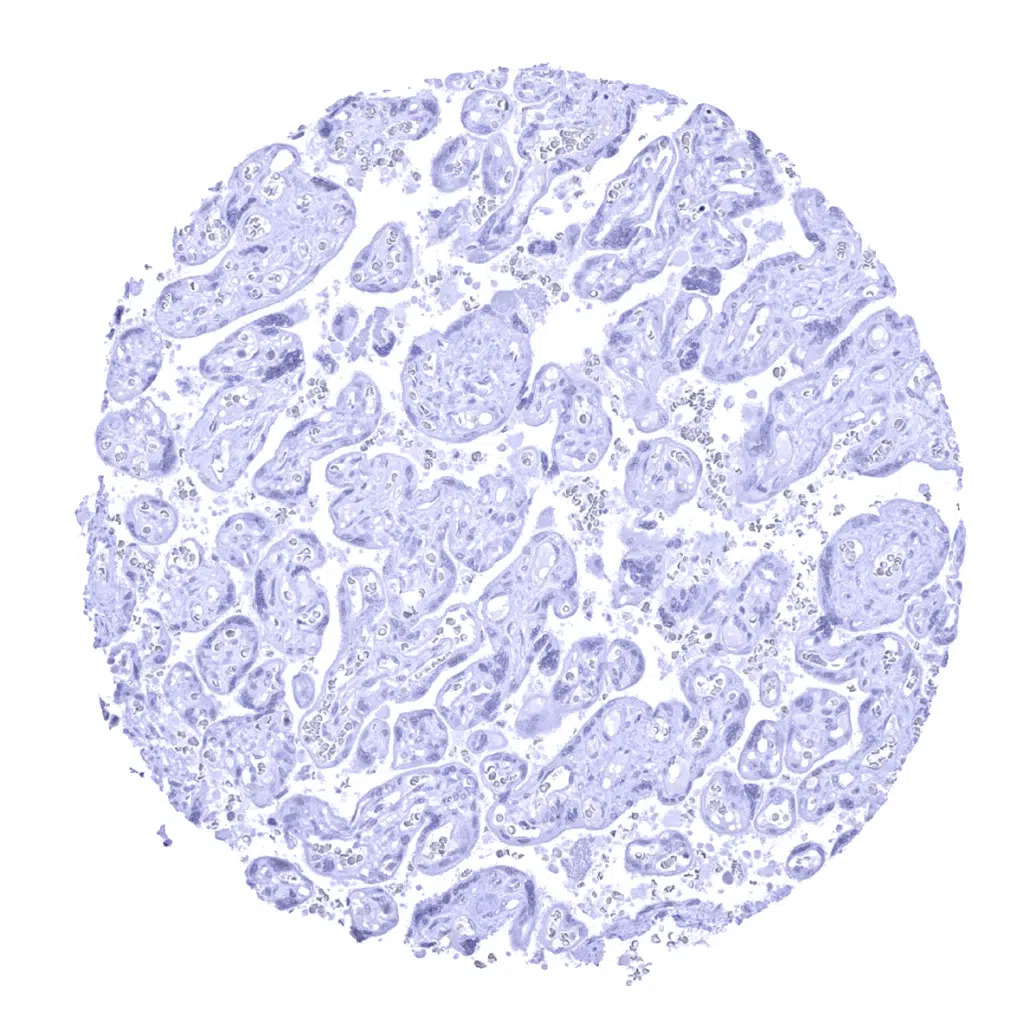
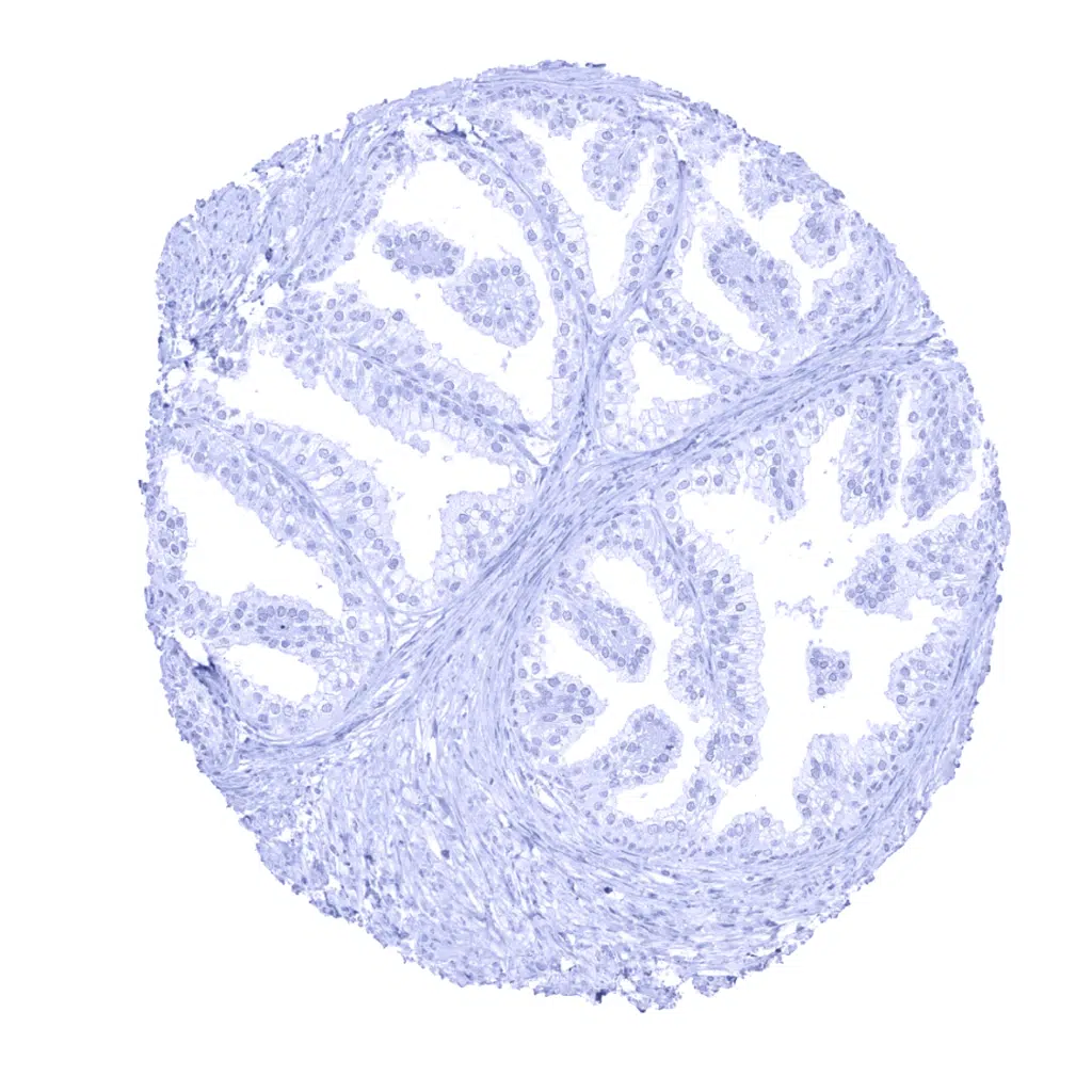
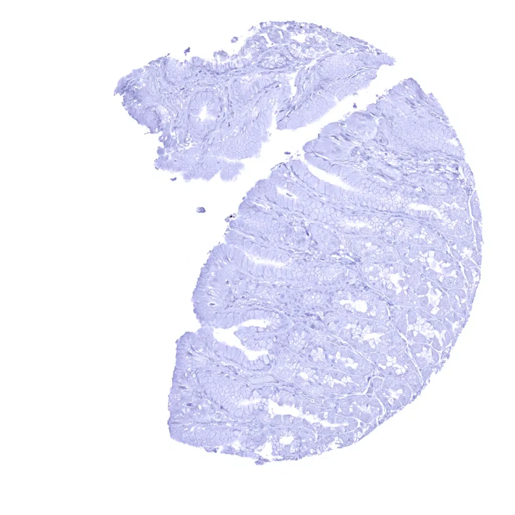
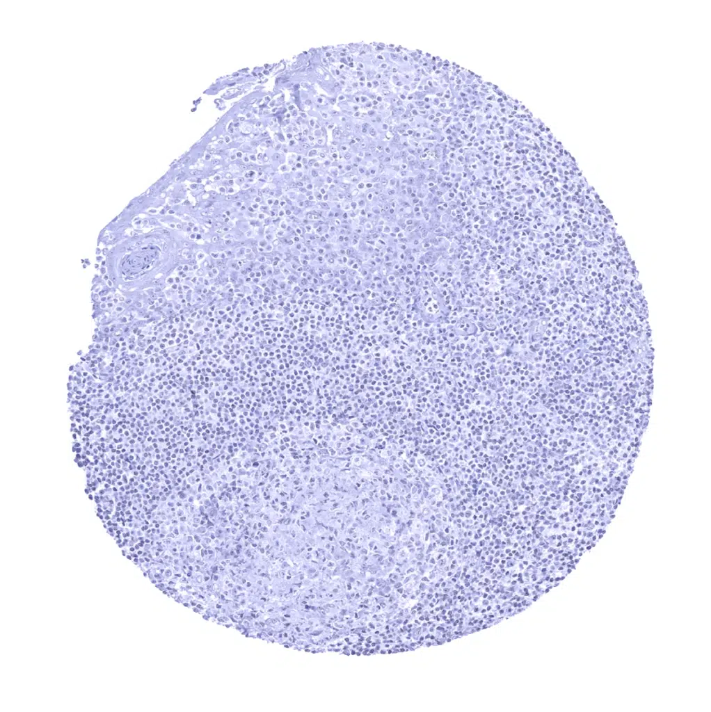
L-FABP antibody [MSVA-501M] HistoMAX


Go to Cancer Tissue Gallery
  
 Go back to product page
  

Go to HistoMAX page


Adrenal gland

Aorta, media

Appendix , mucosa – Strong L-FABP immunostaining of the surface cell layers of the epithelium of the appendix

Appendix, muscular wall


Bone marrow

Breast

Bronchus, mucosa

Cerebellum (molecular, Purkinje and, granule cell layers; white matter)


Cerebellum (molecular layer, Purkinje cell layer, granule cell layer, white matter)1

Cerebrum, grey matter

Cerebrum, white matter

Colon descendens, muscular wall


Colon descendes, mucosa – Strong L-FABP immunostaining of colon epithelial cells. Due to very high levels of L-FABP protein in these epithelial cells, staining of adjacent tissues can occur (contamination artifact)

Duodenum, Brunner gland

Duodenum, mucosa – Strong L-FABP immunostaining of duodenal epithelial cells. Due to very high levels of L-FABP protein in epithelial cells, staining of adjacent tissues can occur (contamination artifact)

Duodenum, mucosa – Strong L-FABP immunostaining of the surface cell layers of the duodenal epithelium


Epididymis

Esophagus, squamous epithelium

Fallopian tube, mucosa

Fat


Gallbladder, epithelium

Heart muscle

Ileum, mucosa – Strong L-FABP immunostaining of epithelial cells. Due to very high levels of L-FABP protein in epithelial cells, some staining of adjacent tissues is seen (contamination artifact)

Kidney, cortex – In the kidney, L-FABP immunostaining preferentially occurs in proximal tubuli


Kidney, medulla

Liver – Strong L-FABP immunostaining of hepatocytes. Due to very high levels of L-FABP protein in these cells, staining of adjacent tissues can occur (contamination artifact)

Lung

Lymph node


Ovary, stroma

Pancreas

Parathyroid

Parathyroid


Pituitary gland, anterior lobe

Pituitary gland, posterior lobe

Placenta (amnion and chorion)

Placenta, early


Placenta, mature

Prostate

Rectum, mucosa – Strong L-FABP immunostaining of epithelial cells of the rectum. Staining is more intense at the surface then at the base of the crypts. Due to high levels of L-FABP protein in these epithelia, staining of adjacent tissues and accumulation in mucosa associated macrophages can occur (contamination artifact).

Seminal vesicle


Sinus paranasales

Skeletal muscle

Skin

Spleen


Stomach, antrum – A focal L-FABP positivity of stomach antrum epithelium can occasionally be seen (1 of 8 samples)

Stomach, antrum

Stomach, corpus

Testis


Thymus

Thyroid gland

Tonsil, surface epithelium

Tonsil


Urinary bladder, muscular wall

Urinary bladder, urothelium

Uterus, ectocervix

Uterus, endocervix


Uterus, endometrium (pregnancy)

Uterus, endometrium (pregnancy)

Uterus, endometrium (secretion)

Uterus, myometrium


| TOP |