Normal Tissue Gallery- GTX04409

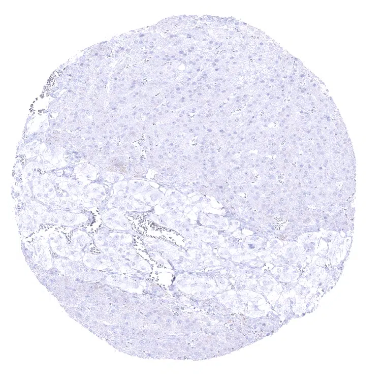
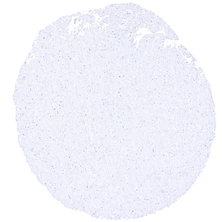
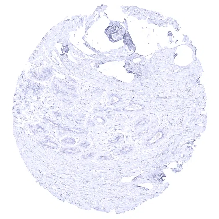
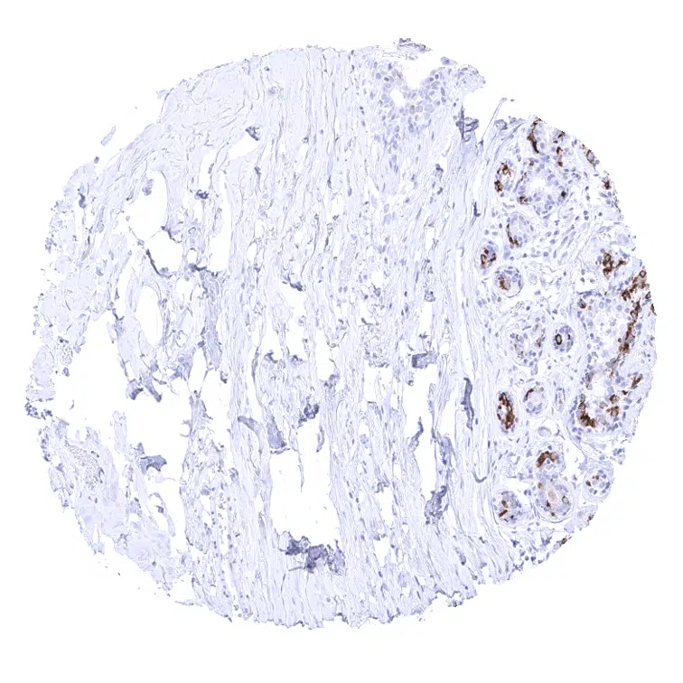
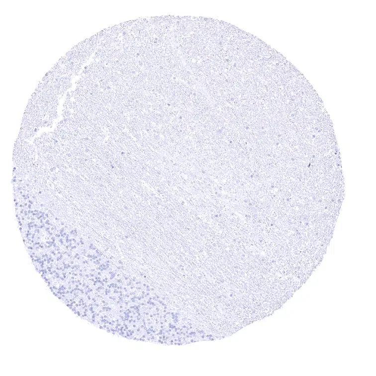
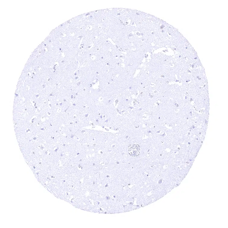
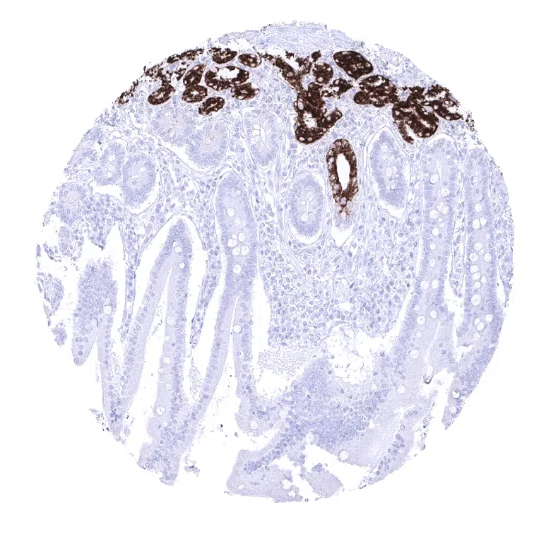
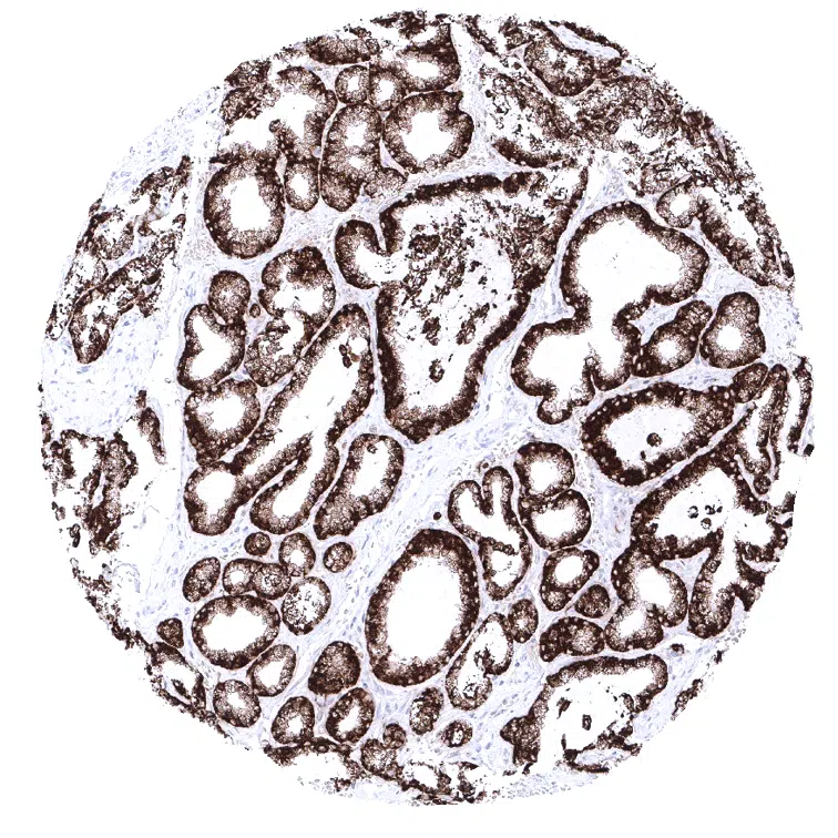
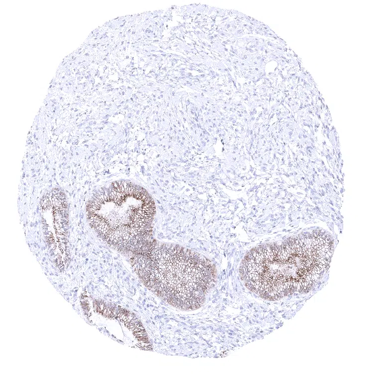
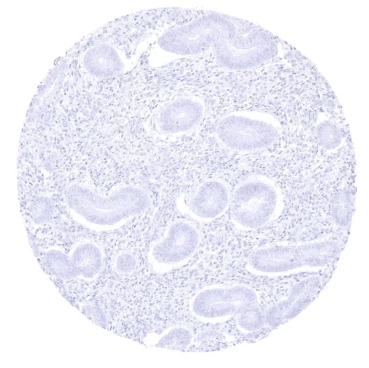
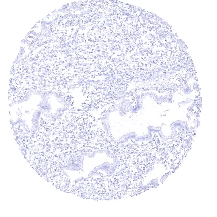
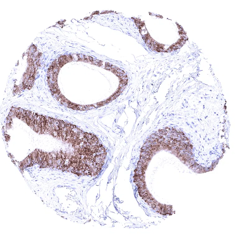
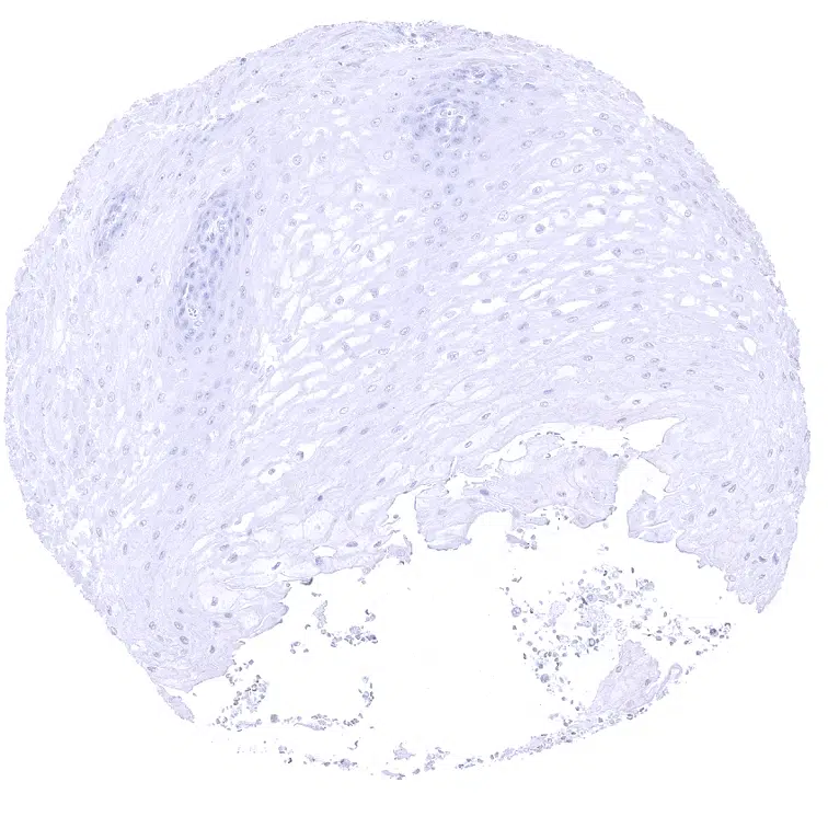
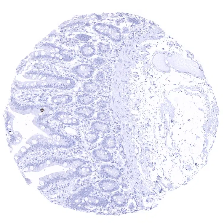
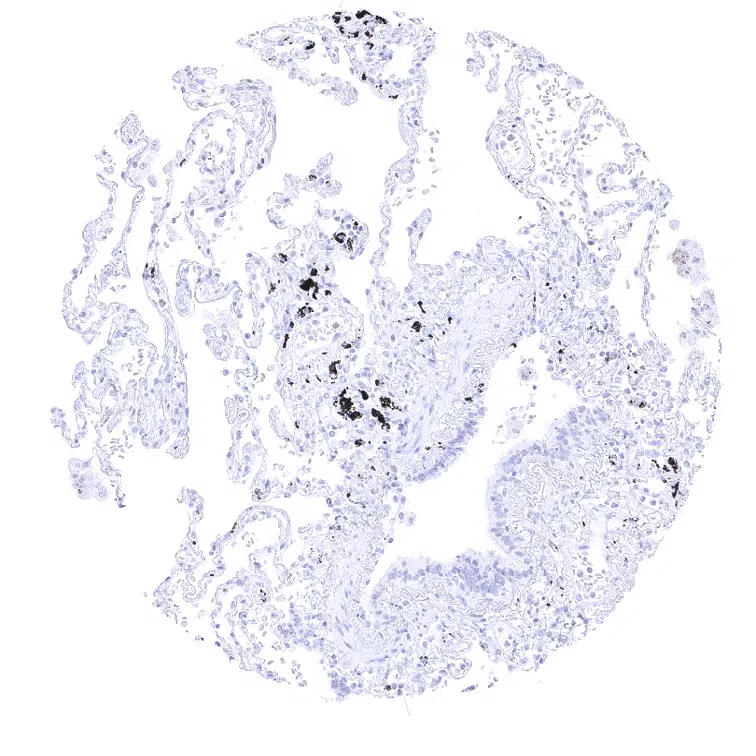
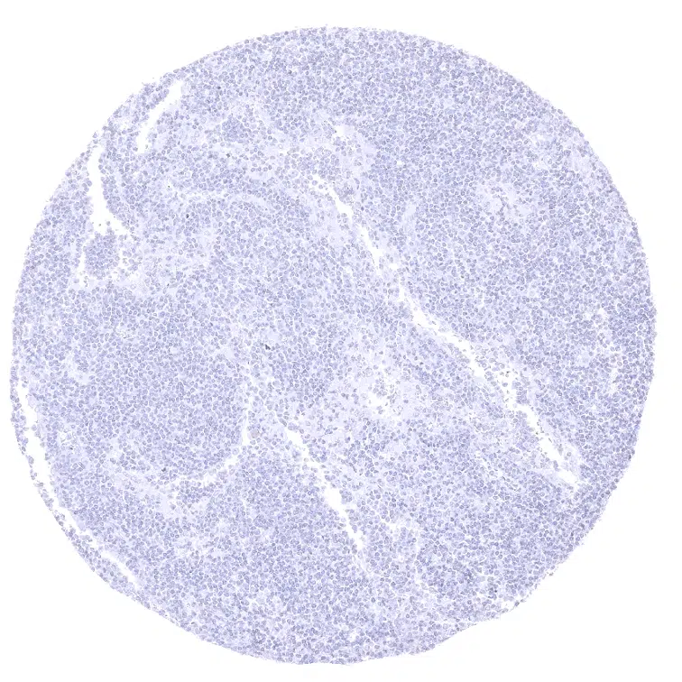
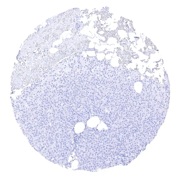
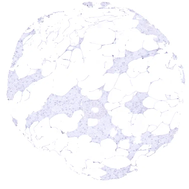
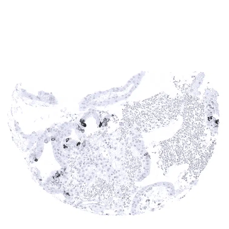
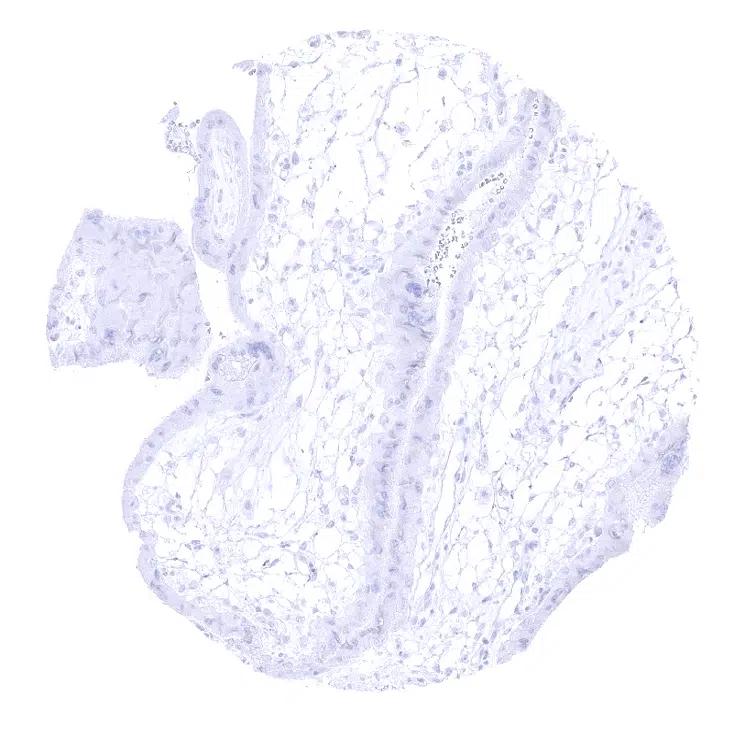
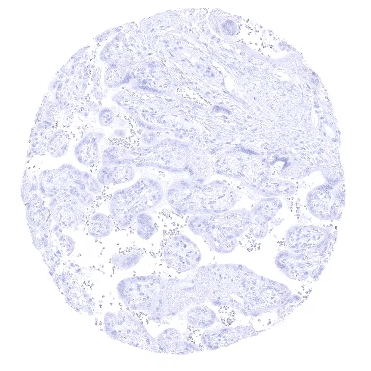
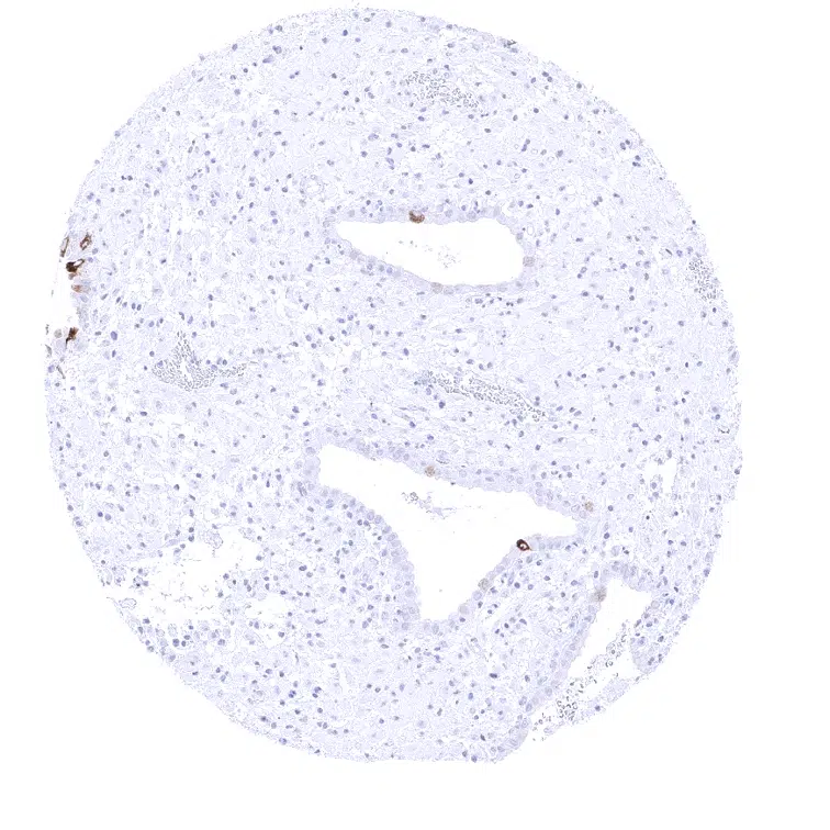
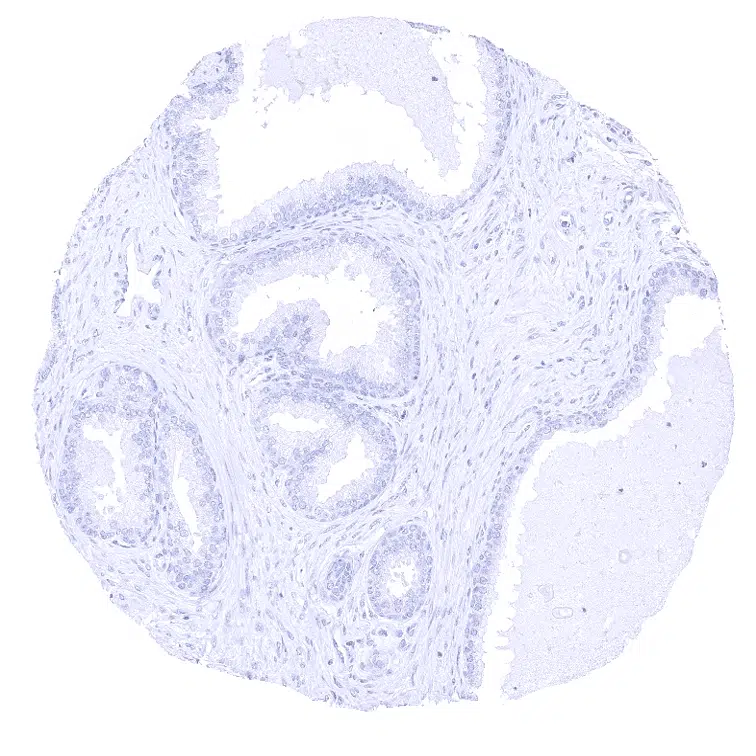
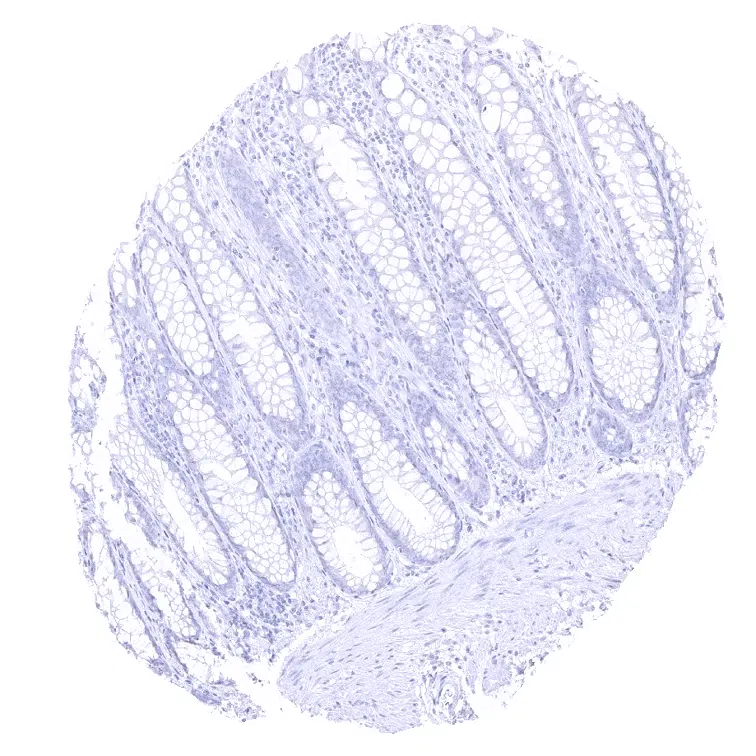
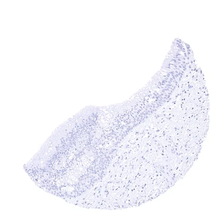
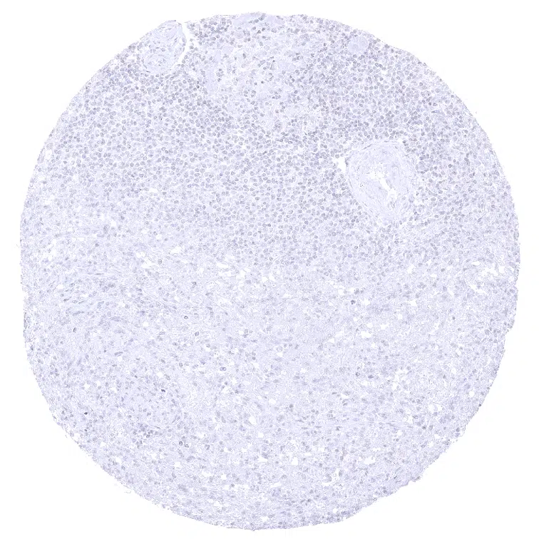
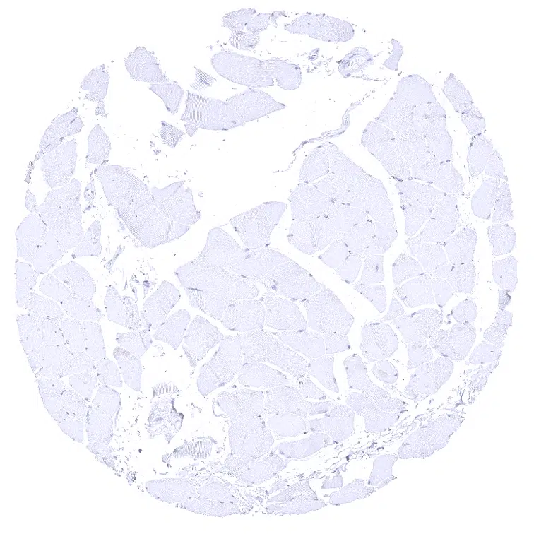
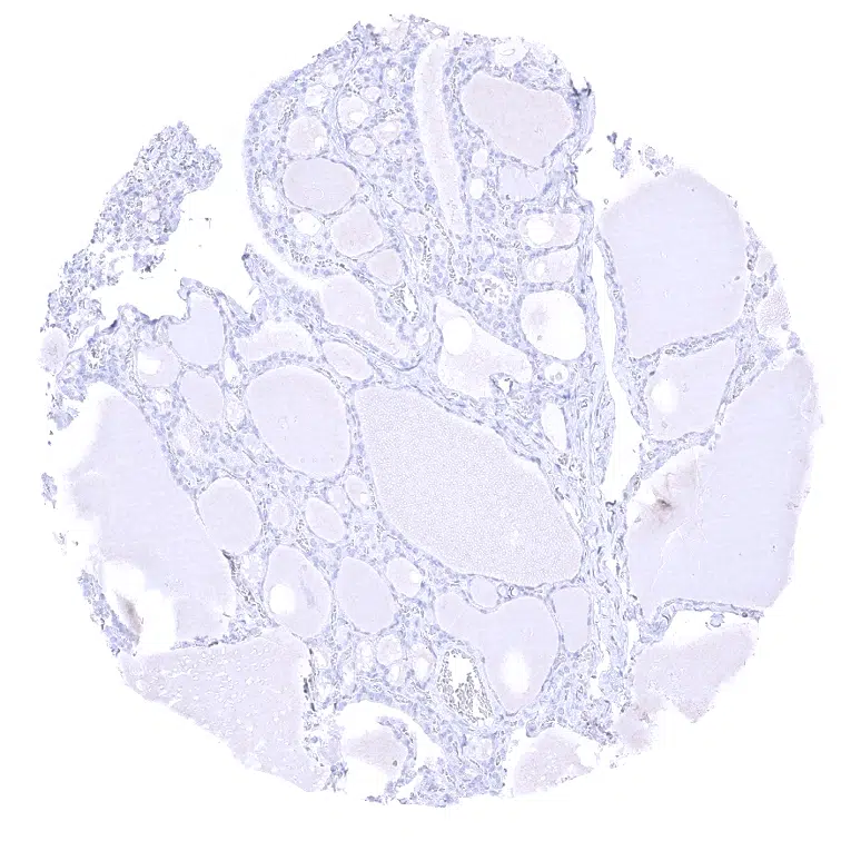
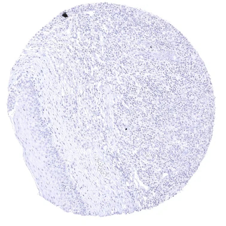
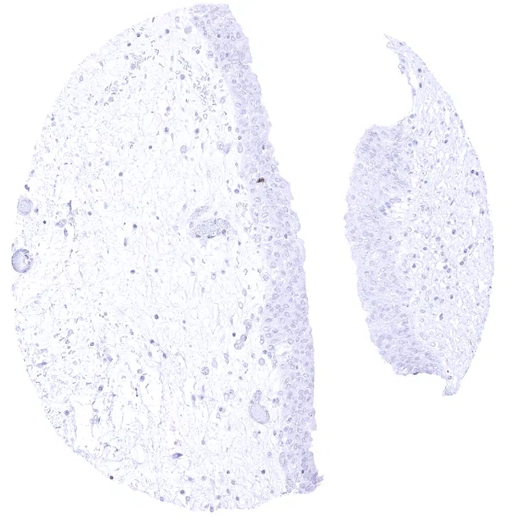
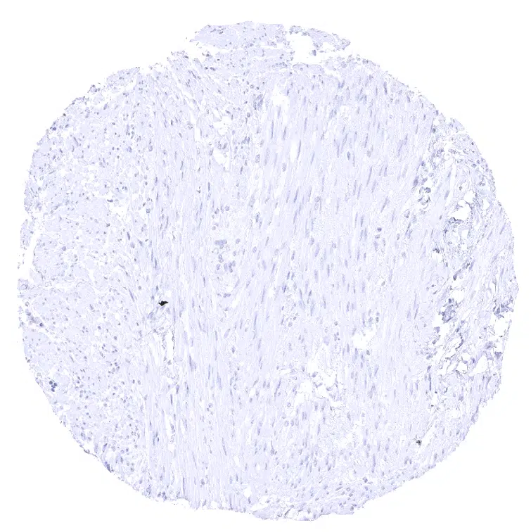
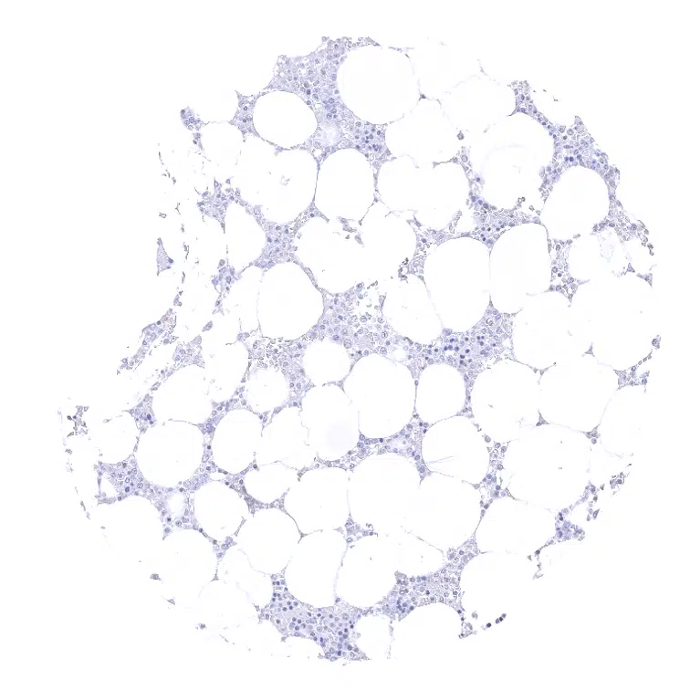
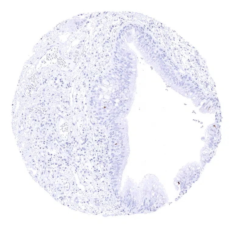
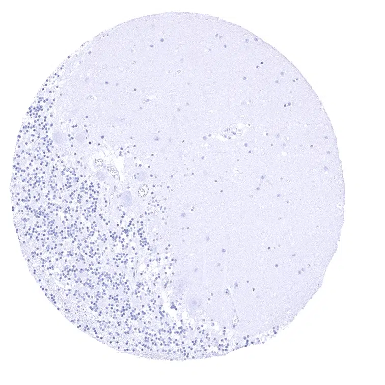
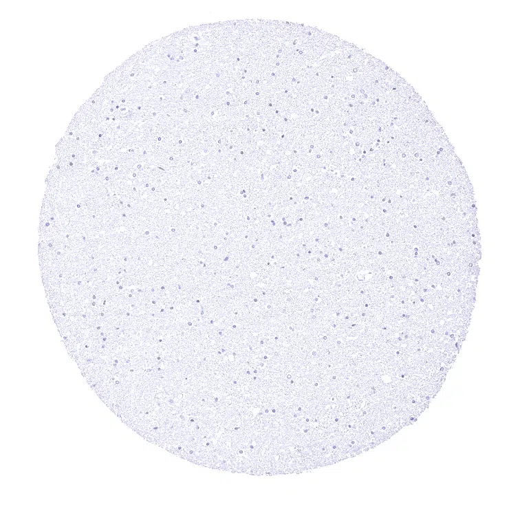
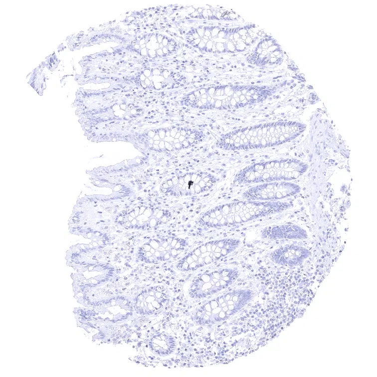
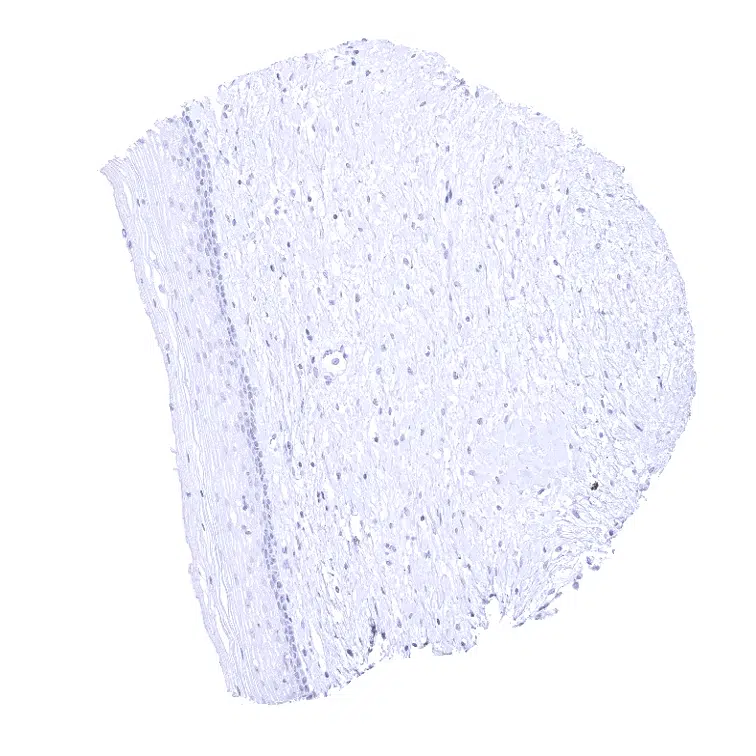
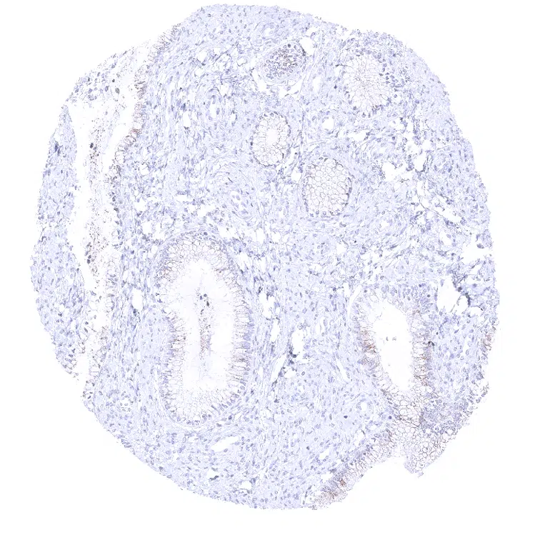
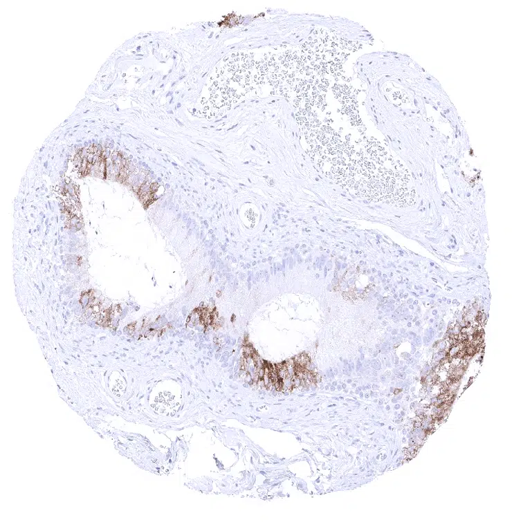
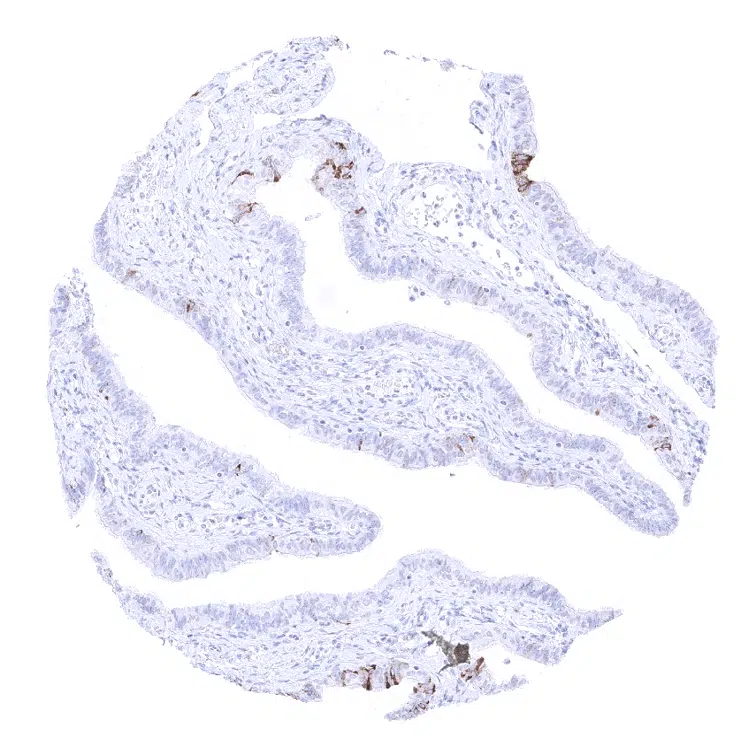
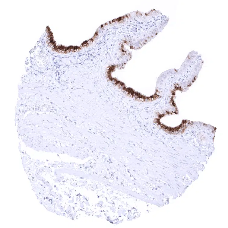
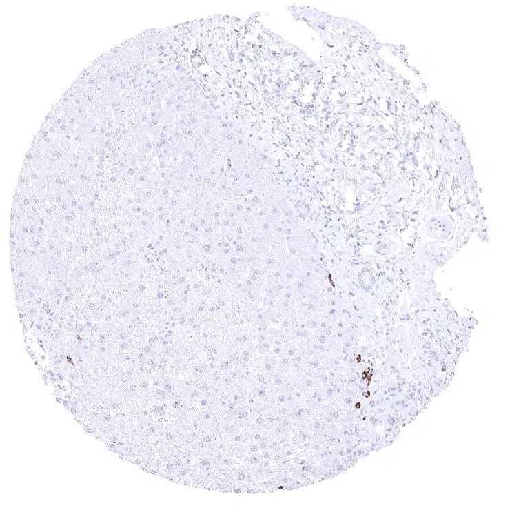
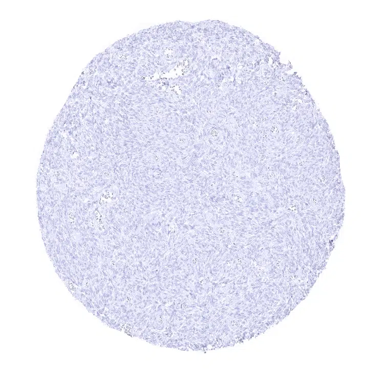
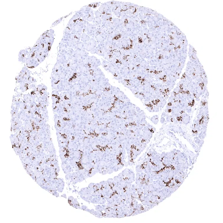
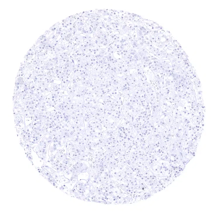
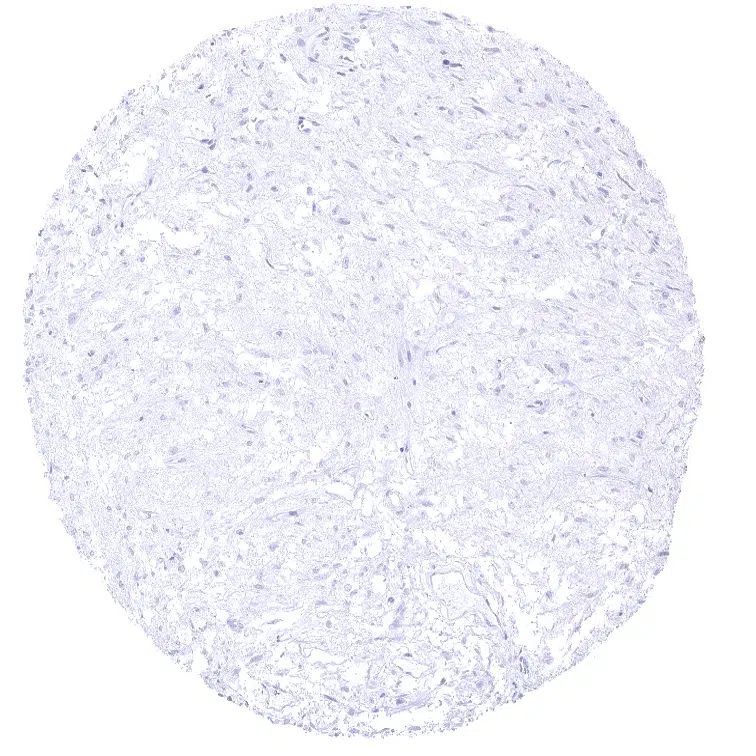
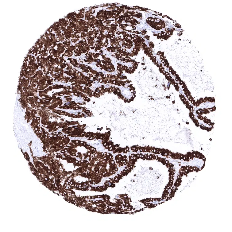
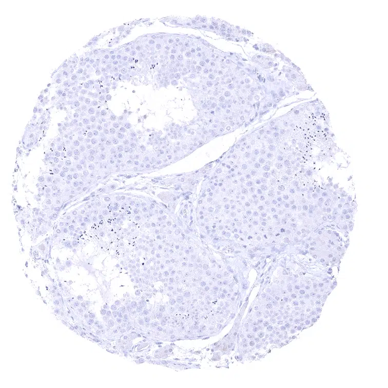
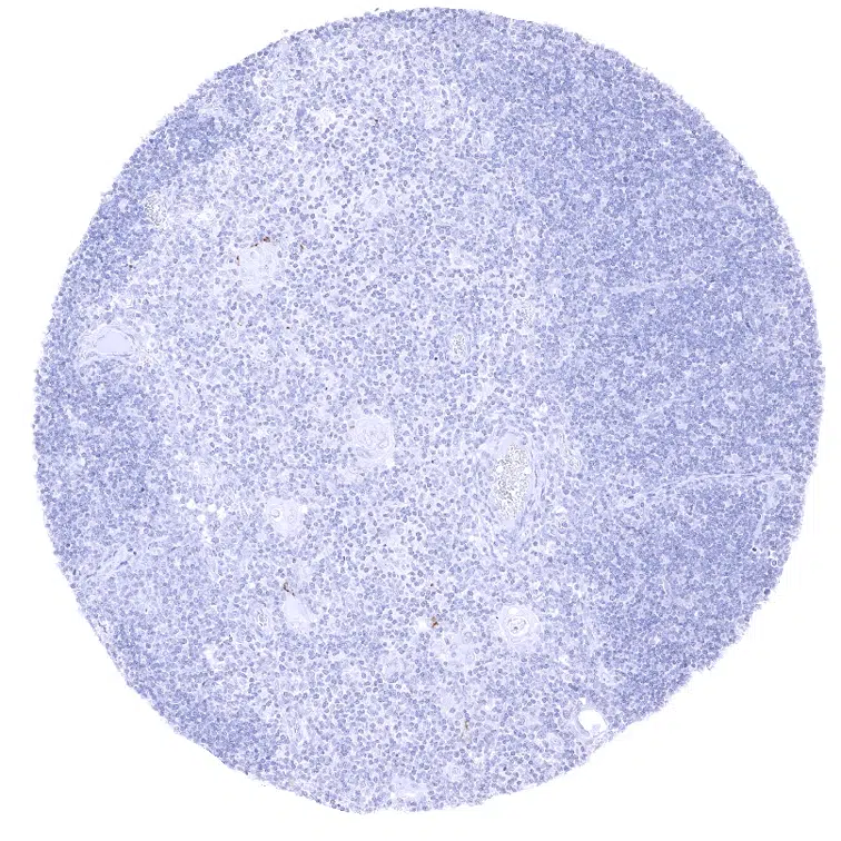
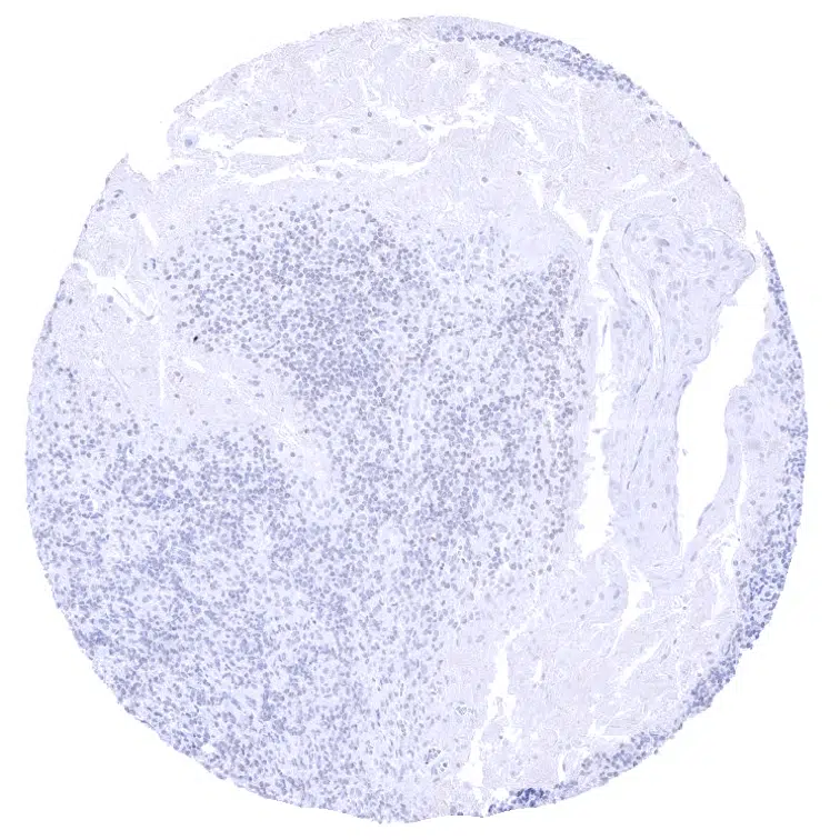
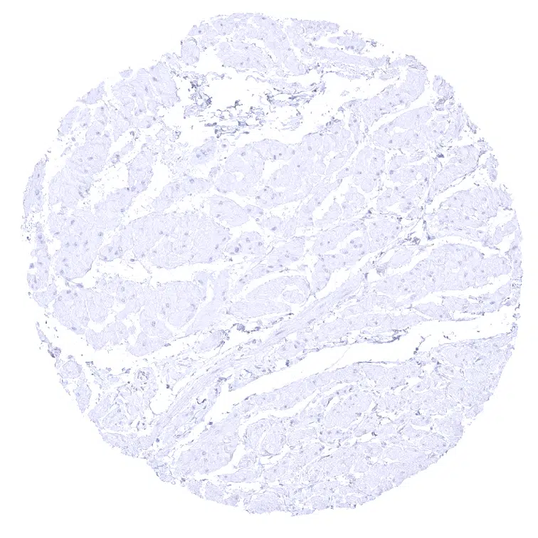
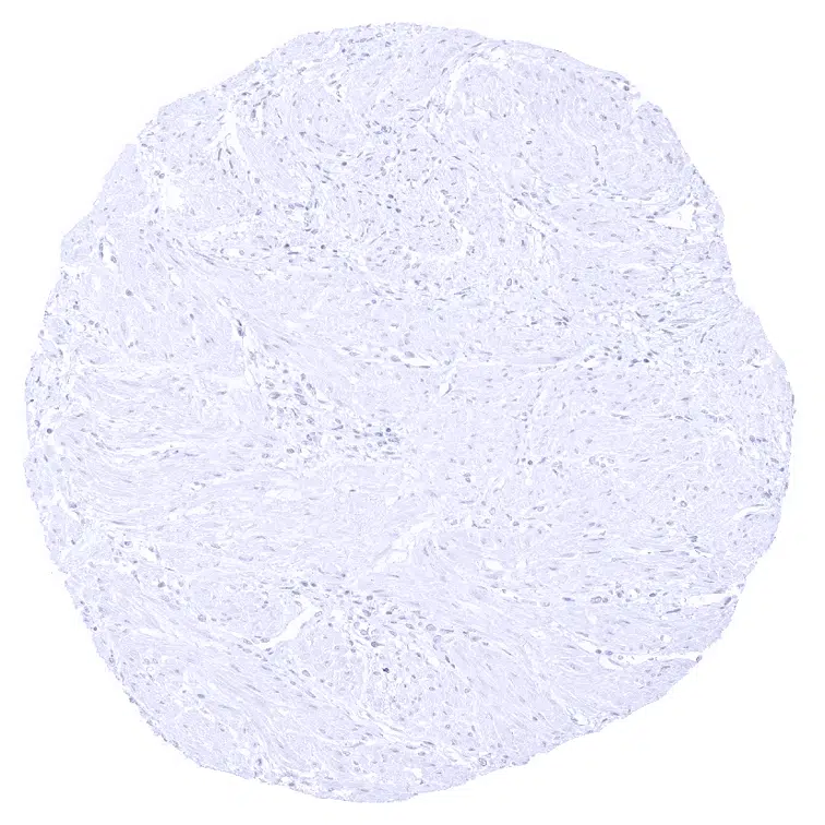
Mucin 6 antibody [MSVA-806R] HistoMAX


Go to Cancer Tissue Gallery
  
 Go back to product page
  

Go to HistoMAX page


Adrenal gland

Aorta, Media

Appendix, Mucosa

Appendix, muscular wall


Bone marrow

Breast

Breast – MUC6 immunostaining can be seen in luminal cells of breast glands (not in all samples).

Bronchus, mucosa


Cerebellum (molecular layer, Purkinje cell layer, granule cell layer)

Cerebellum (granule cell layer, white matter)

Cerebrum, grey matter

Cerebrum, white matter


Colon descendens, Mucosa

Colon descendens, muscular wall

Duodenum mucosa – Strong MUC6 immunostaining at the transition zone towards Brunner glands.

Duodenum, Brunner gland – Strong MUC6 immunostaining of all glands.


Ectocervix

Endocervix – A weak to moderate MUC6 immunostaining may be seen in individual endocervical glands.

Endocervix – MUC6 immunostaining ranges from weak to moderate in endocervical glands.

Endometrium, proliferation


Endometrium, secretion

Epididymis – MUC6 immunostaining is variable in the Epididymis. It is more intense in the cauda than in the caput, where positivity can be focal.

Epididymis – MUC6 immunostaining is variable in the Epididymis. It is most intense in the cauda.

Esophagus, squamous epithelium


Fallopian tube, mucosa

Fat

Gallbladder, epithelium – Strong MUC6 immunostaining of some but not all cells of the surface epithelium.

Heart


Ileum, mucosa

Kidney, cortex

Kidney, medulla

Liver – A strong MUC6 staining is regularly seen in small juxtaportal bile ducts while large bile ducts are MUC6 negative.


Lung

Lymph node

Ovary, stroma

Pancreas – Strong MUC6 staining of intercalated and interlobular ducts.


Parathyroid

Parotid gland

Pituitary gland, anterior lobe

Pituitary gland, posterior lobe


Placenta, early – Few scattered MUC6 positive cells can be seen in the trophoblast of the first trimenon placenta.

Placenta, early

Placenta, mature

Pregnant uterus (decidua)


Prostate

Rectum, mucosa

Seminal vesicle – Strong MUC6 immunostaining of all epithelial cells.

Sinus paranasales


Skin

Spleen

Stomach, antrum – Moderate to strong MUC6 staining of mucus producing cells, but not of surface epithelial cells.

Stomach, corpus – Moderate to strong MUC6 staining of mucus producing cells, but not of surface epithelial cells.


Striated muscle

Testis

Thymus

Thyroid gland


Tonsil, surface epithelium

Tonsil

Urinary bladder, muscular wall

Urinary bladder, urothelium


Uterus, myometrium


| TOP |