Normal Tissue Gallery- GTX640561

NFIX antibody [HMV329] HistoMAXTM


Go to Cancer Tissue Gallery Go back to product page Go to HistoMAX page


Adrenal gland (Cortex) – Weak to moderate nuclear NFIX staining staining of adrenocortical cells
Adrenal gland (Medulla) – Distinct nuclear NFIX staining of few interspersed stromal (sustentacular cells_ endothelial_) cells while medullary cells remain negative
Aorta, media
Appendix, mucosa
Appendix, muscular wall – Distinct nuclear staining of smooth muscle cells and of other cell types
Bone marrow – Strong nuclear MRE11 staining of a rather small subset of cells (often arranged in groups)
Breast – Strong nuclear NFIX staining of a large subset of basal and luminal epithelial cells
Bronchus, mucosa – Distinct nuclear NFIX staining of most respiratory epithelial cells, but the staining intensity decreases markedly from the basal layer towards the more superficial cell layers
Cerebellum (white matter) – Weak to moderate nuclear NFIX staining of glial cells
Cerebellum, cortex (molecular layer, Purkinje cell layer, granule cell layer) – Weak nuclear NFIX staining of glial and granular cells. Lack of staining NFIX in Purkinje cell
Cerebrum, grey matter – Strong nuclear NFIX staining of glial cells. Weak or absent staining in neurons
Cerebrum, white matter – Strong nuclear NFIX staining of glial cells
Colon descendens, mucosa – Moderate nuclear NFIX staining of epithelial cells
Colon descendens, muscular wall – Distinct nuclear staining of smooth muscle cells and of other cell types
Duodenum, Brunner gland – Distinct nuclear MRE11 staining of all epithelial cells, including Brunner glands
Duodenum, mucosa – Nuclear NFIX staining is moderate in crypt bases and weak-faint or even absent in the surface epithelium
Epididymis (Caput and cauda) – Strong nuclear NFIX staining of epithelial cells in the cauda. In the caput, the NFIX staining is only faint in chief cells but strong in basal cells
Epididymis (Cauda) – Strong nuclear NFIX staining of epithelial cells
Esophagus, squamous epithelium – Significant decrease of nuclear NFIX staining of squamous epithelium from the basal-suprabasal to the superficial cell layers
Fallopian tube, mucosa – Strong nuclear NFIX staining of all cells
Fat
Gallbladder, epithelium – Nuclear NFIX staining is weak to moderate in epithelial cells and stronger in stromal cells
Heart muscle – Distinct nuclear staining of heart muscle cells and of other cell types
Ileum, mucosa – Nuclear NFIX staining is moderate in crypt bases and weak-faint or even absent in the surface epithelium
Kidney, cortex – Nuclear NFIX staining is generally low in all cell types and ranges from absent to faint-weak
Kidney, medulla – Nuclear NFIX staining is generally low in all cell types and ranges from absent to faint-weak
Kidney, pelvis, urothelium – Distinct nuclear NFIX staining of urothelial cells although there is a significant decrease of NFIX staining intensity from the basal to the superficial cell layers. Umbrella cells are largely NFIX negative
Liver – Nuclear NFIX staining is largely absent in hepatocytes. NFIX staining is only seen in stromal cells
Lung – Strong nuclear NFIX staining of all cell types (except macrophages- these remain NFIX negative)
Ovary, corpus luteum – Strong nuclear NFIX staining of stromal cells. Corpus luteum cells are NFIX negative
Ovary, stroma – Strong nuclear NFIX staining of stromal cells
Pancreas – Strong nuclear NFIX staining of epithelial cells of excretory ducts and of intercalated ducts. NFIX staining is markedly weaker in acinar cells
Pancreas – Strong nuclear NFIX staining of stromal and intercalated duct cells. NFIX staining is faint or absent in acinar cells
Parathyroid gland – Moderate nuclear NFIX staining of all epithelial cells
Parotid gland
Pituitary gland, anterior lobe – Only few cells show a moderate to strong nuclear NFIX staining
Pituitary gland, posterior lobe – Strong nuclear NFIX staining of all pituicytes
Placenta (amnion)
Placenta (chorion)
Placenta, early – Strong nuclear MRE11 staining of stromal cells while trophoblast cells remain negative
Placenta, mature – Strong nuclear NFIX staining of stromal cells while trophoblast cells remain negative
Prostate – Strong nuclear NFIX staining of stromal and all epithelial cells
Rectum, mucosa – Weak to moderate nuclear NFIX staining of epithelial cells. Slight but significant decrease of NFIX staining intensity from the crypt base to the superficial epithelial cell layers
Seminal vesicle – Strong nuclear NFIX staining of stromal and basal epithelial cells. NFIX staining is weak or absent in most luminal epithelial cells
Sinus paranasales – Distinct nuclear NFIX staining of respiratory epithelial cells but the staining intensity decreases from the basal layer towards the more superficial cell layers
Skeletal muscle – Distinct nuclear staining of skeletal muscle cells and of other cell types
Skin – Strong nuclear NFIX staining of squamous epithelial cells
Skin, hairfollicel and sebaceous glands – Weak to moderate nuclear NFIX staining of sebaceous gland cells. The staining intensity decreases towards the centre of the glands
Spleen – Distinct nuclear NFIX staining of some stromal and endotheliel cells. Lymphocytes are NFIX negative
Stomach, antrum – Moderate to strong nuclear NFIX staining of most epithelial cells but few glandular cells do only show faint or even absent NFIX staining
Stomach, muscular – Distinct nuclear staining of smooth muscle cells and of other cell types
Testis – Distinct nuclear NFIX staining of intertubular cells (including Leydig cells). Complete absence of NFIX staining in tubular cells (Sertoli cells, germ cells)
Thymus – Lymphocytes are NFIX negative. Distinct nuclear NFIX staining of epithelial cells. Maturing squamous epithelial cells of corpuscles of Hassall's stain weakly or remain negative
Thymus – Lymphocytes are NFIX negative. Distinct nuclear NFIX staining of thymic epithelial cells
Thyroid gland – Strong nuclear MRE11 staining of follicular cells
Tonsil – Distinct nuclear NFIX staining of a subset of squamous epithelial cells. Decreasing staining intensity from basal to the superficial cell layers. Some stromal cells and endothelial cells are strongly positive. Lymphocytes are largely negativ
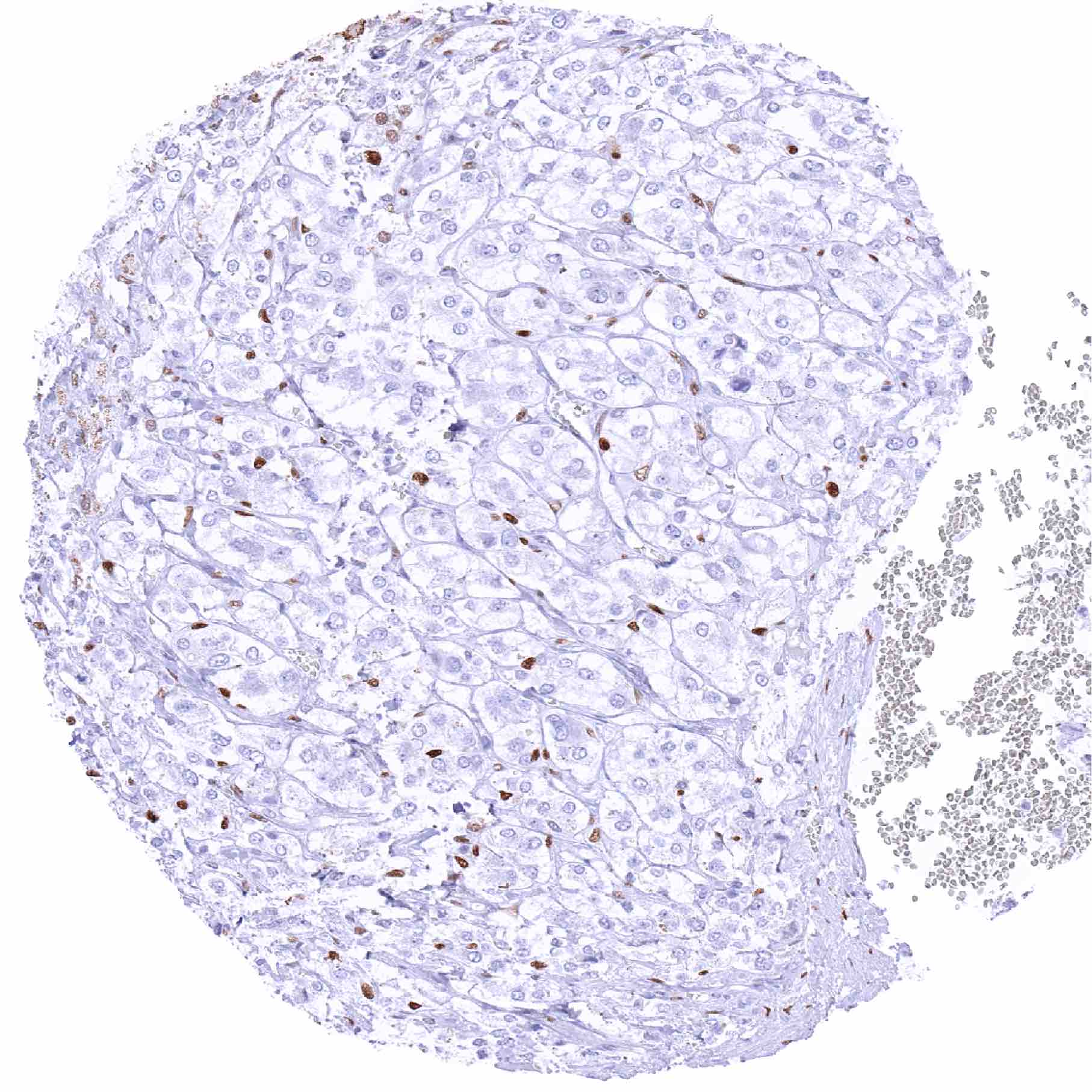
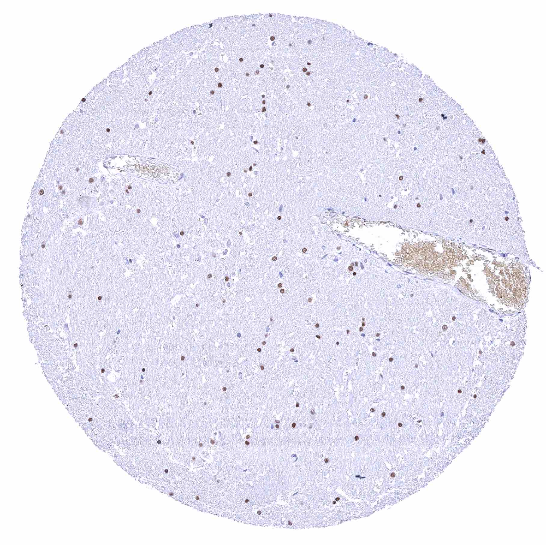
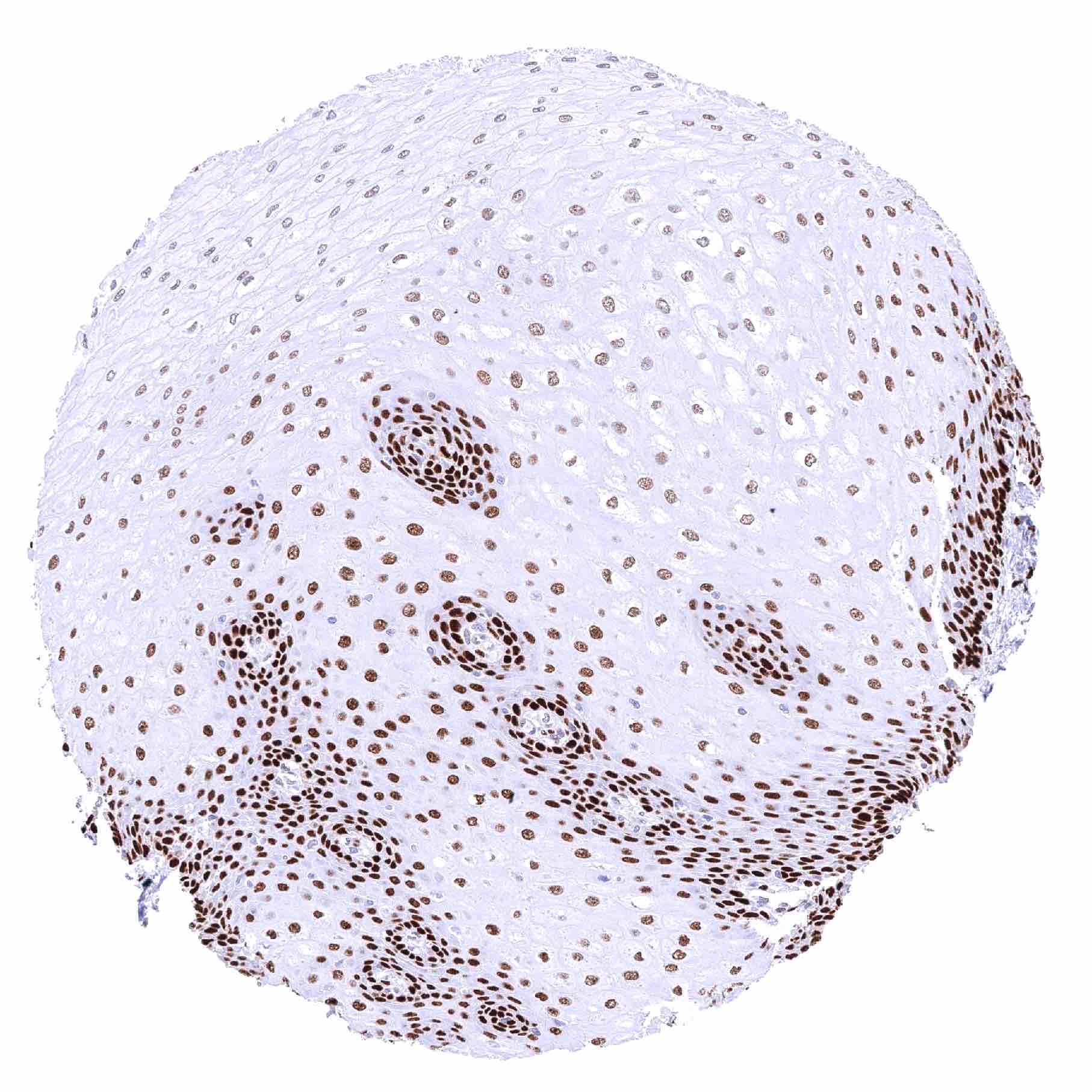
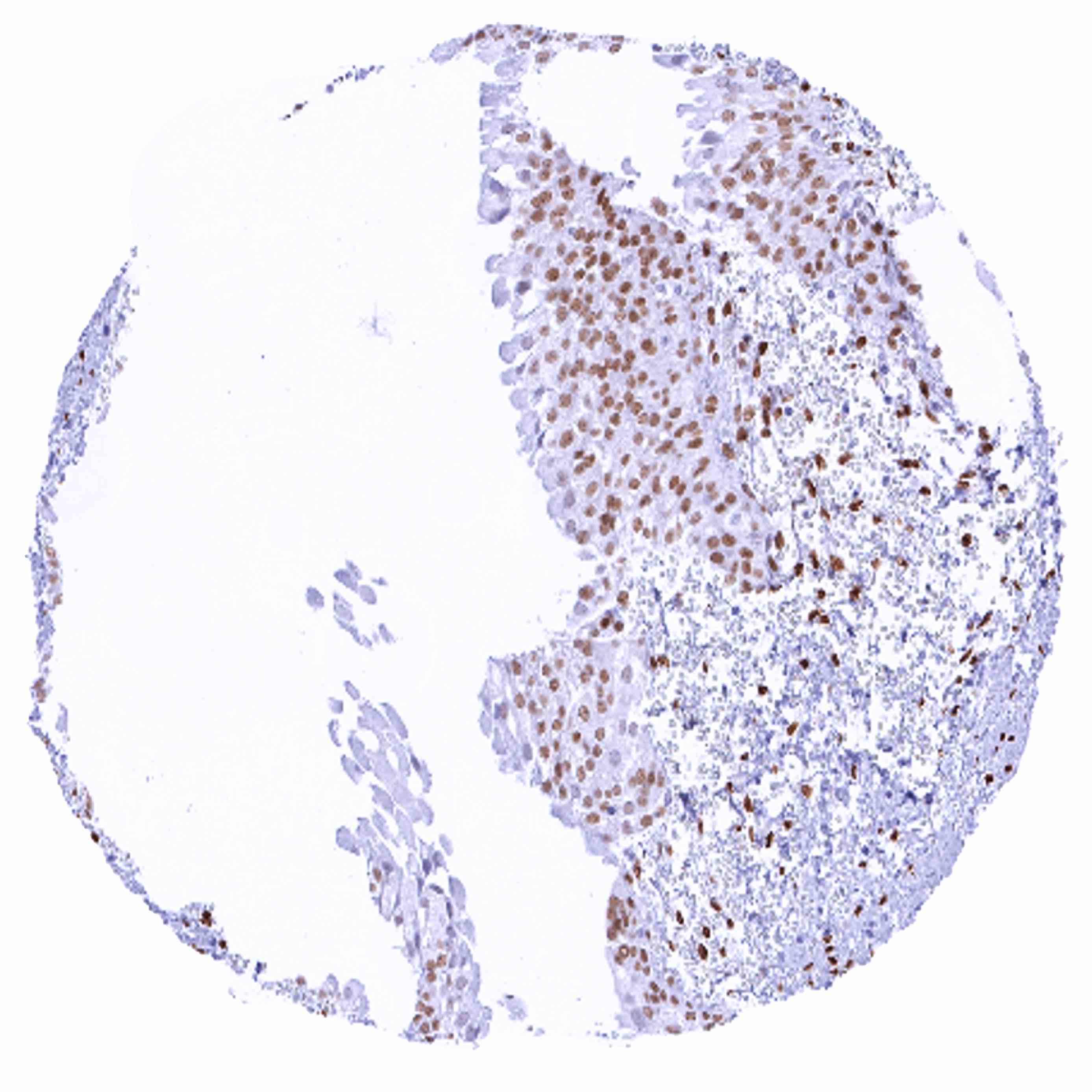
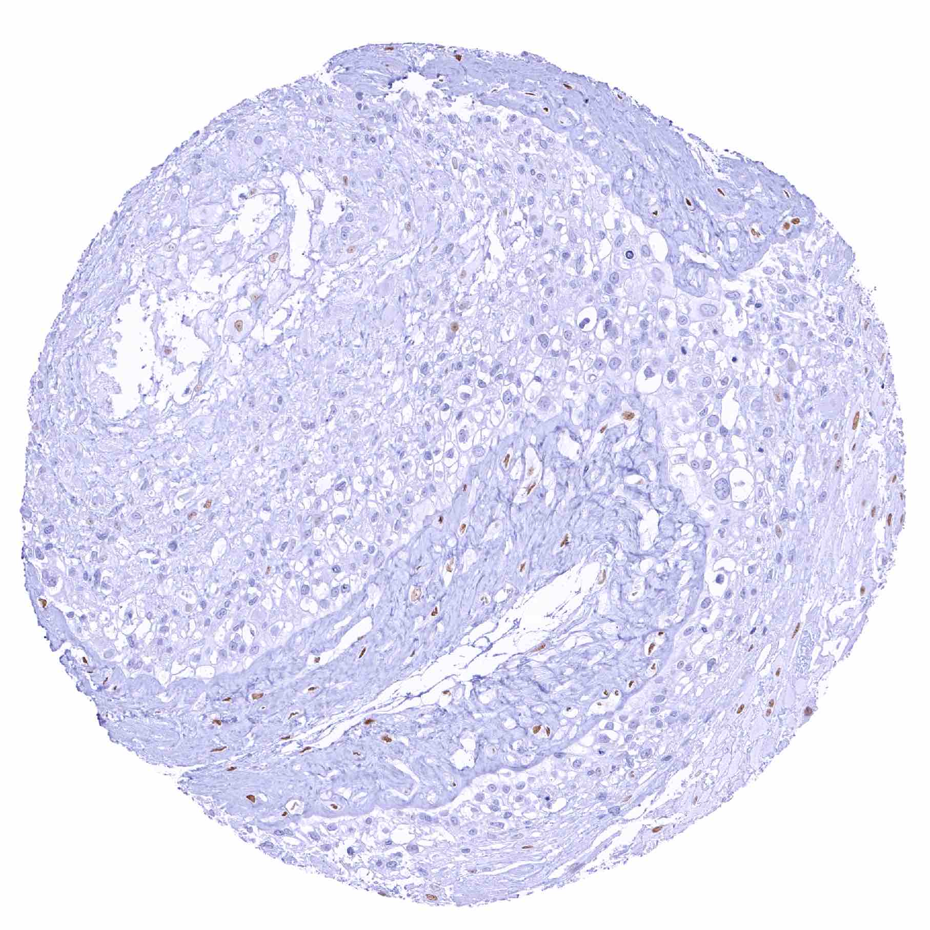
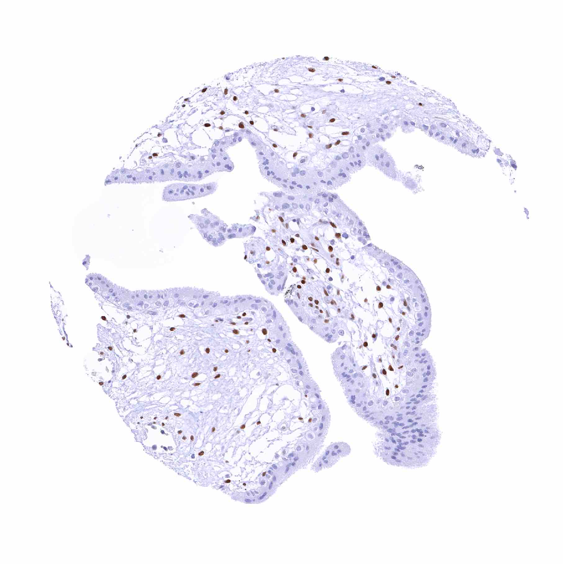
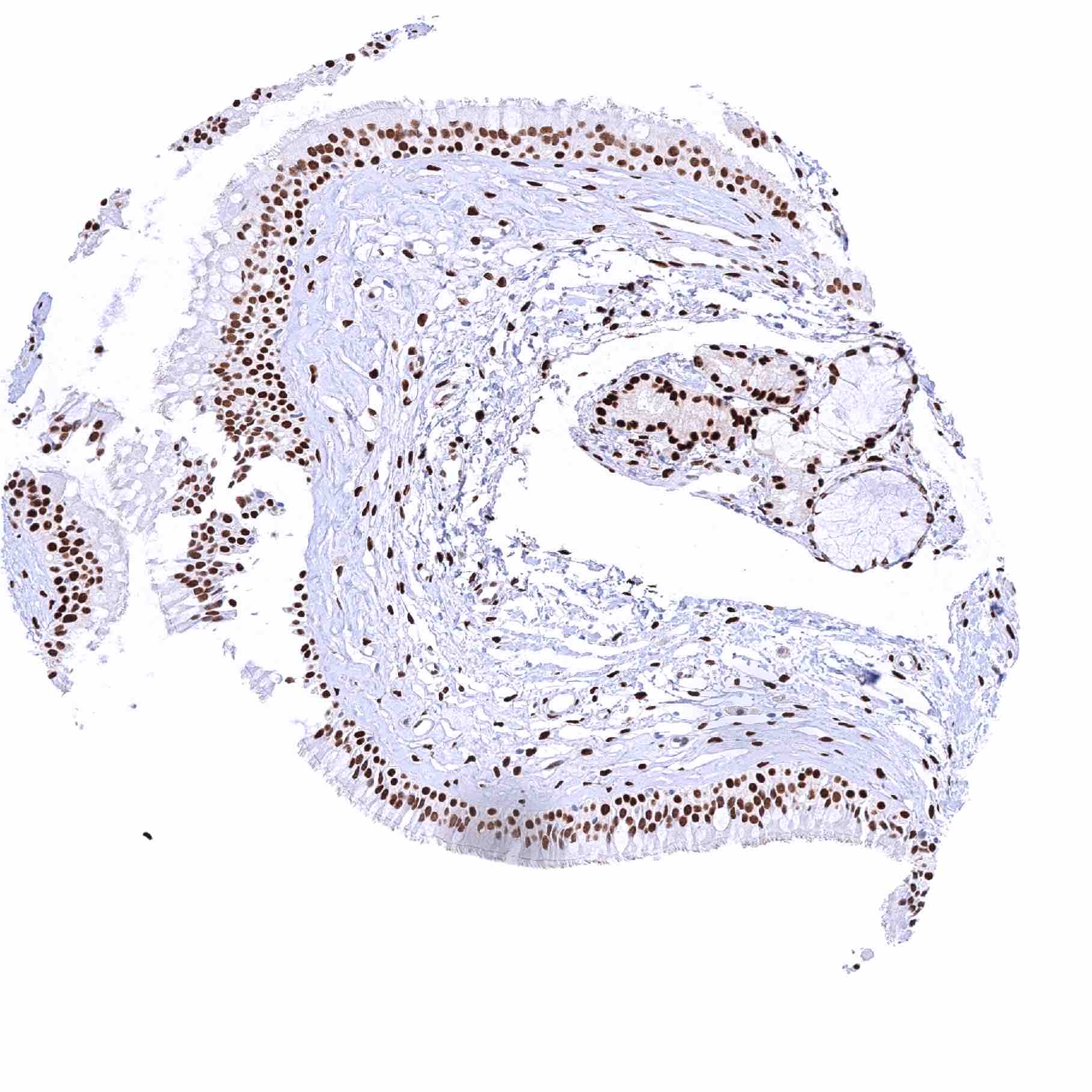
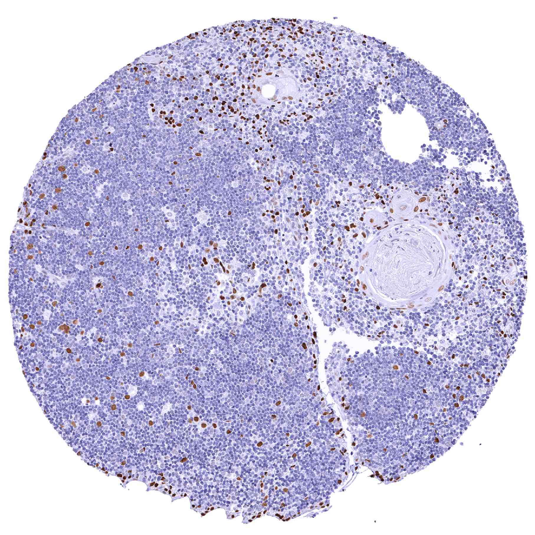
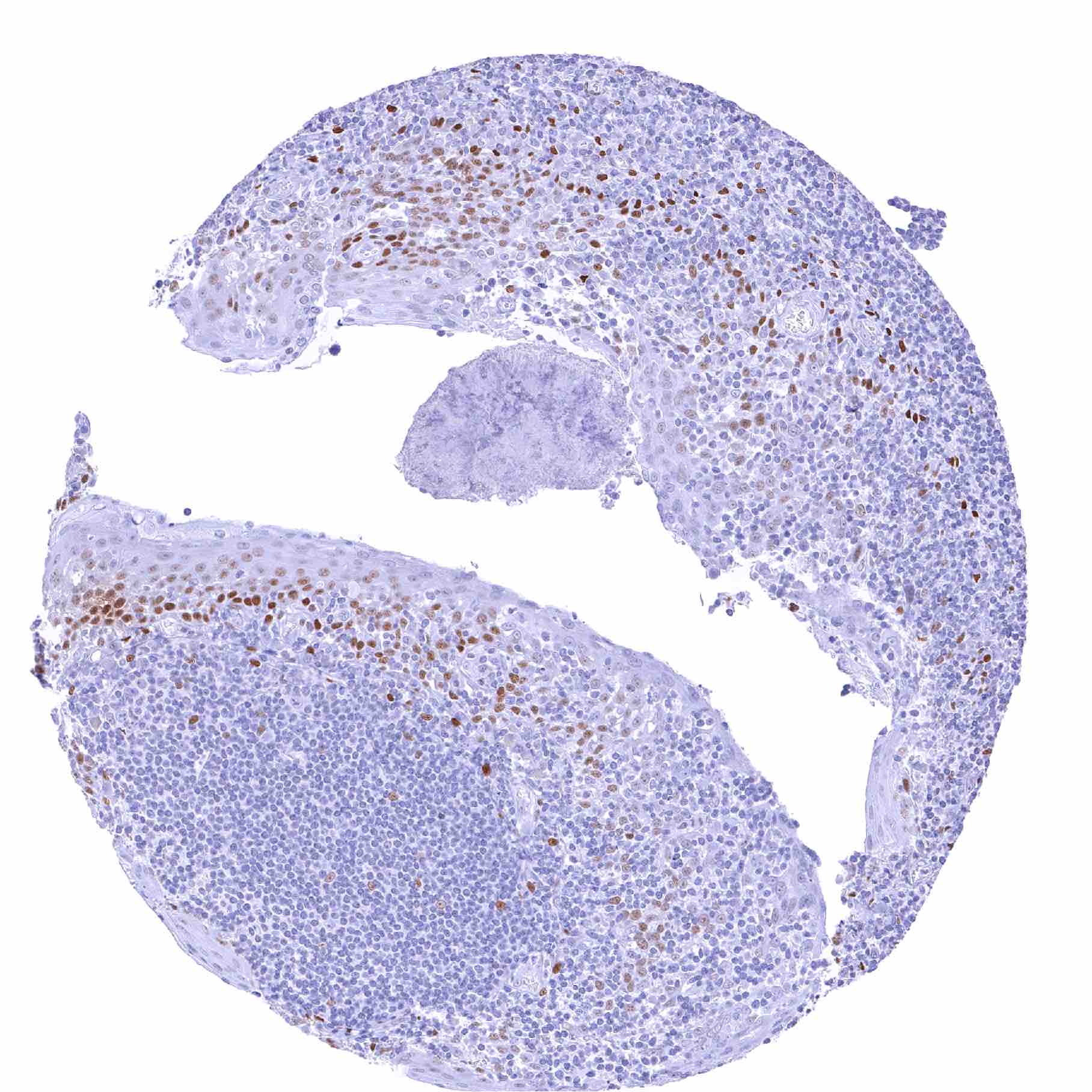
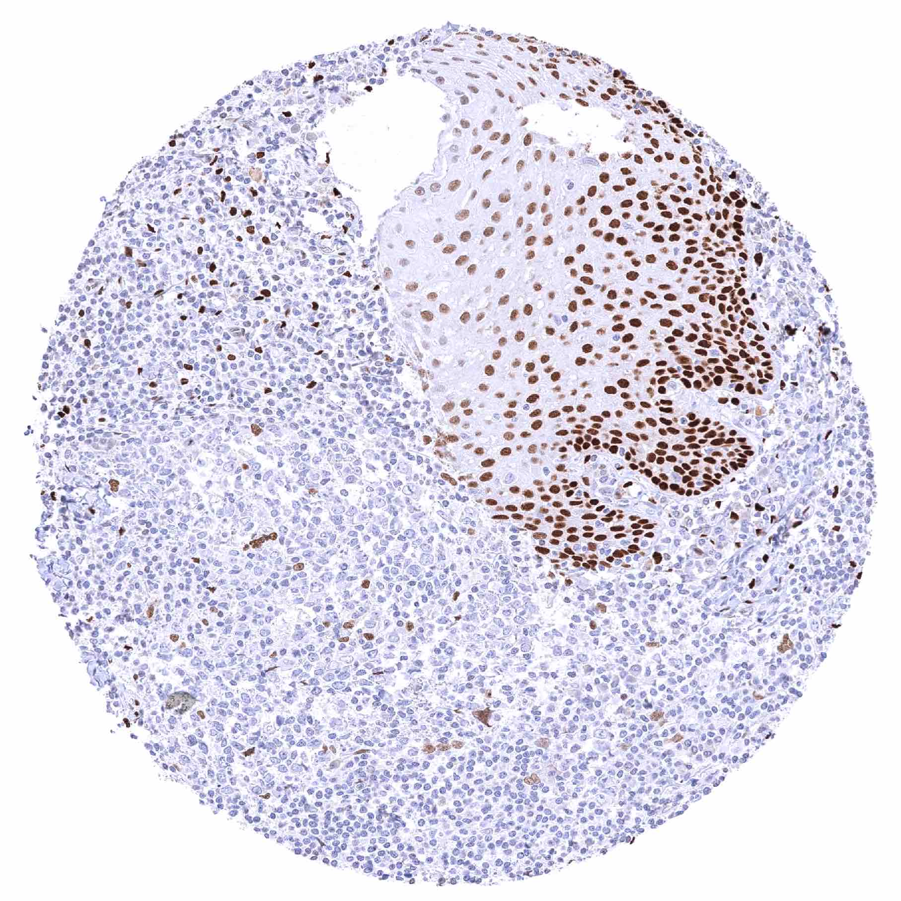
Tonsil, surface epithelium – Significant decrease of nuclear NFIX staining of squamous epithelium from the basal-suprabasal to the superficial cell layers
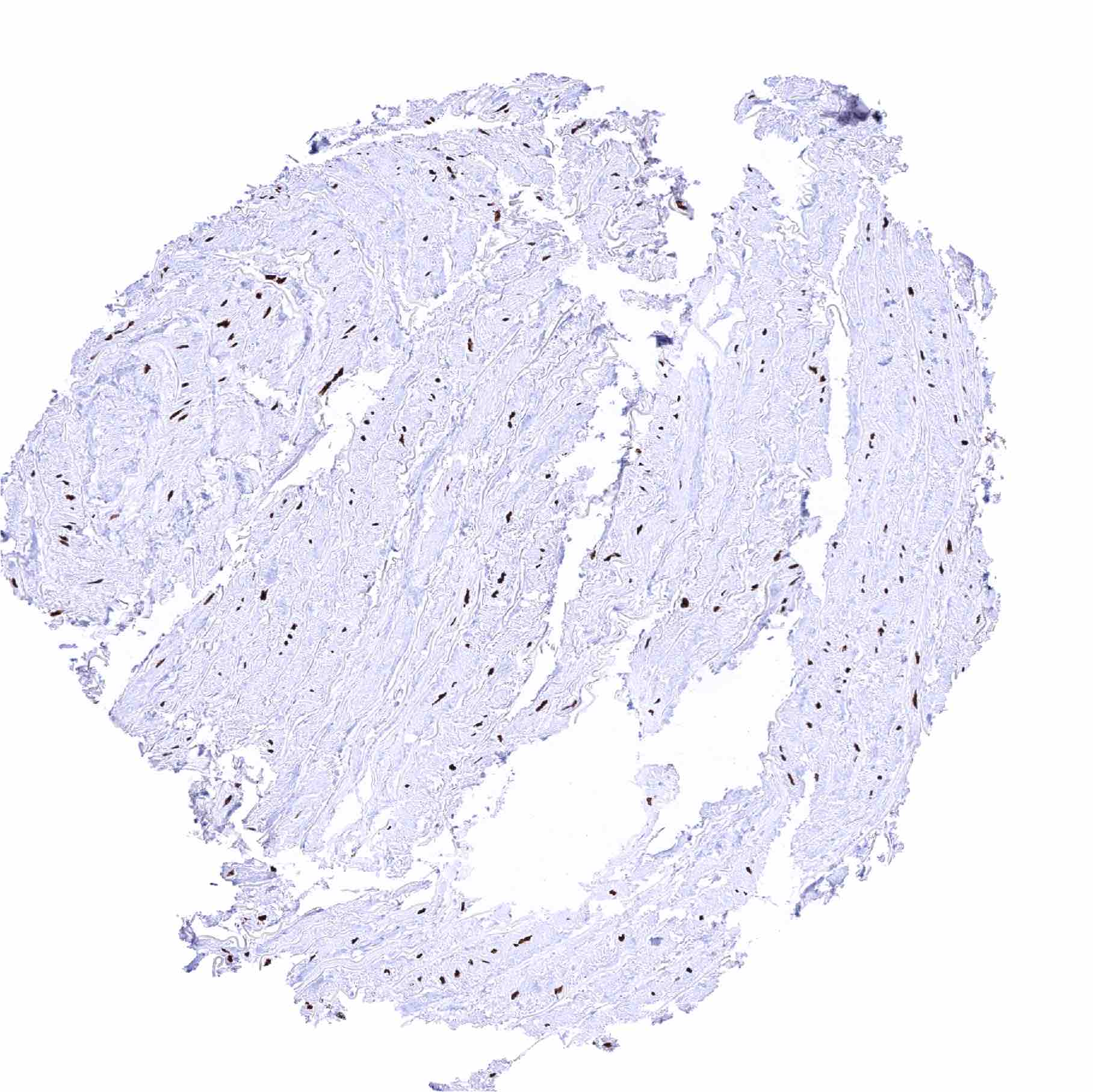
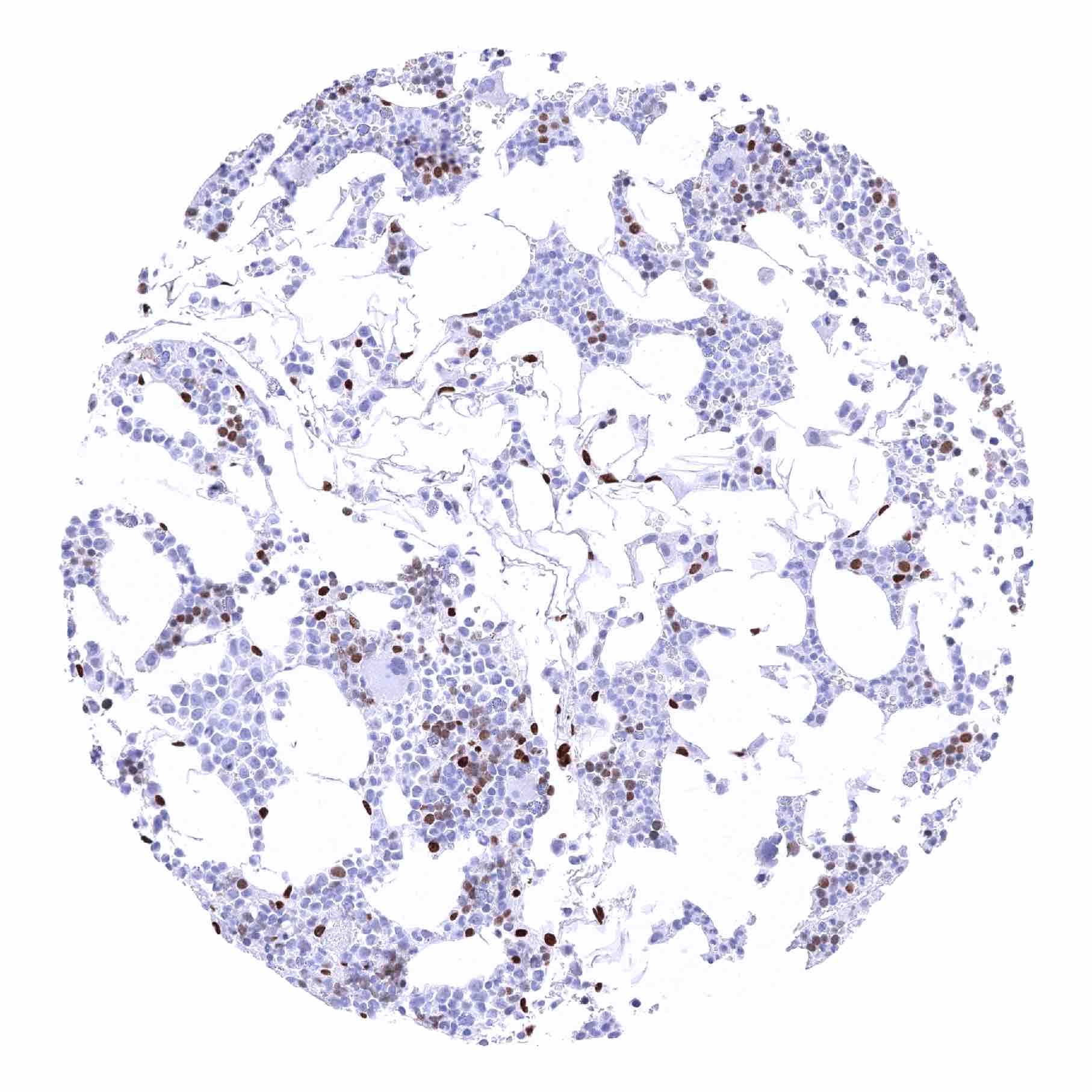
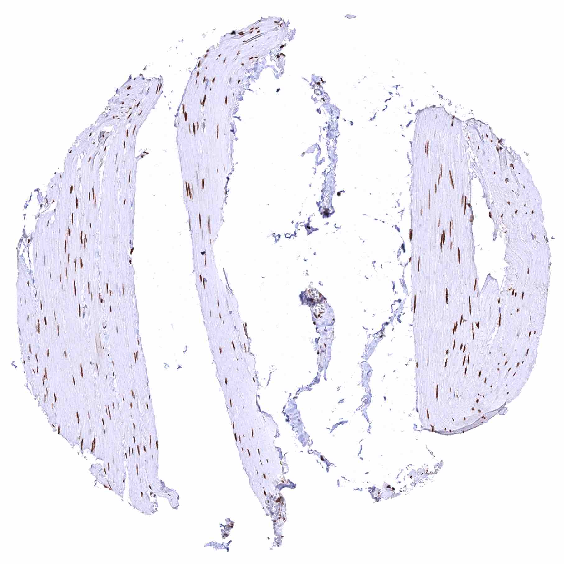
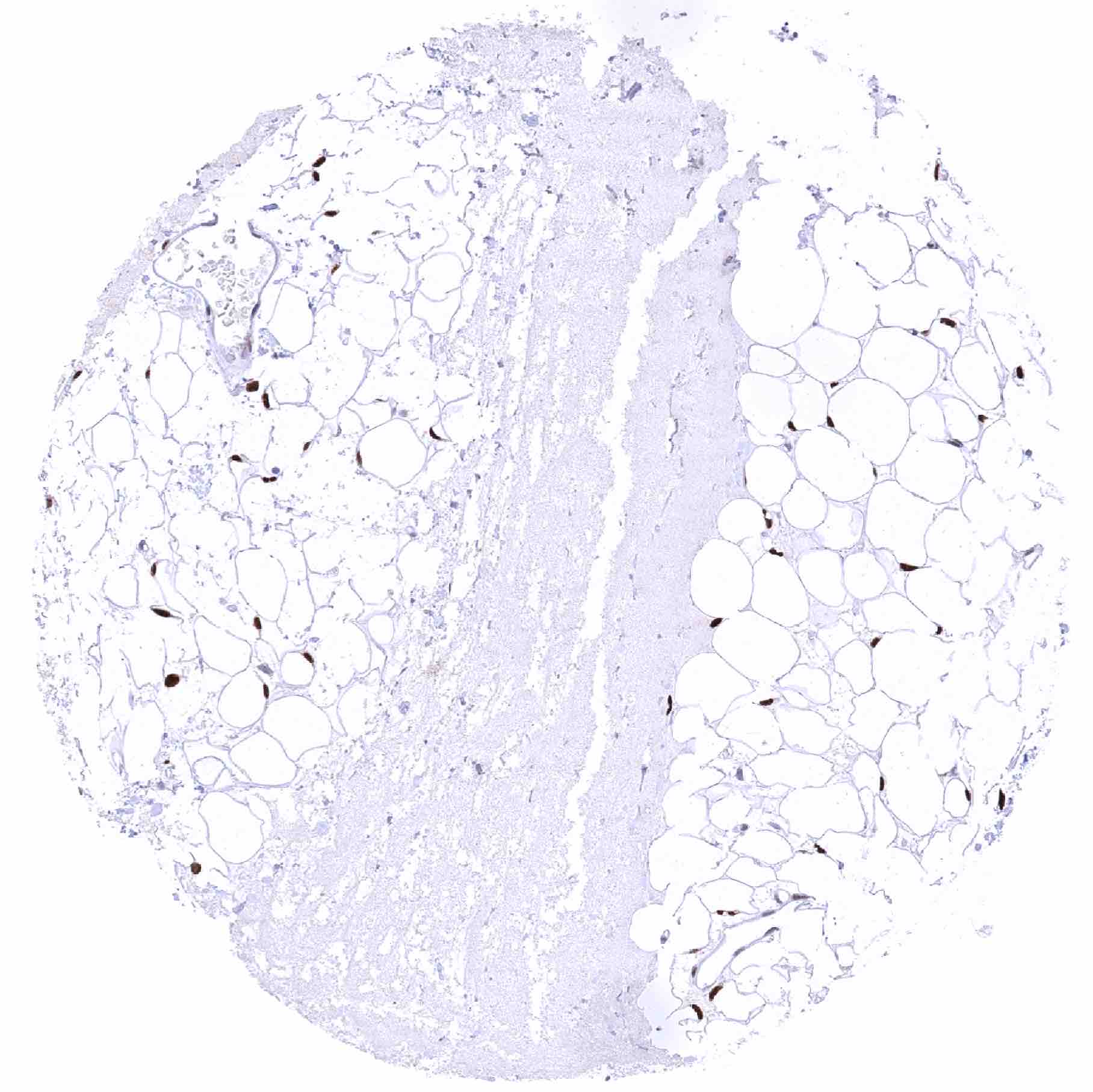
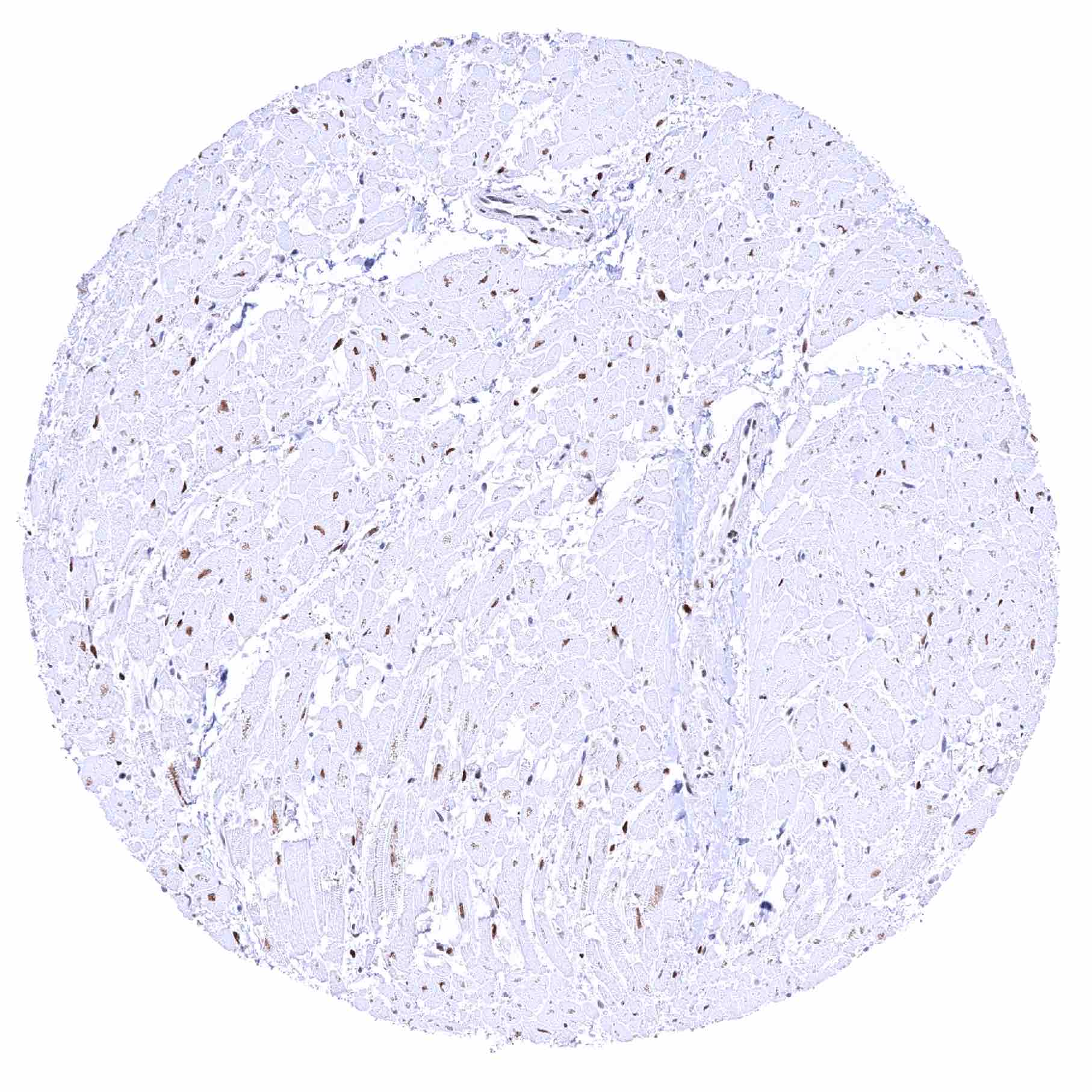
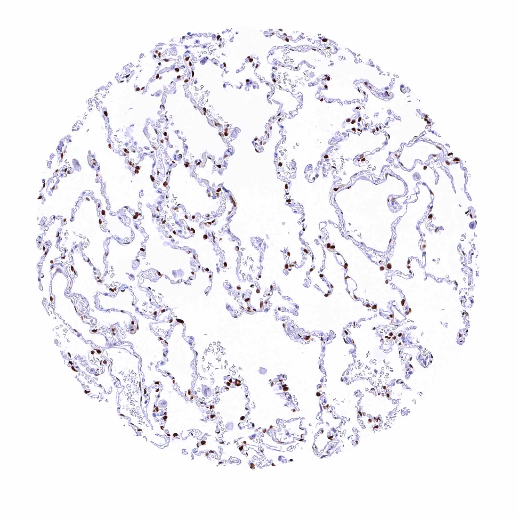
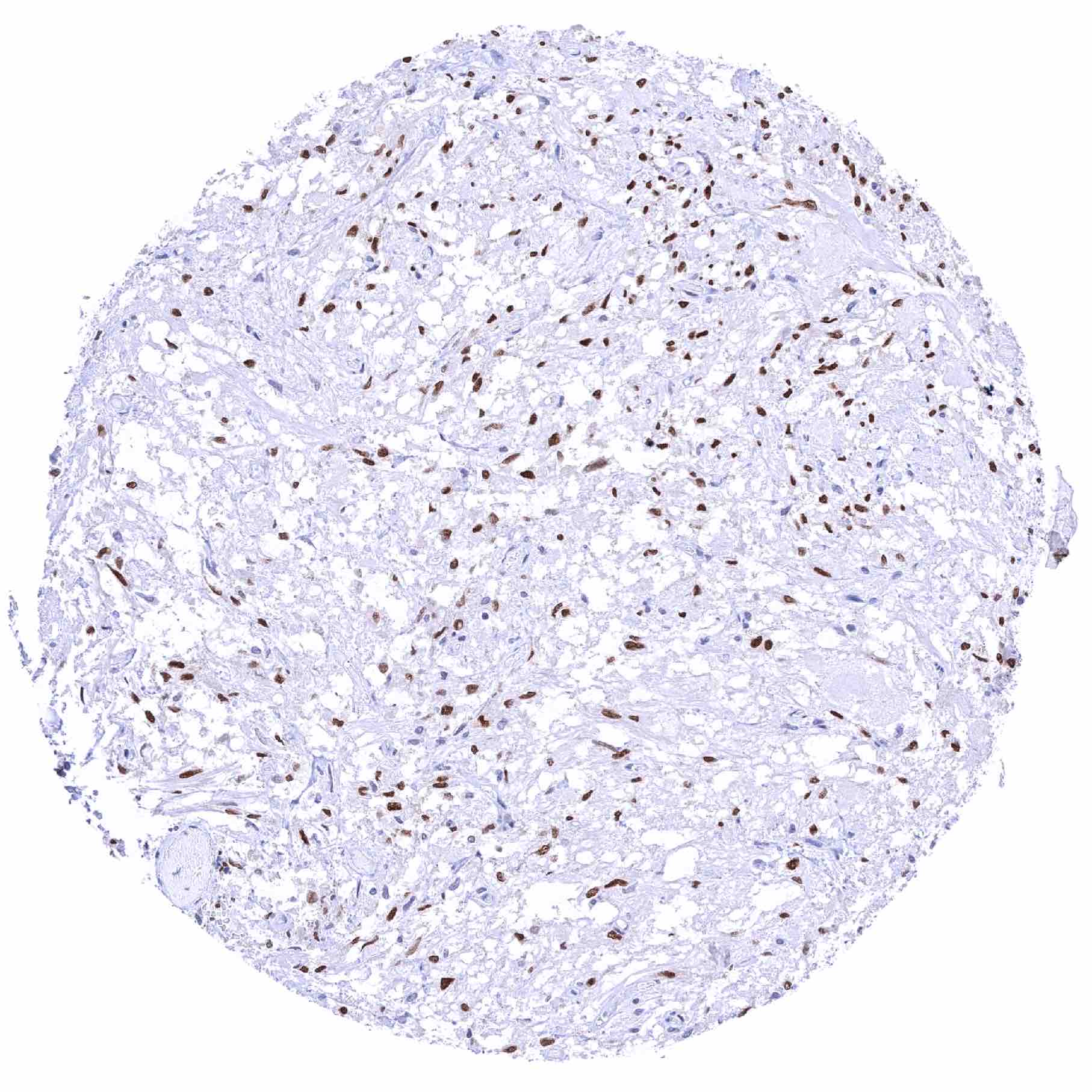
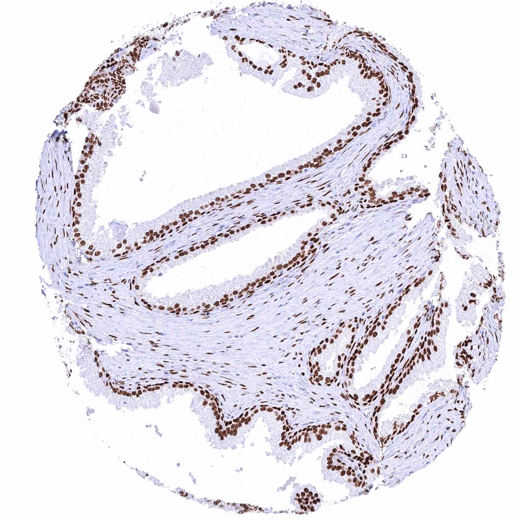
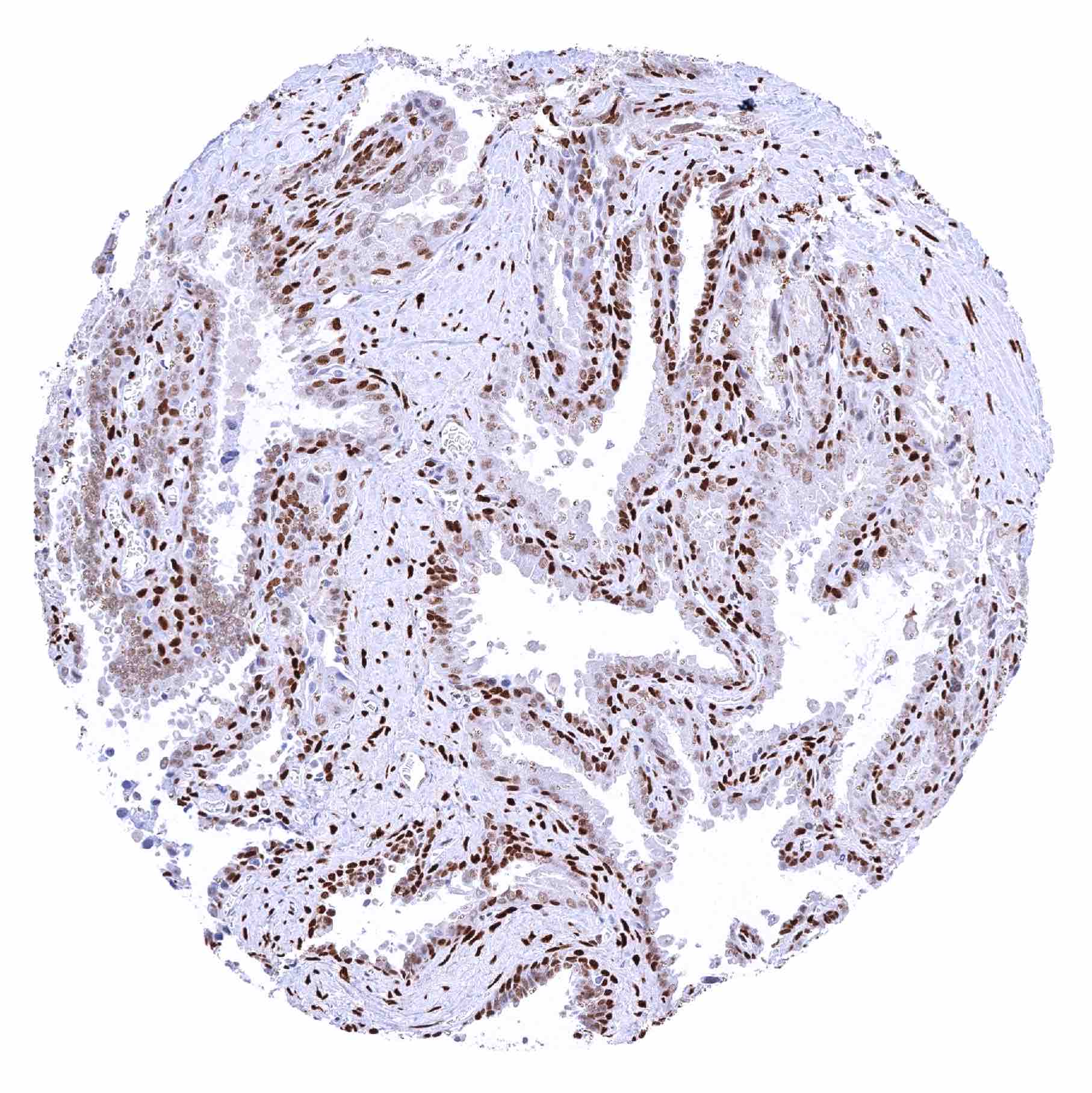
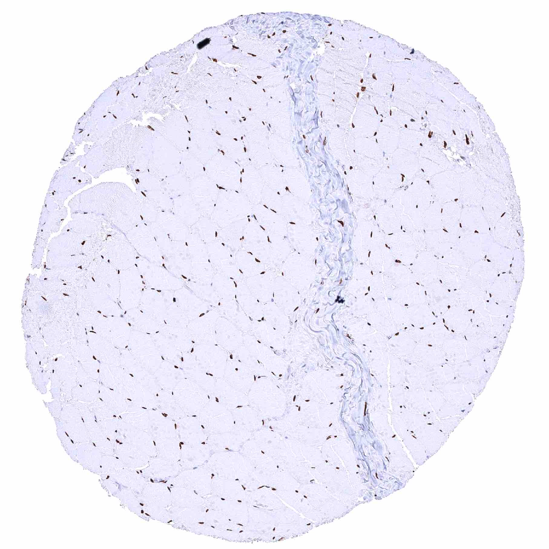
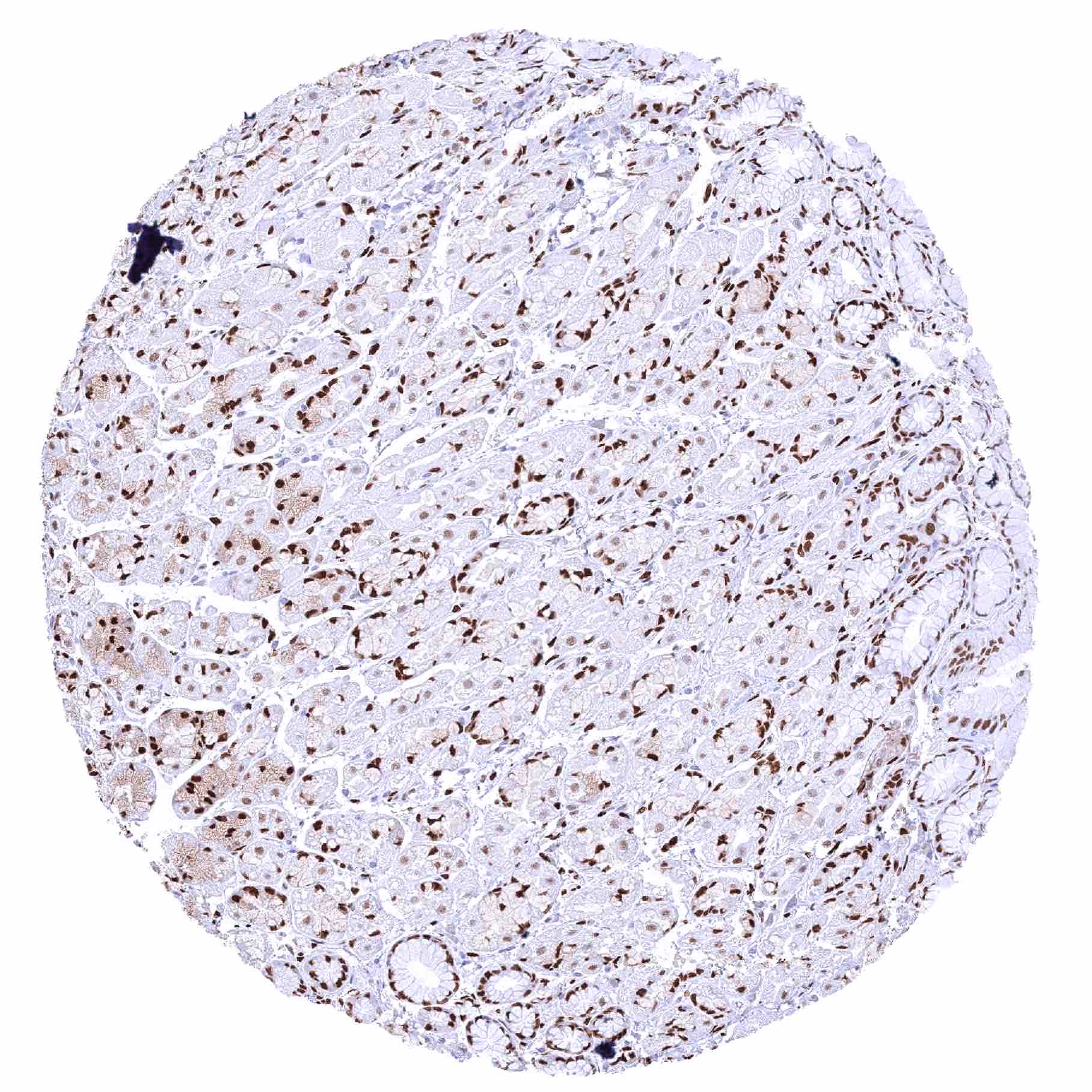
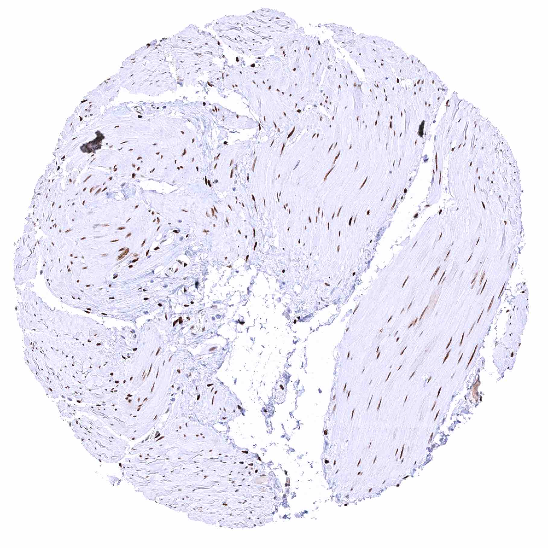
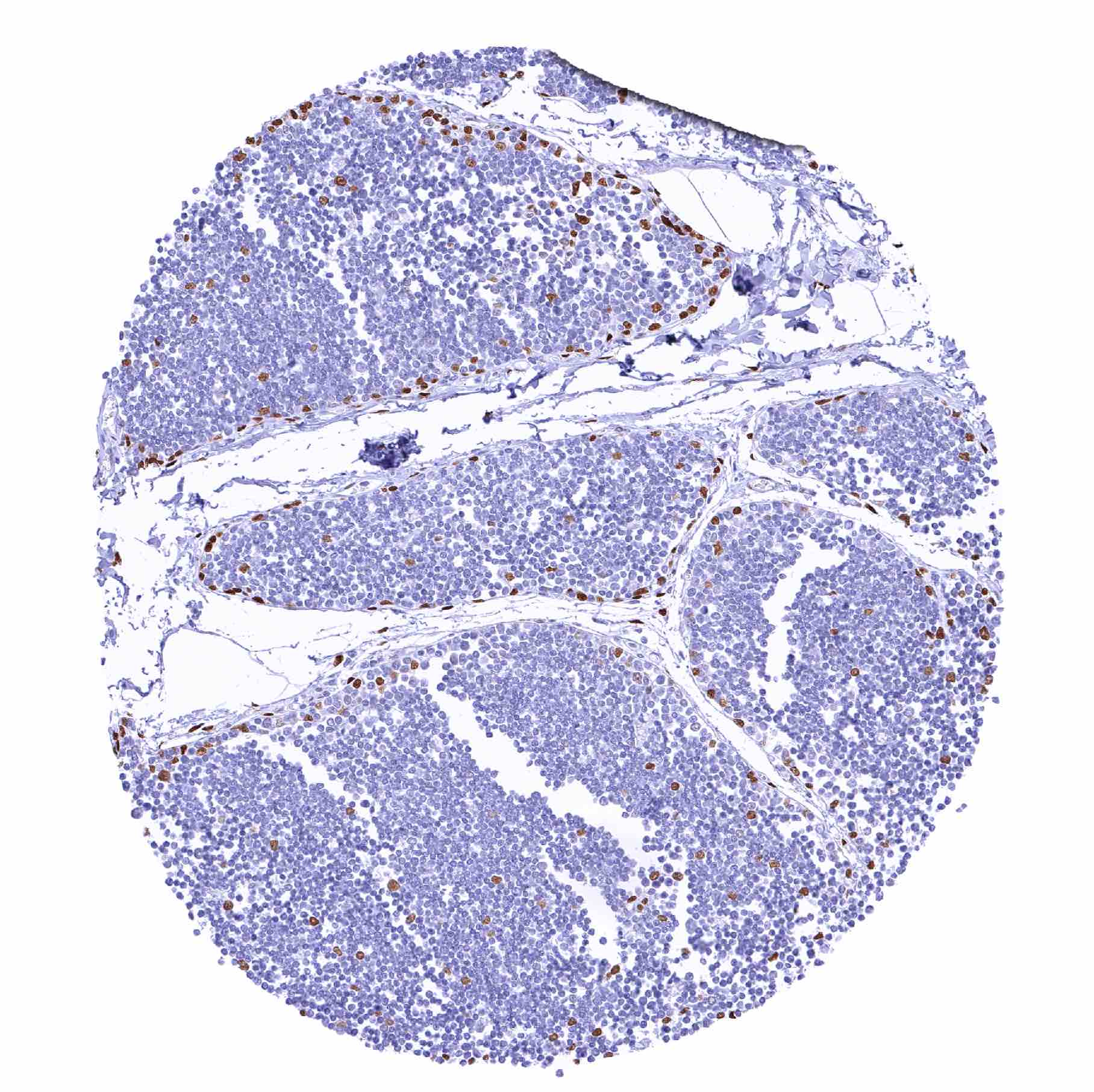
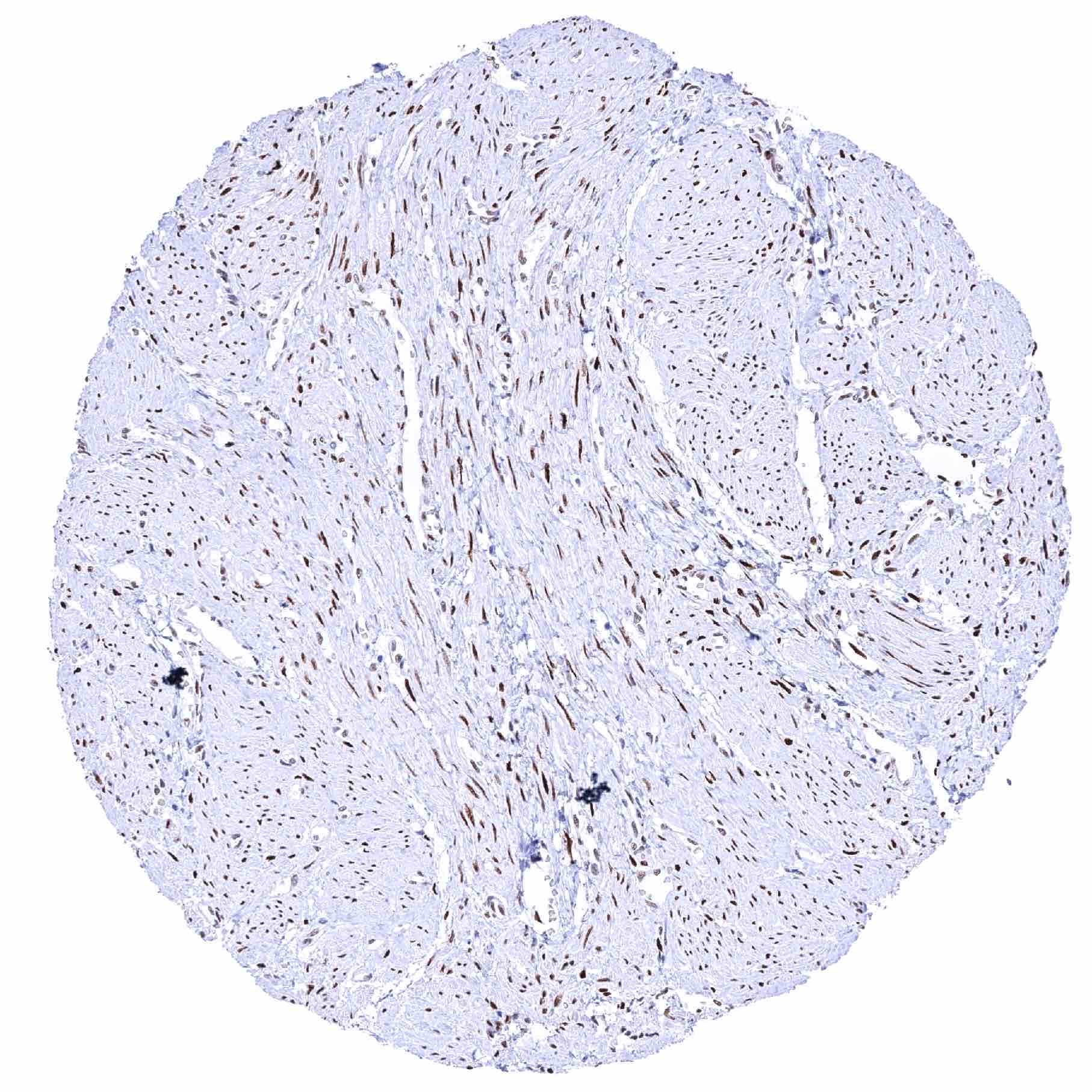
Urinary bladder, muscular wall – Distinct nuclear staining of smooth muscle cells and of other cell types
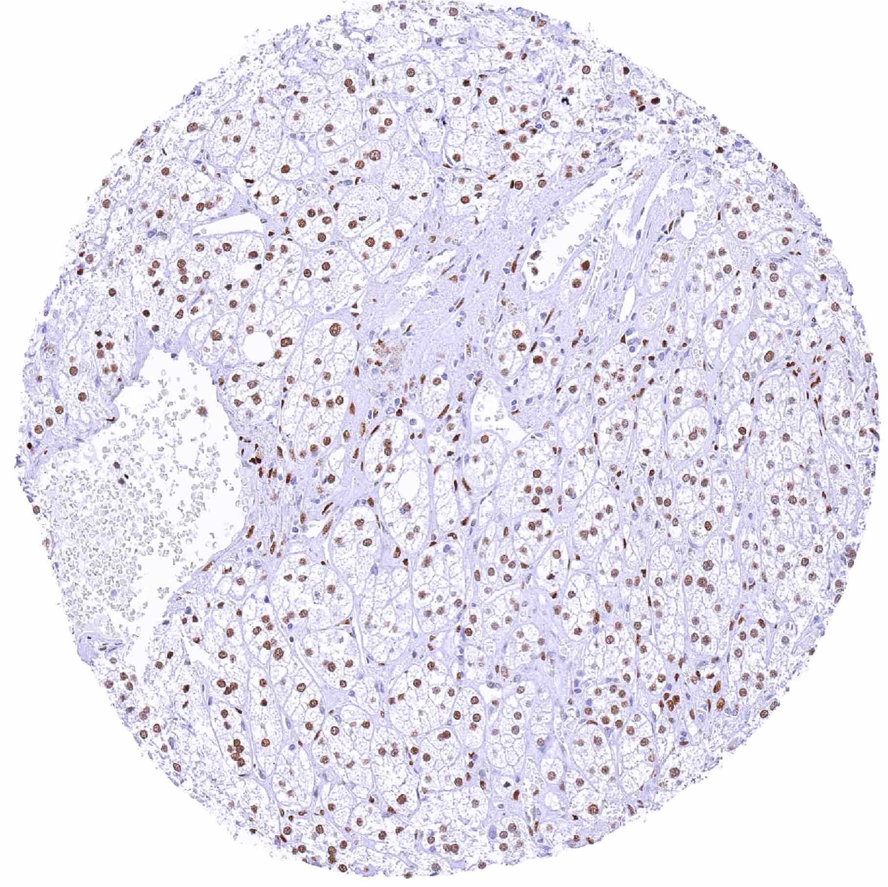
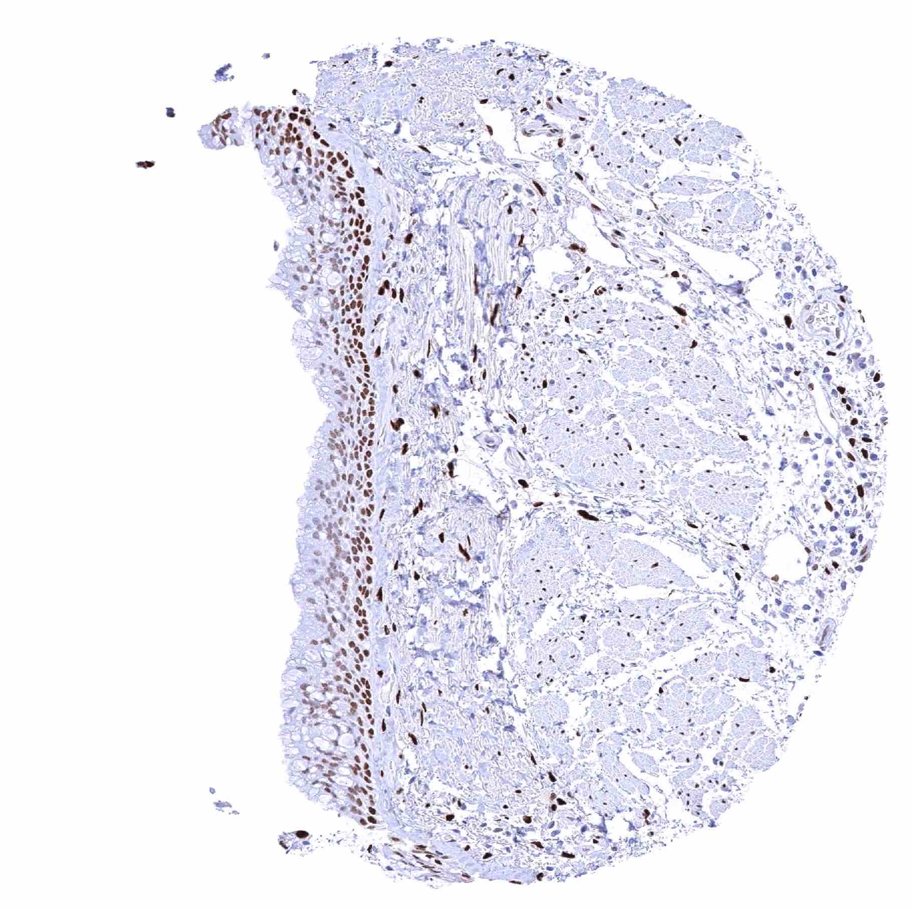
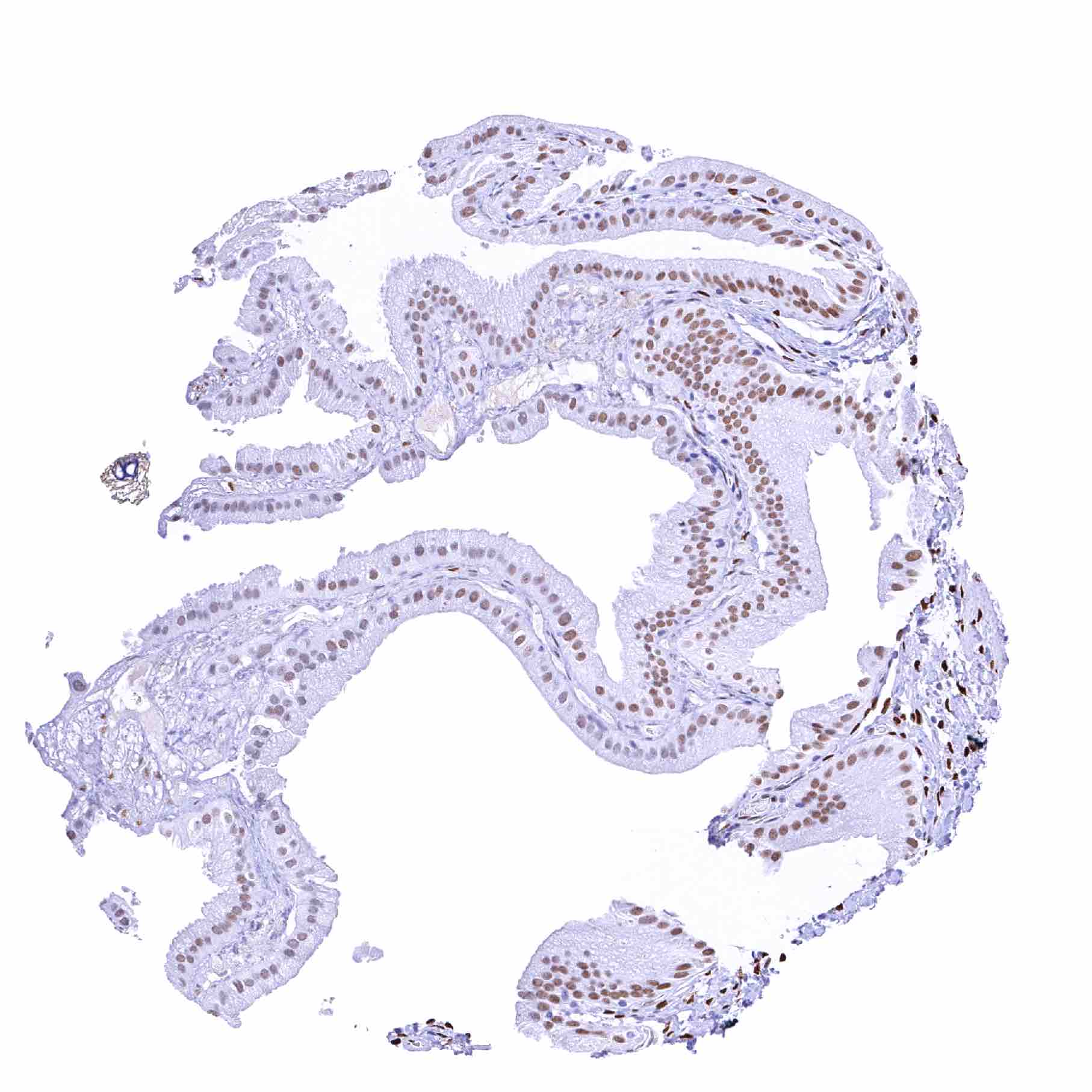
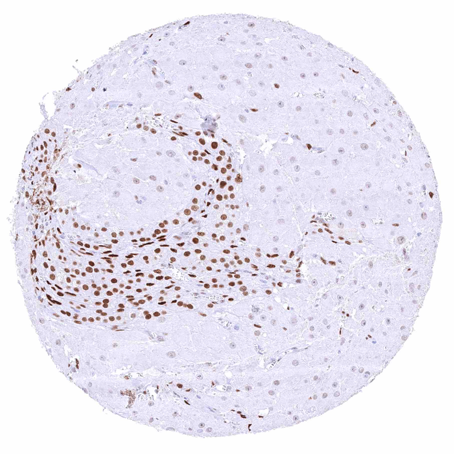
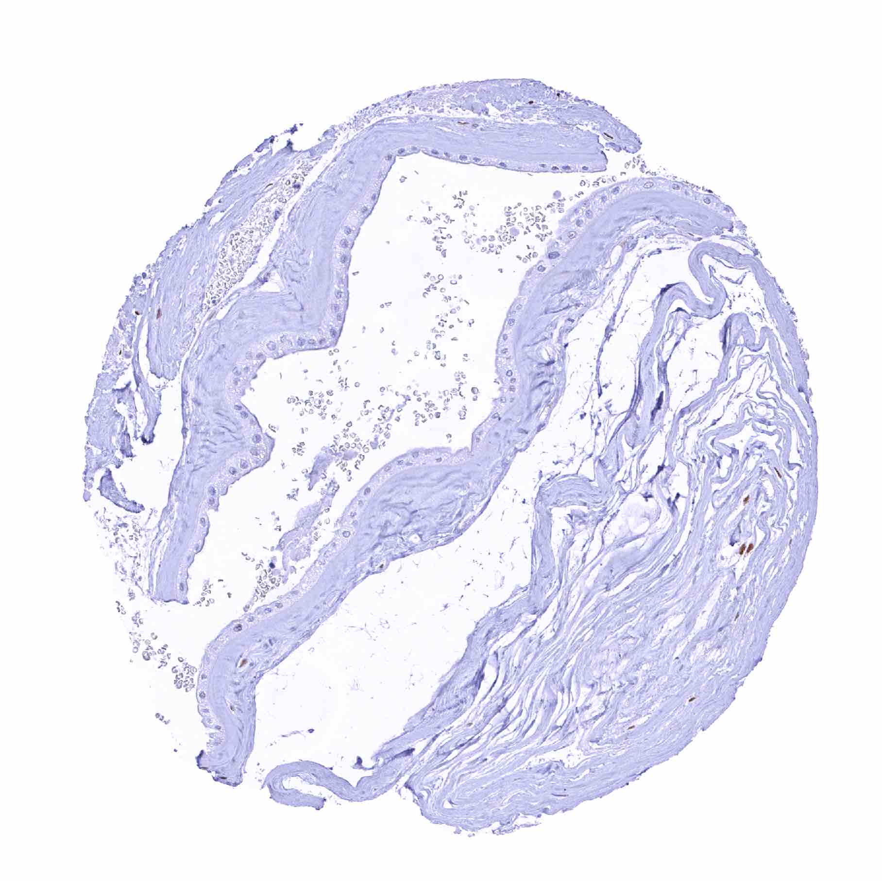
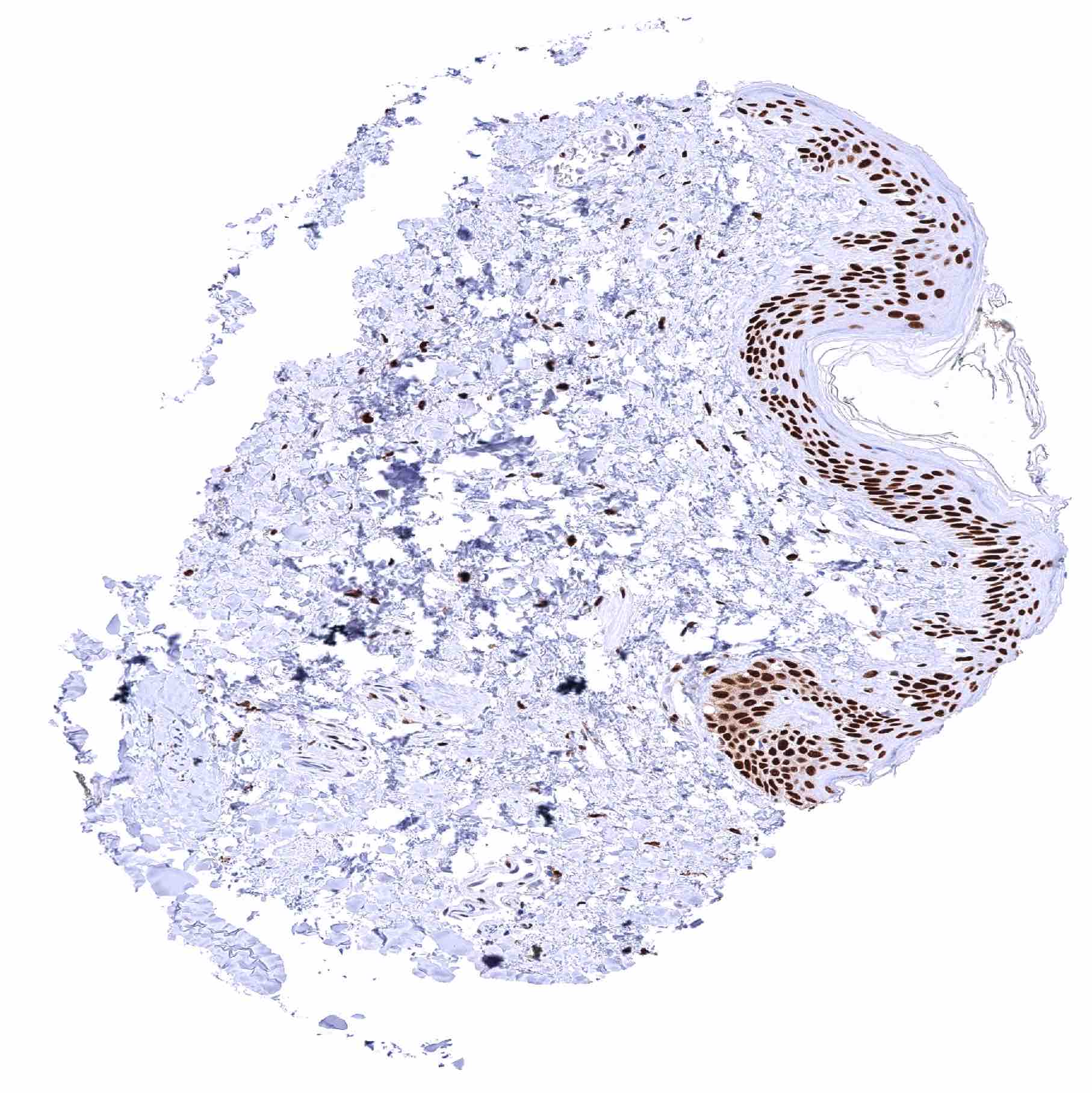
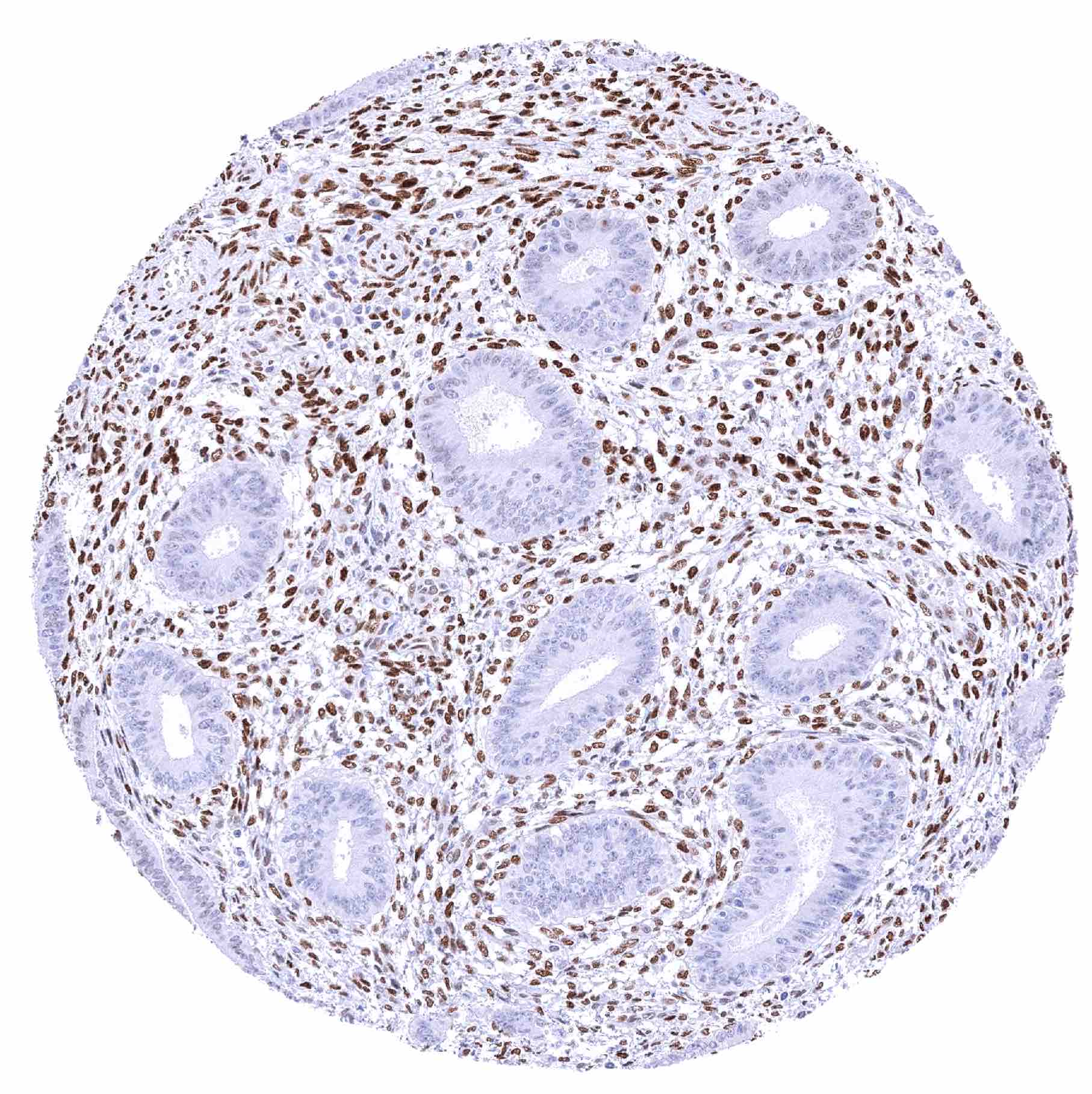
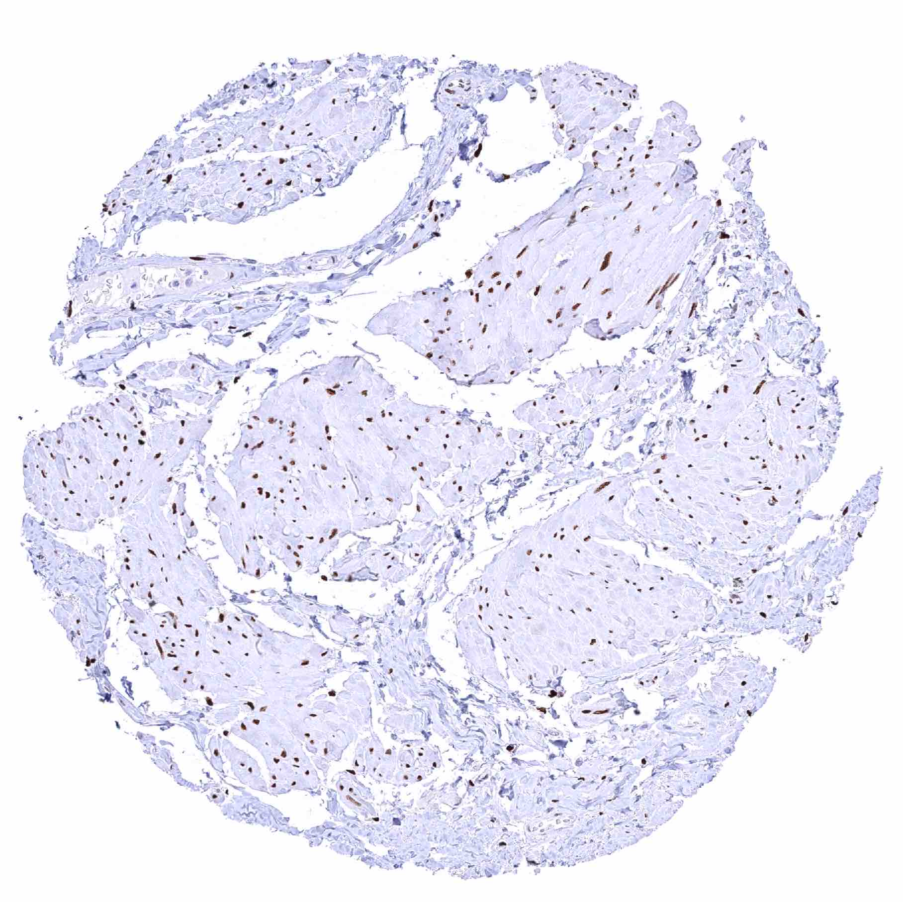
Urinary bladder, urothelium – Distinct nuclear NFIX staining of urothelial cells although there is a significant decrease of NFIX staining intensity from the basal to the superficial cell layers. Umbrella cells are largely NFIX negative
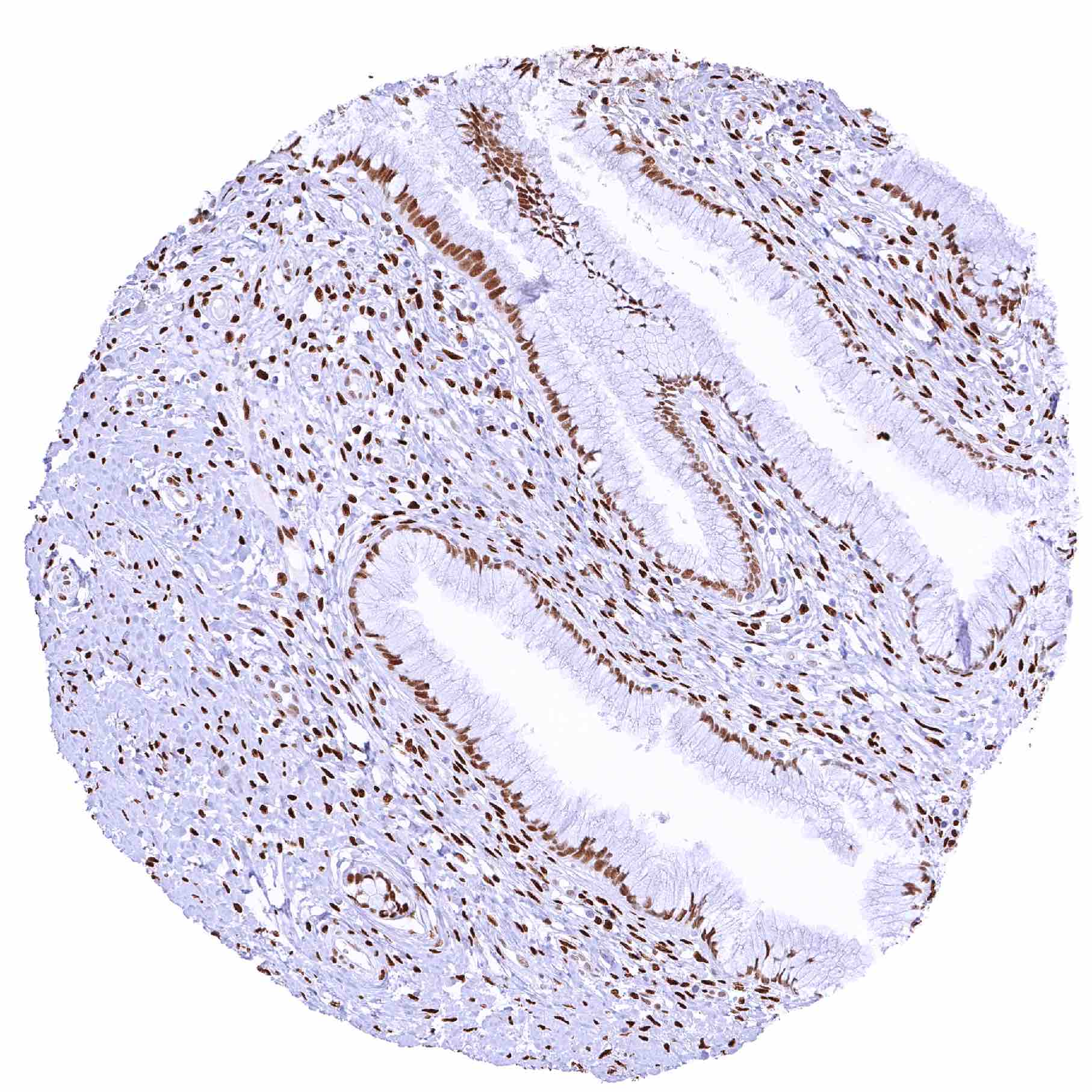
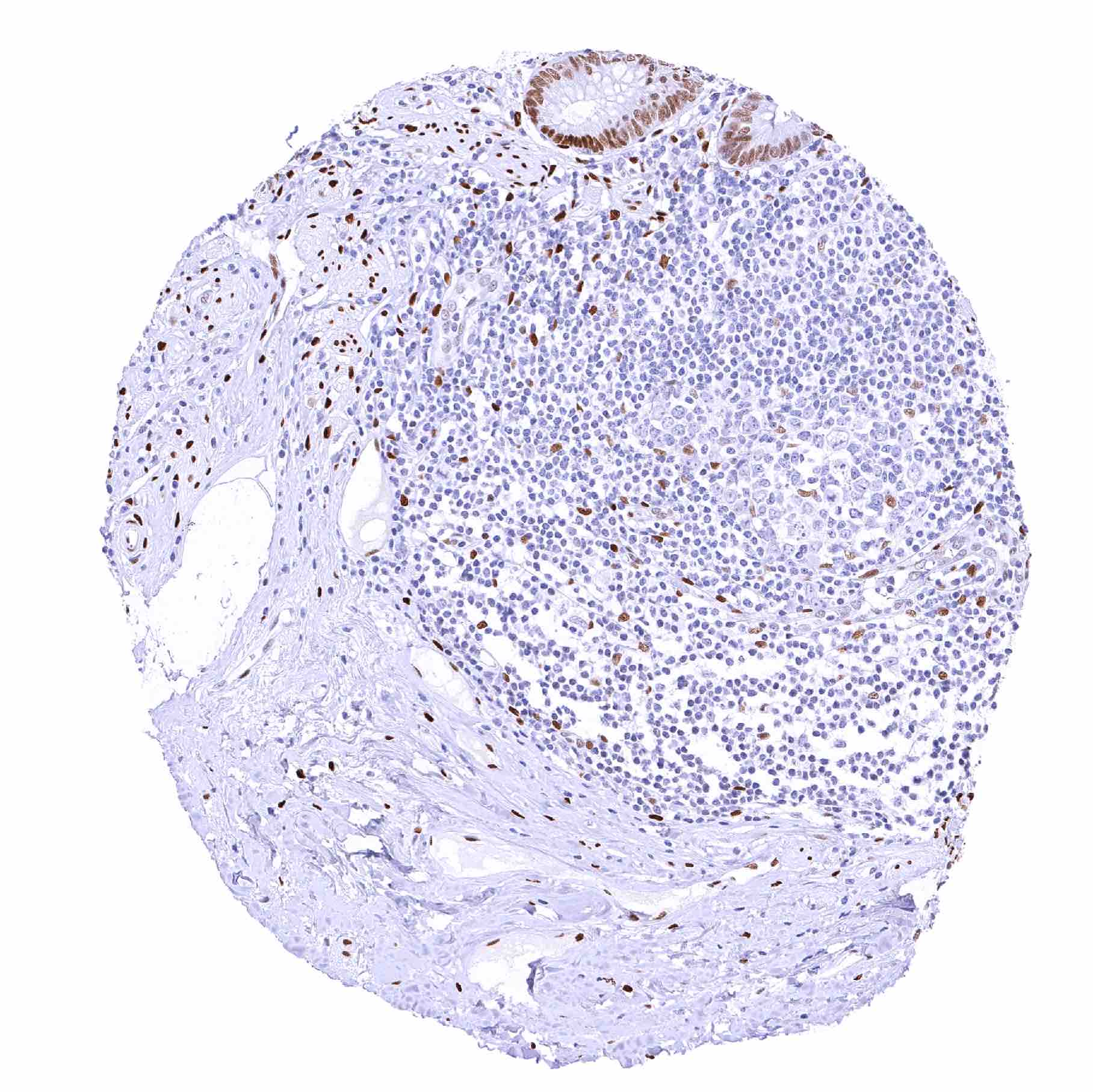
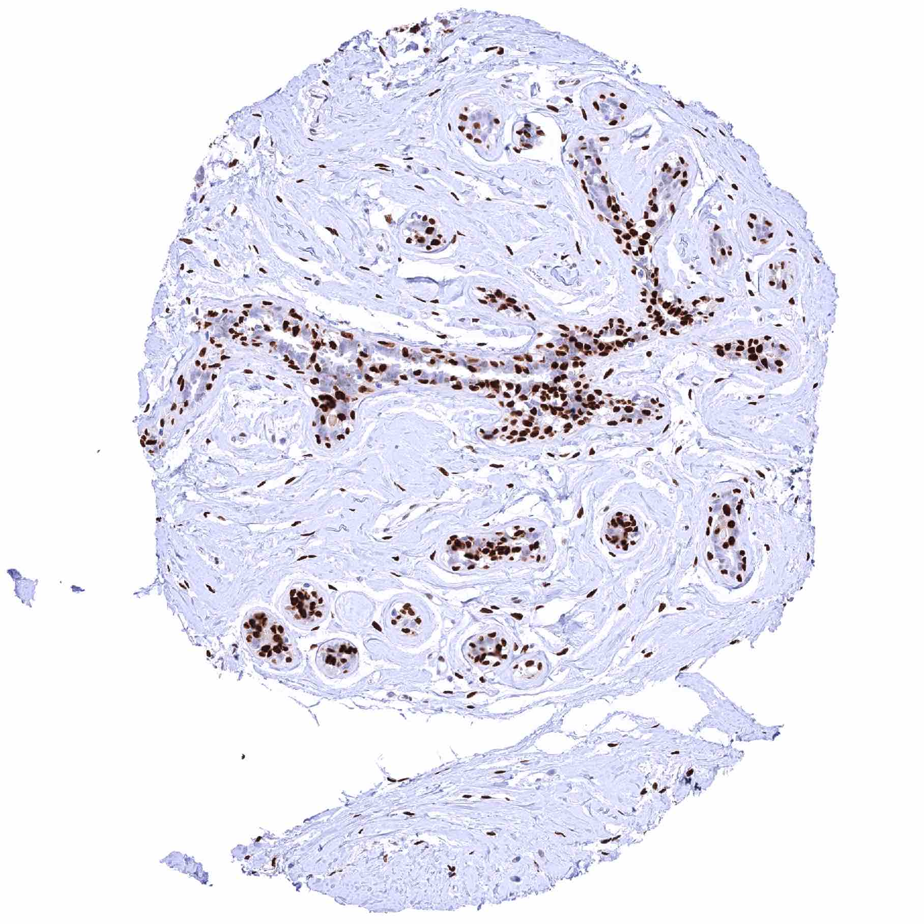
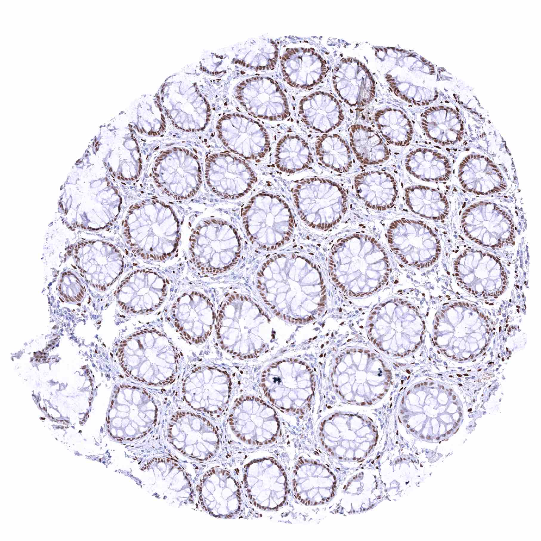
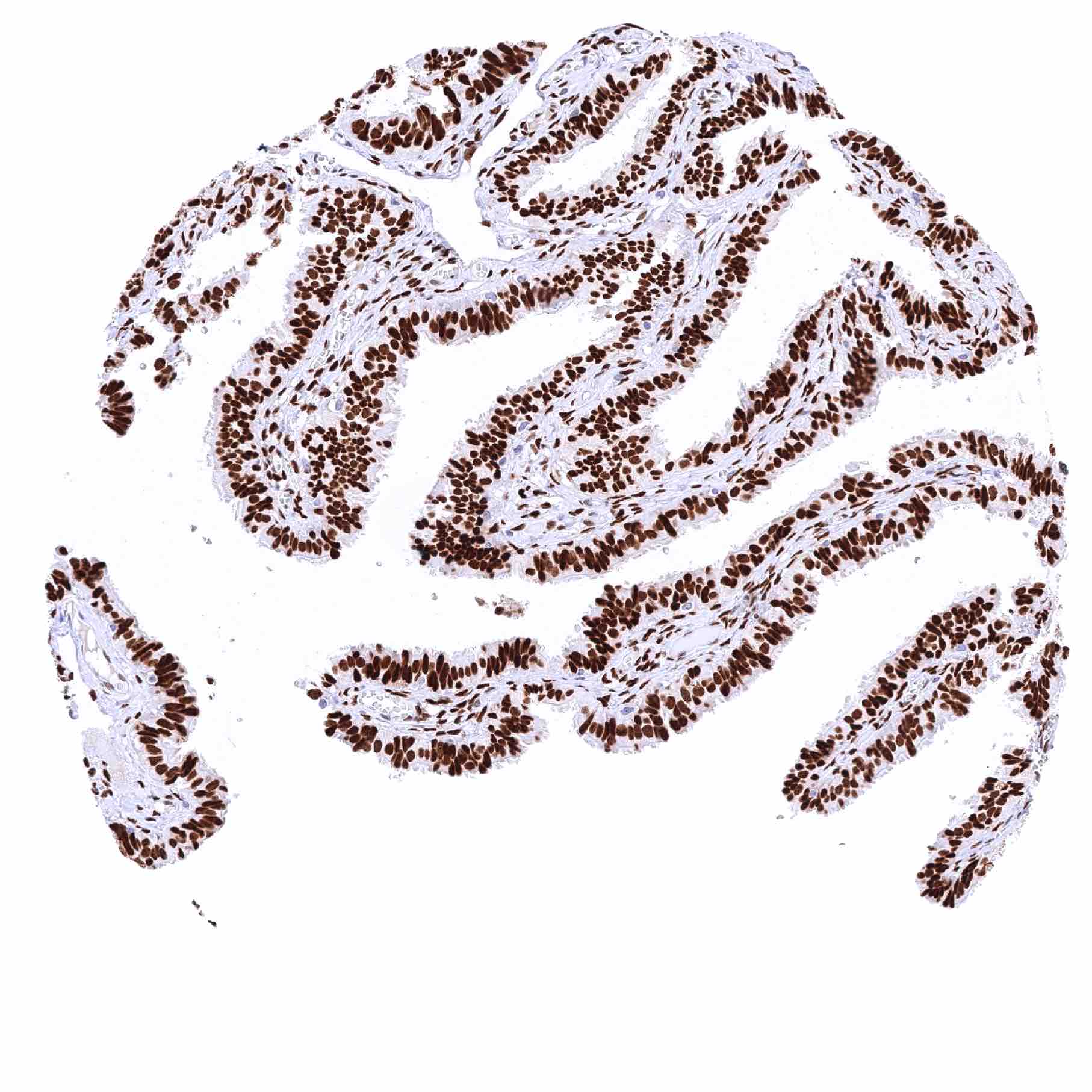
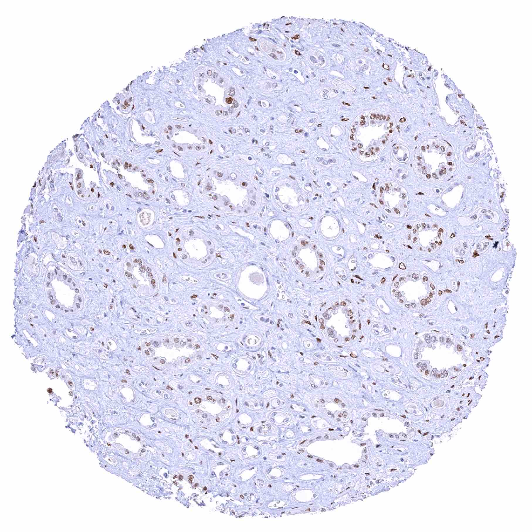
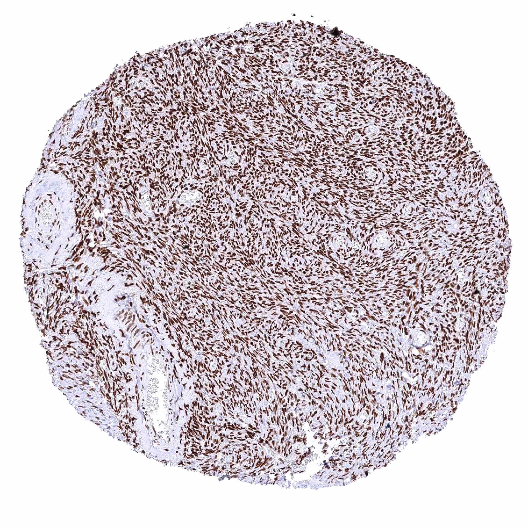
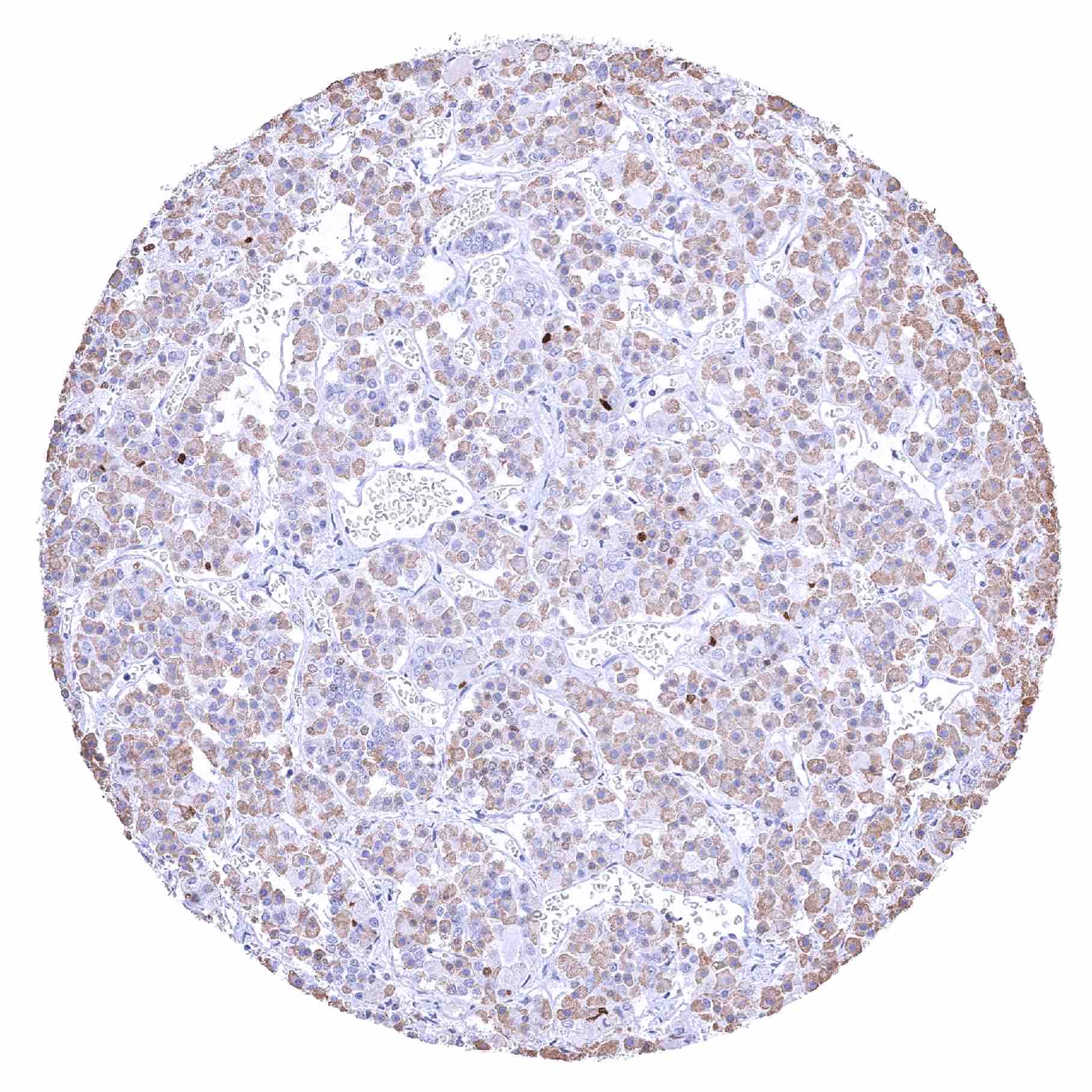
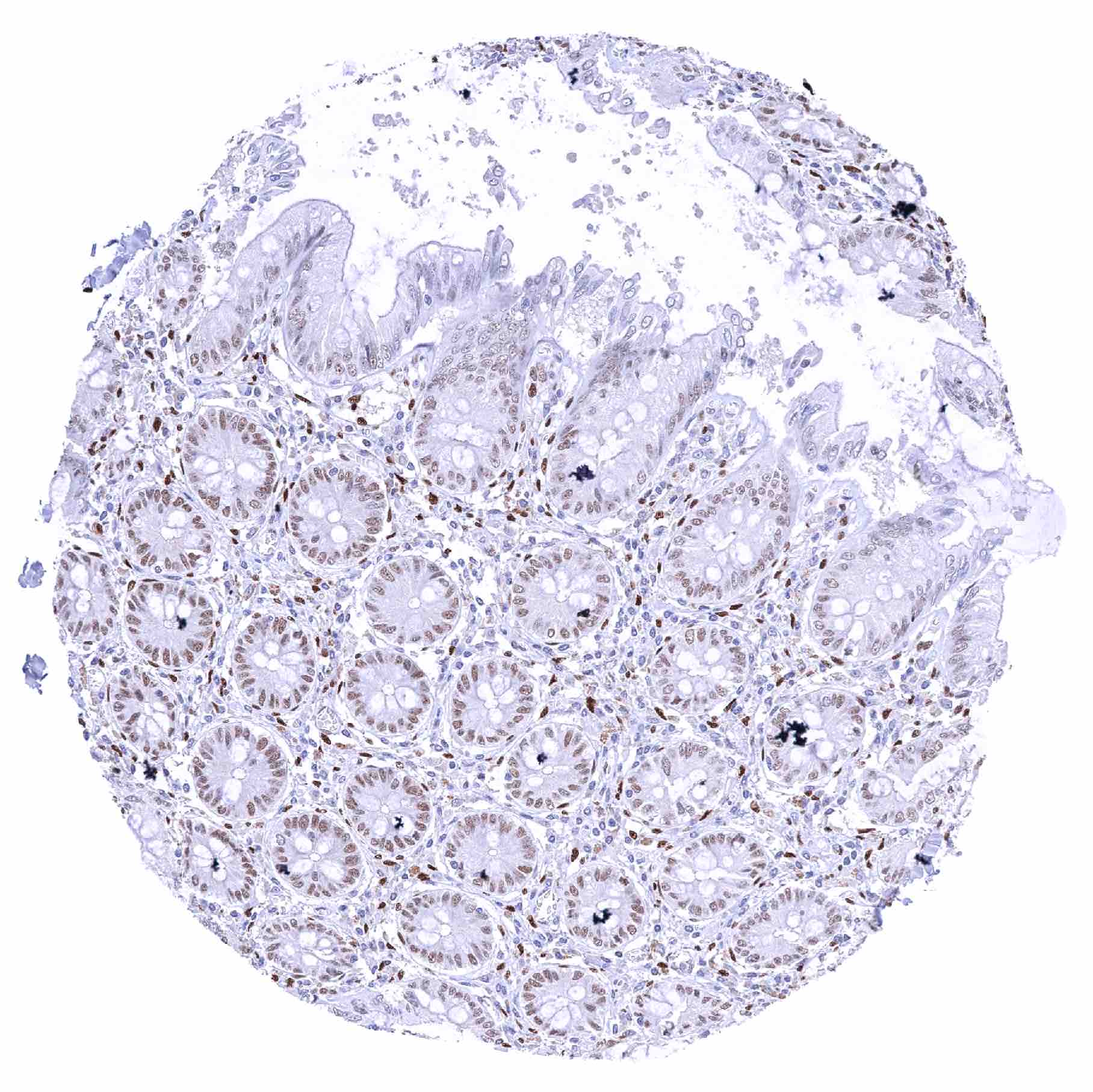
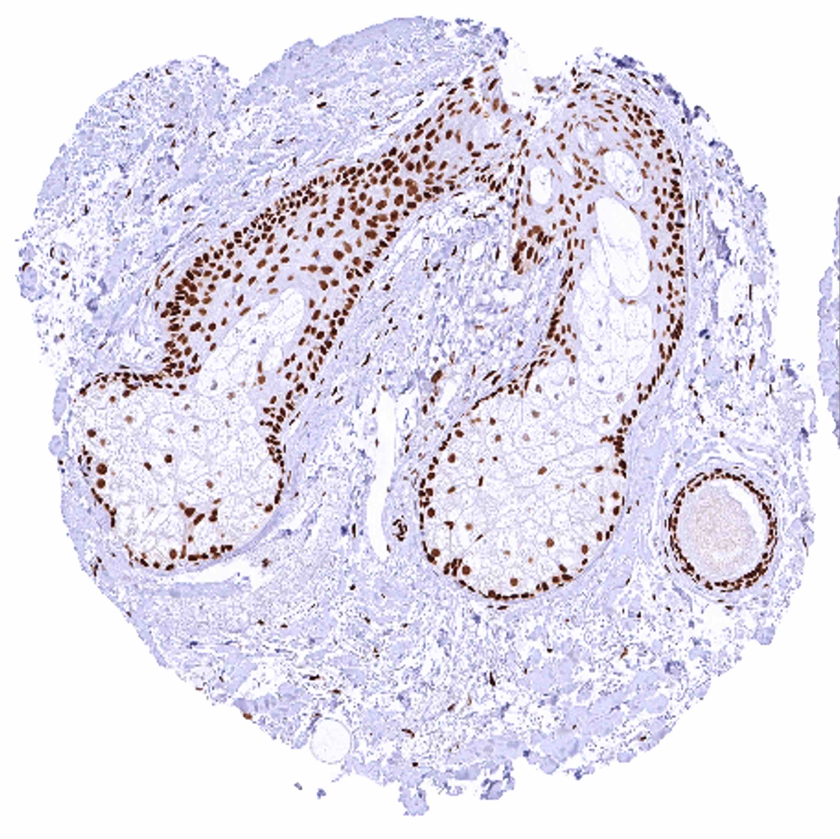
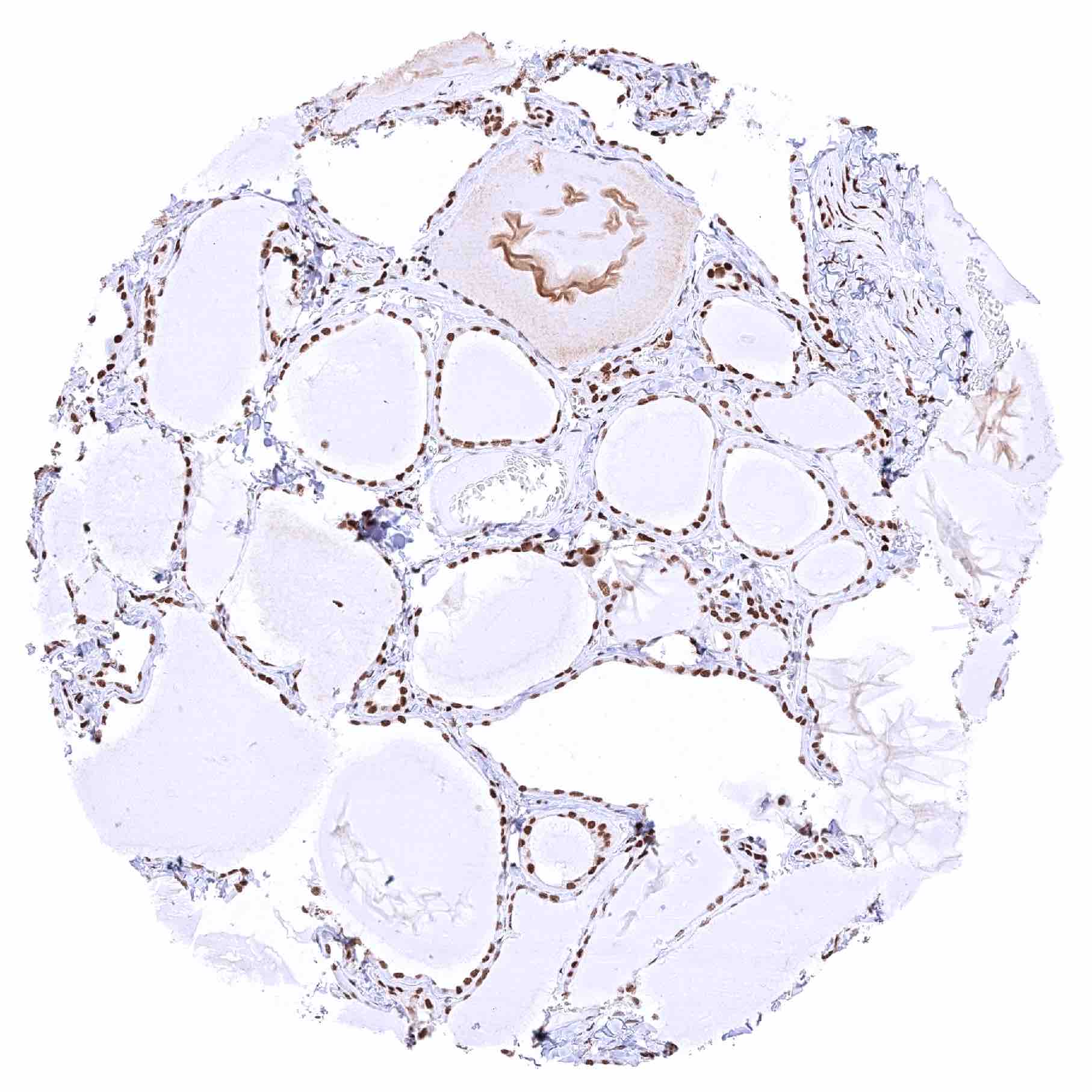
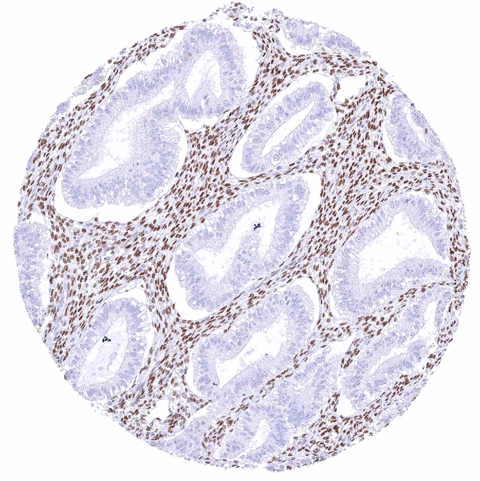
Uterus, endocervix – Moderate nuclear NFIX staining of glandular cells. Strong nuclear NFIX staining of stromal cells
Uterus, endometrium (pregnancy)
Uterus, endometrium (proliferation) – Strong nuclear NFIX staining of stromal cells while endometrium cells remain NFIX negative
Uterus, endometrium (secretion) – Strong nuclear NFIX staining of stromal cells while endometrium cells remain NFIX negative
Uterus, myometrium – Distinct nuclear staining of smooth muscle cells of the myometrium

| TOP |