Normal Tissue Gallery- GTX04491

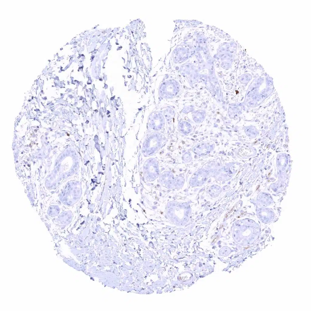
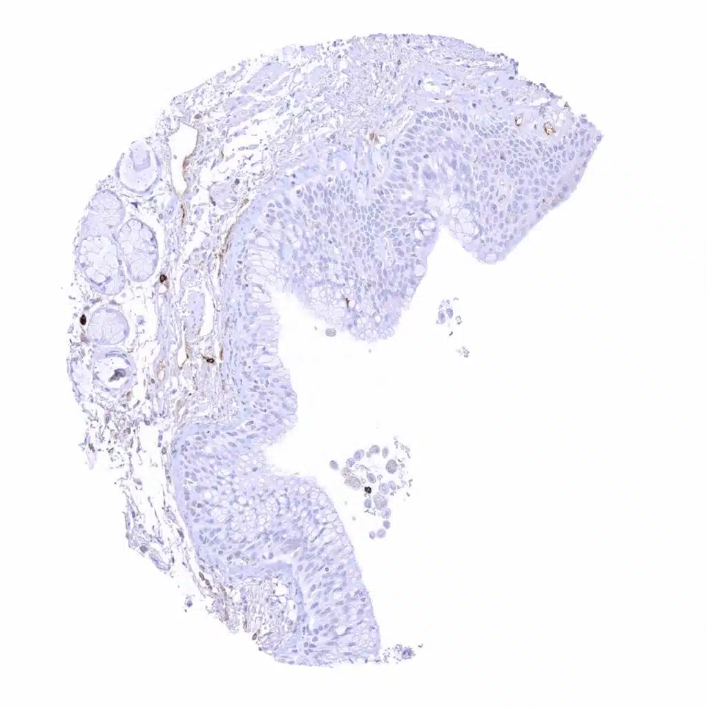
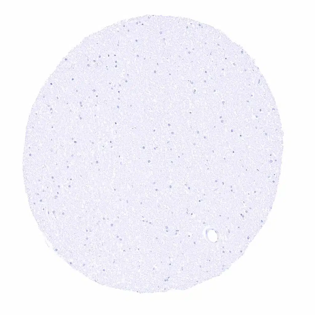
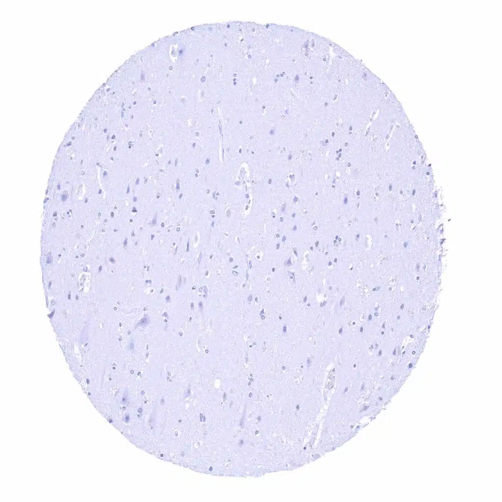
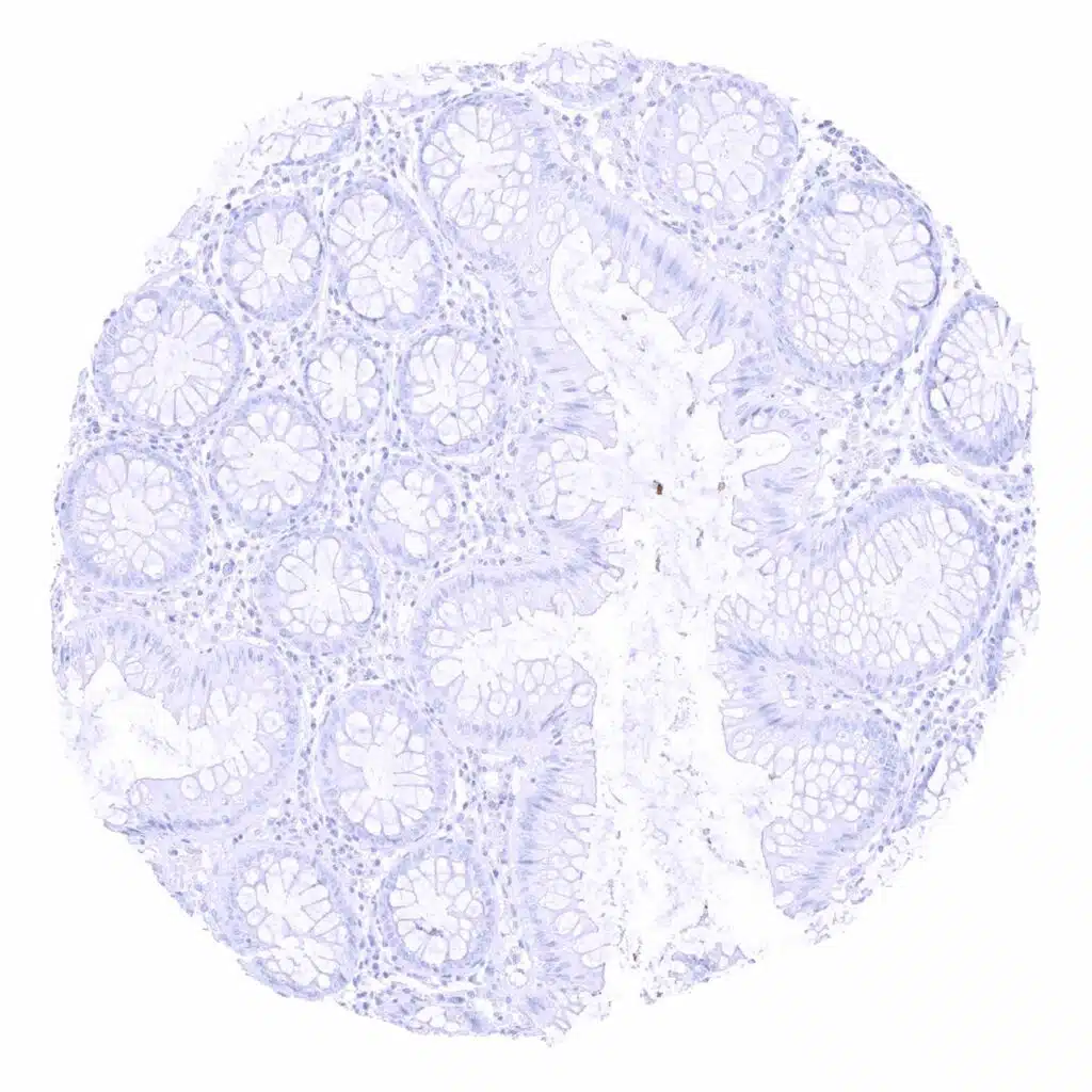
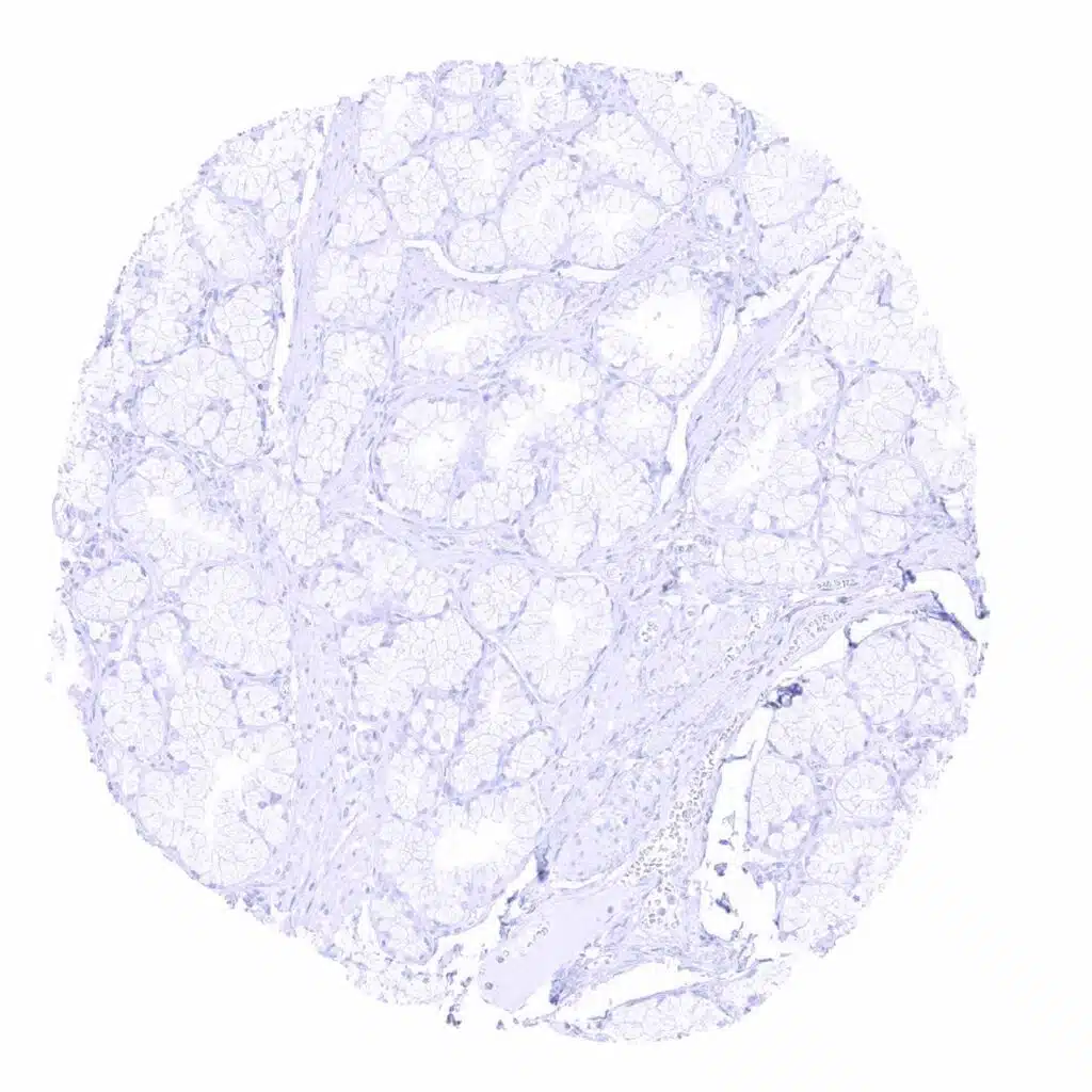
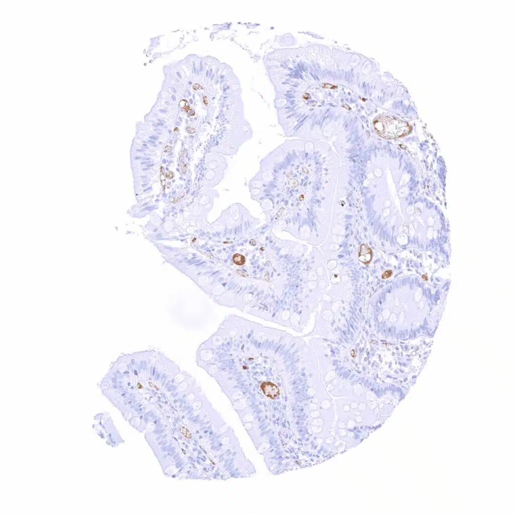
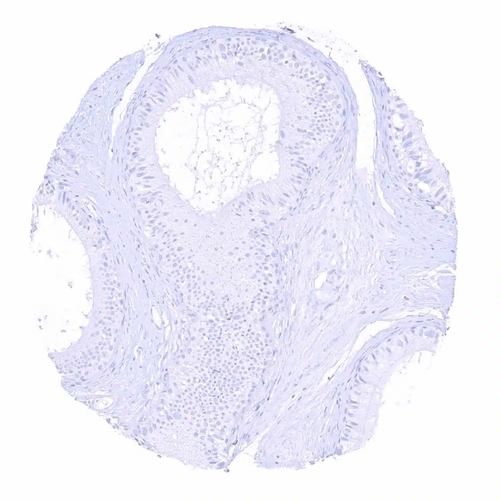
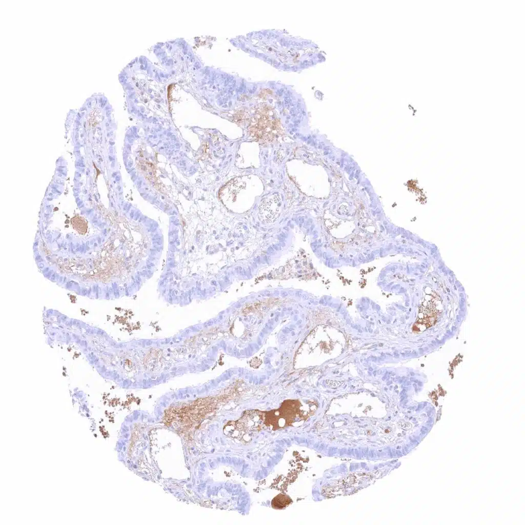
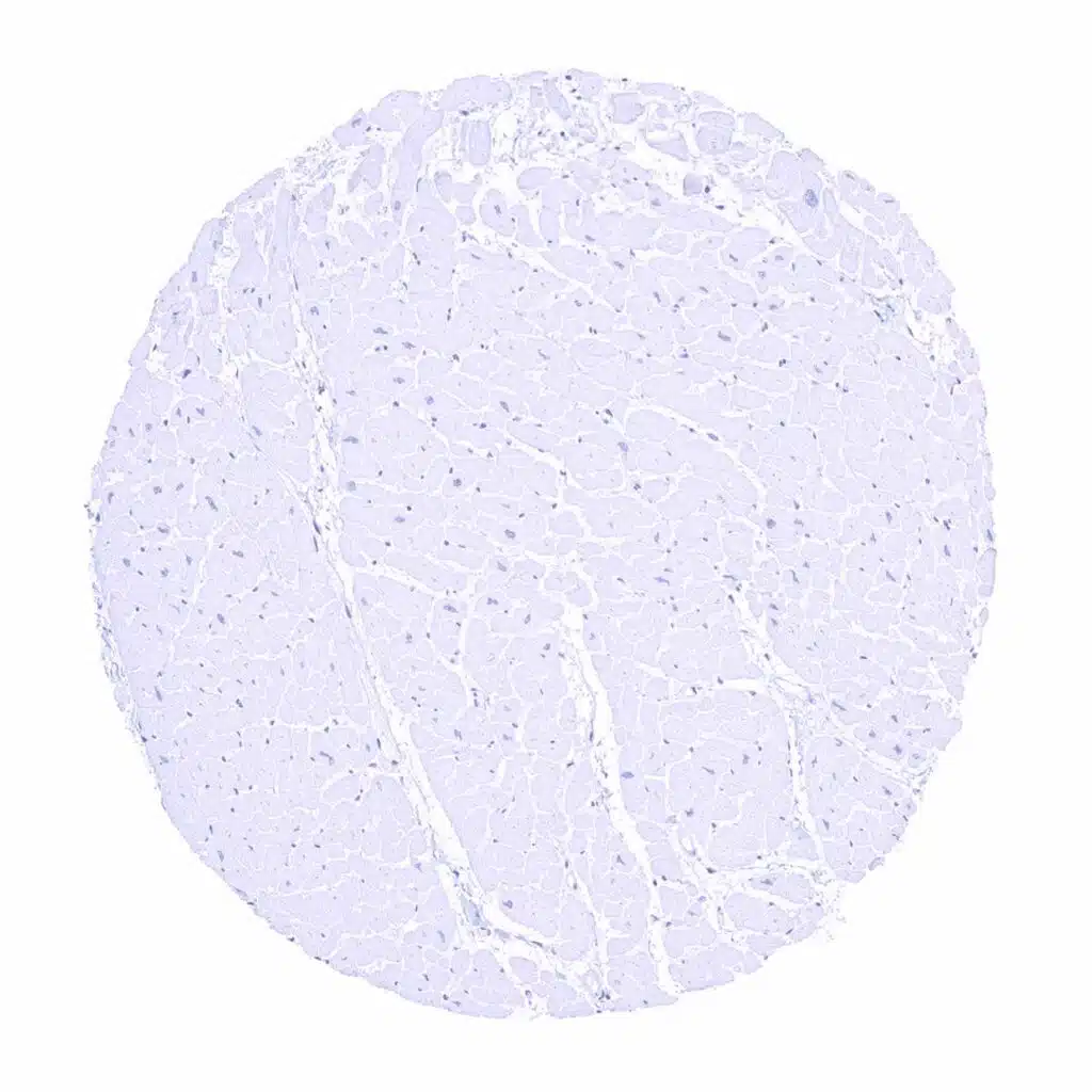
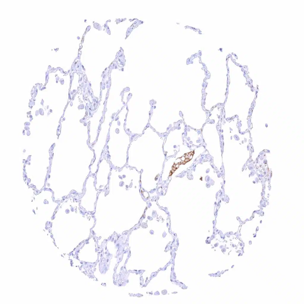
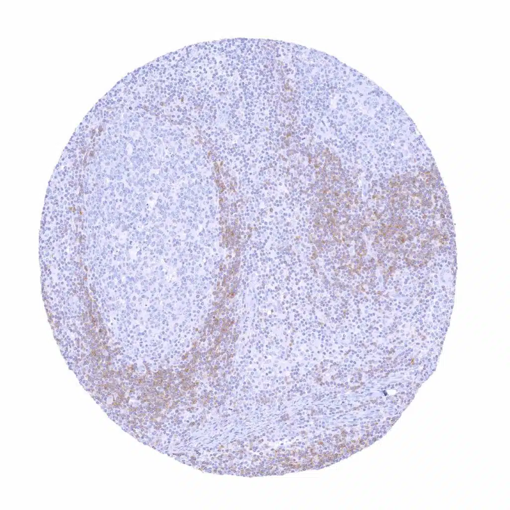
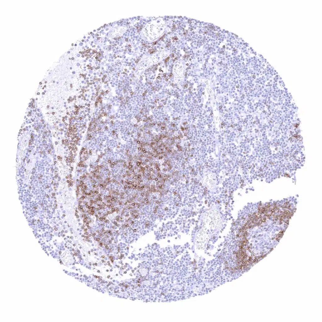
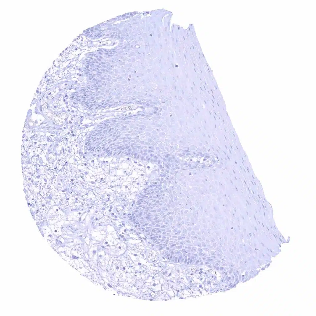
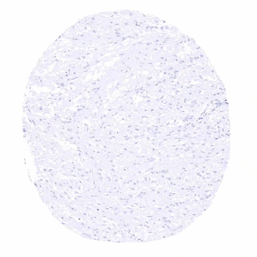
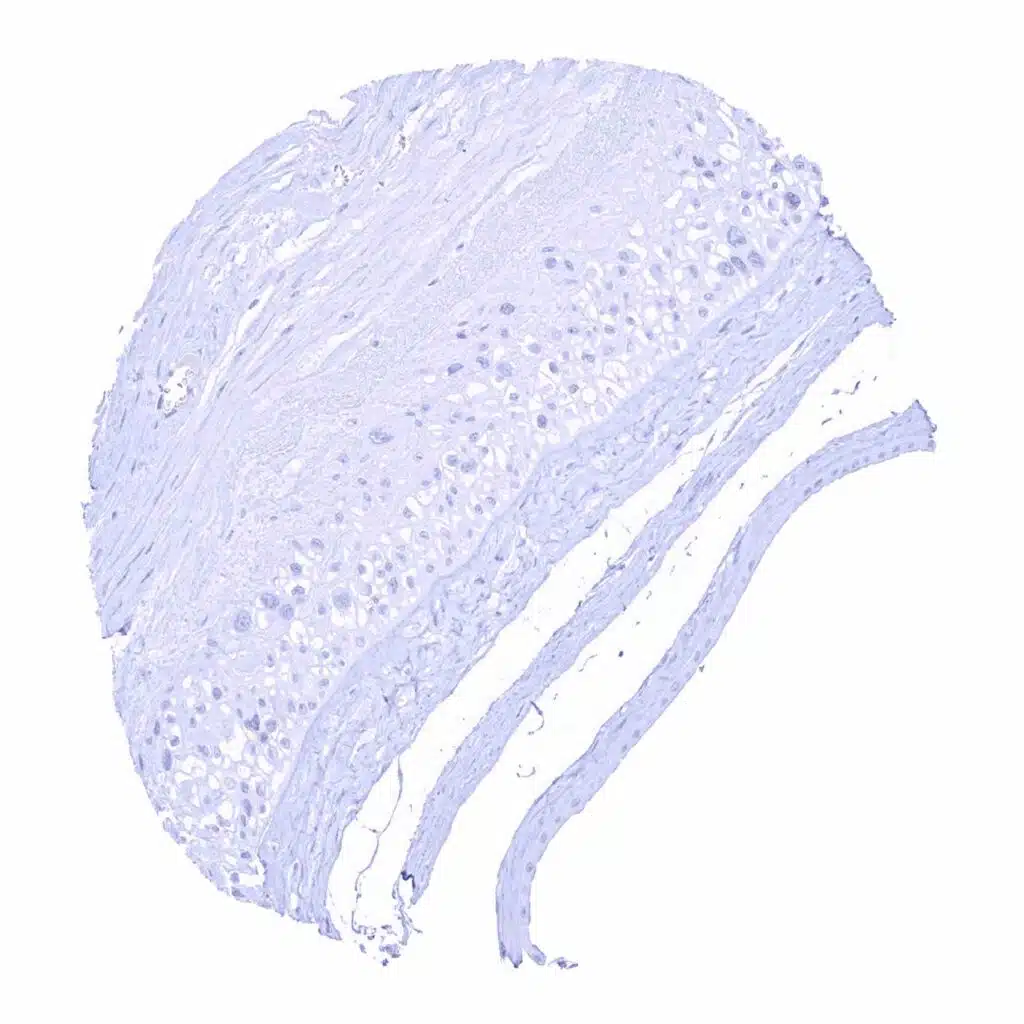
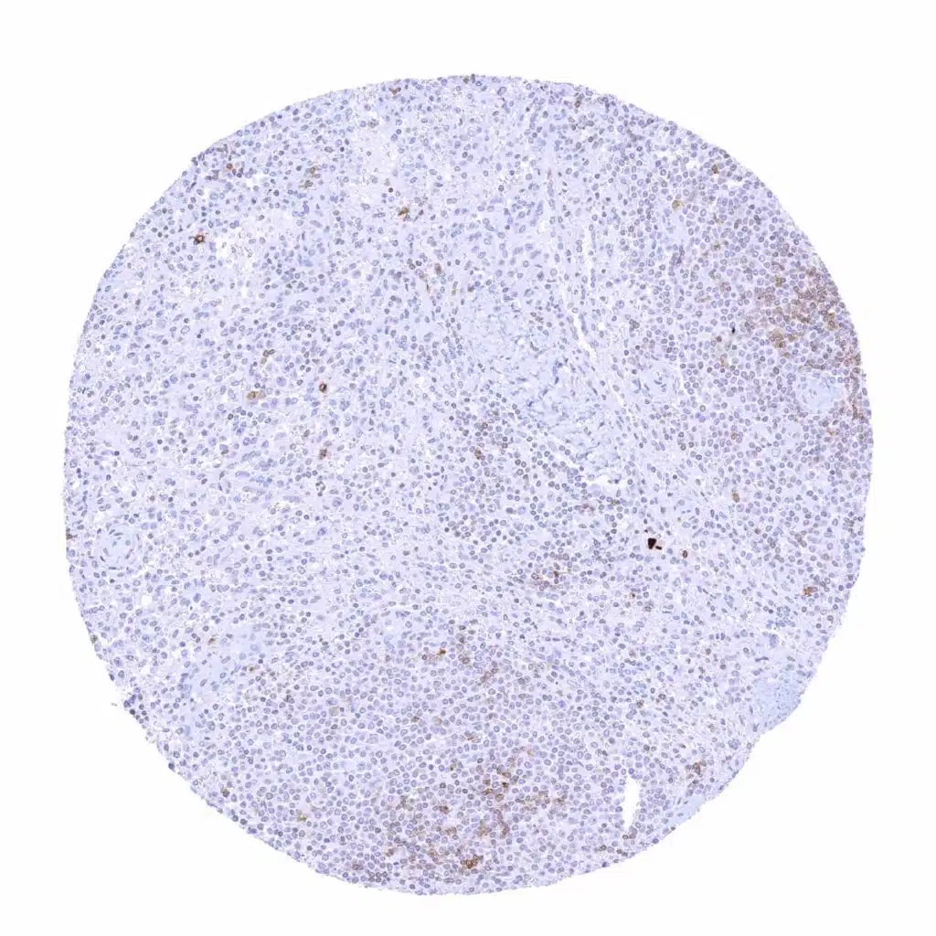
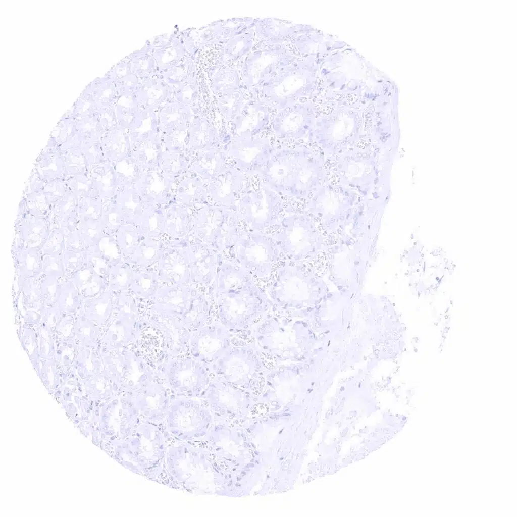
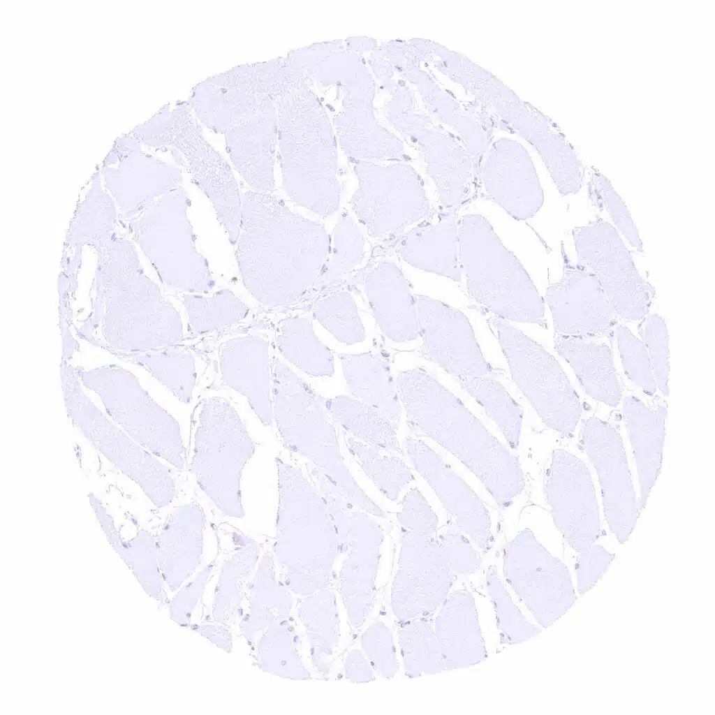
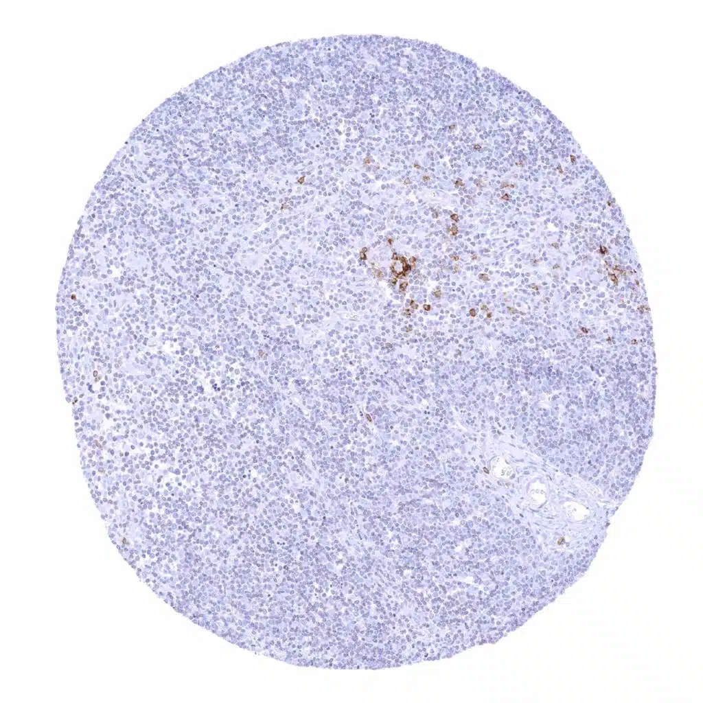
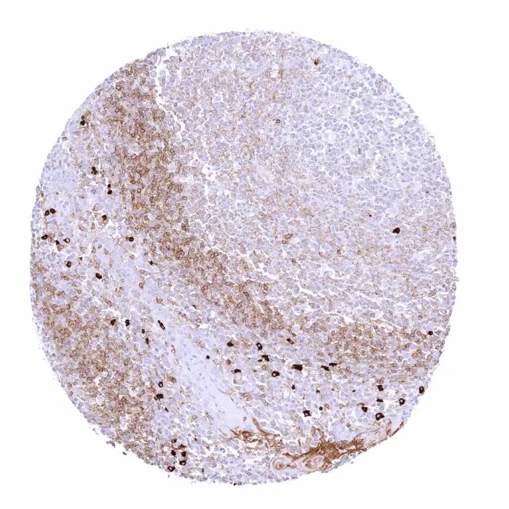
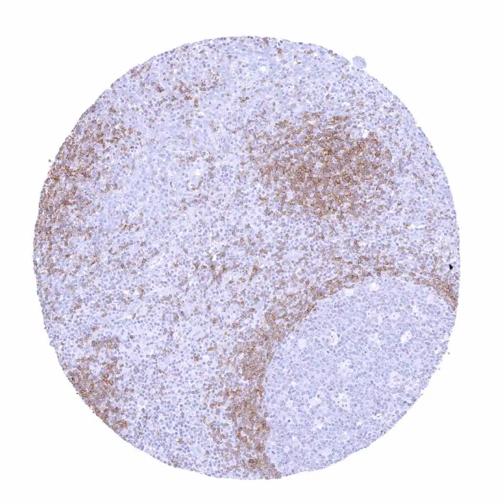
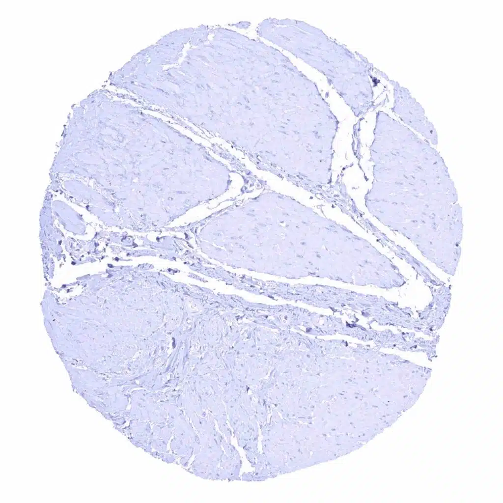
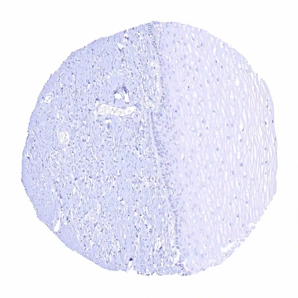
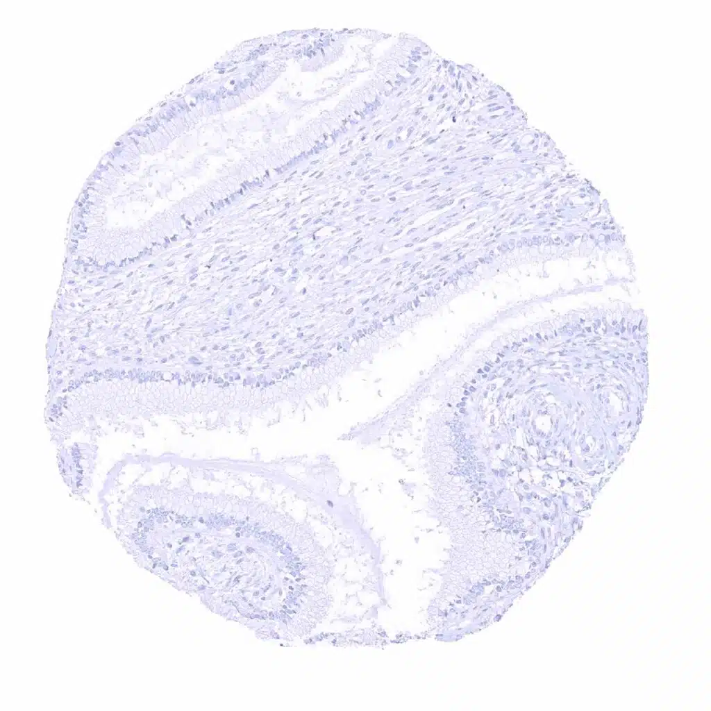
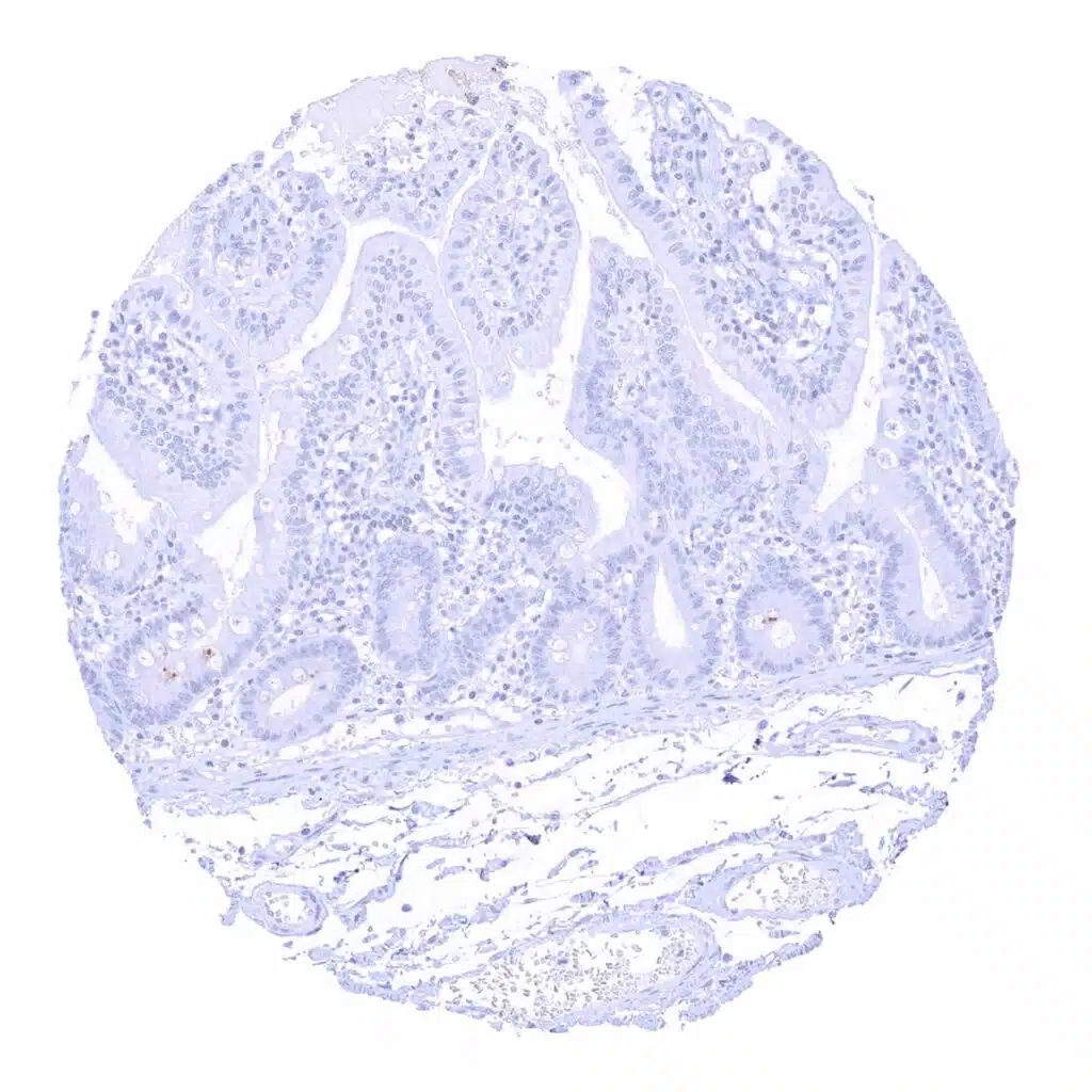
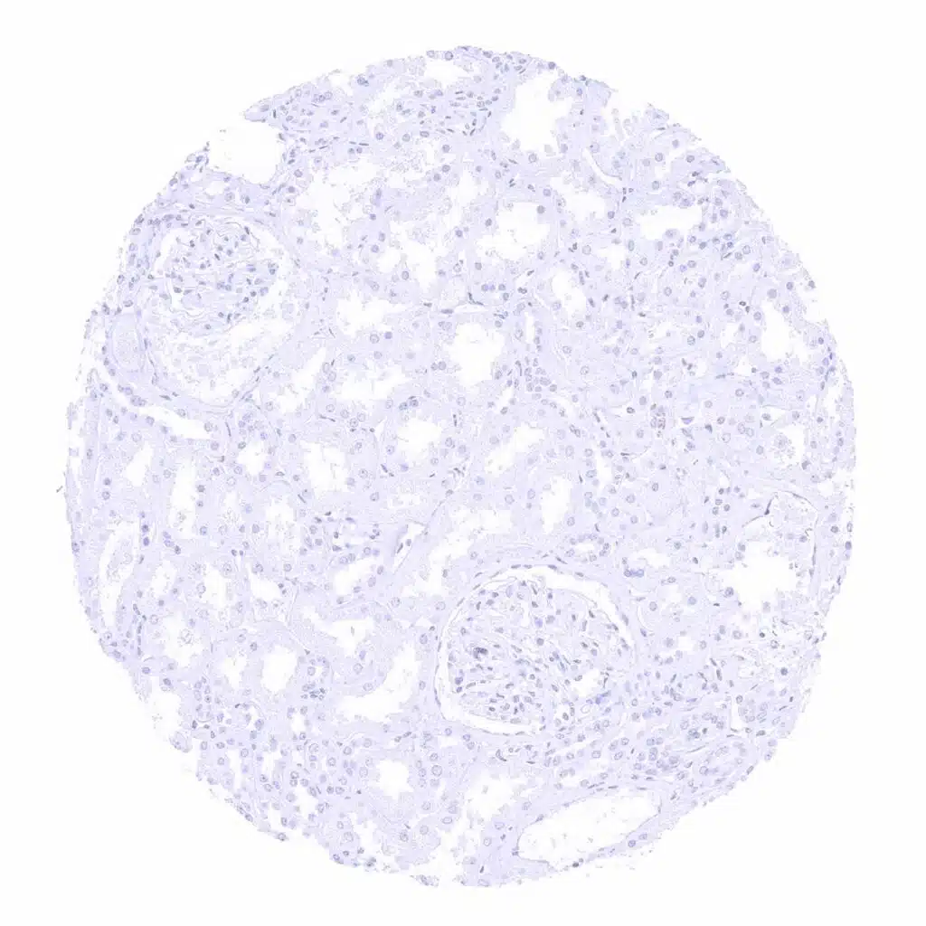
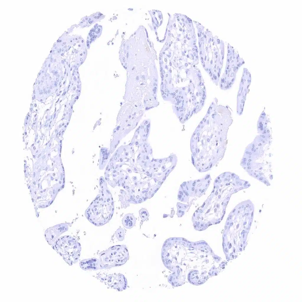
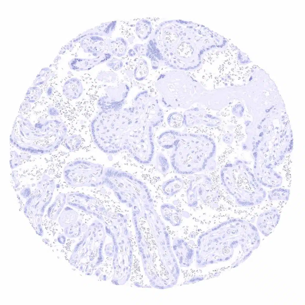
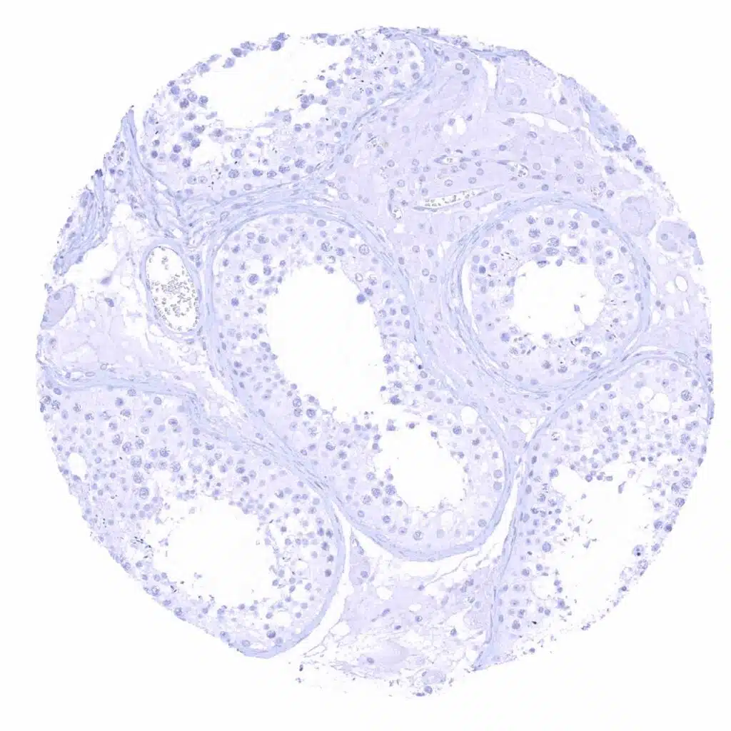
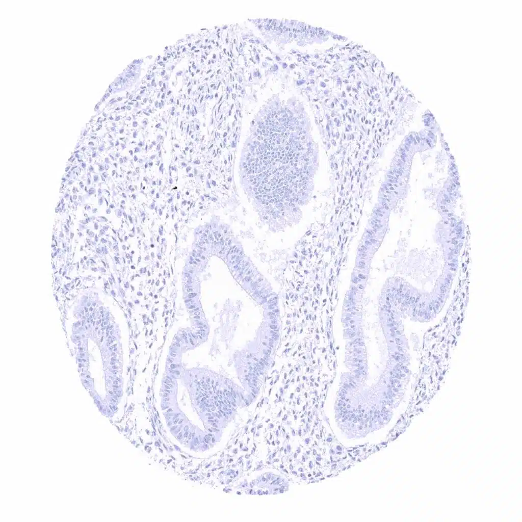
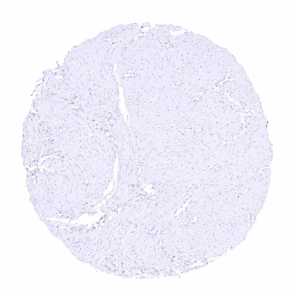
Rabbit anti-human IgD antibody [MSVA-701R] HistoMAX


Go to Cancer Tissue Gallery
  
 Go back to product page
  

Go to HistoMAX page


Adrenal gland

Aorta, media

Appendix, mucosa – Rabbit anti-human IgD immunostaining of mantle cells surrounding a germinal centre of the appendical mucosa

Appendix, mucosa – Rabbit anti-human IgD staining is largely limited to mantle cells surrounding a germinal centre of the appendical mucosa


Appendix, mucosa

Appendix, muscular wall

Bone marrow

Breast


Bronchus, mucosa – Few plasma cells show strong Rabbit anti-human IgD staining

Cerebellum, cortex (Stratum moleculare)

Cerebellum, grey (Stratum neuronorum)

Cerebrum, grey matter


Cerebrum, white matter

Colon descendens, mucosa

Colon descendens, muscular wall

Duodenum, Brunner gland


Duodenum, mucosa – Rabbit anti-human IgD staining of few inflammatory cells and of intravascular material

Epididymis

Esophagus, squamous epithelium

Fallopian tube, mucosa – Rabbit anti-human IgD staining is largely lacking in this sample


Fallopian tube, mucosa – Substantial Rabbit anti-human IgD staining of intravascular liquids. Weaker staining is also seen in adjacent stroma

Fat

Gallbladder, epithelium

Heart muscle


Ileum, mucosa

Kidney, cortex

Kidney, medulla – Weak Rabbit anti-human IgD staining of intratubular liquid

Kidney, pelvis, urothelium


Liver

Lung – Moderate to strong Rabbit anti-human IgD staining of intravascular liquid

Lymph node – Significant Rabbit anti-human IgD immunostaining in B-lymphocytes of the mantle zone

Lymph node – Strong Rabbit anti-human IgD staining in a fraction of B-lymphocytes, especially in the mantle zone


Oral cavity

Ovary, stroma

Pancreas

Parathyroid gland


Parotid gland – Strong Rabbit anti-human IgD staining of intravascular plasma

Parotid gland

Pituitary gland, anterior lobe

Pituitary gland, posterior lobe


Placenta (amnion and chorion)

Placenta early, decidua

Placenta, early – Substantial Rabbit anti-human IgD staining of intervillous liquids. Weaker staining of adjacent syncytiotrophoblast may reflect a contamination artifact

Placenta, early


Placenta, mature

Prostate

Rectum, mucosa

Seminal vesicle


Sinus paranasales

Skin

Spleen – Moderate Rabbit anti-human IgD staining of a fraction of B-lymphocytes

Stomach, antrum


Stomach, corpus

Striated muscle

Testis – Rabbit anti-human IgD staining is not seen in this sample

Testis – Weak Rabbit anti-human IgD staining of intravascular liquid


Thymus – Only few cells show Rabbit anti-human IgD staining, especially in the medulla

Thyroid gland

Tonsil – Rabbit anti-human IgD staining of variable intensity in B-lymphocytes. Staining is particularly prominent in the mantle zone of germinal centres. Staining intensity is highest in interspersed plasma cells

Tonsil – Strong Rabbit anti-human IgD staining in a fraction of B-lymphocytes, especially in the mantle zone


Tonsil, surface epithelium – Numerous Rabbit anti-human IgD positive inflammatory cells

Urinary bladder, muscular wall

Uterus, ectocervix

Uterus, endocervix


Uterus, endometrium (proliferation)

Uterus, endometrium (proliferation)

Uterus, endometrium (secretion)

Uterus, myometrium


| TOP |