Normal Tissue Gallery- GTX04483

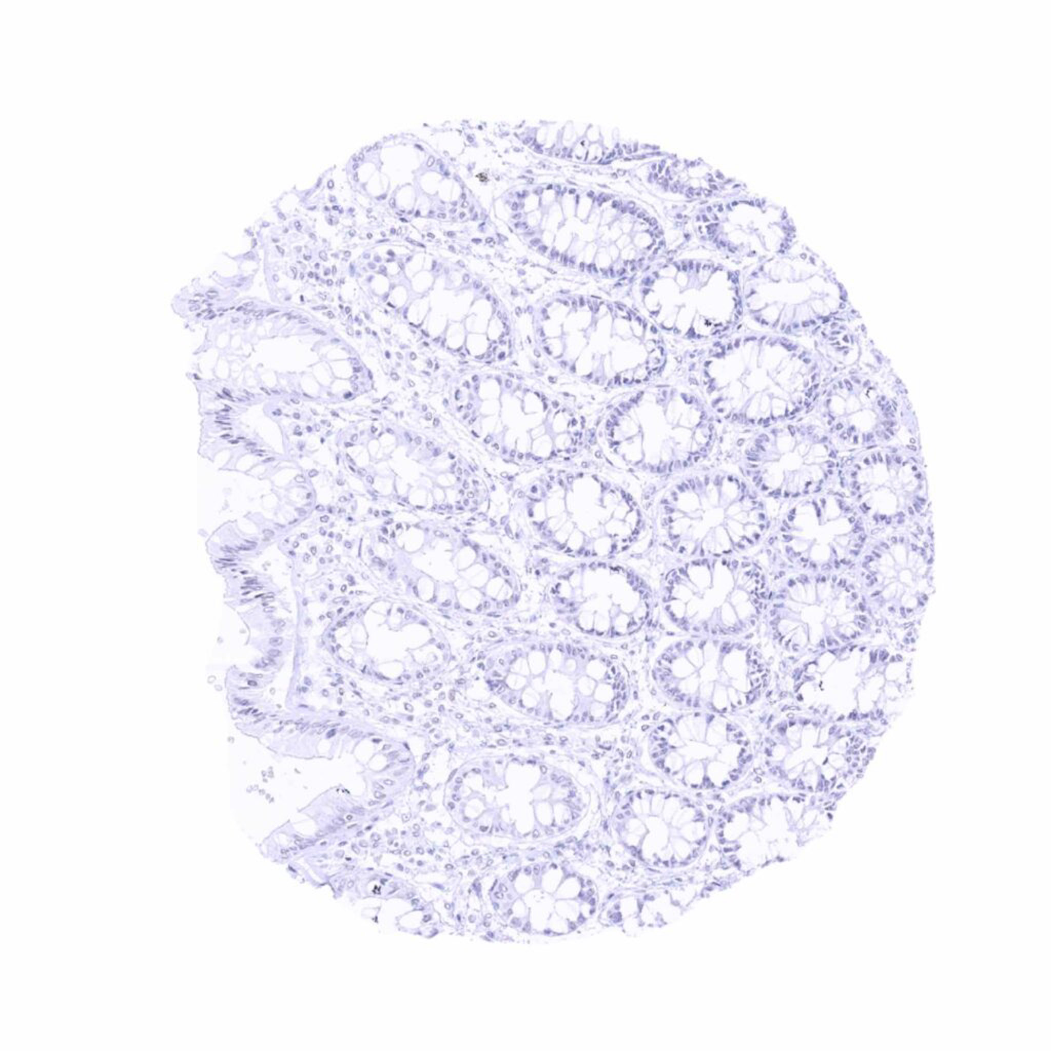
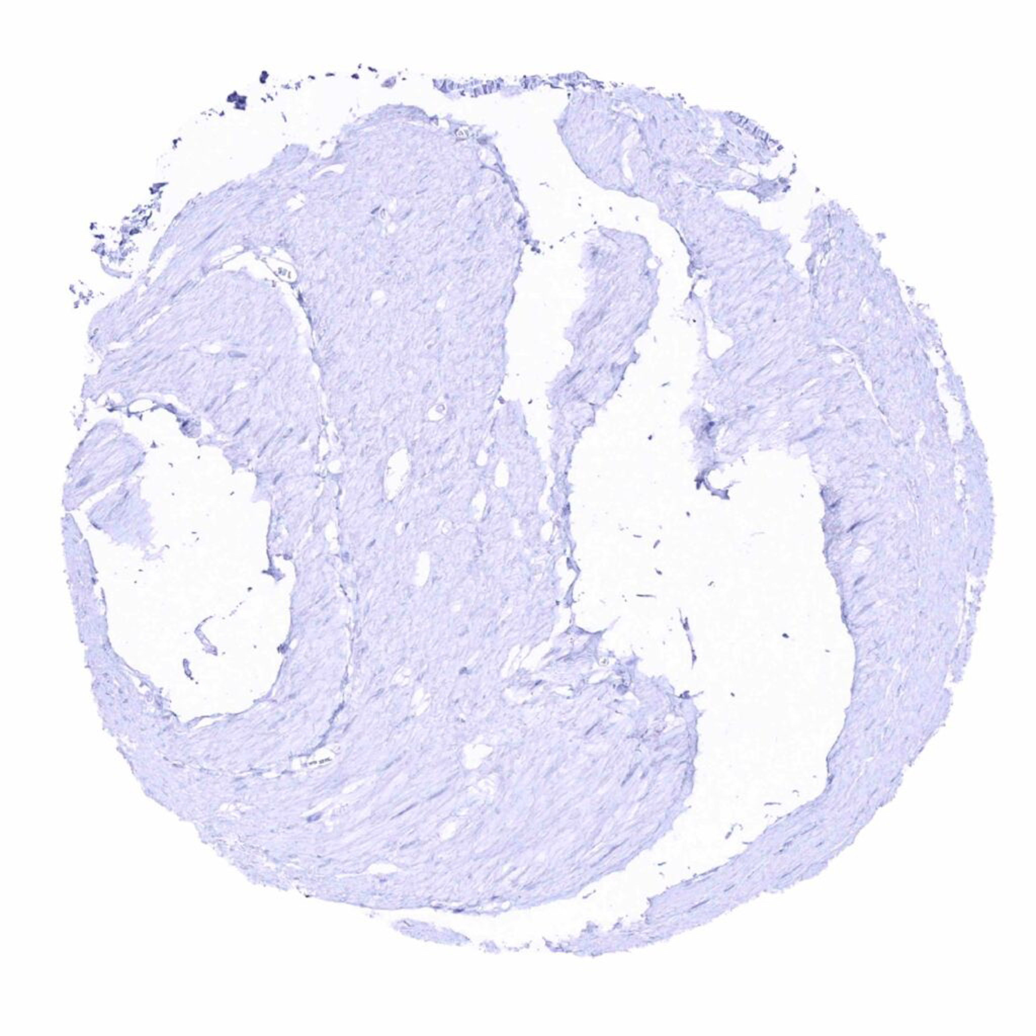
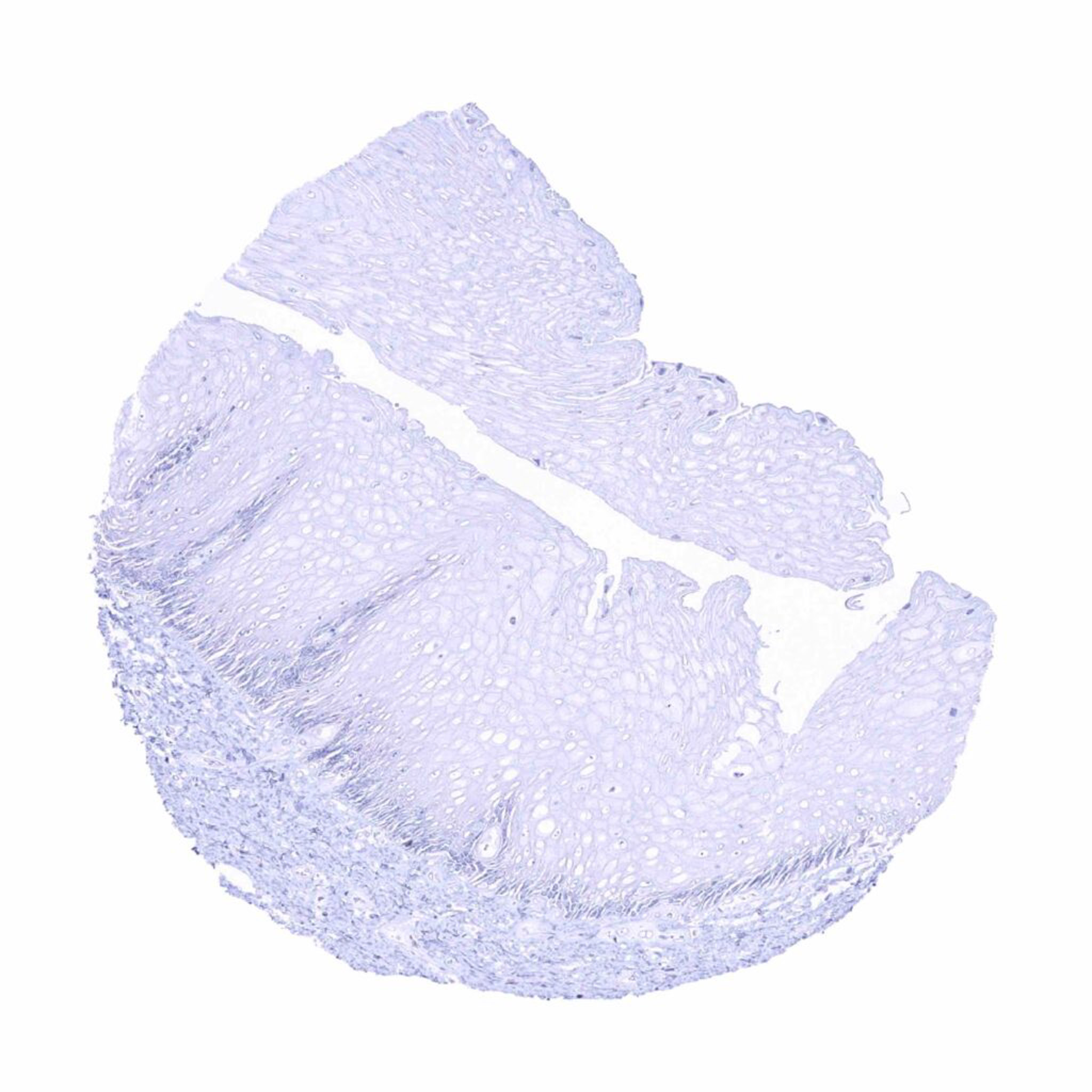
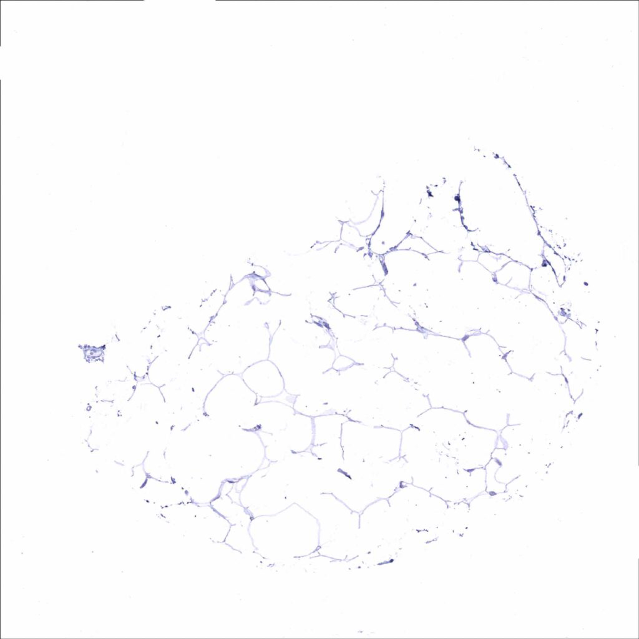
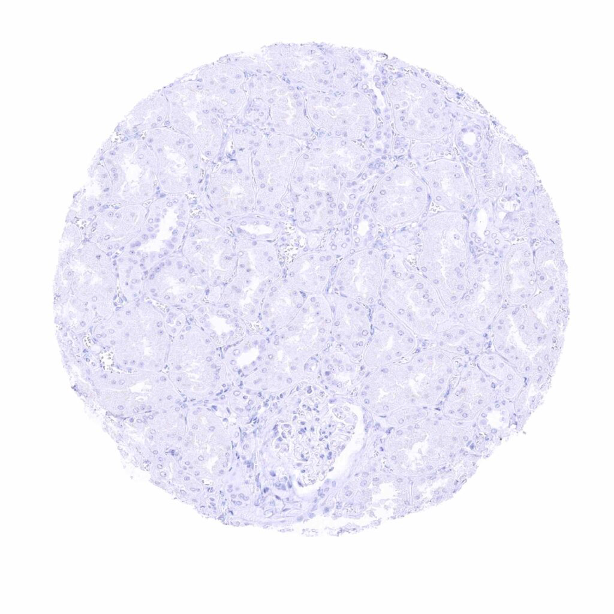
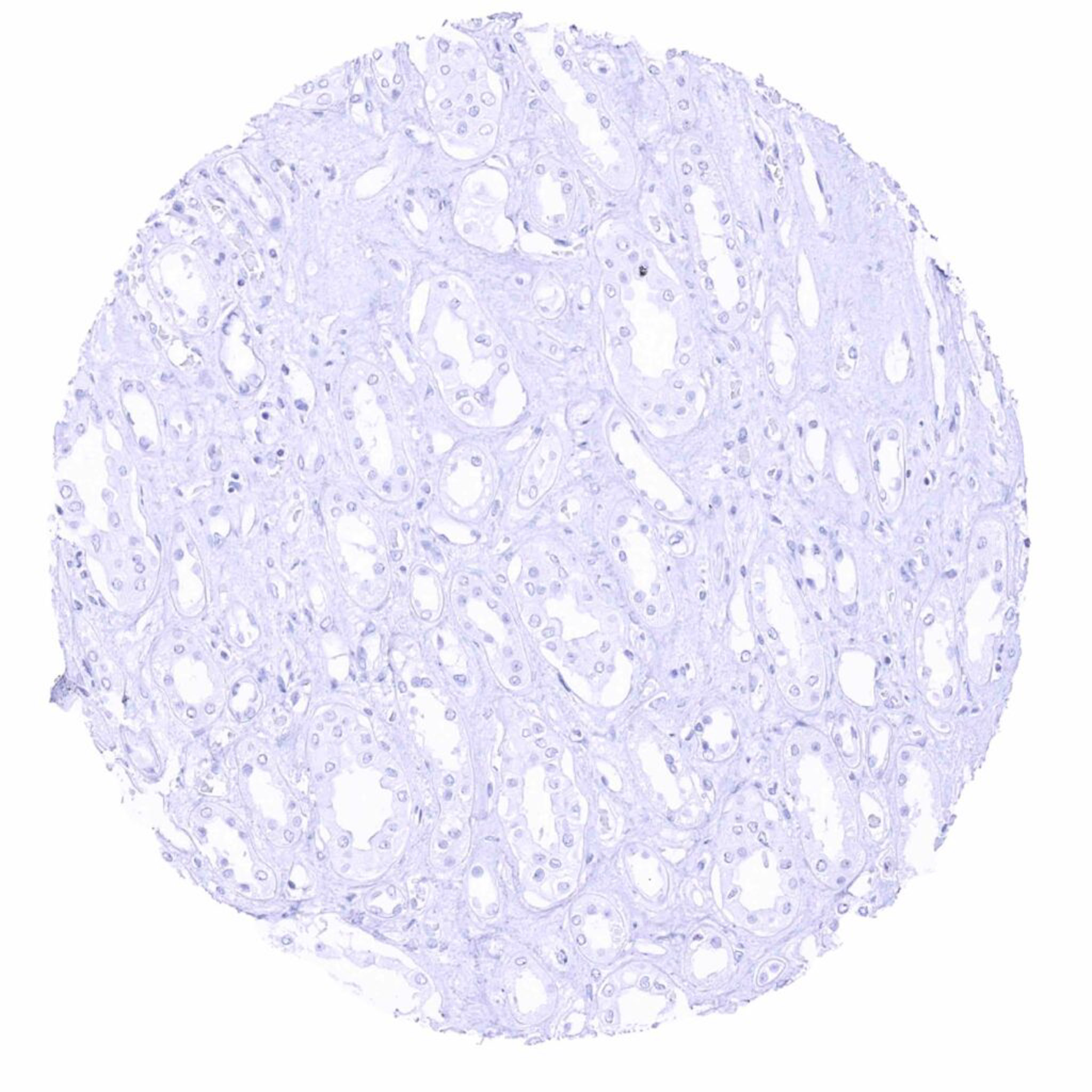
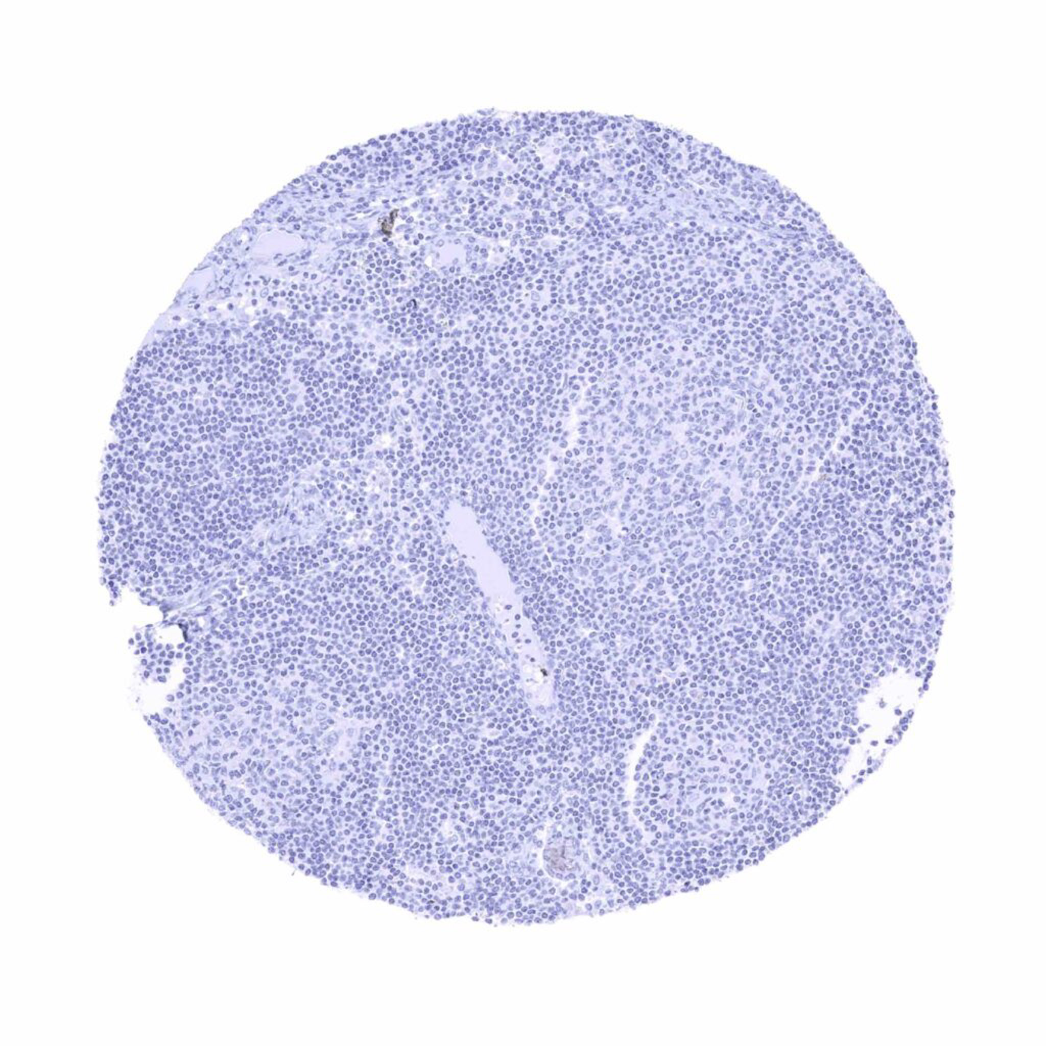
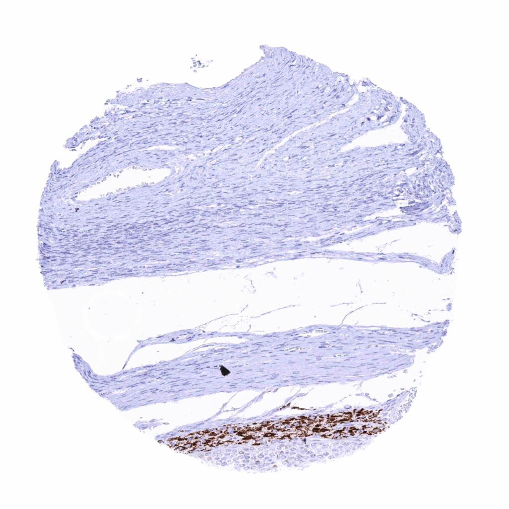
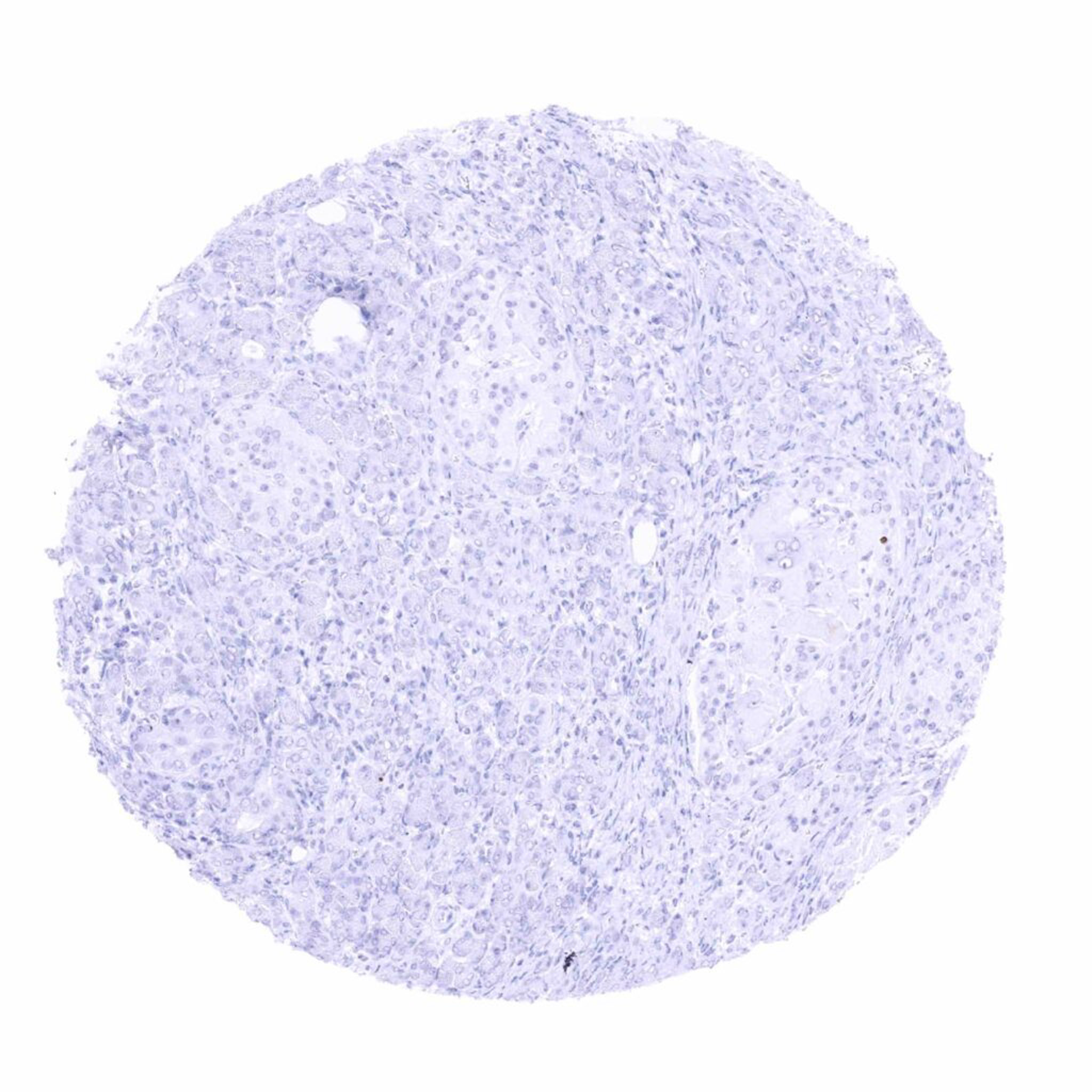
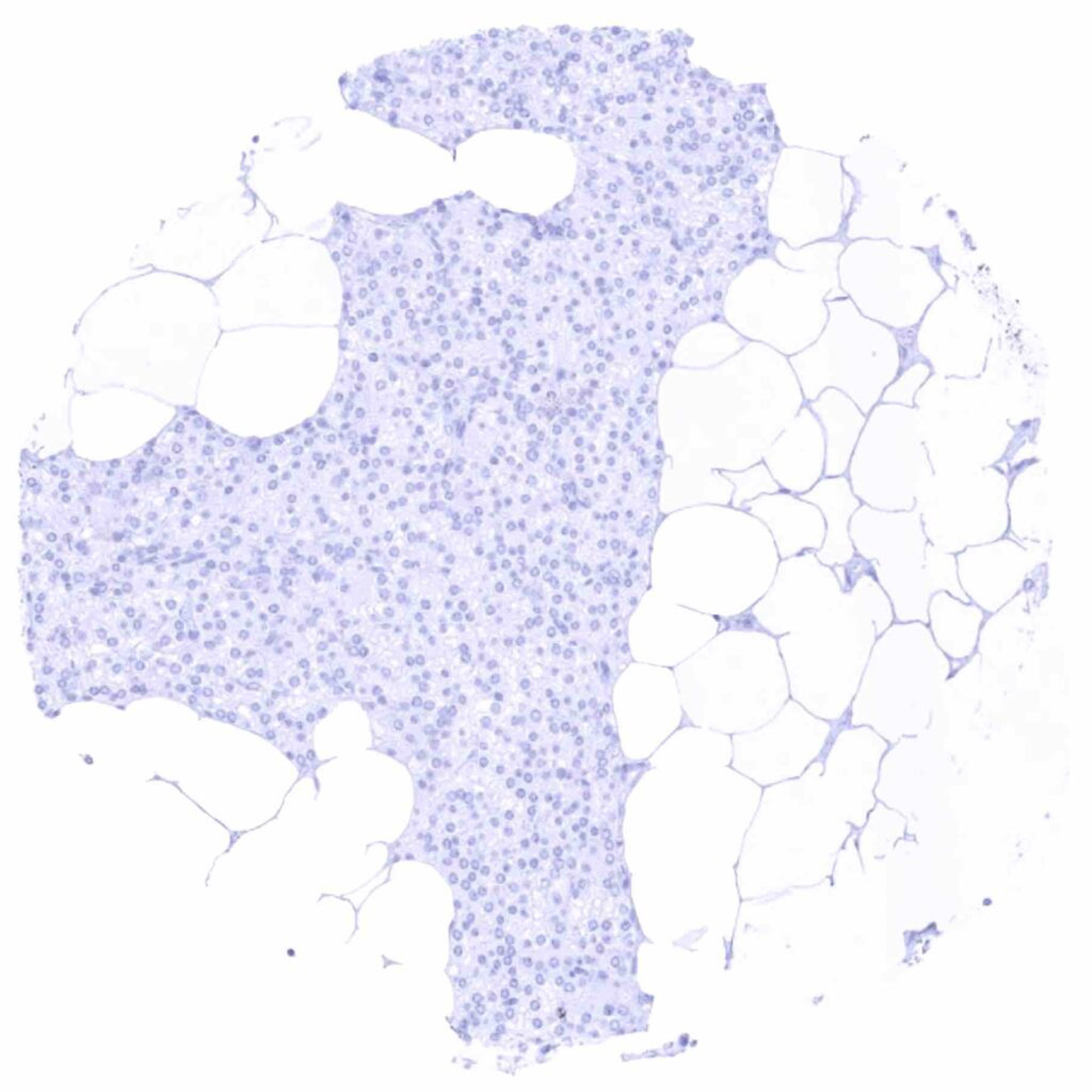
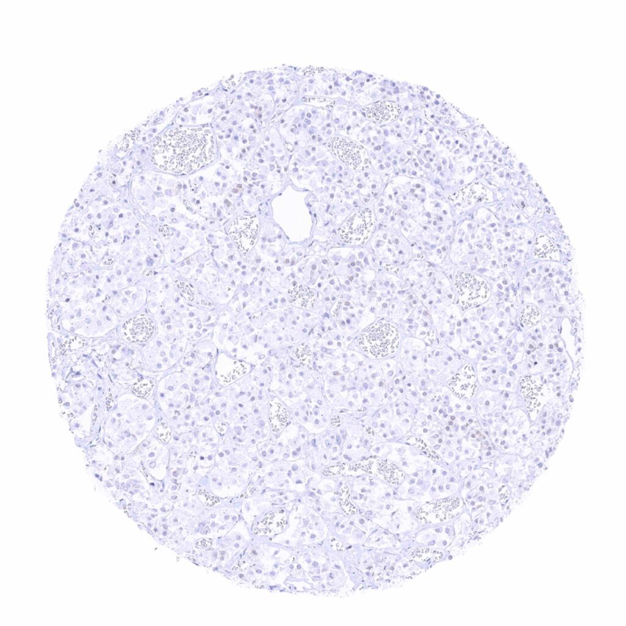
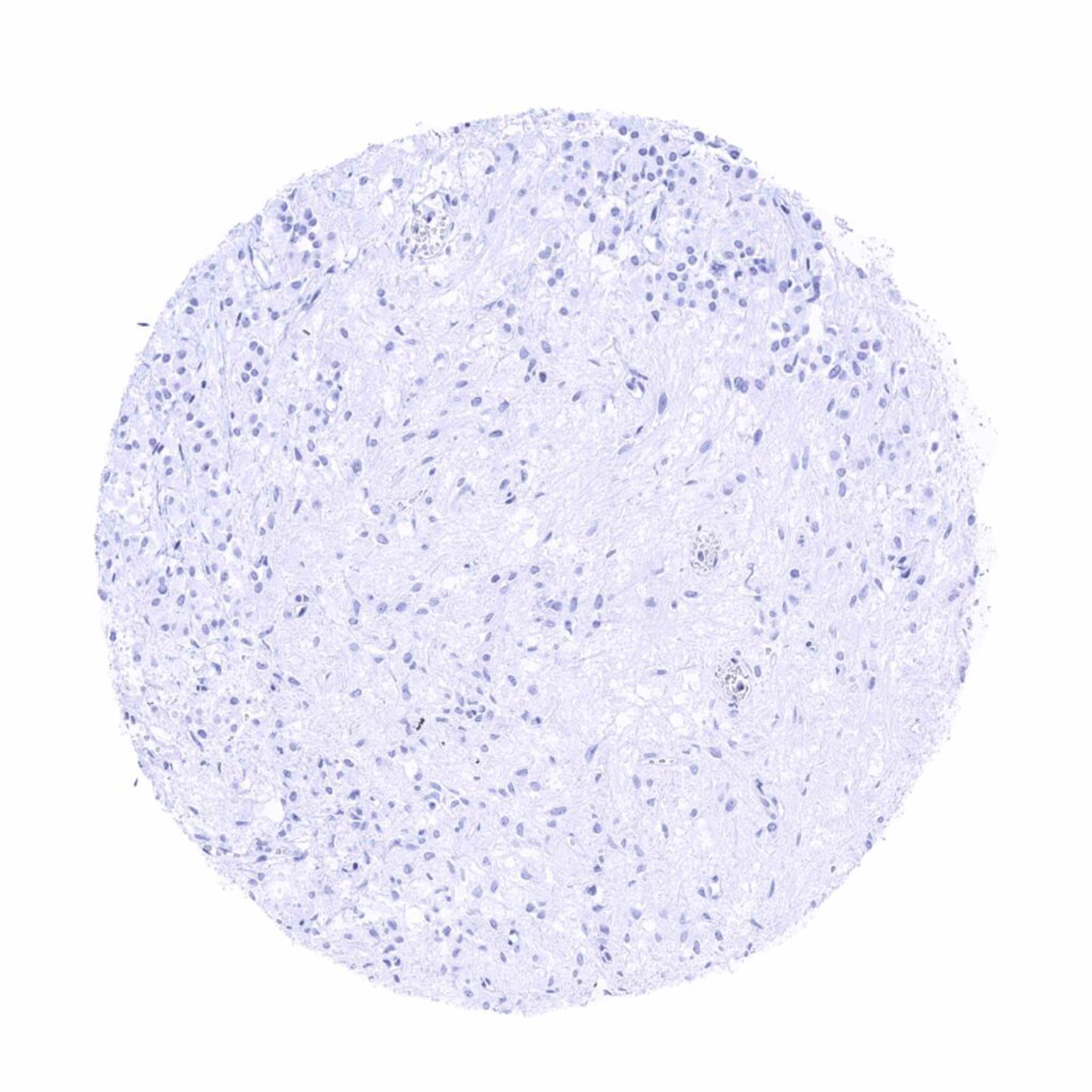
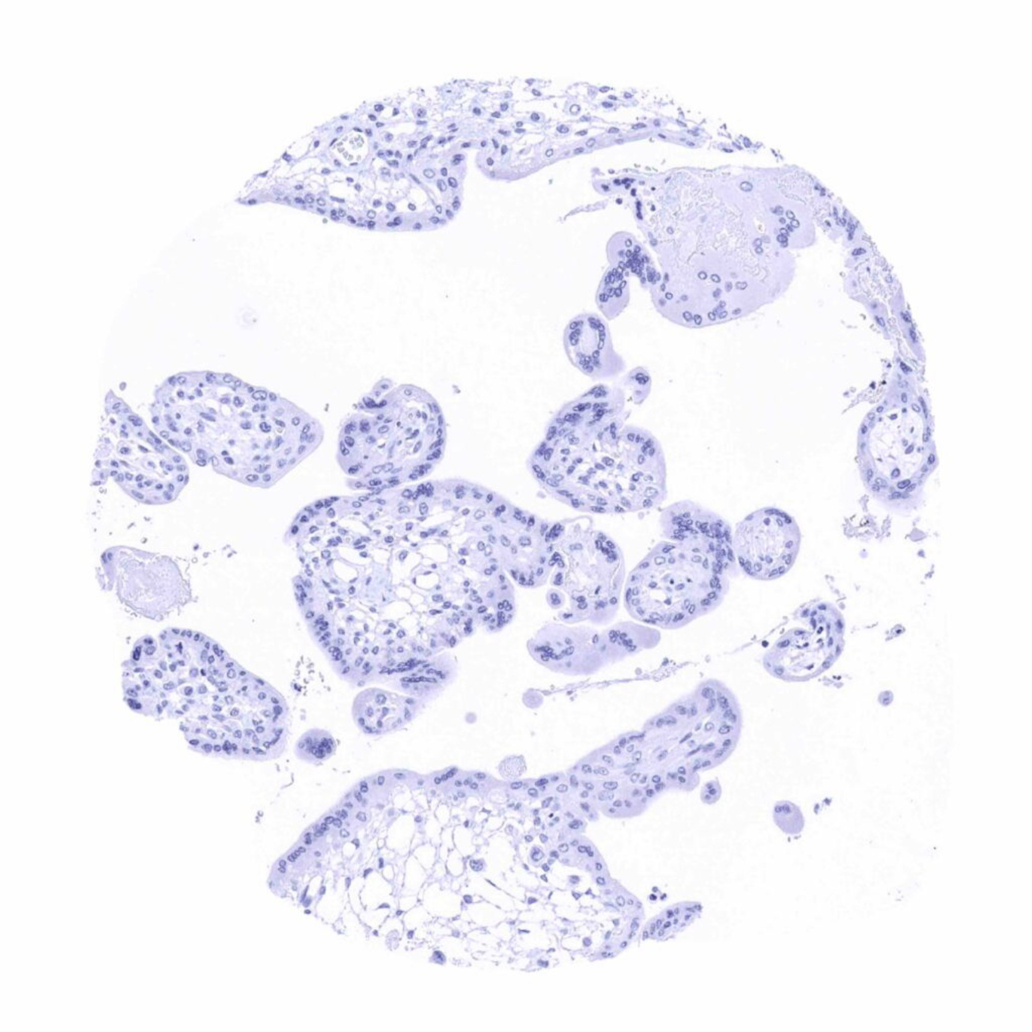
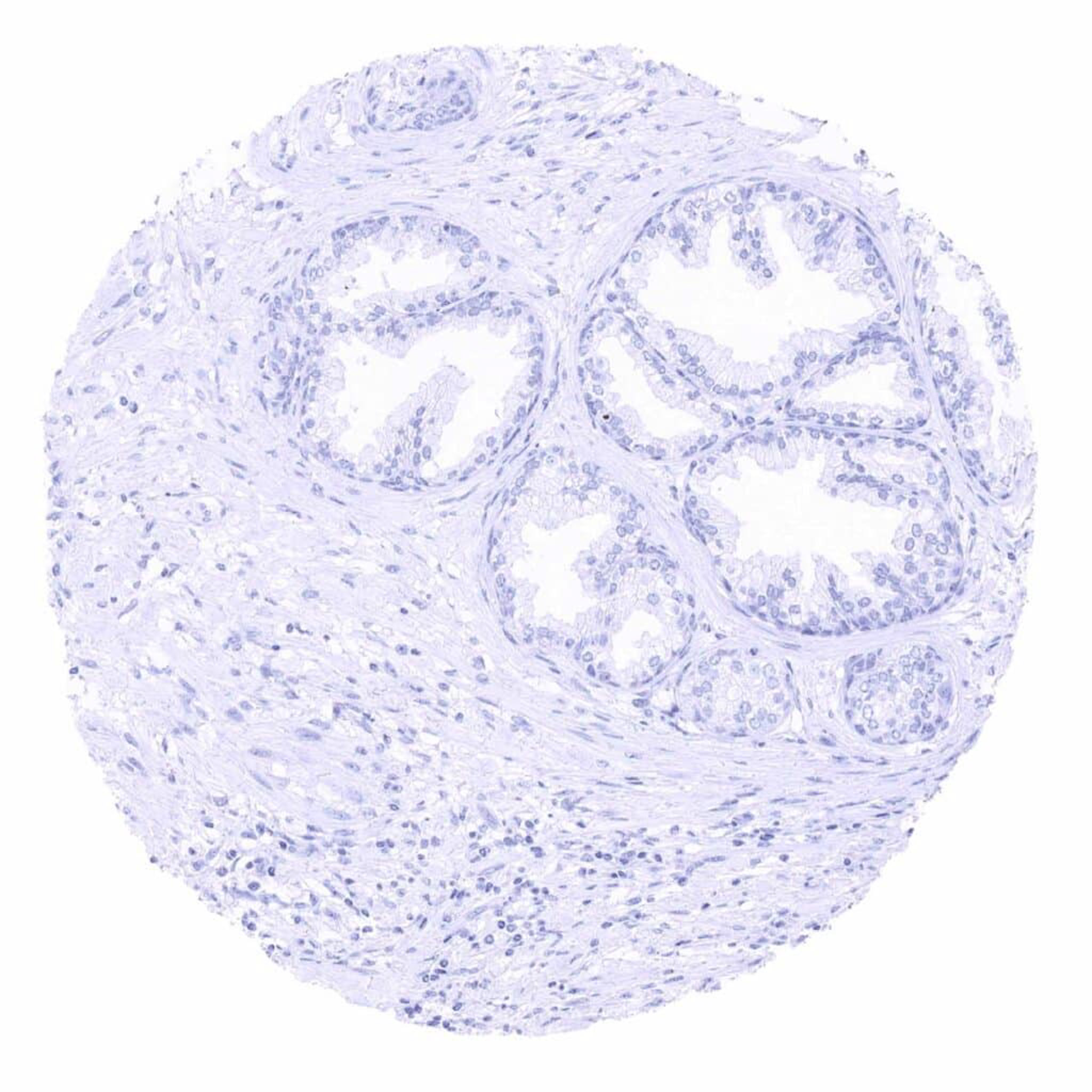
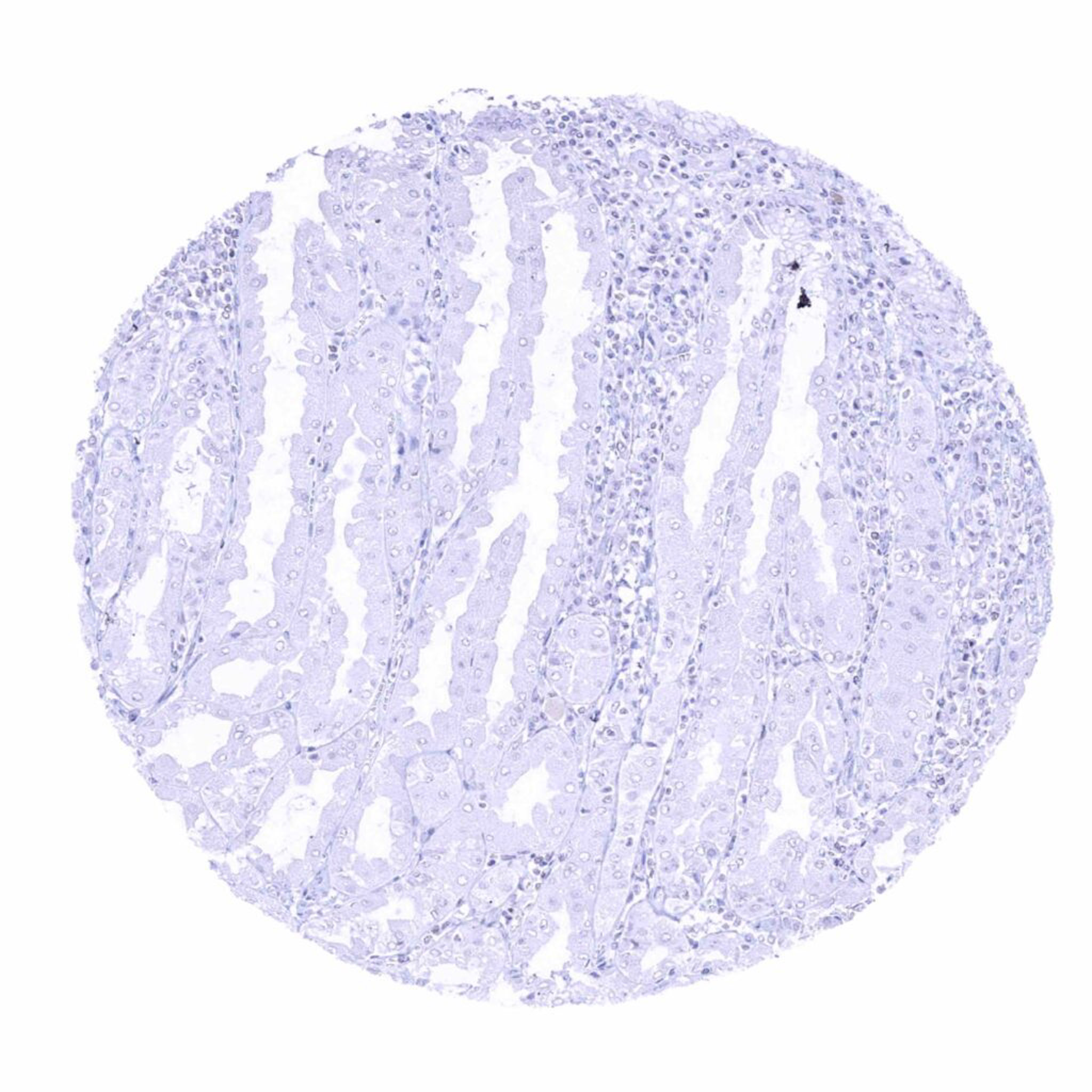
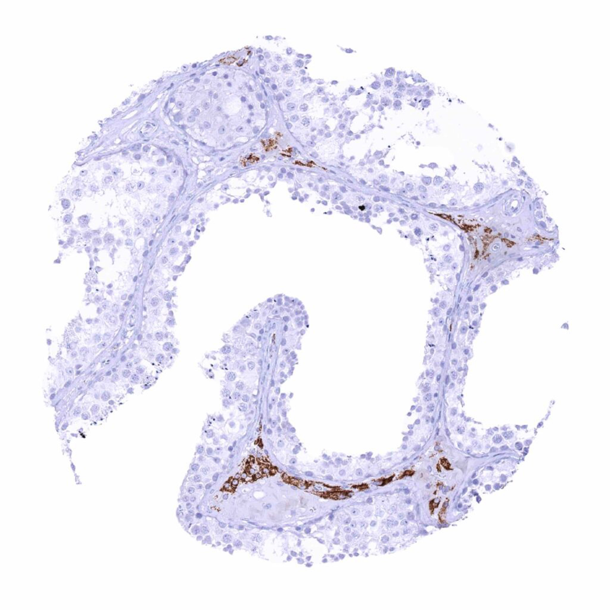
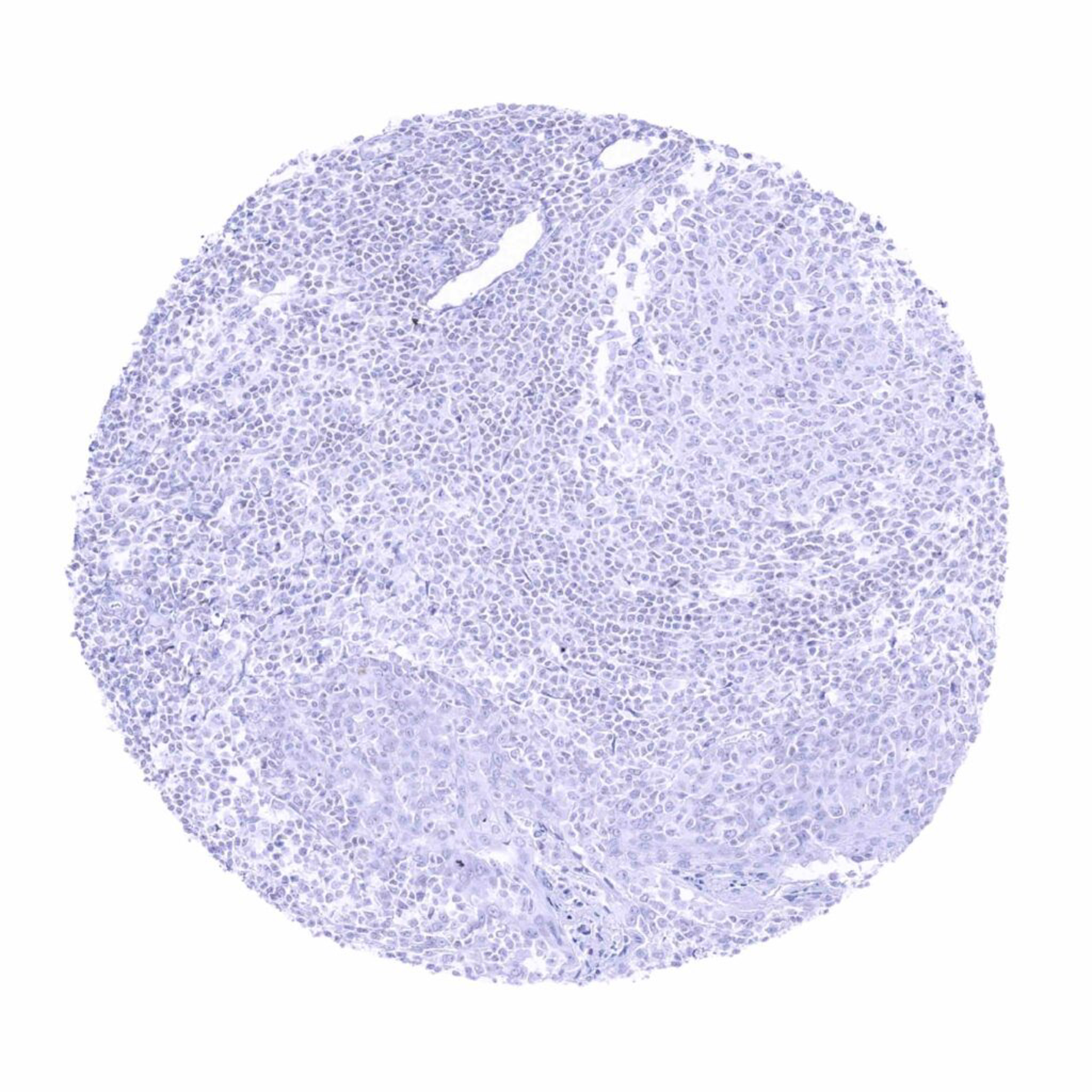
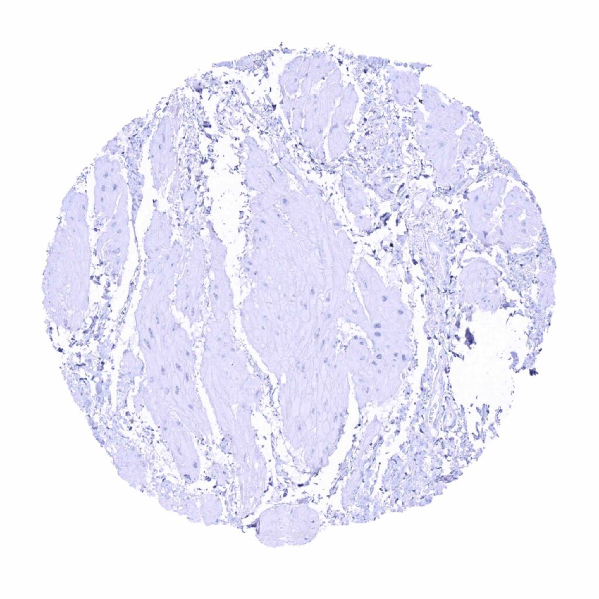
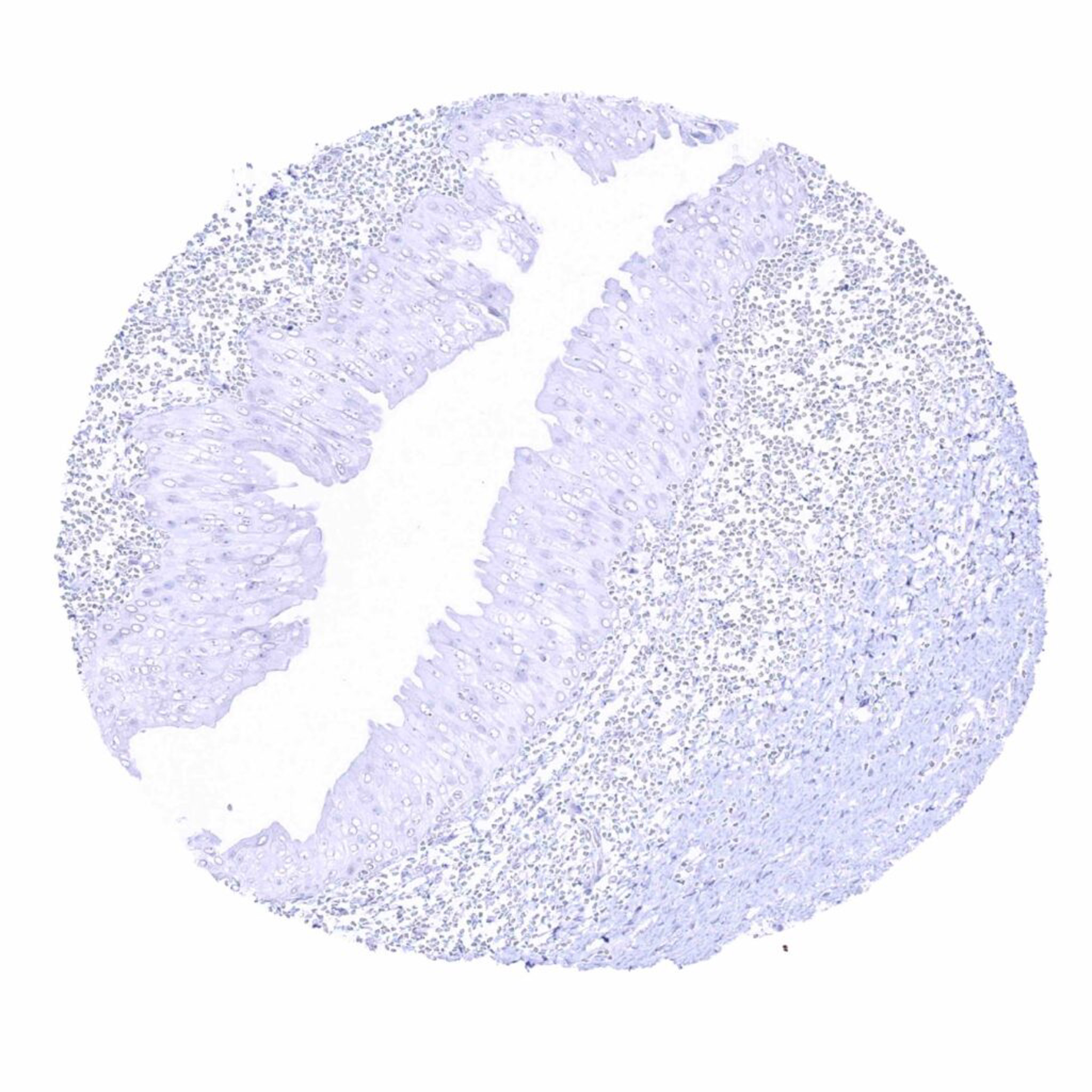
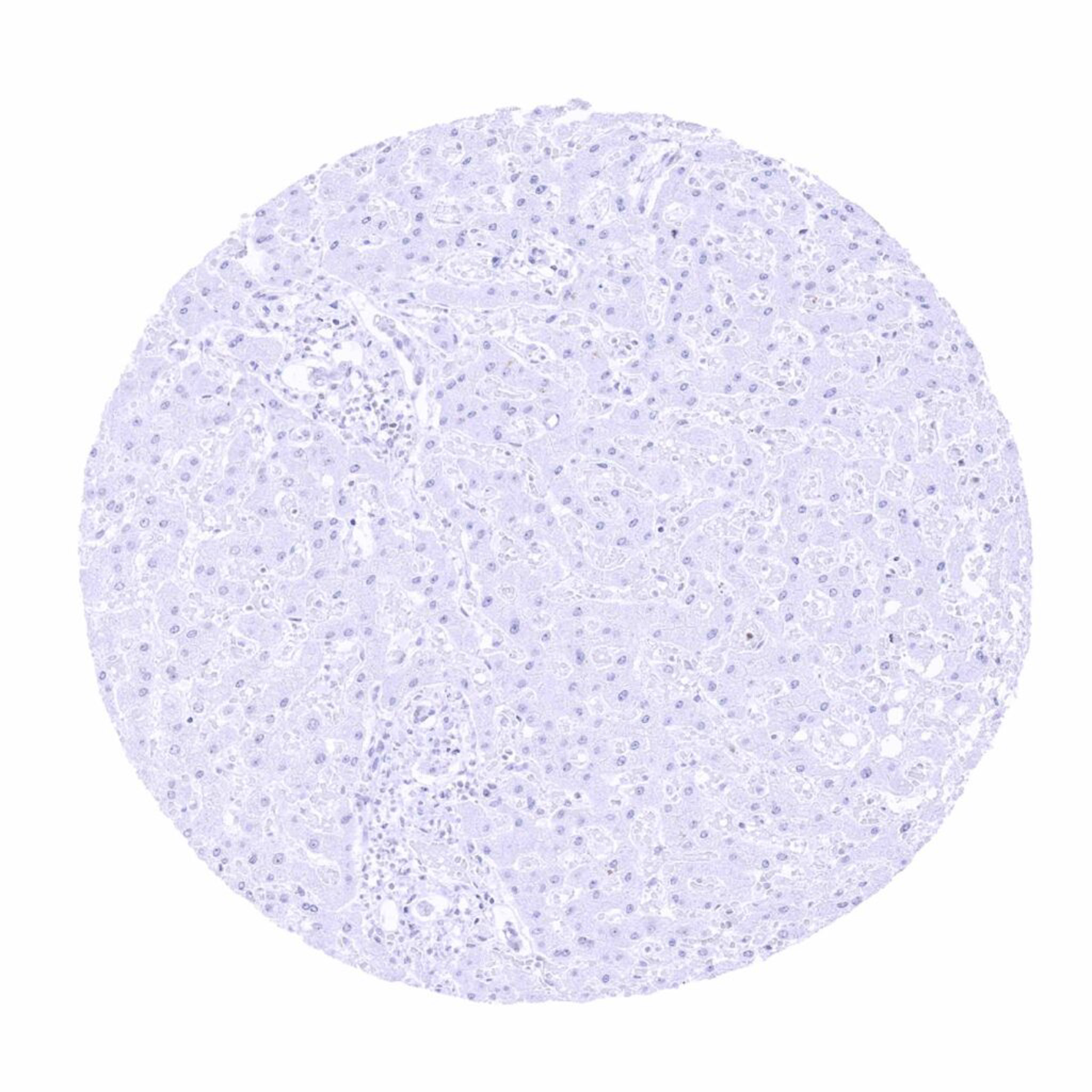
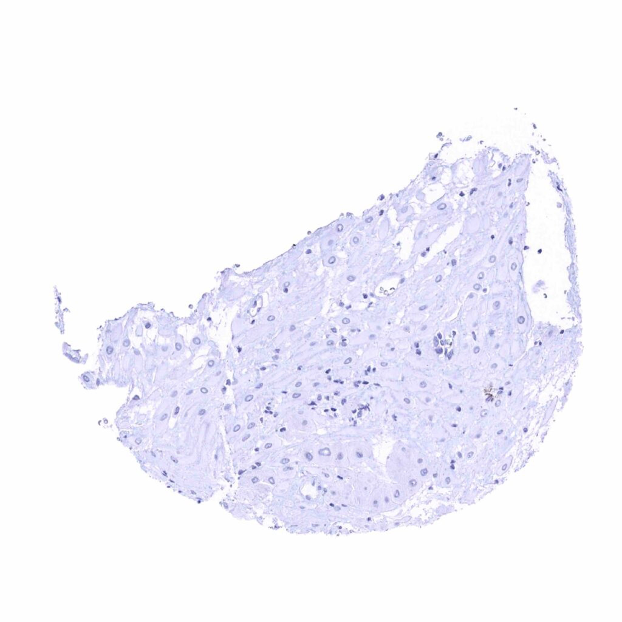
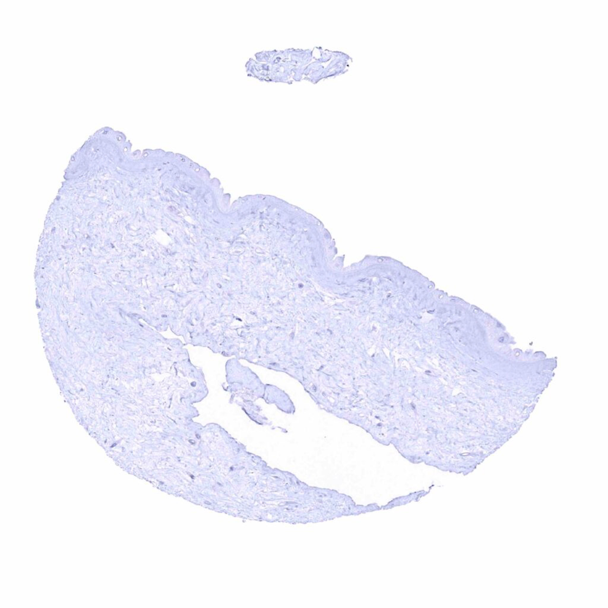
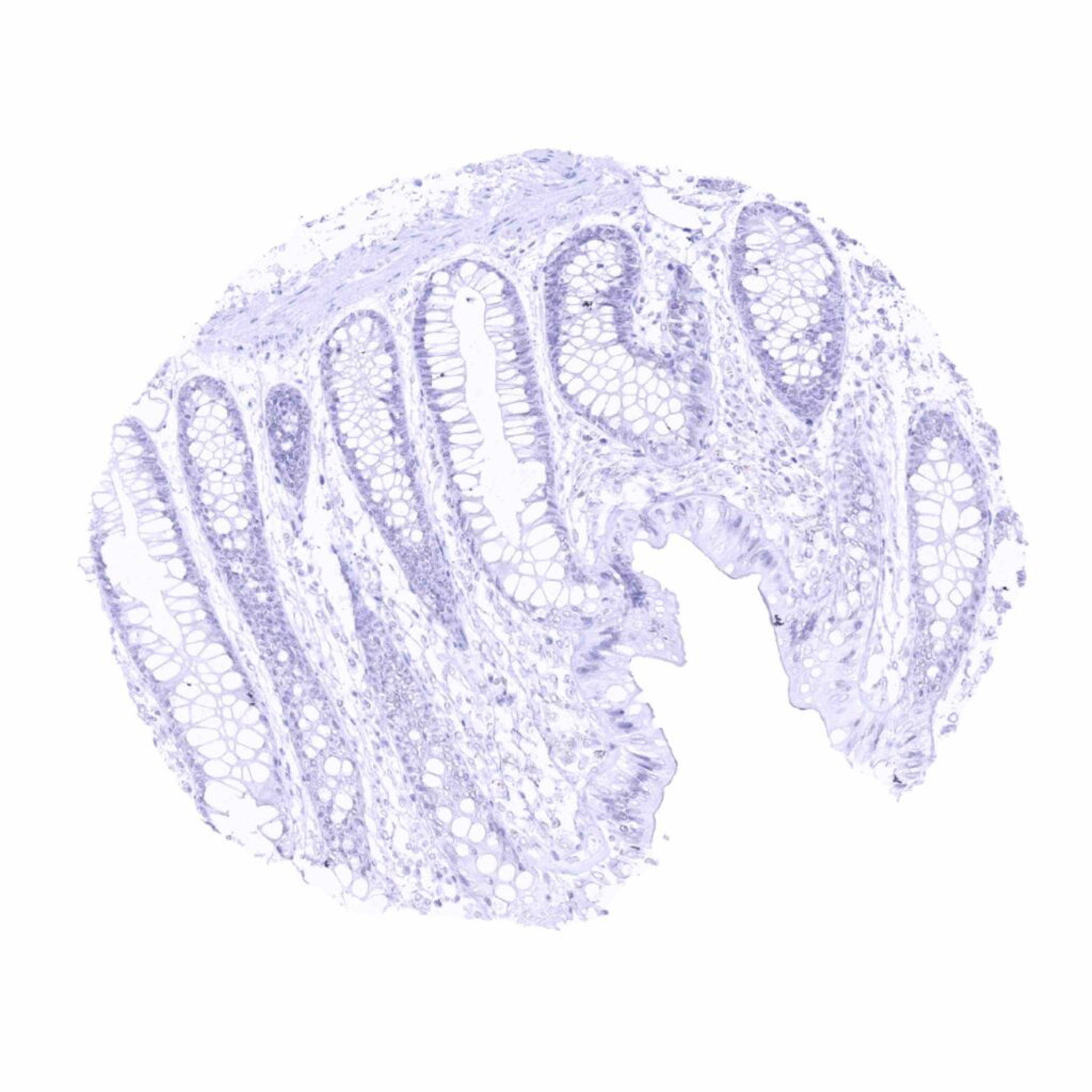
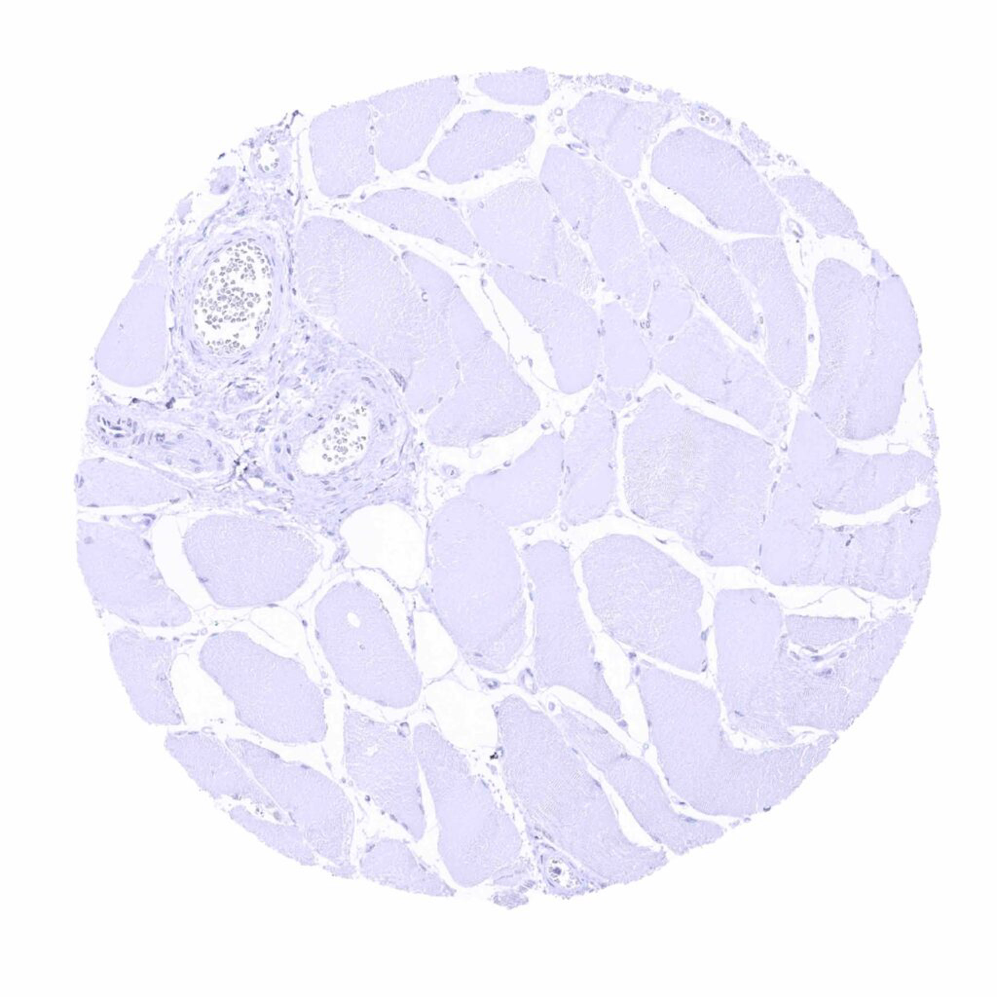
StAR antibody [MSVA-740R] HistoMAXTM


a
Go to Cancer Tissue Gallery
  
 Go back to product page
  

Go to HistoMAX page


Adrenal gland – StAR staining of variable intensity of adrenocortical cells. Staining variability may be due to the functional status of the cells

Adrenal gland – Strong StAR staining of adrenocortical cells

Aorta, media

Appendix, muscular wall


Bone marrow

Breast

Bronchus, mucosa

Cerebellum (molecular layer, Purkinje cell layer, granule cell layer, white matter)


Cerebellum (molecular layer, Purkinje cell layer, granule cell layer)

Cerebrum, grey

Cerebrum, white

Colon descendens, mucosa


Colon descendens, muscular wall

Duodenum, mucosa

Esophagus, squamous epithelium

Fallopian tube, mucosa


Fat

Gallbladder, epithelium

Heart muscle

Ileum, mucosa


Kidney, cortex

Kidney, medulla

Kidney, pelvis, muscular wall

Kidney, pelvis, urothelium


Liver

Lung

Lymph node

Ovary (corpus luteum) – Strong cytoplasmic StAR staining of corpus luteum cells


Ovary (follicular cyst) – Strong cytoplasmic StAR staining of theca interna cells of a follicular cyst. A weaker staining can be seen in granulosa cells

Pancreas

Pancreas

Parathyroid gland


Parotid gland

Pituitary, anterior lobe

Pituitary, posterior lobe- infundibulum

Placenta (amnion and chorion)


Placenta (amnion and chorion)

Placenta, early

Placenta, mature

Prostate


Rectum, mucosa

Seminal vesicle

Skeletal muscle

Skin


Spleen

Stomach, antrum

Stomach, corpus

Testis – Strong cytoplasmic StAR immunostaining of Leydig cells


Thymus

Thymus

Thyroid gland

Tonsil, surface epithelium


Tonsil

Urinary bladder, muscular wall

Urinary bladder, urothelium

Uterus, ectocervix


Uterus, endocervix

Uterus, endometrium (pregnancy)

Uterus, endometrium (proliferation)

Uterus, endometrium (secretion)


Uterus, myometrium


| TOP |