Normal Tissue Gallery- GTX639929

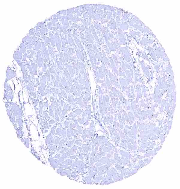
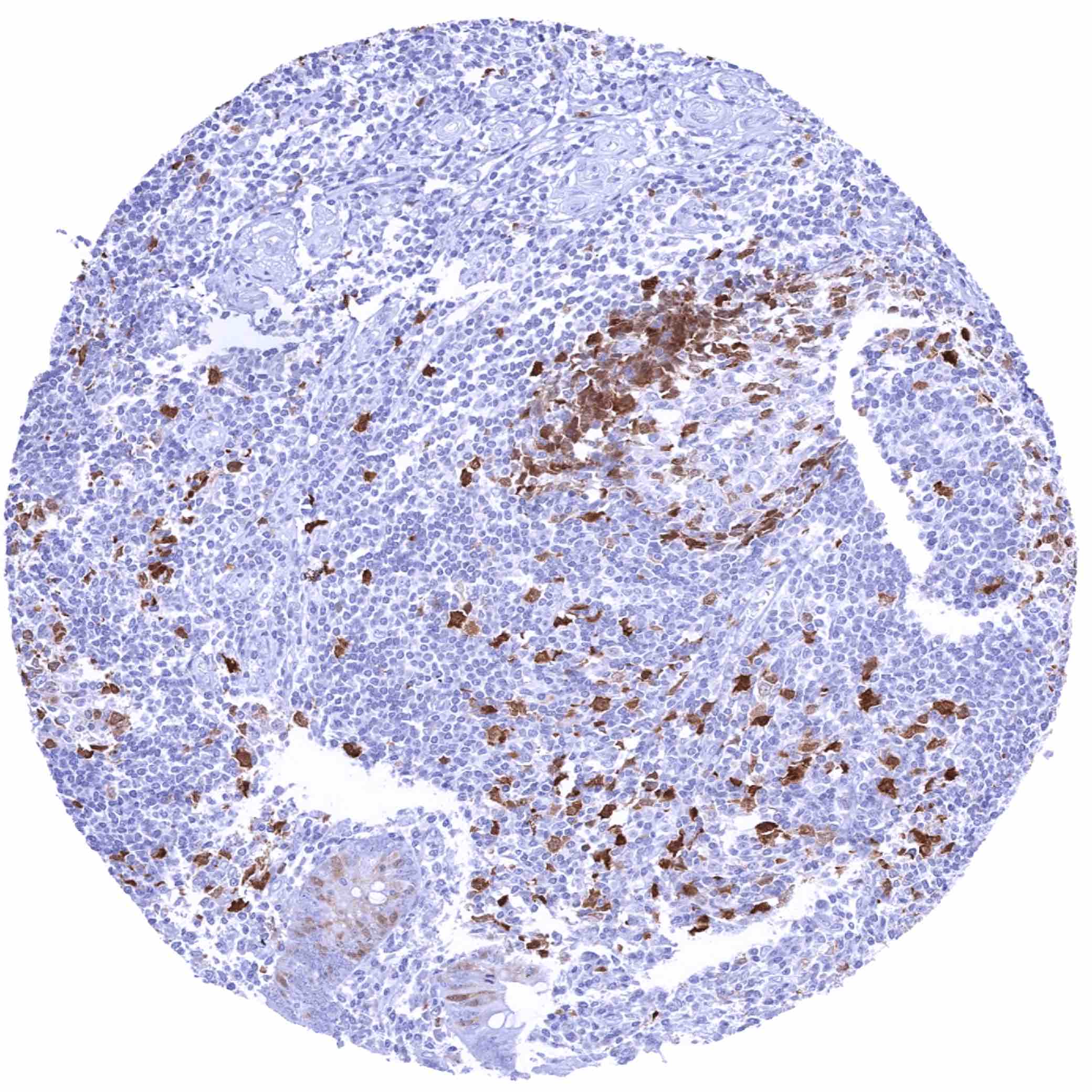
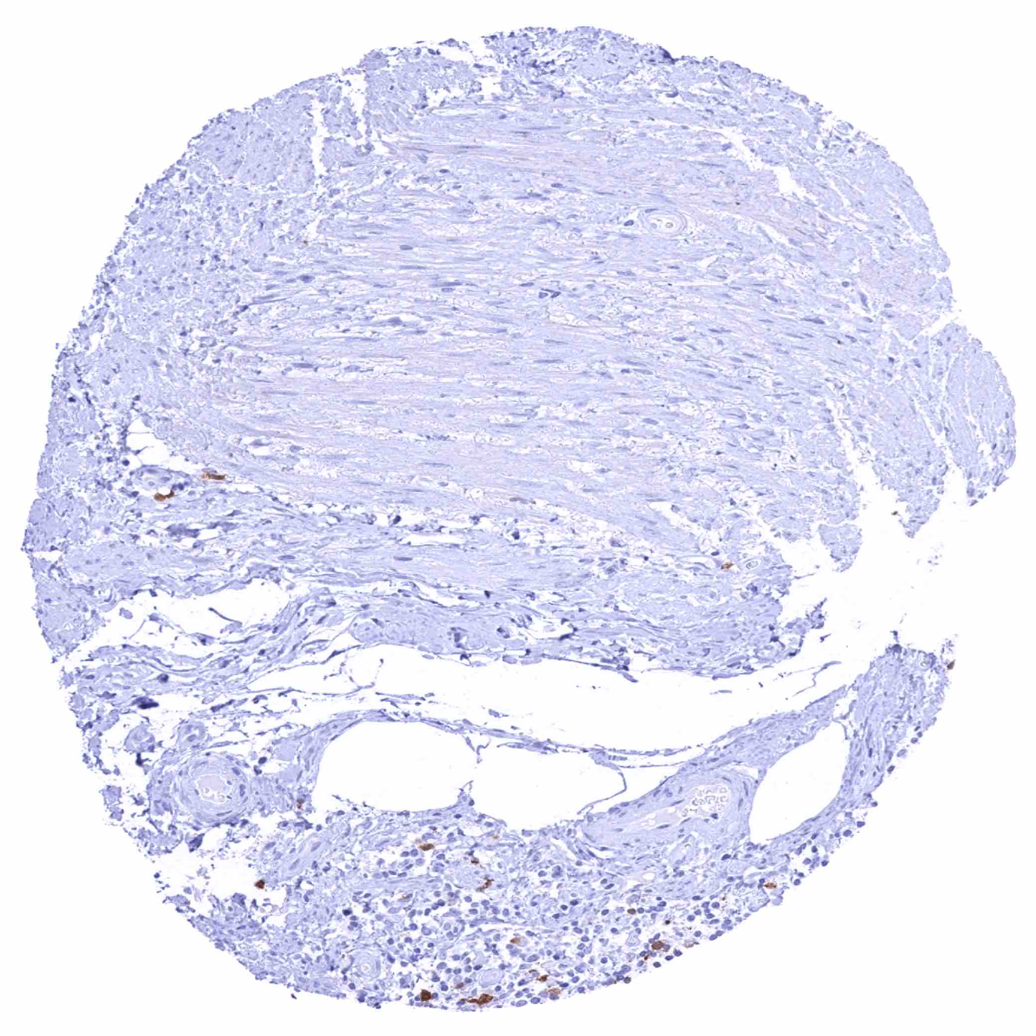
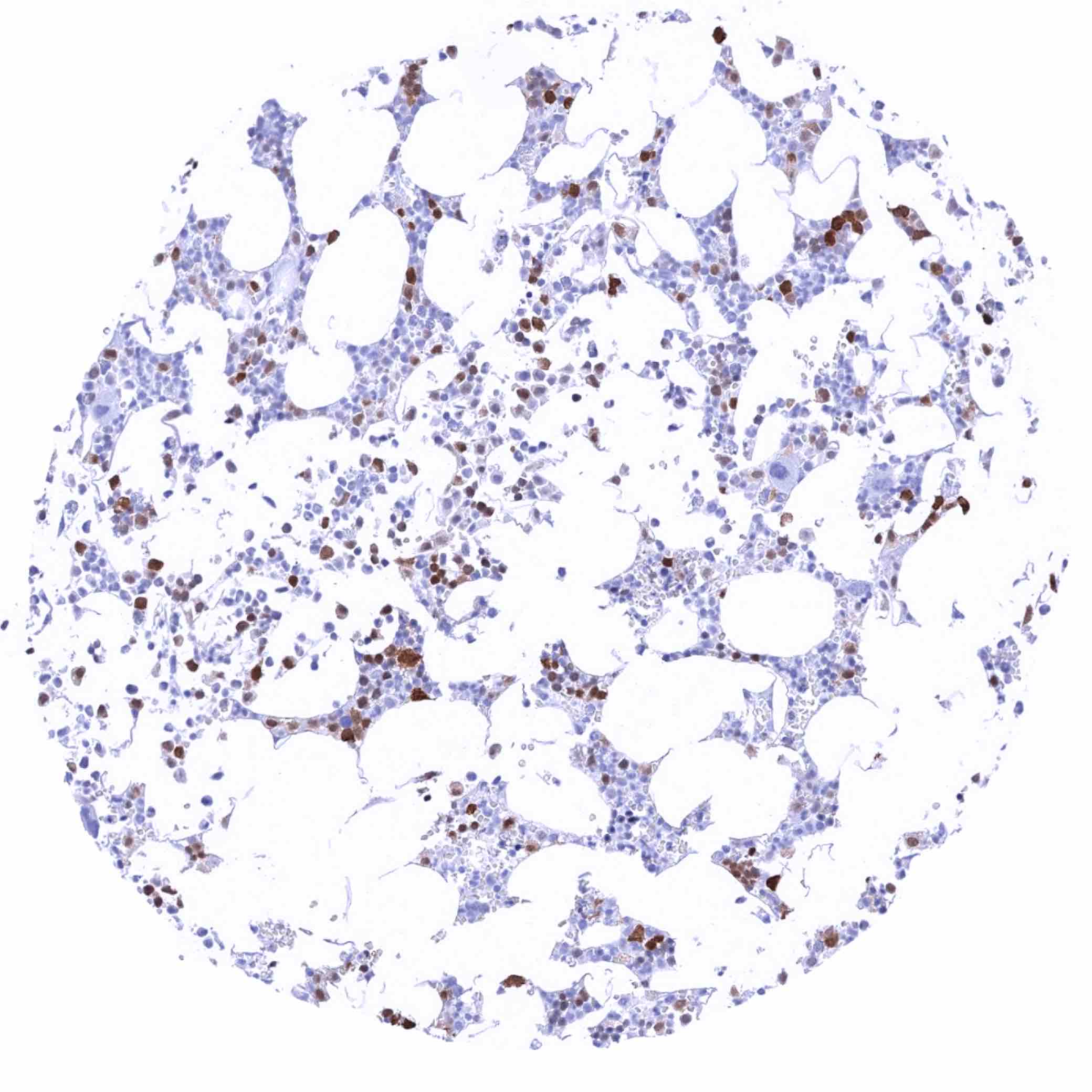
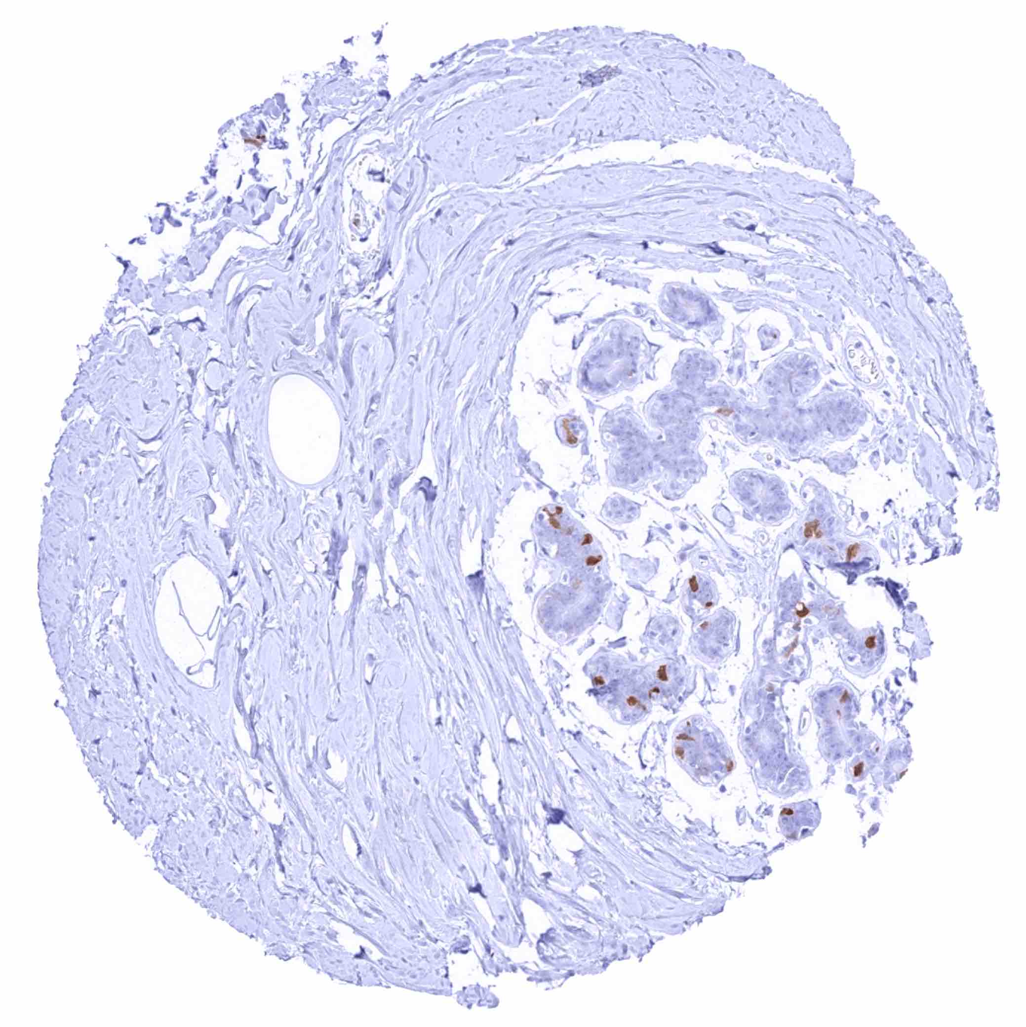
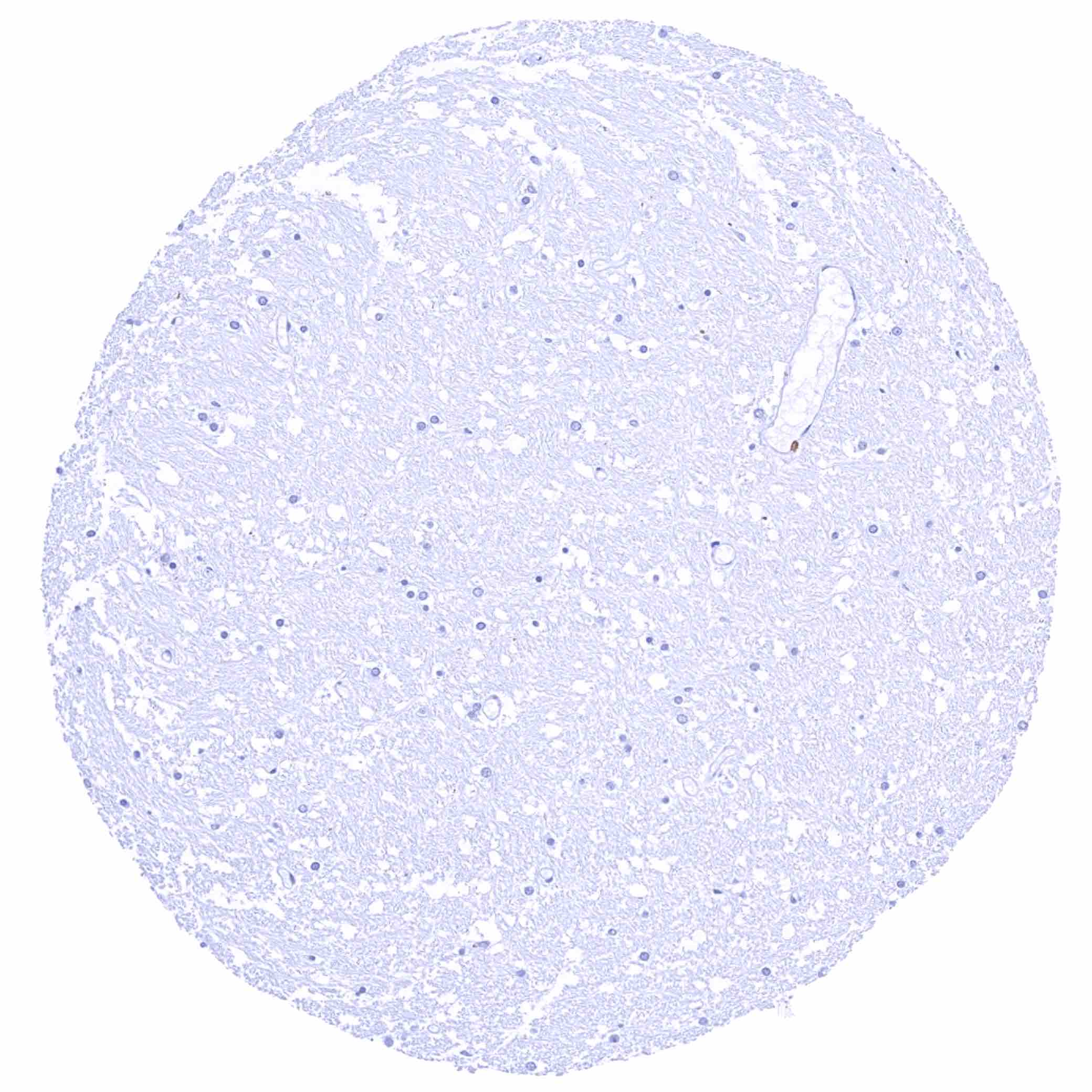
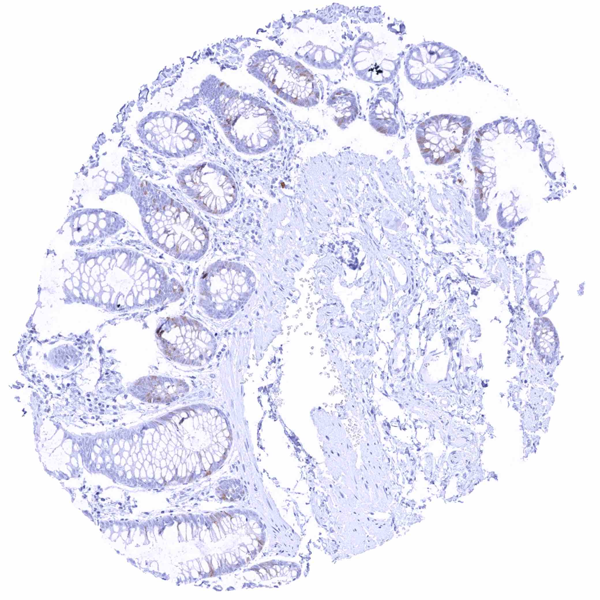
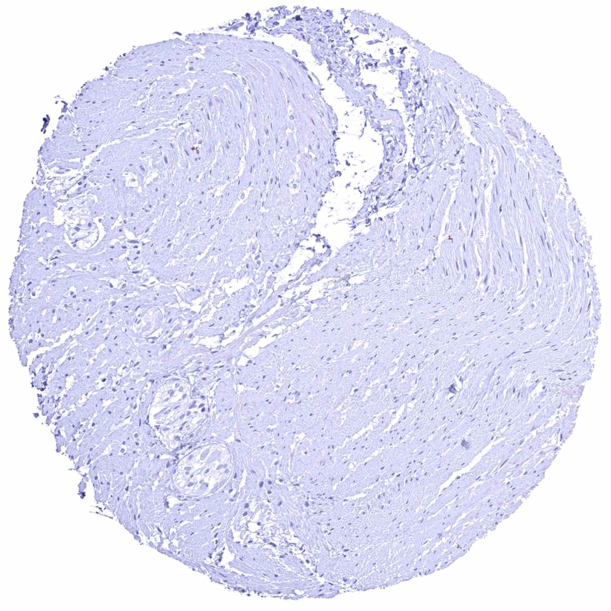
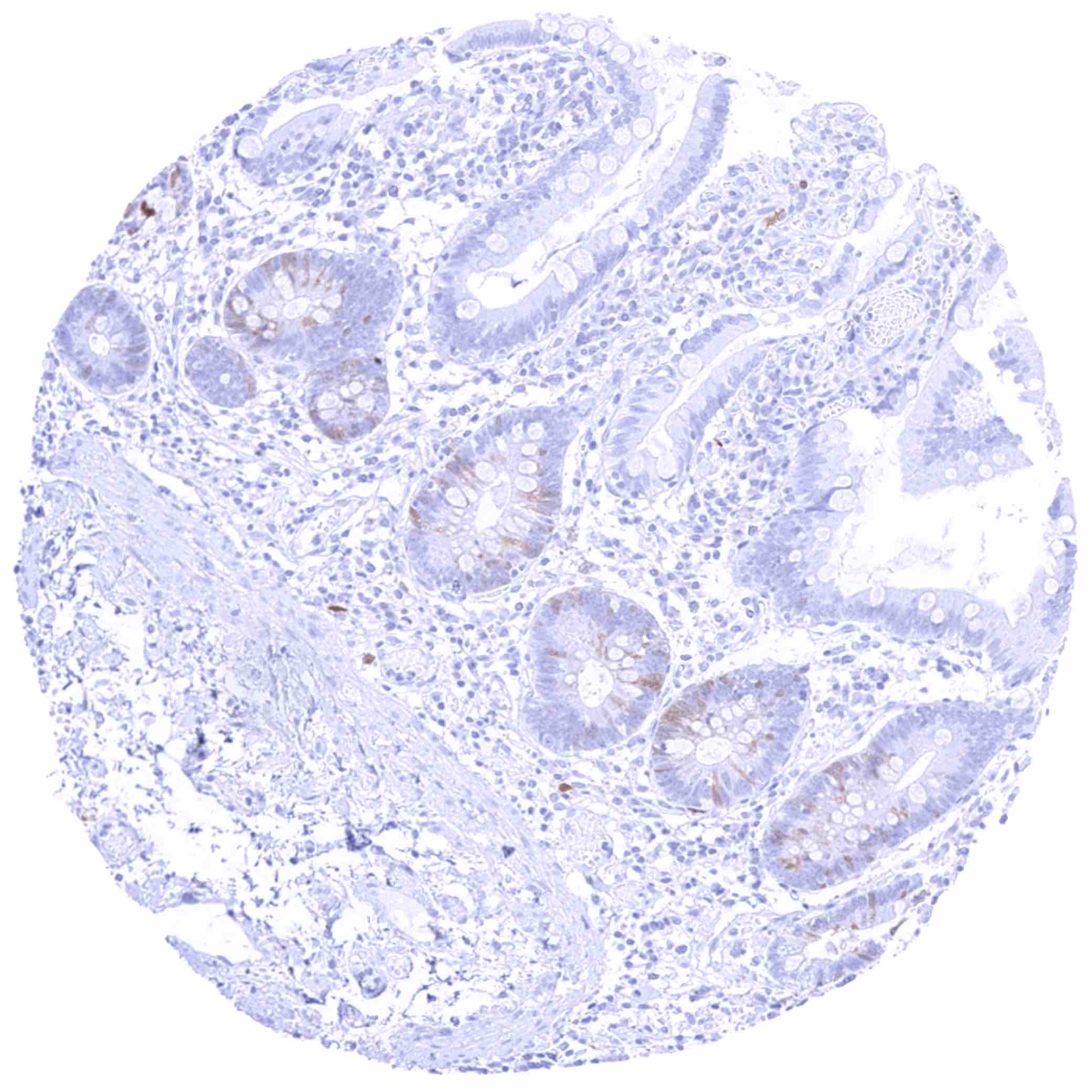
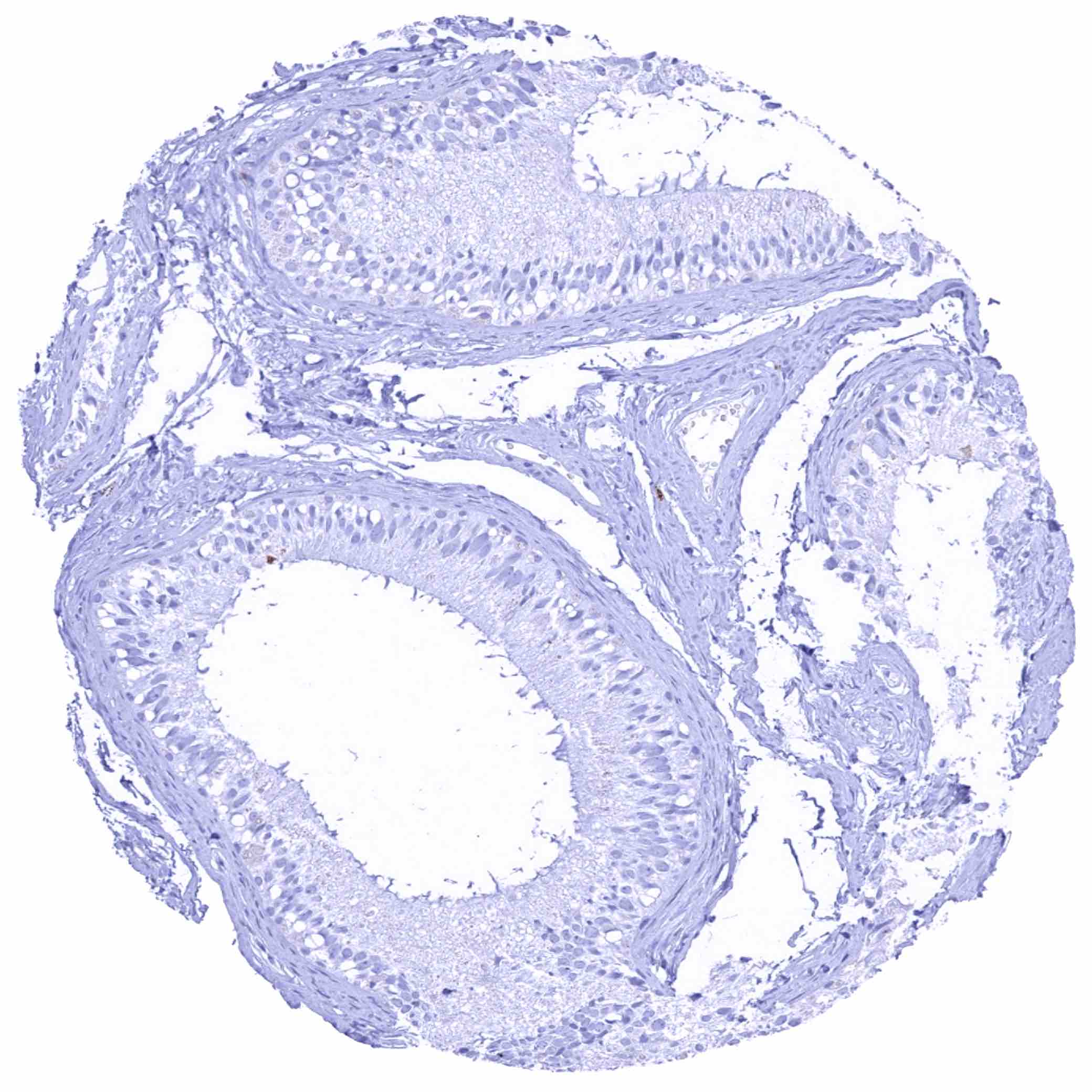
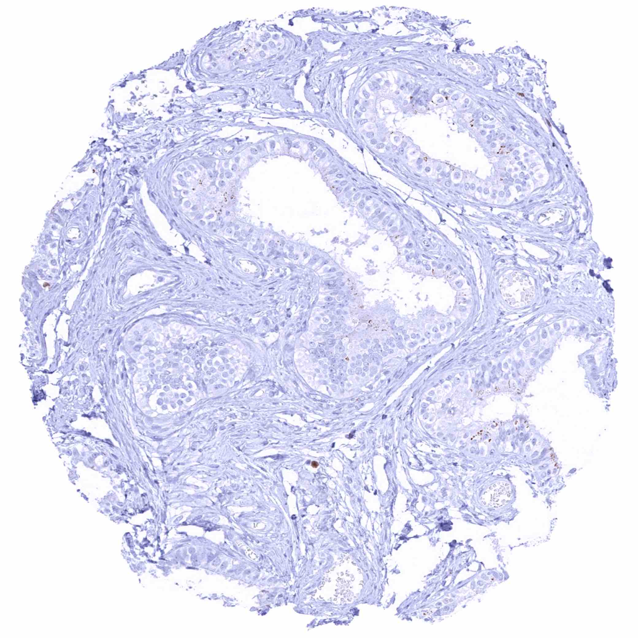
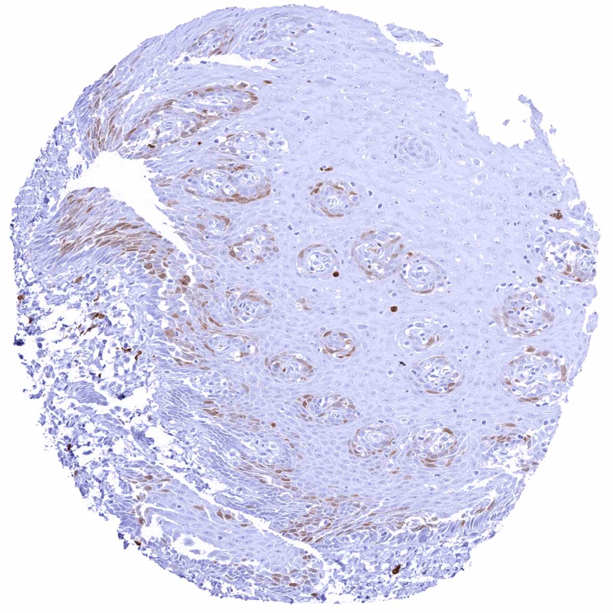
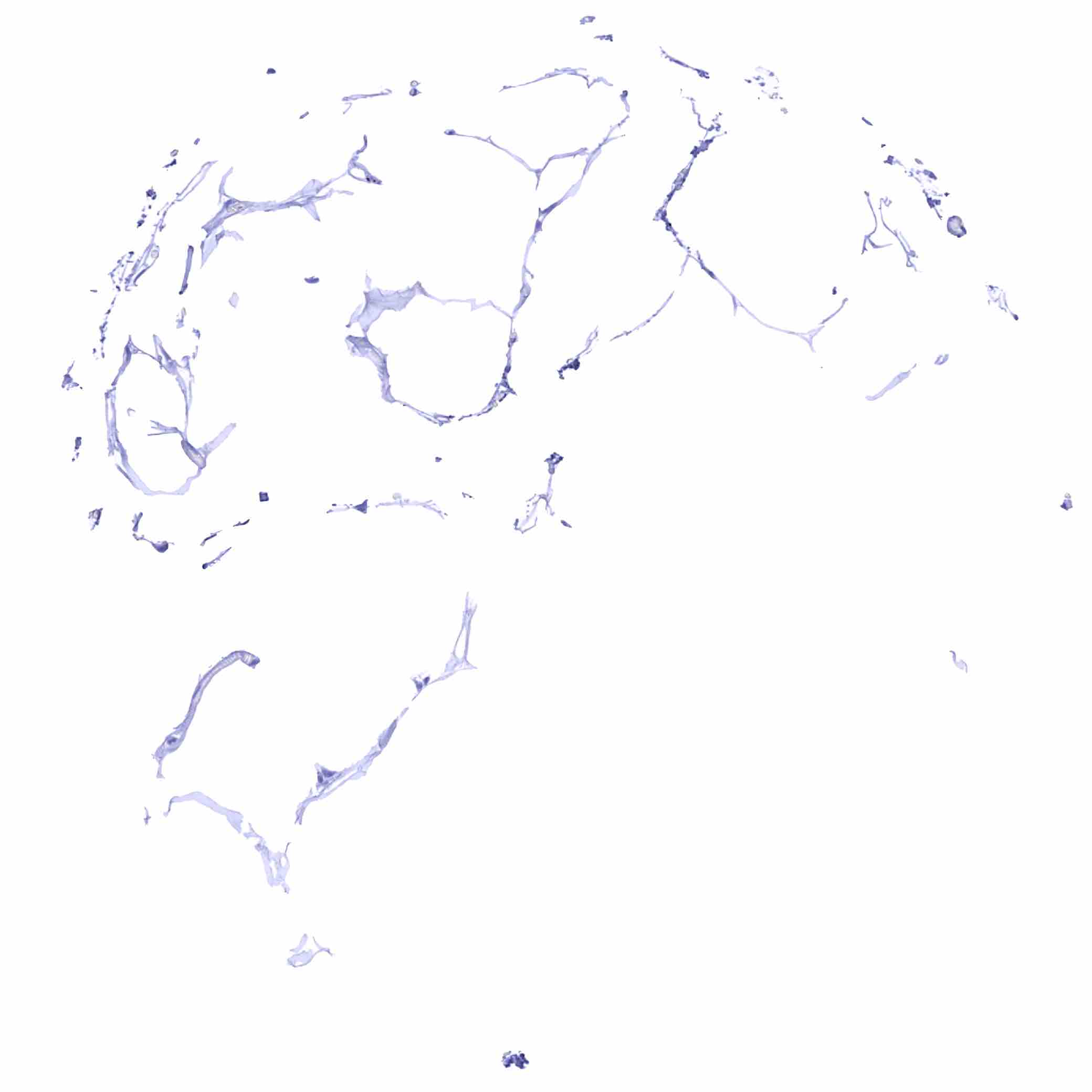
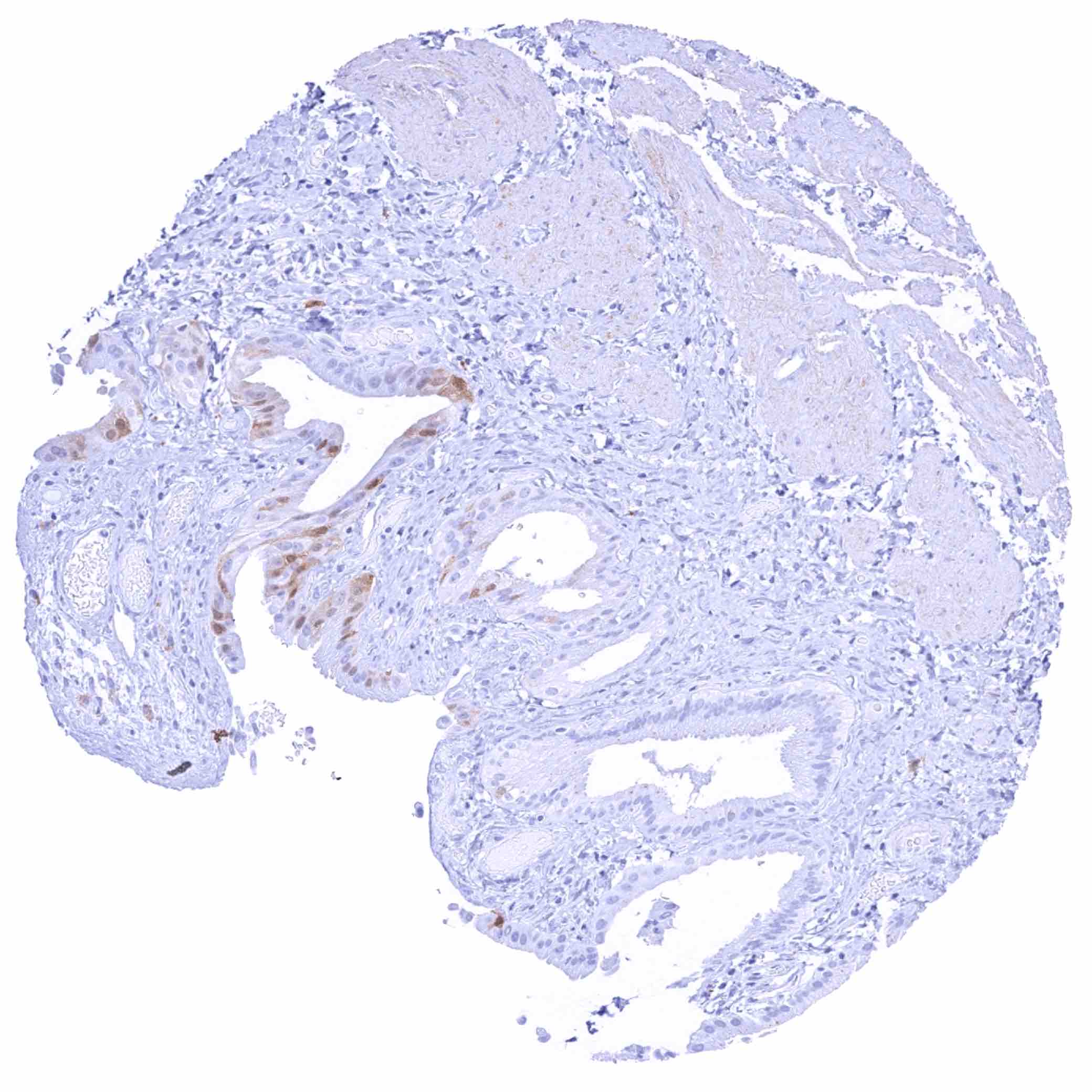
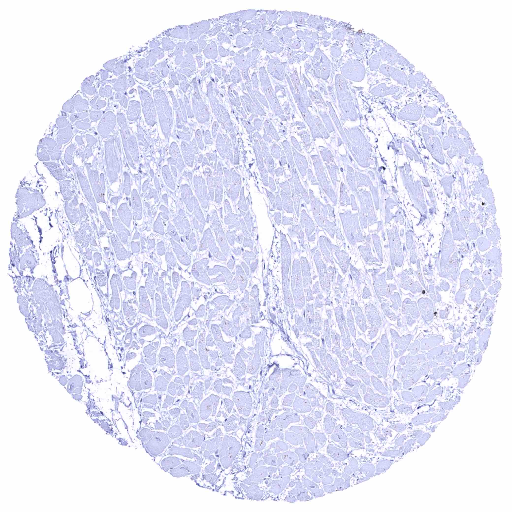
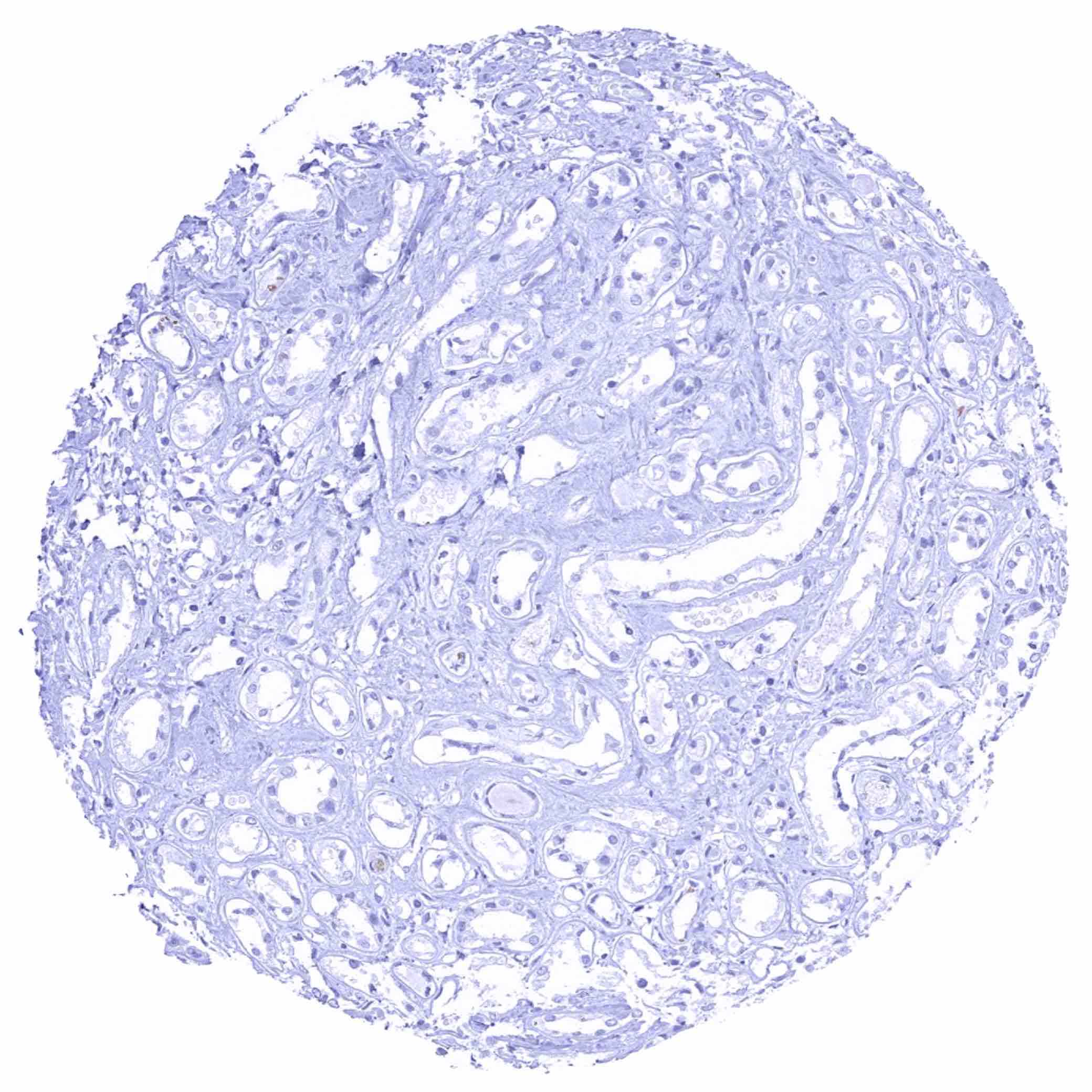
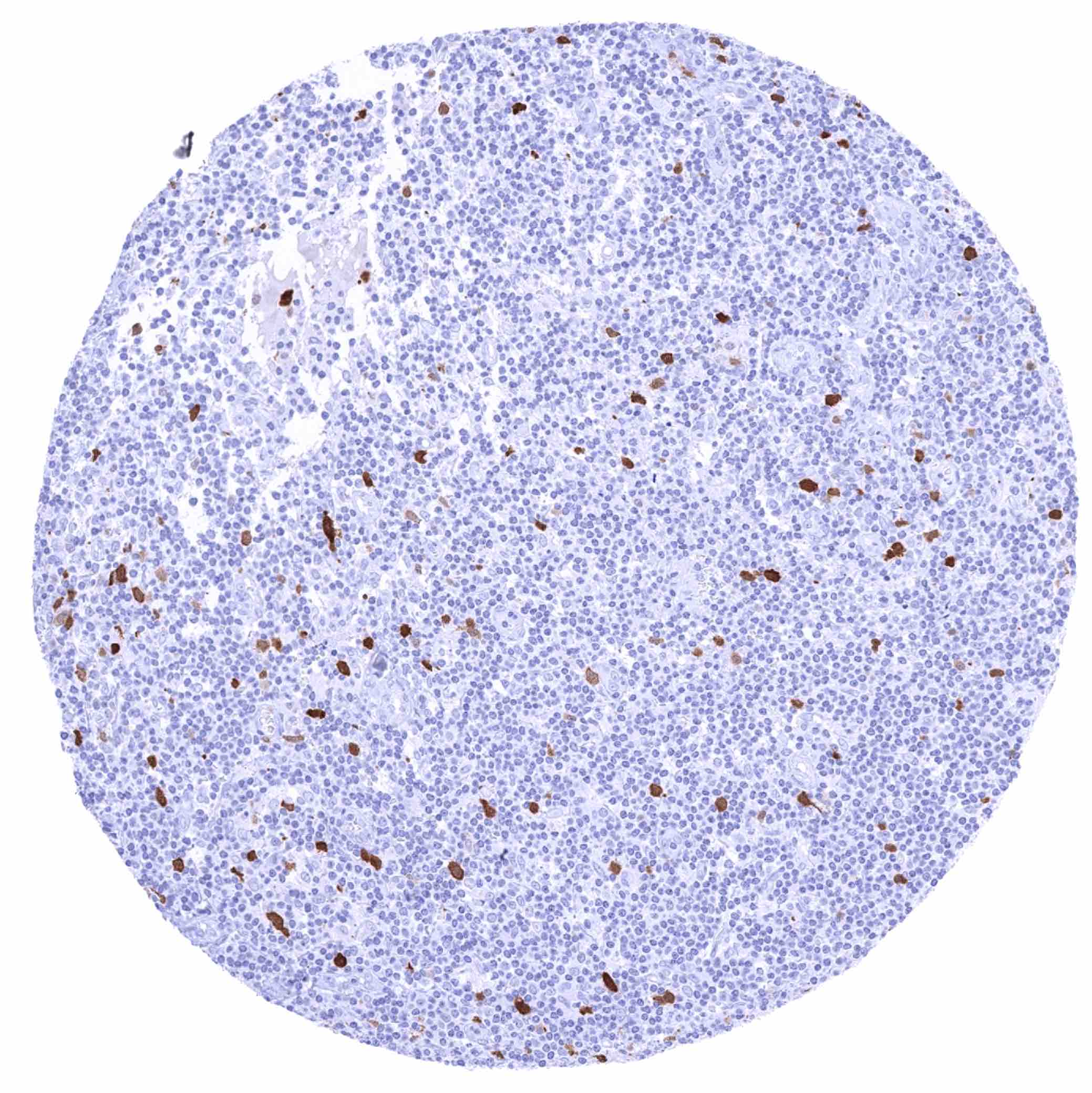
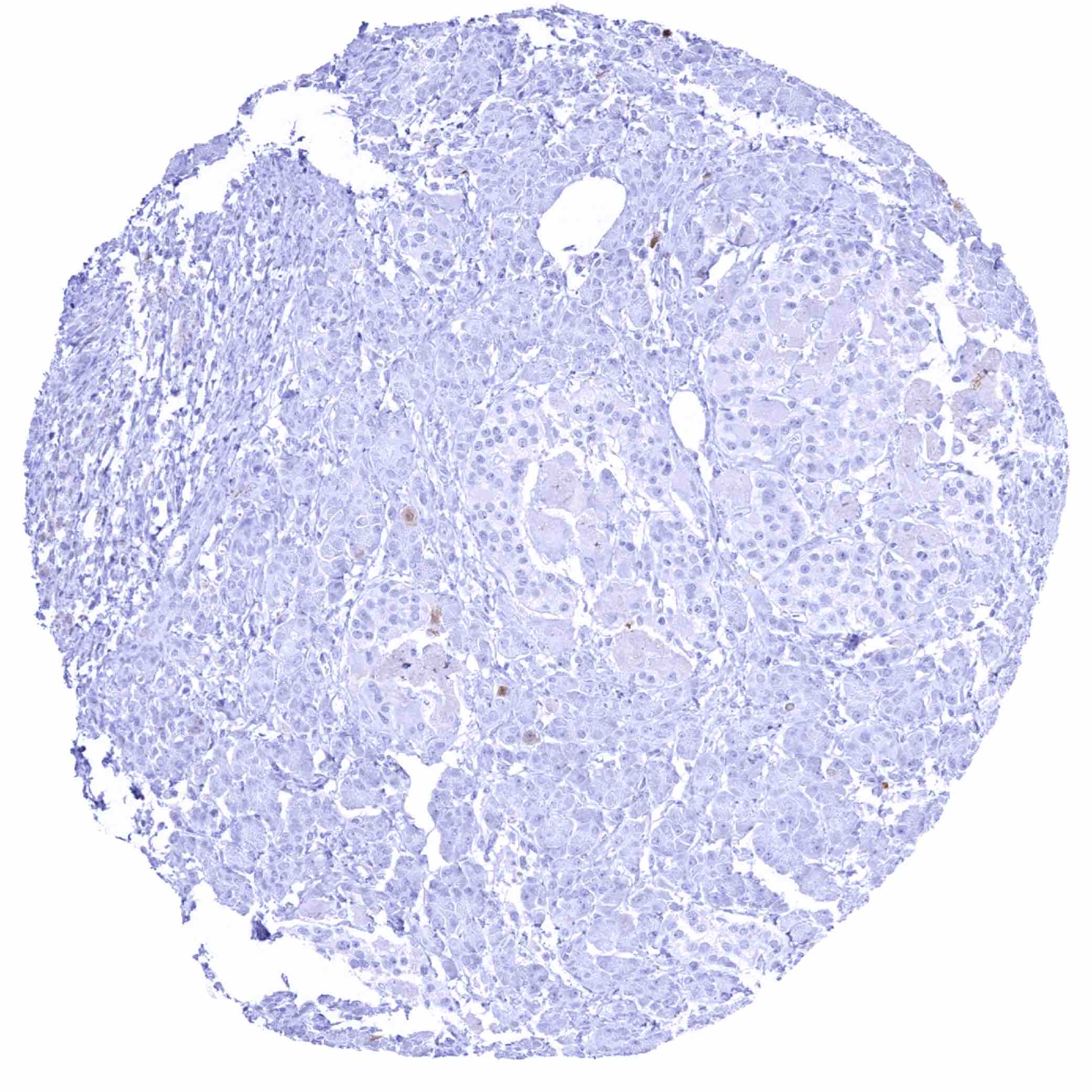
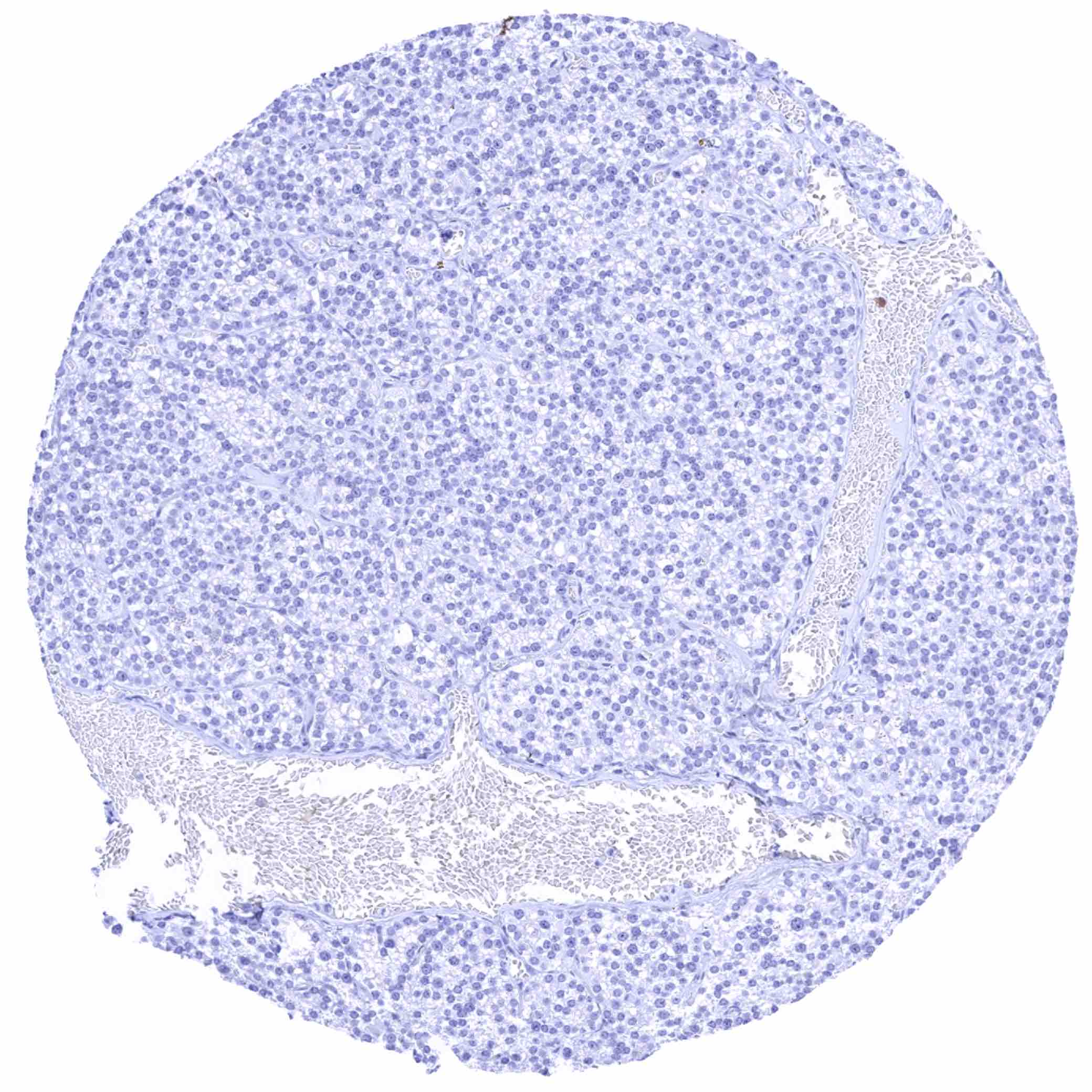
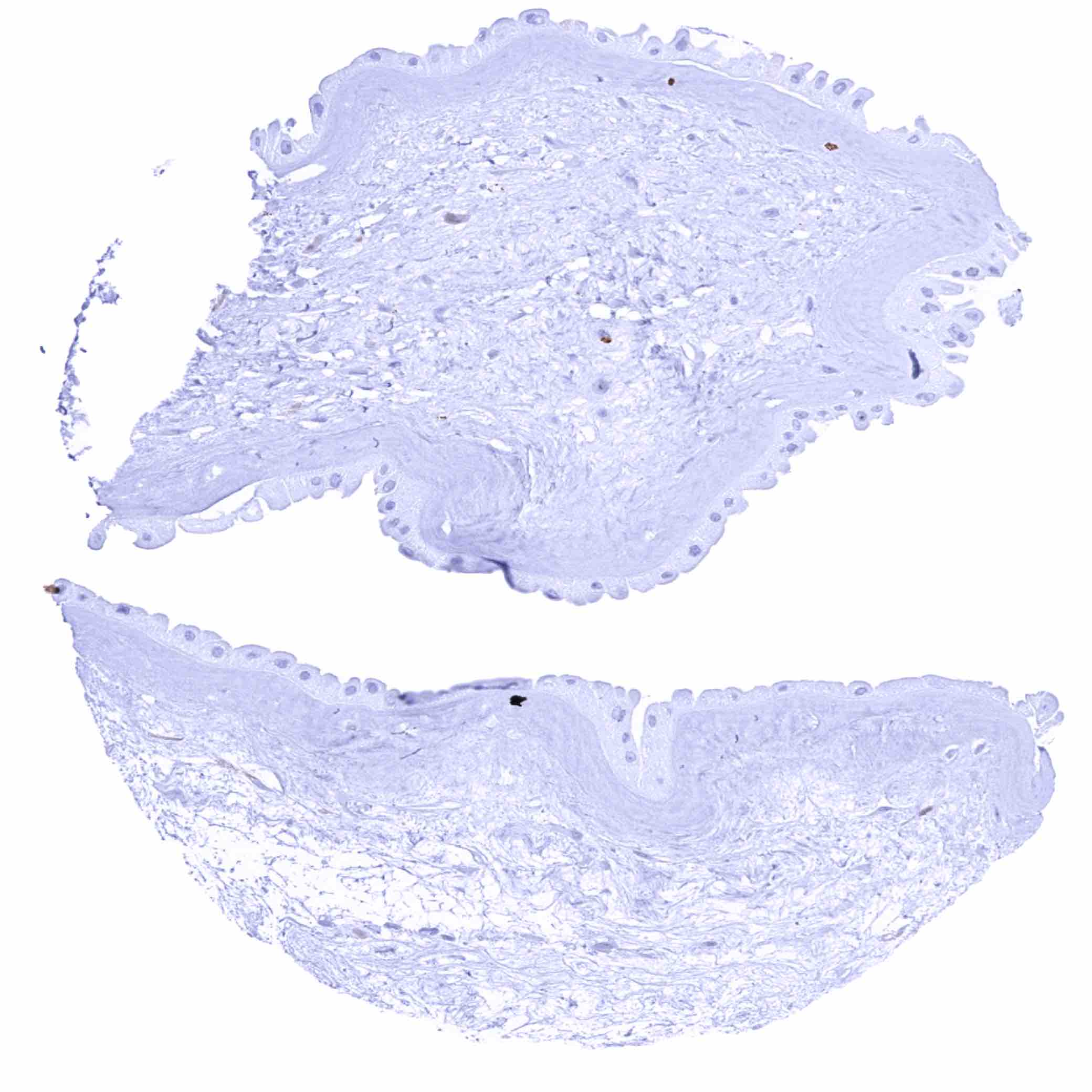
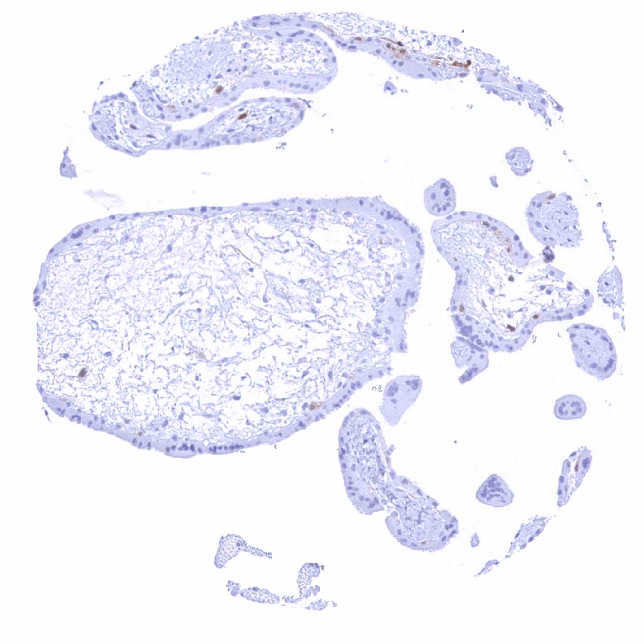
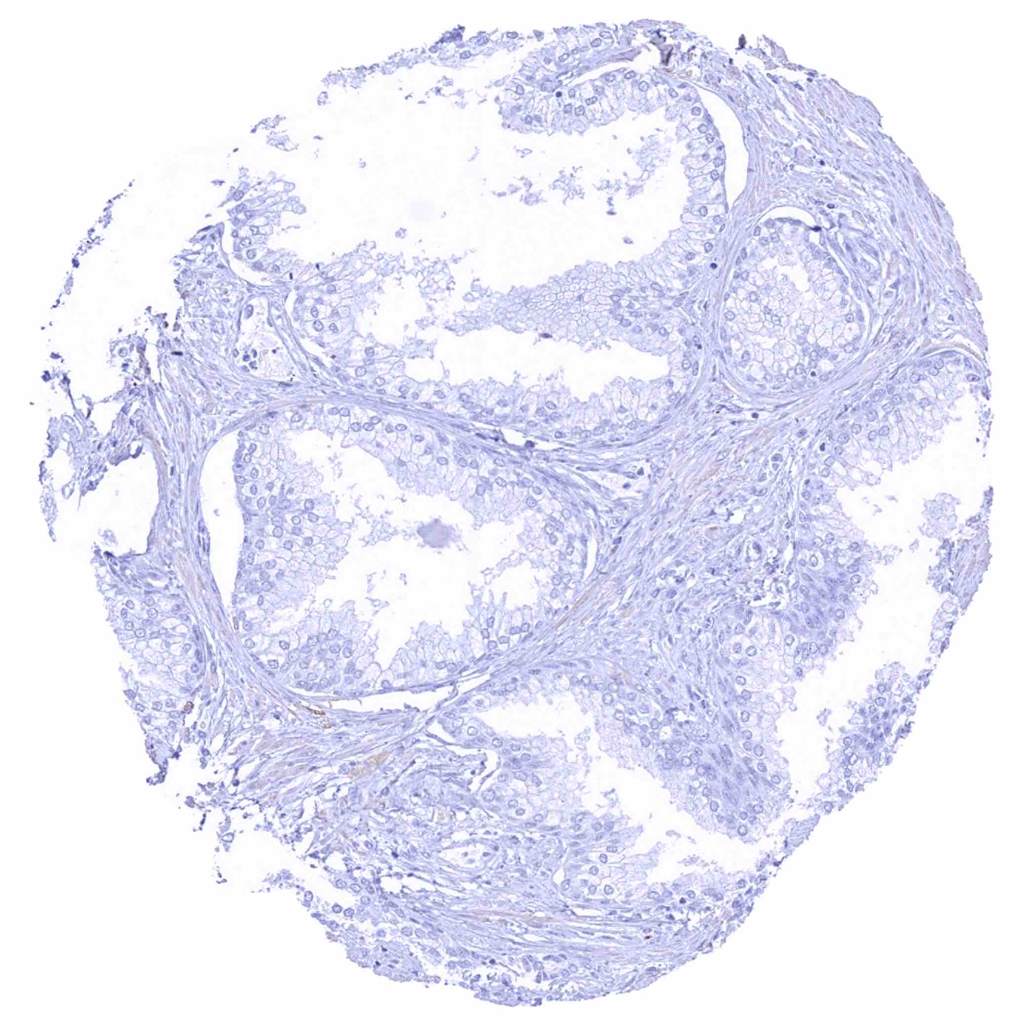
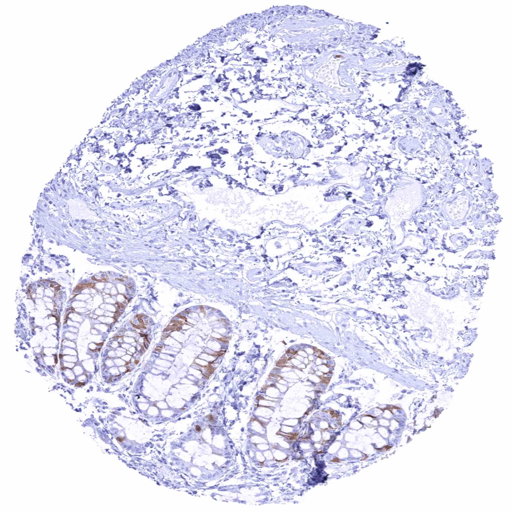
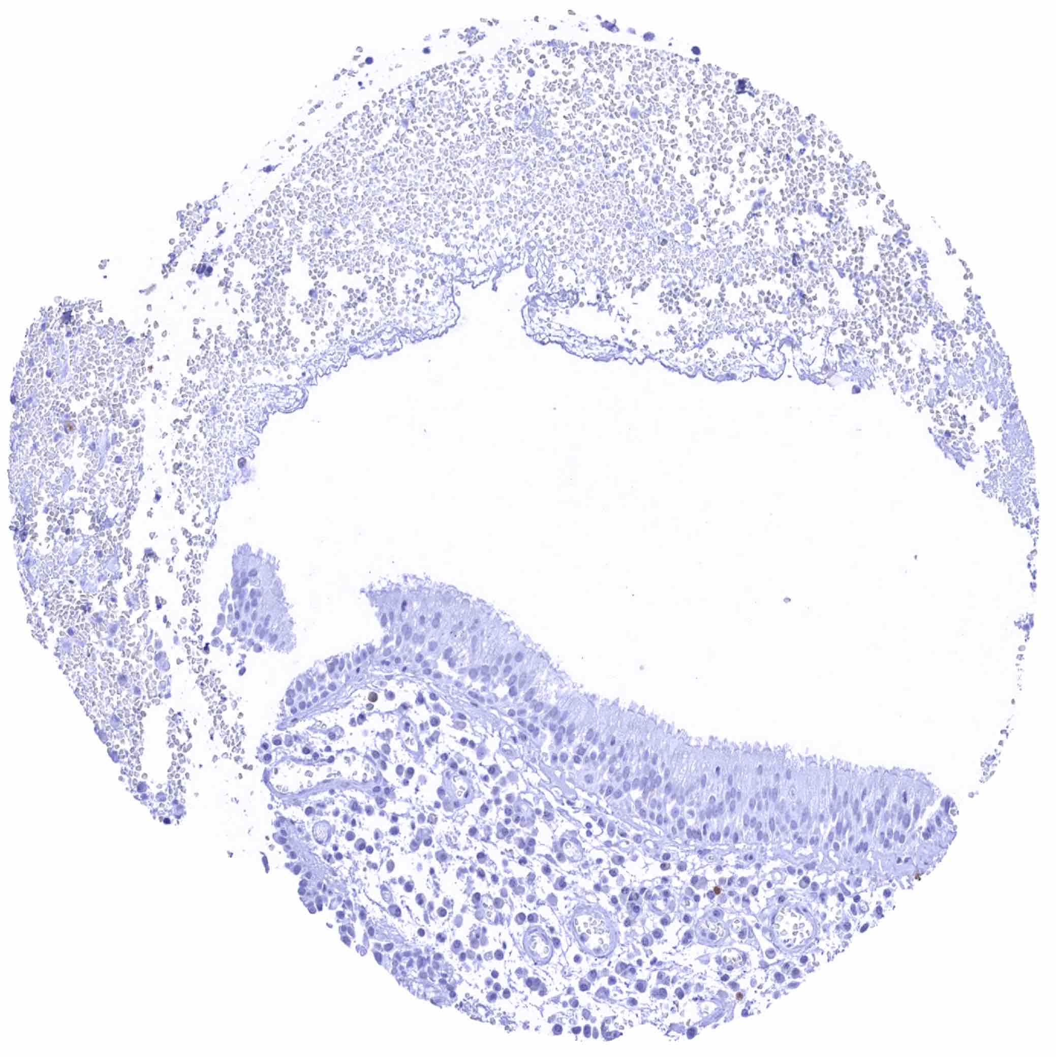
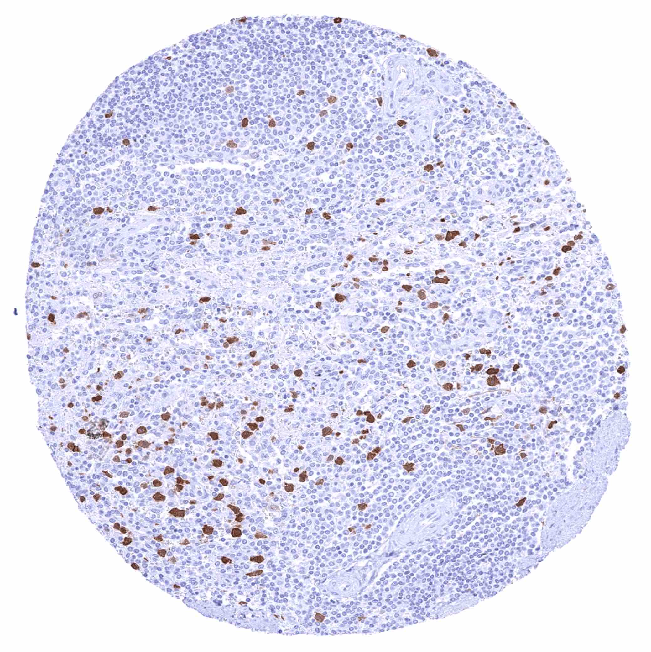
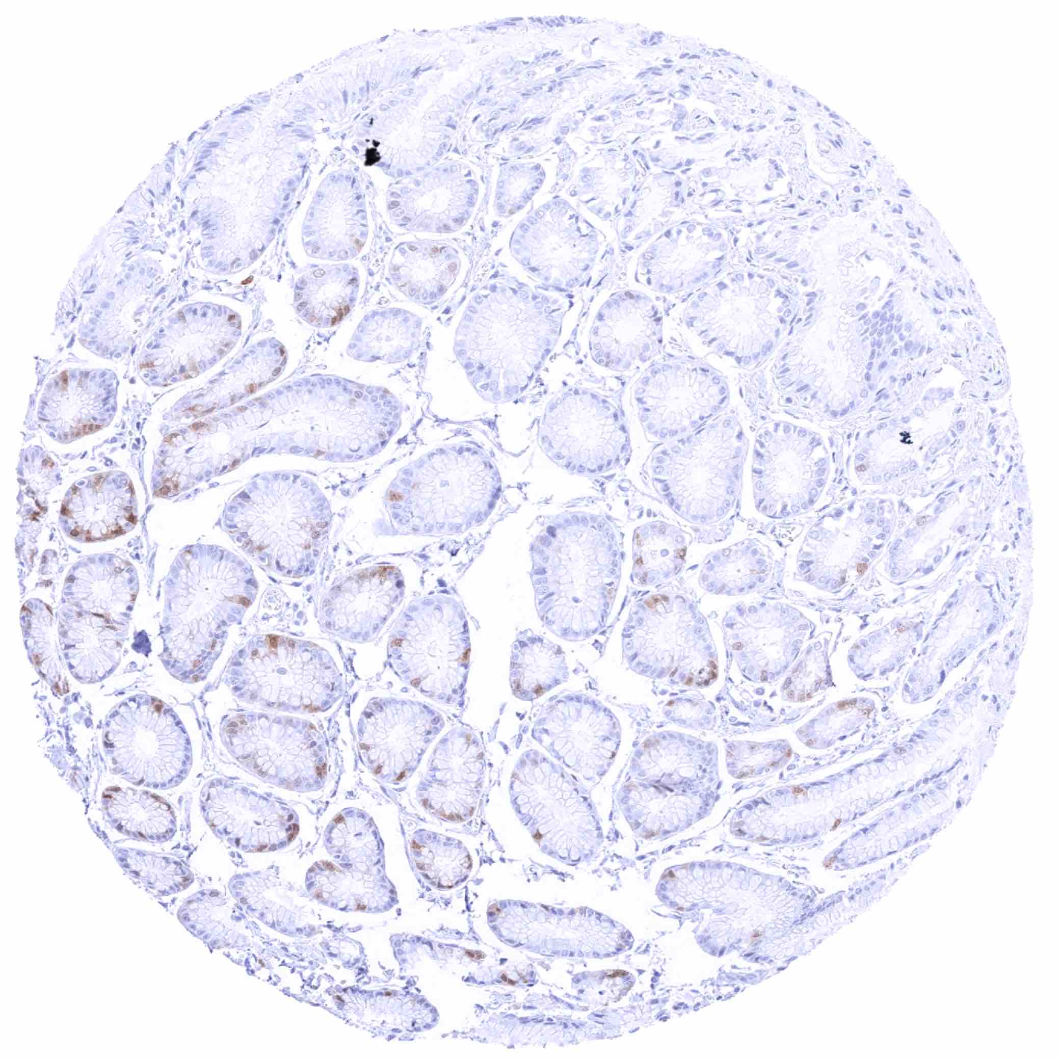
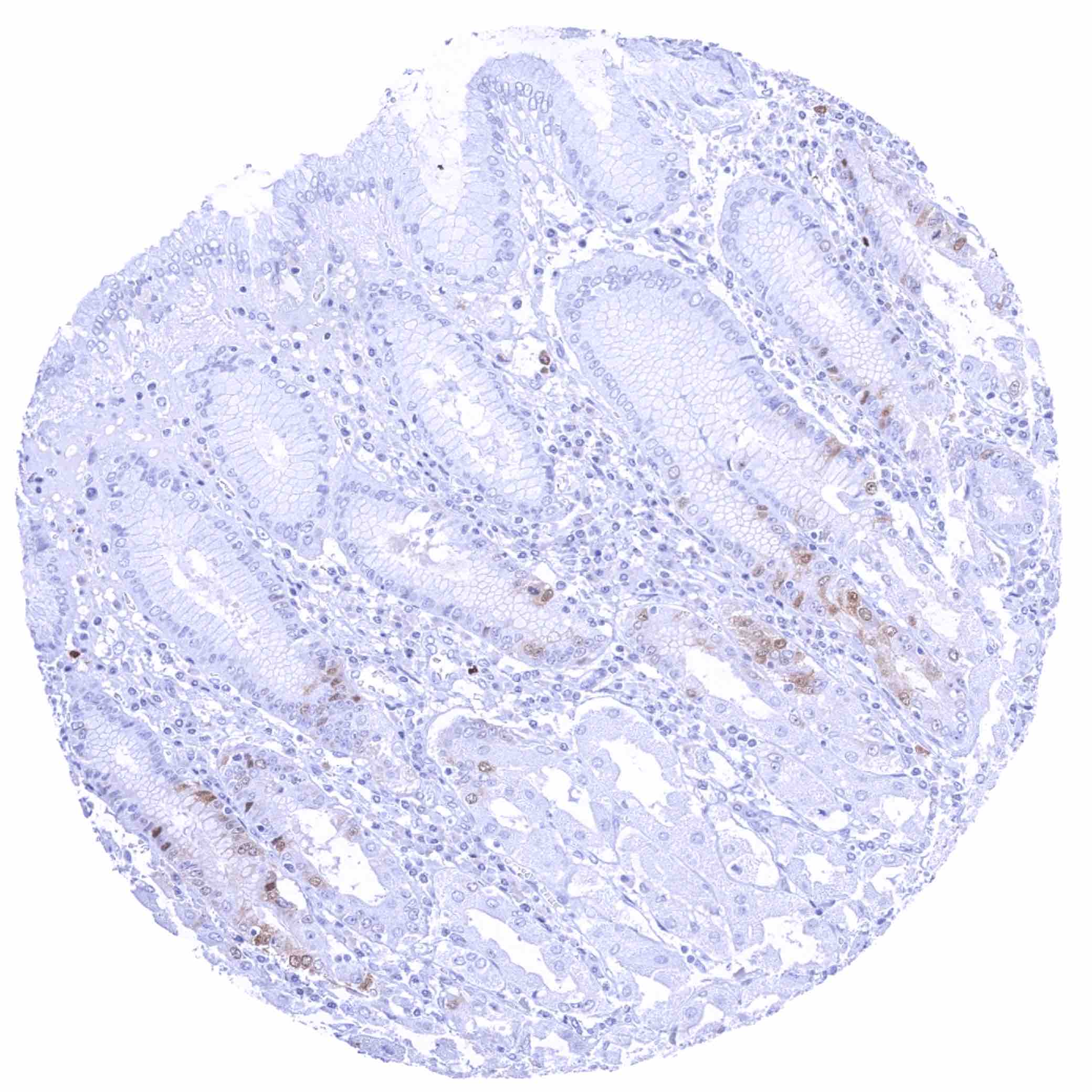
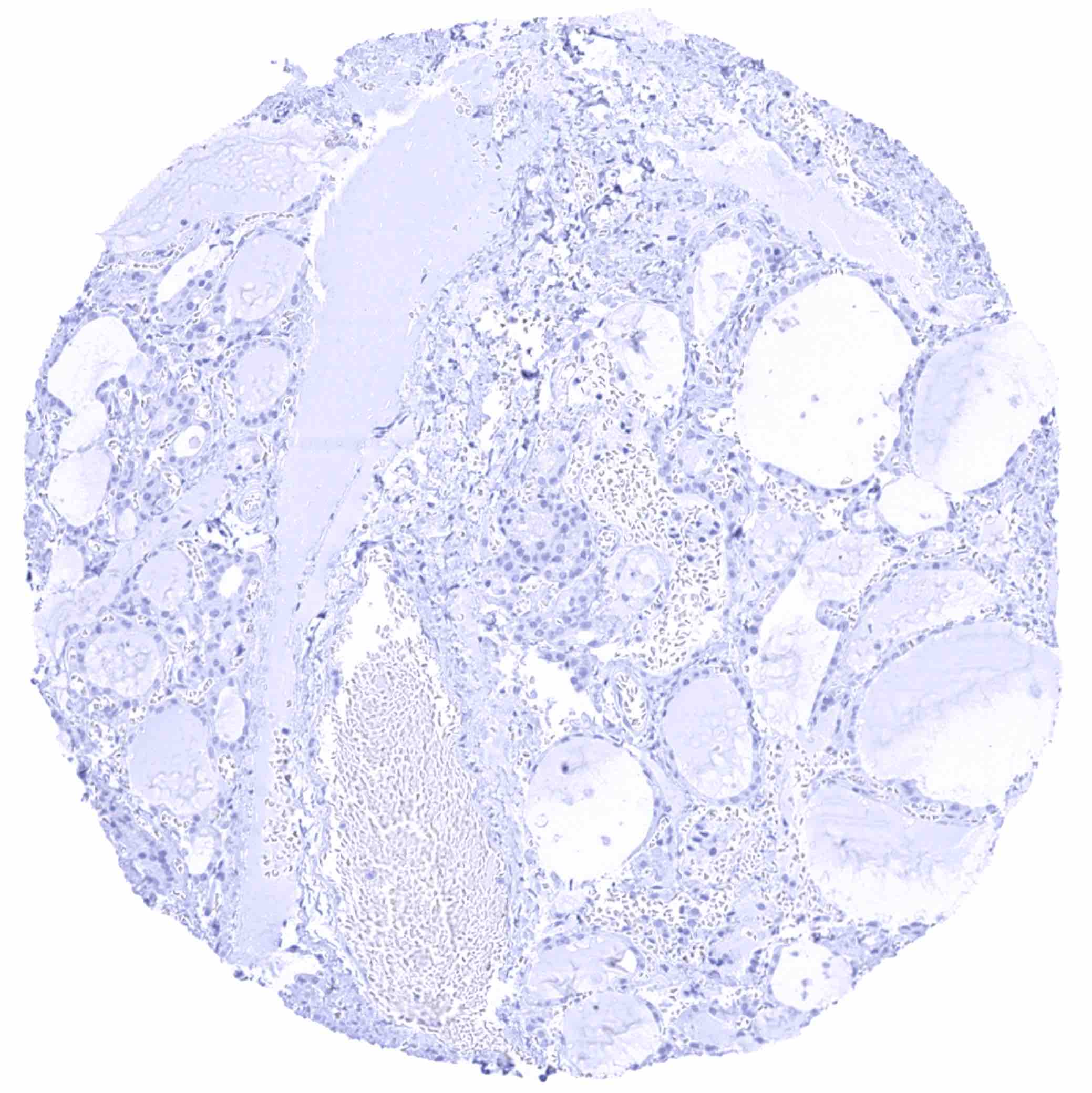
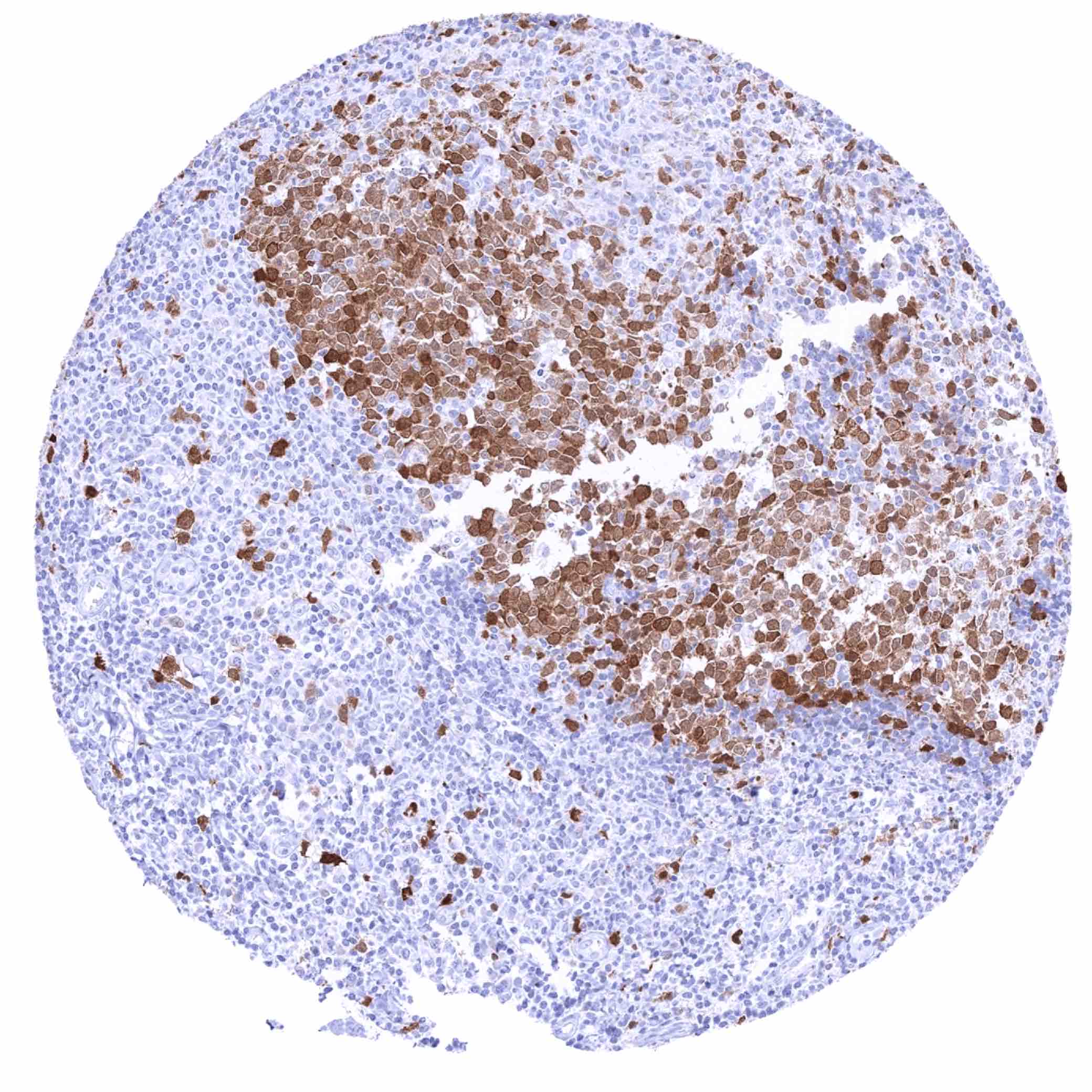
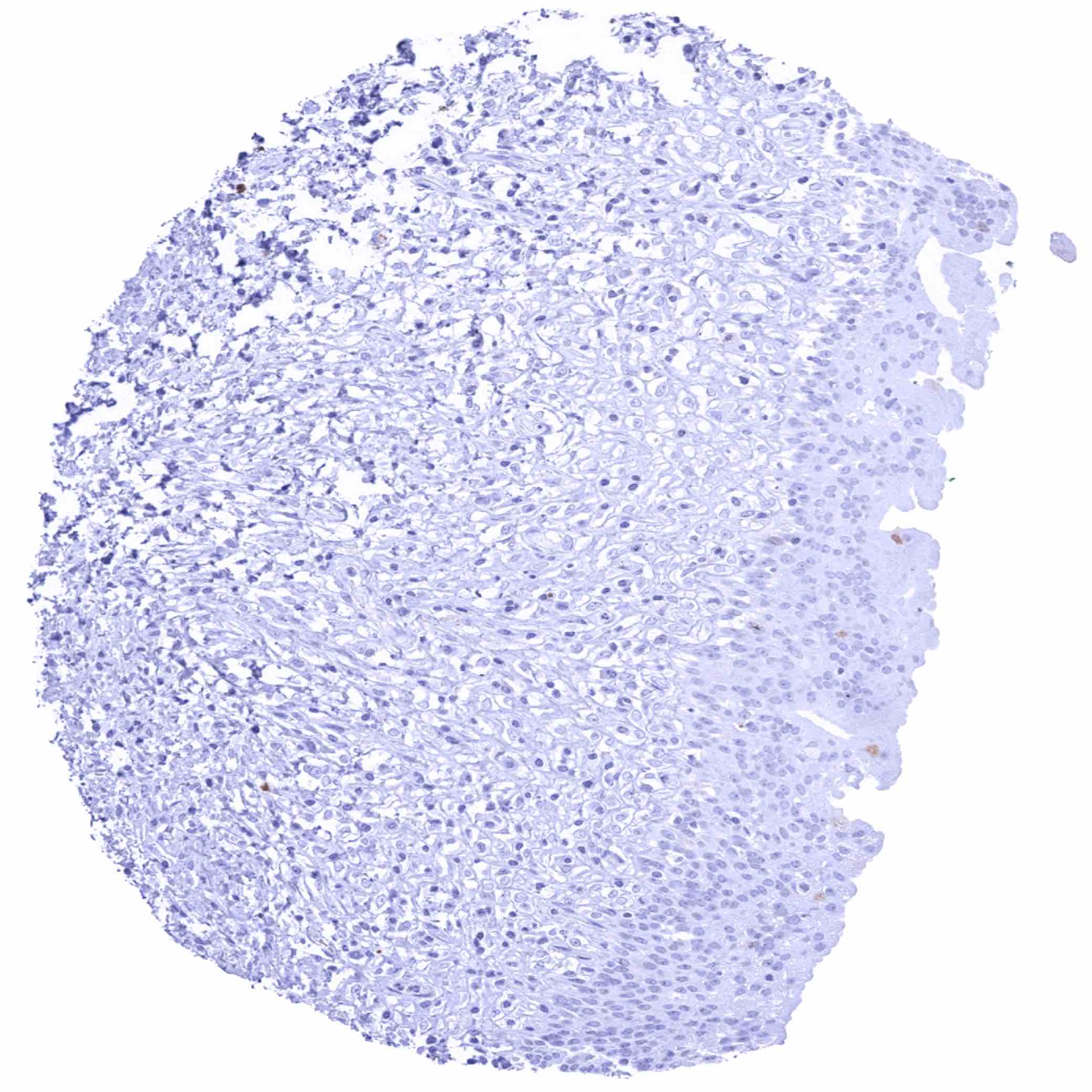
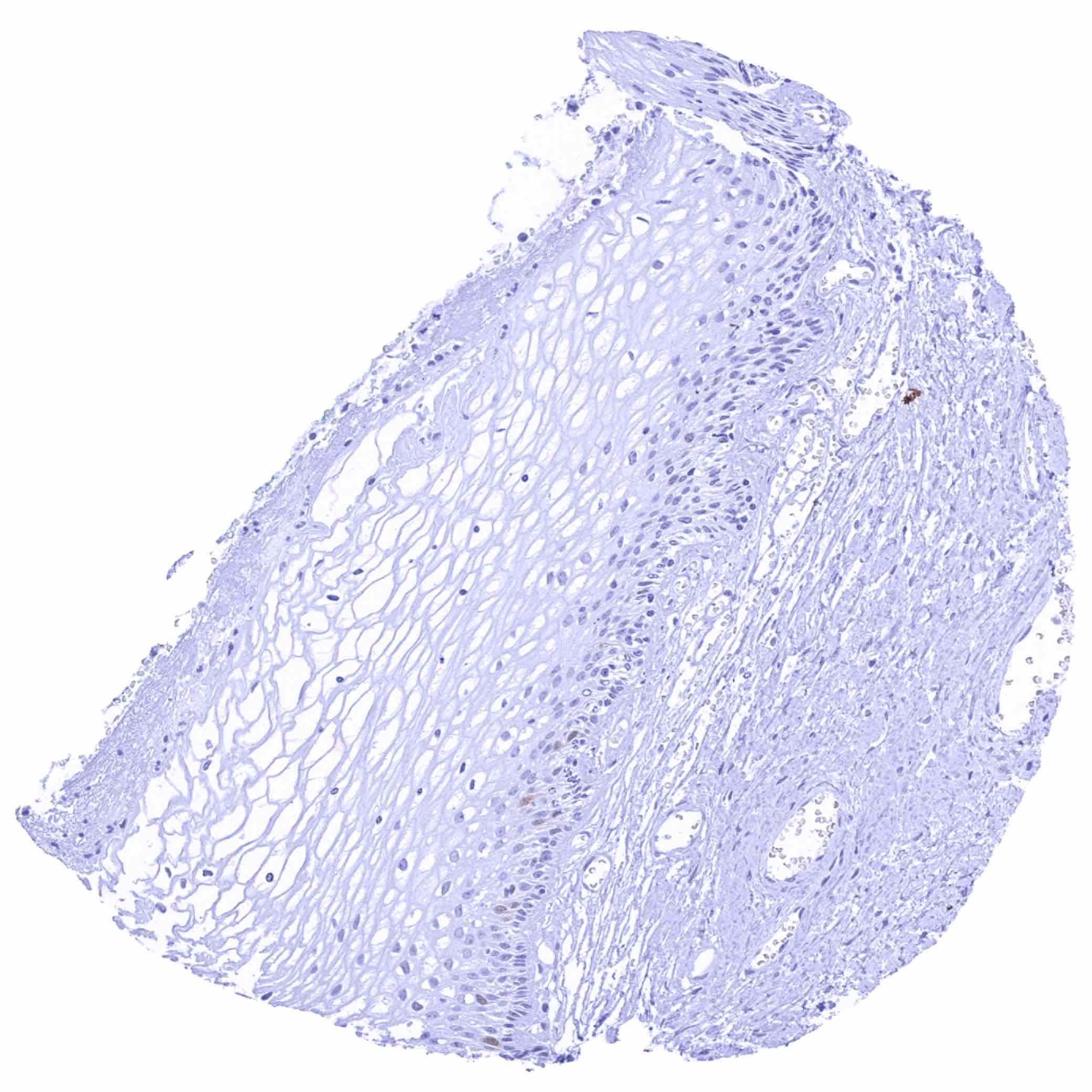
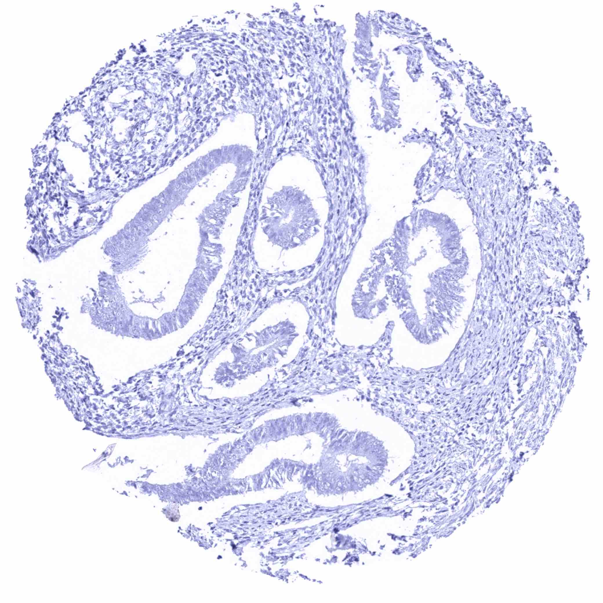
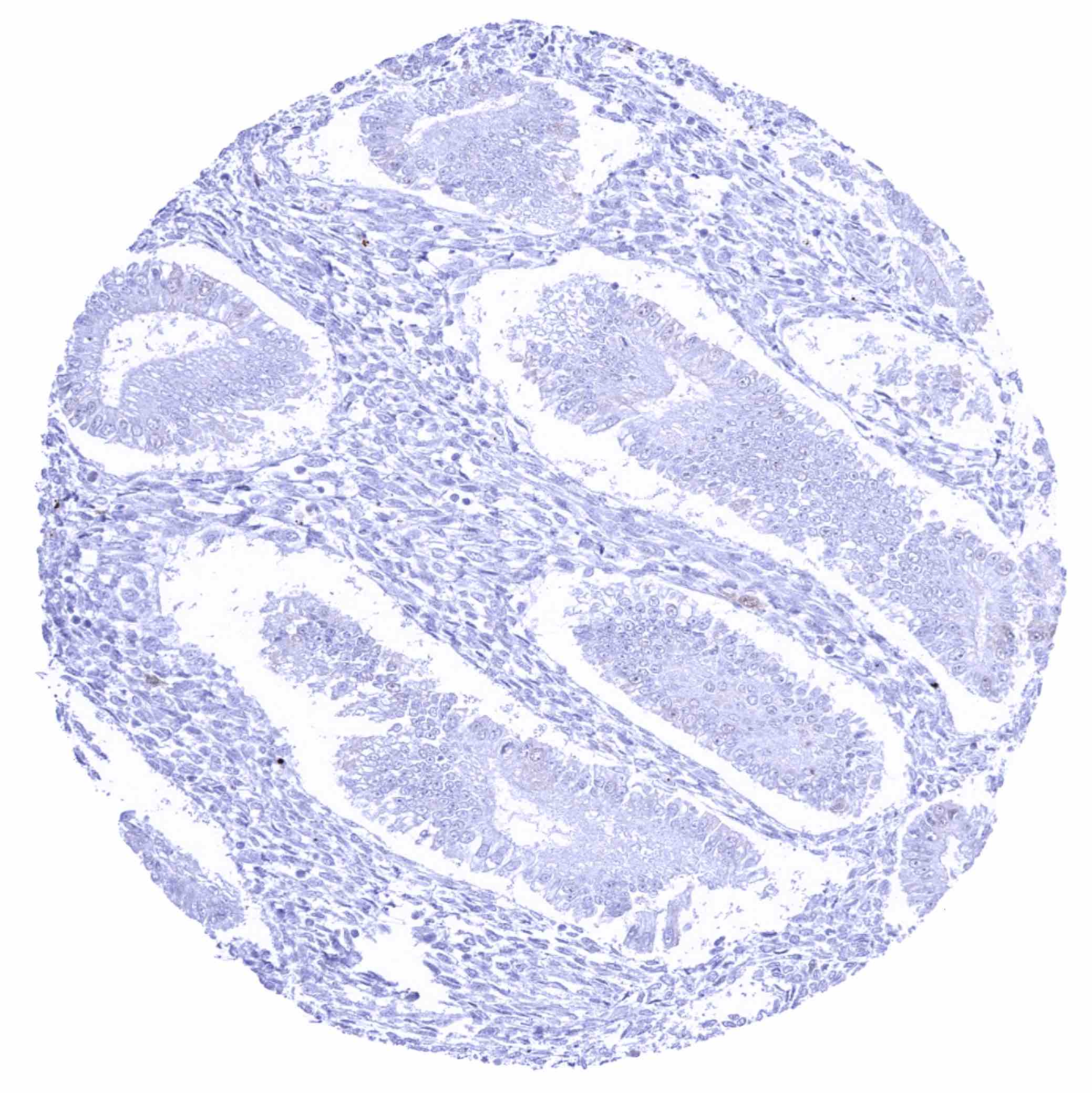
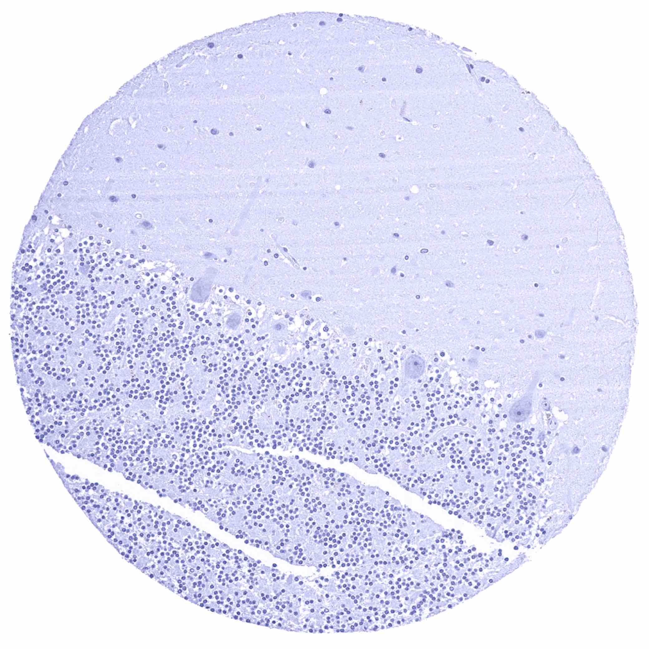
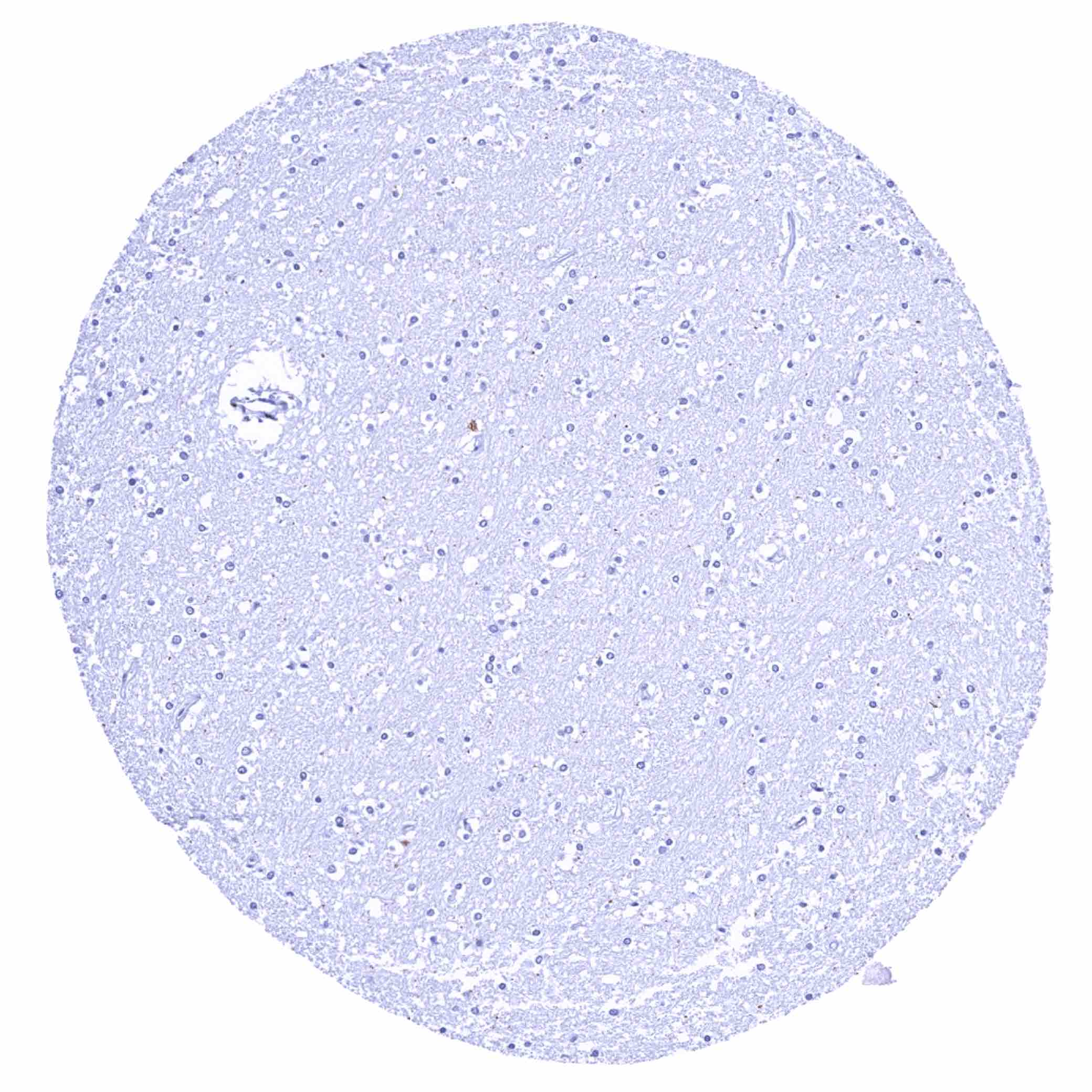
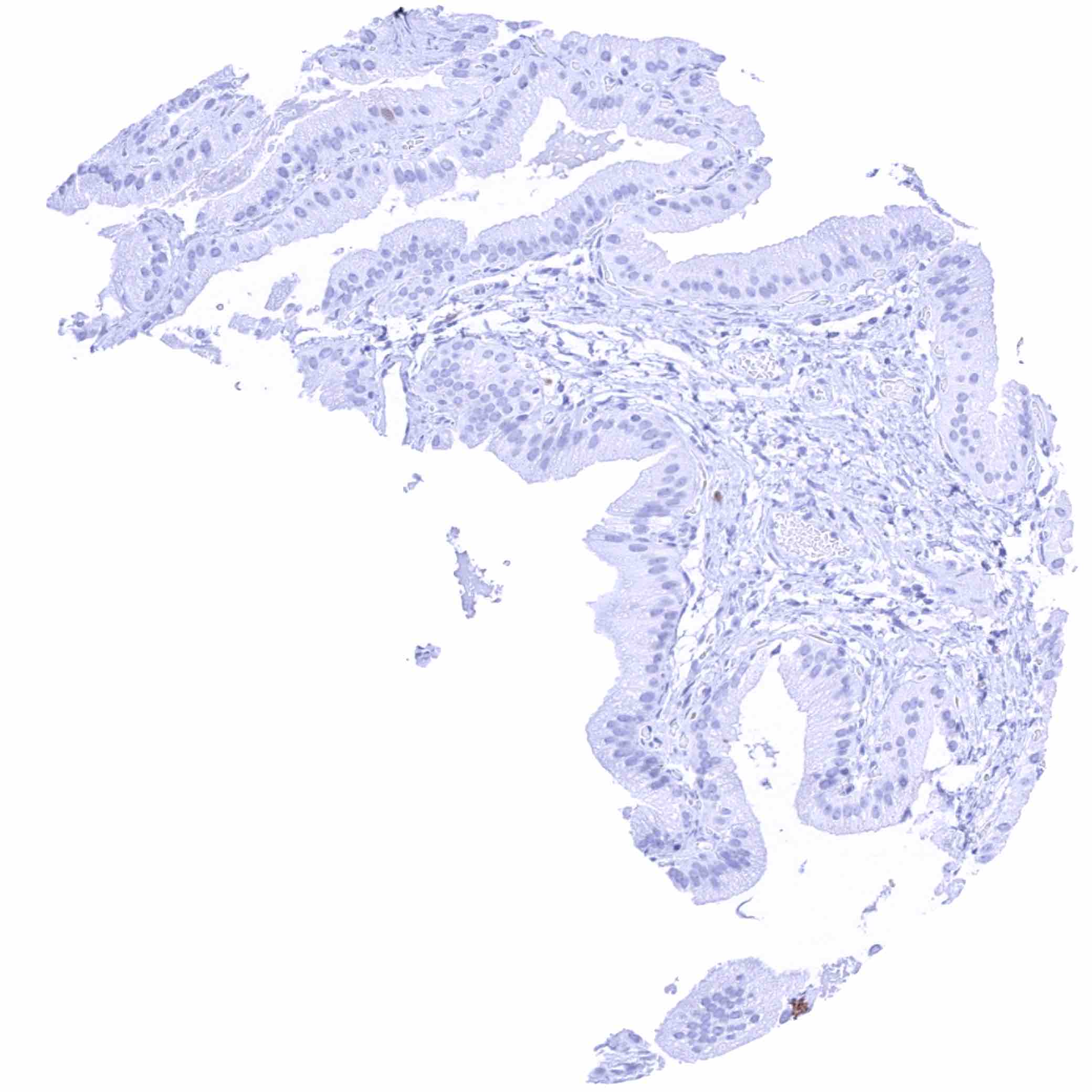
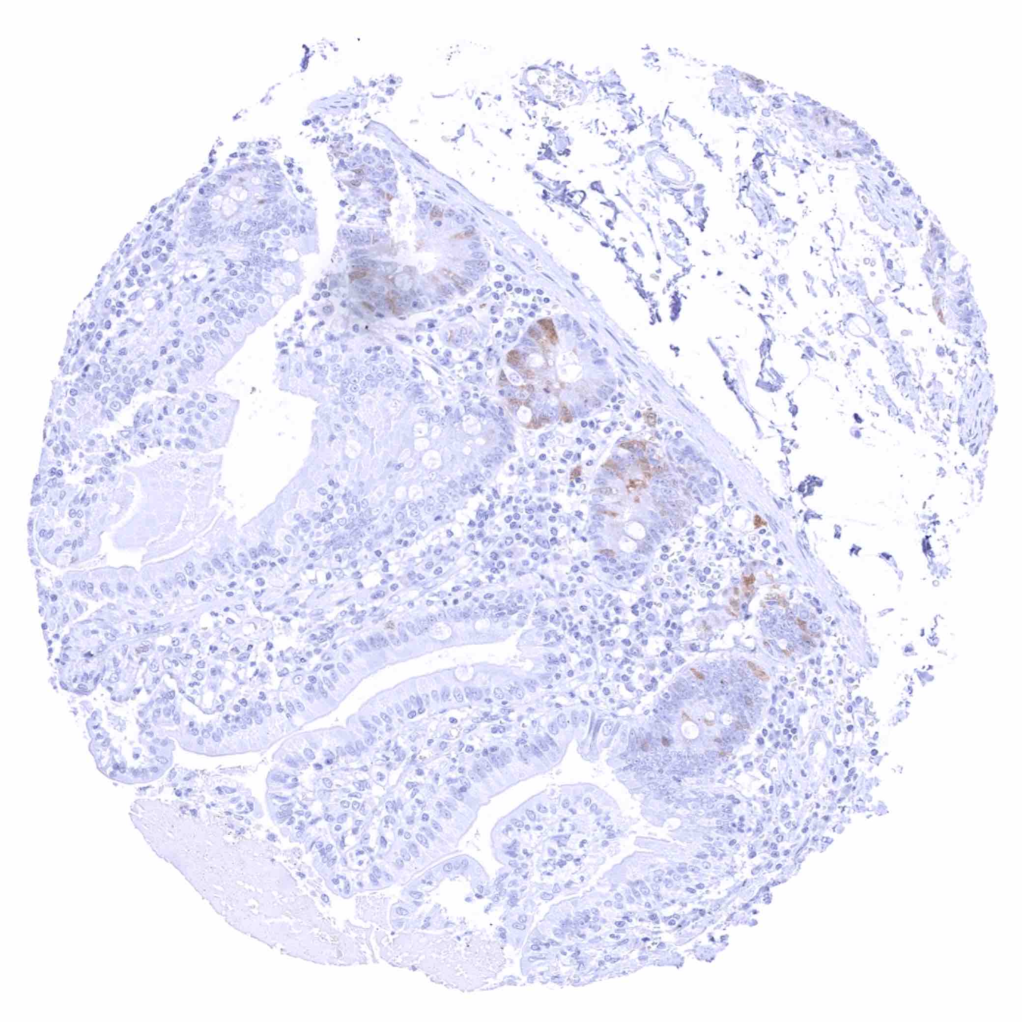
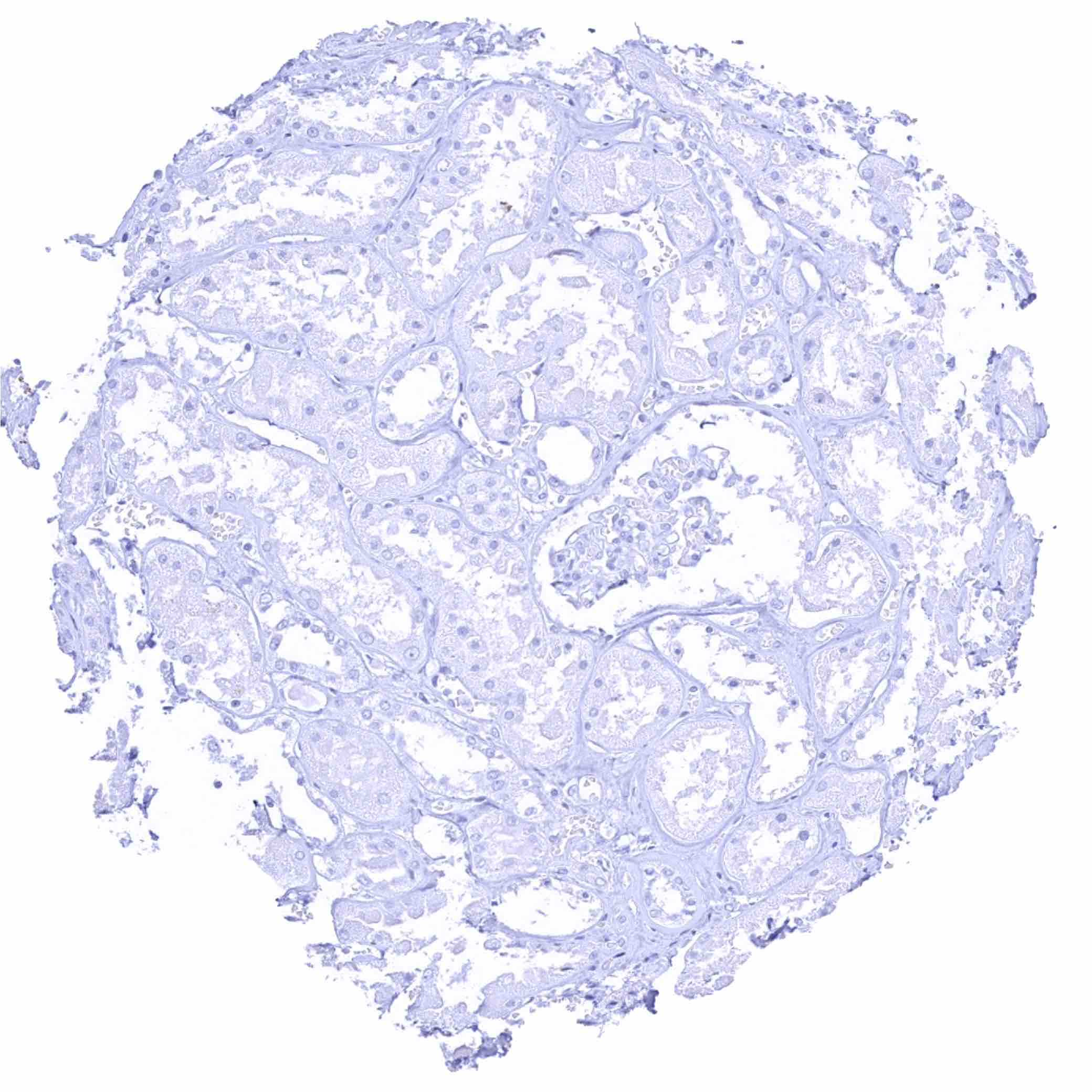
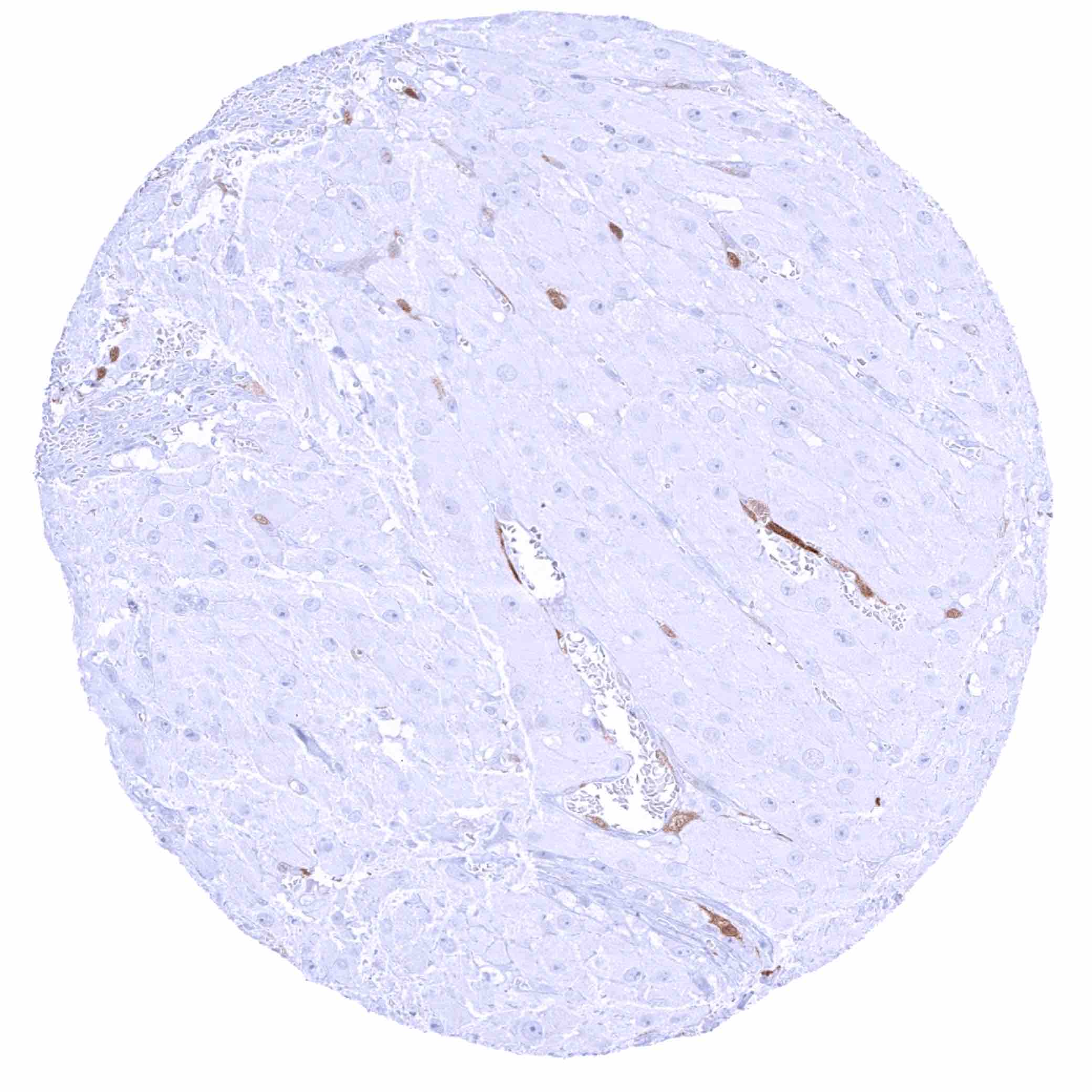
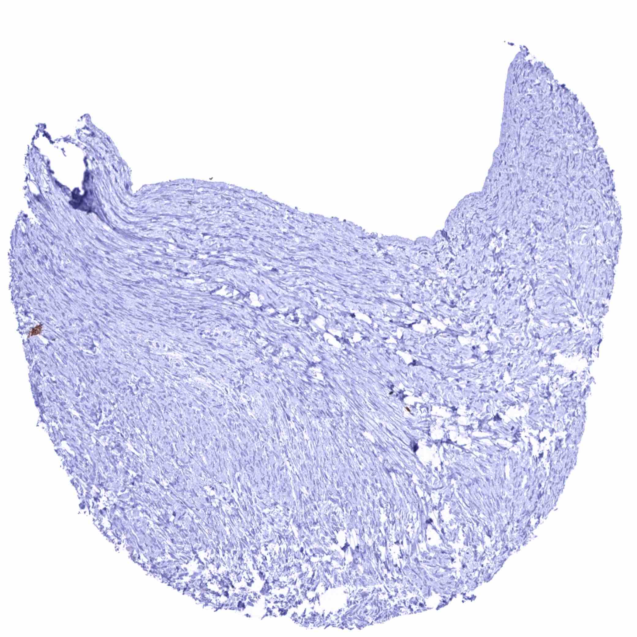
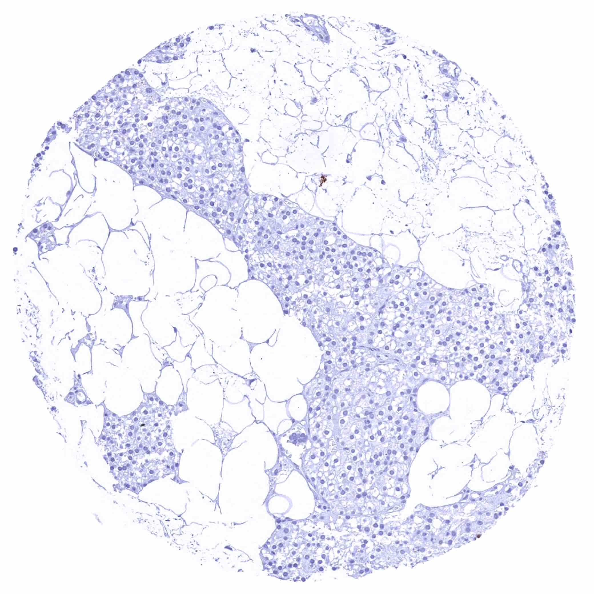
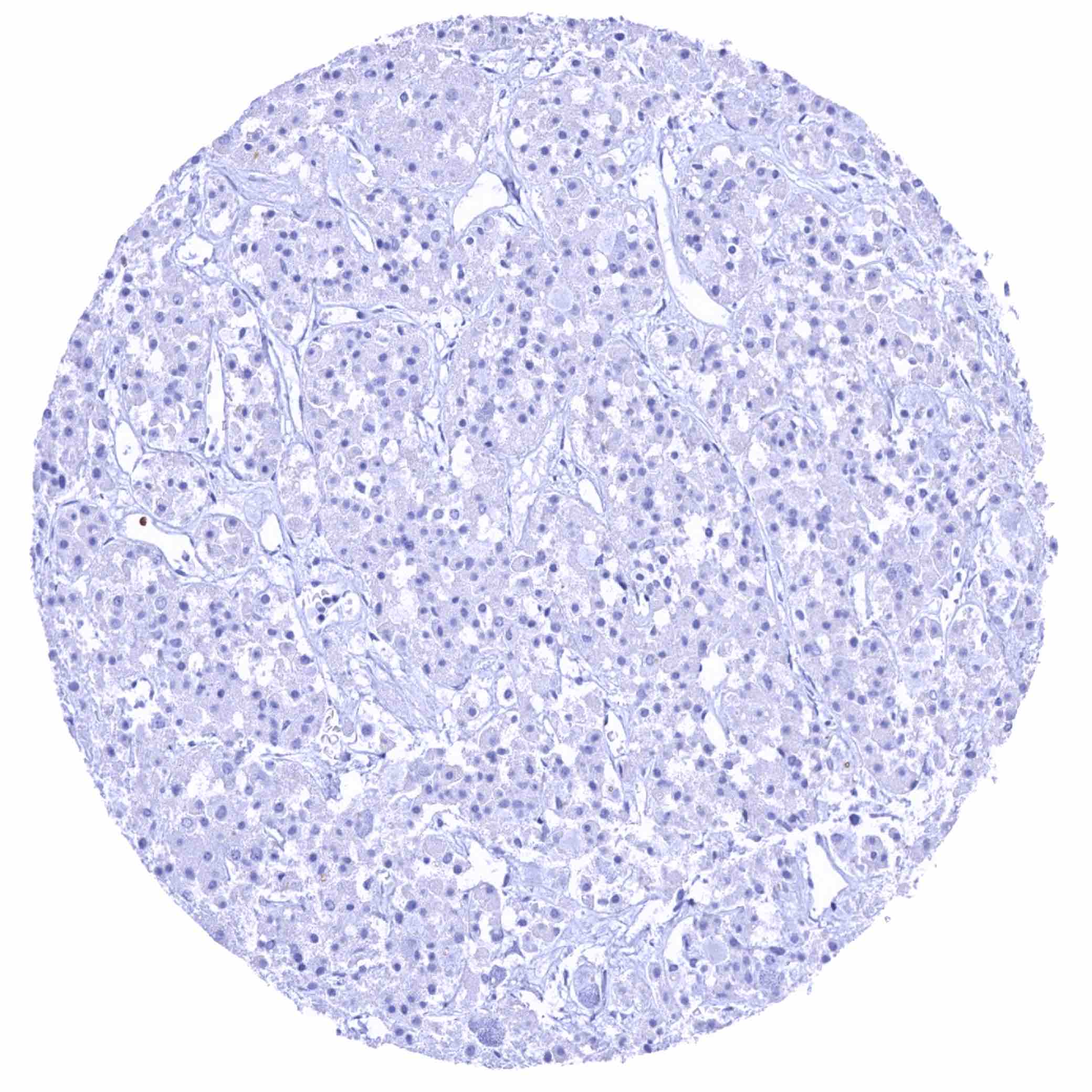
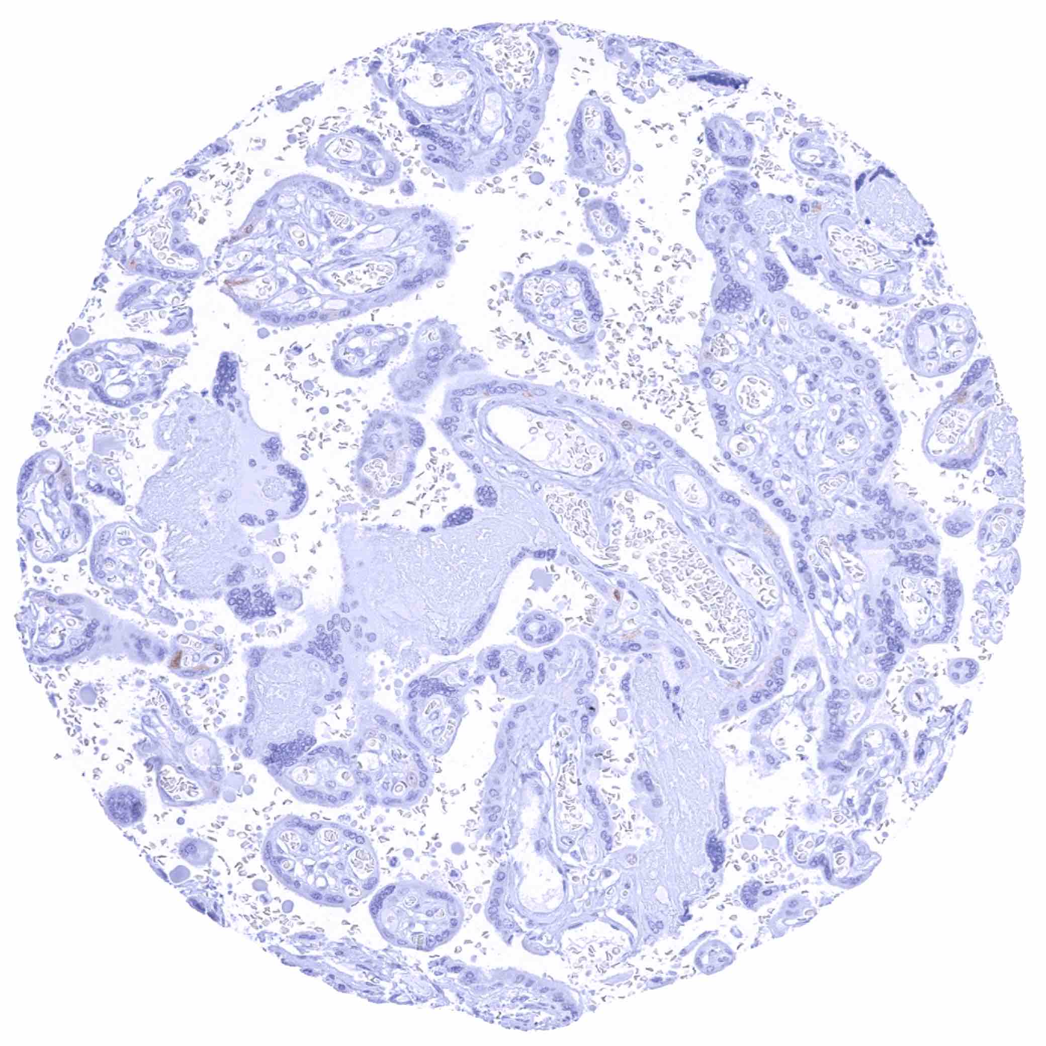
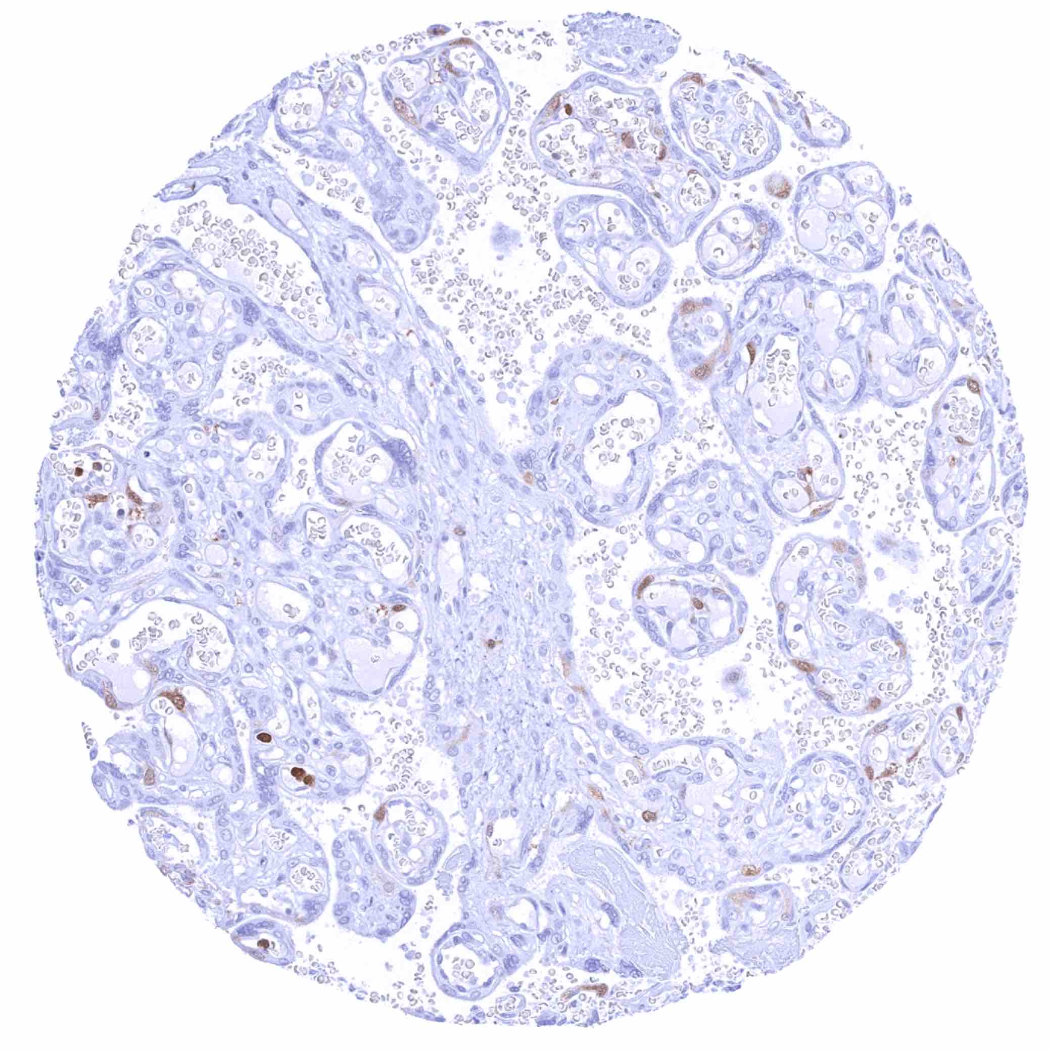
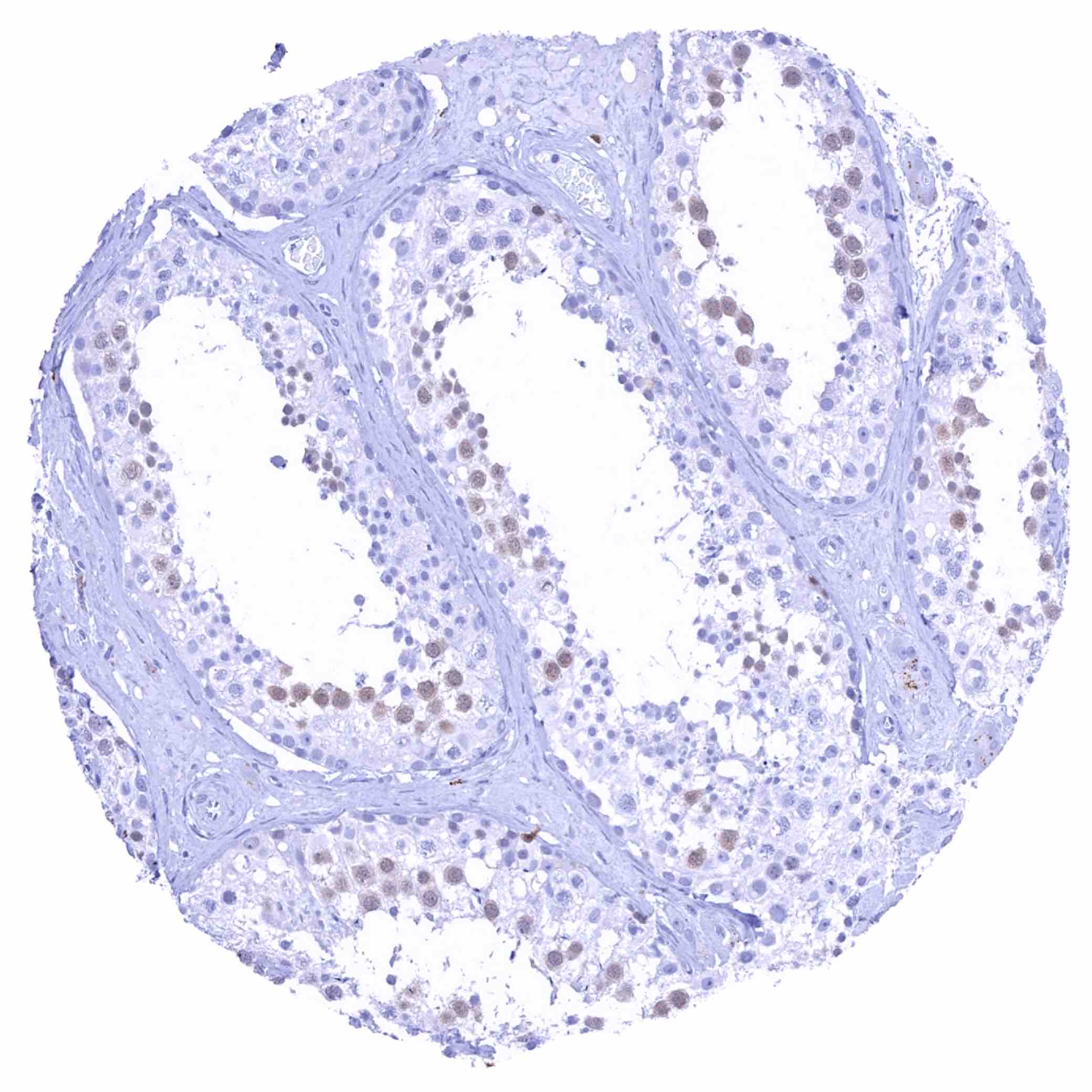
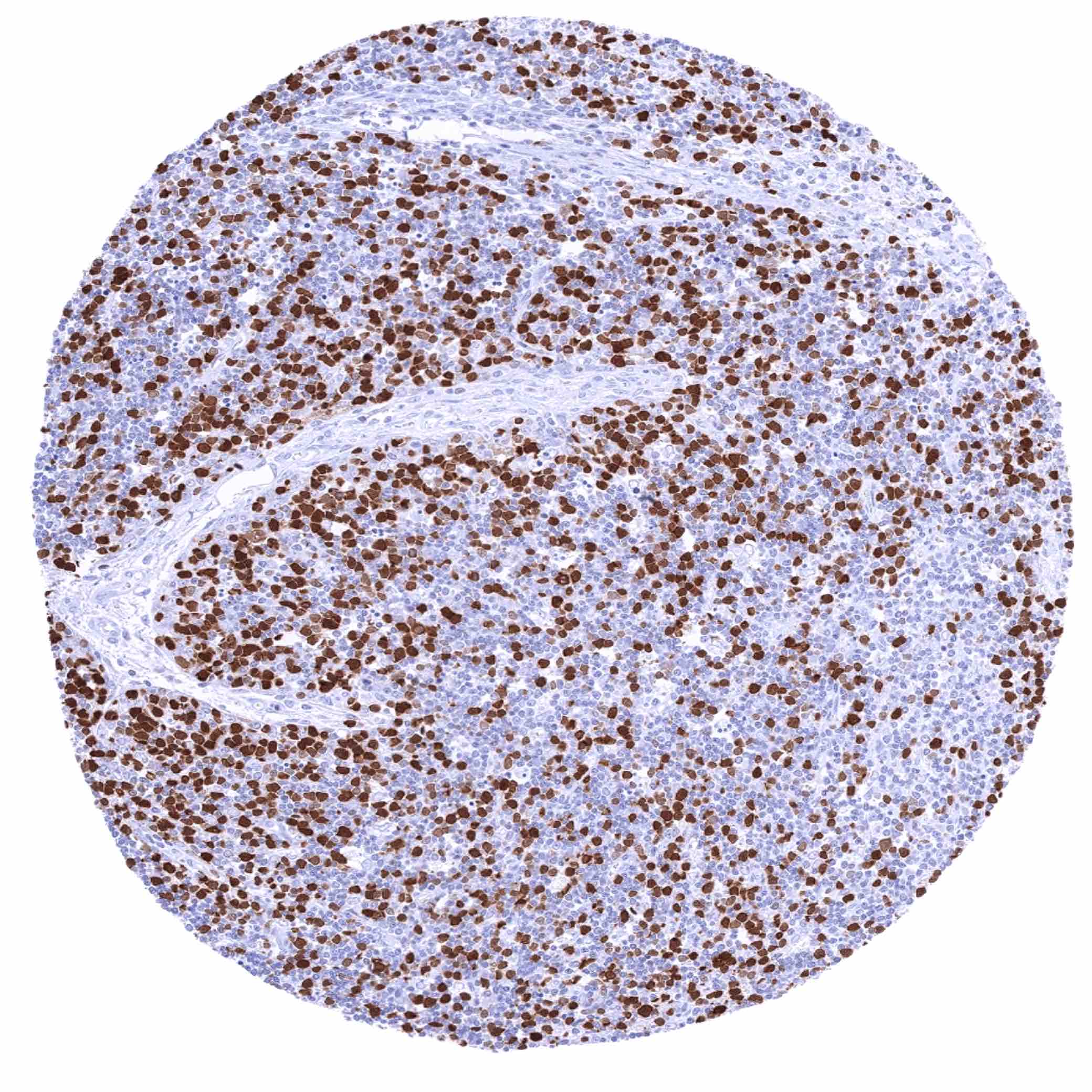
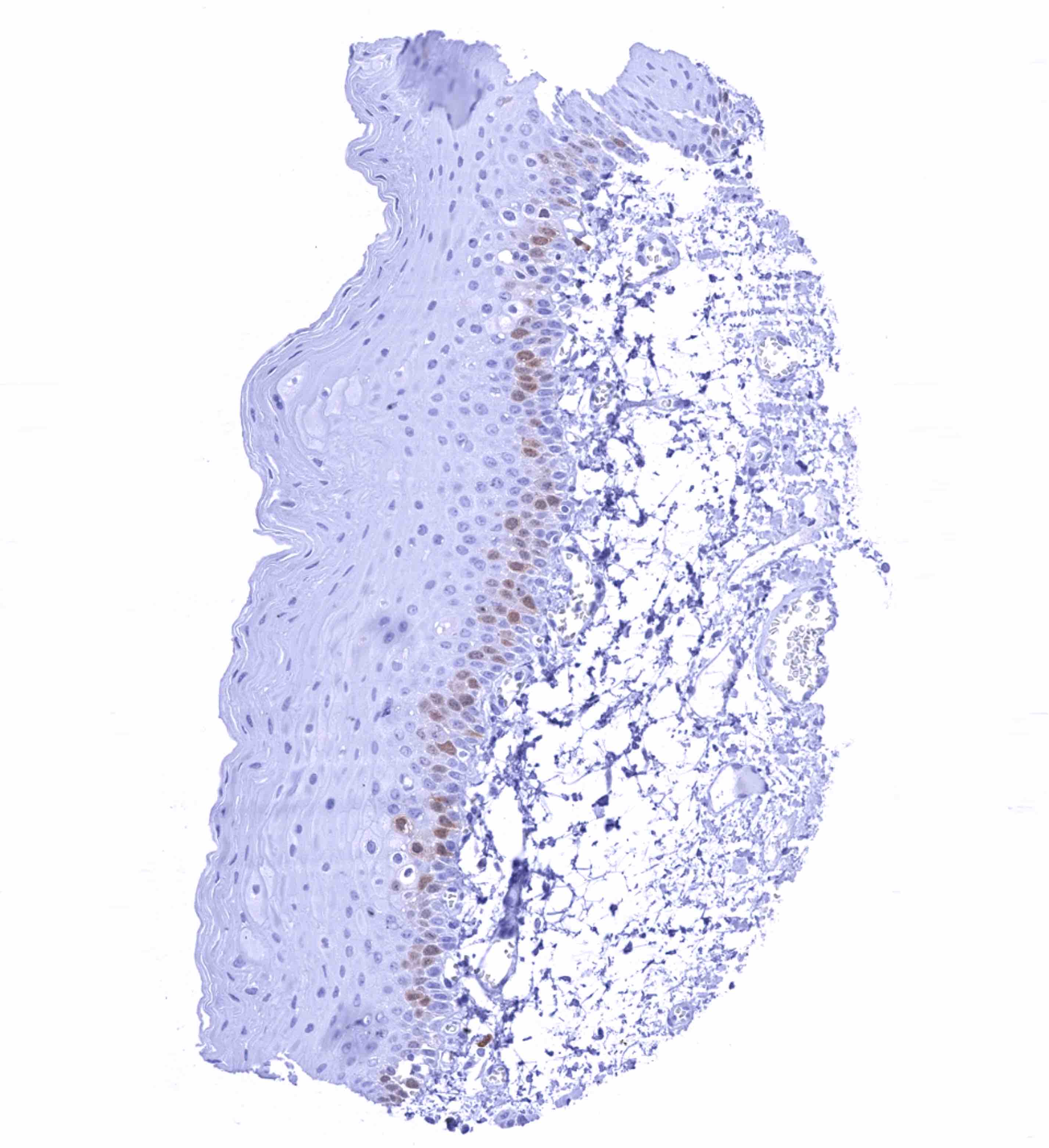
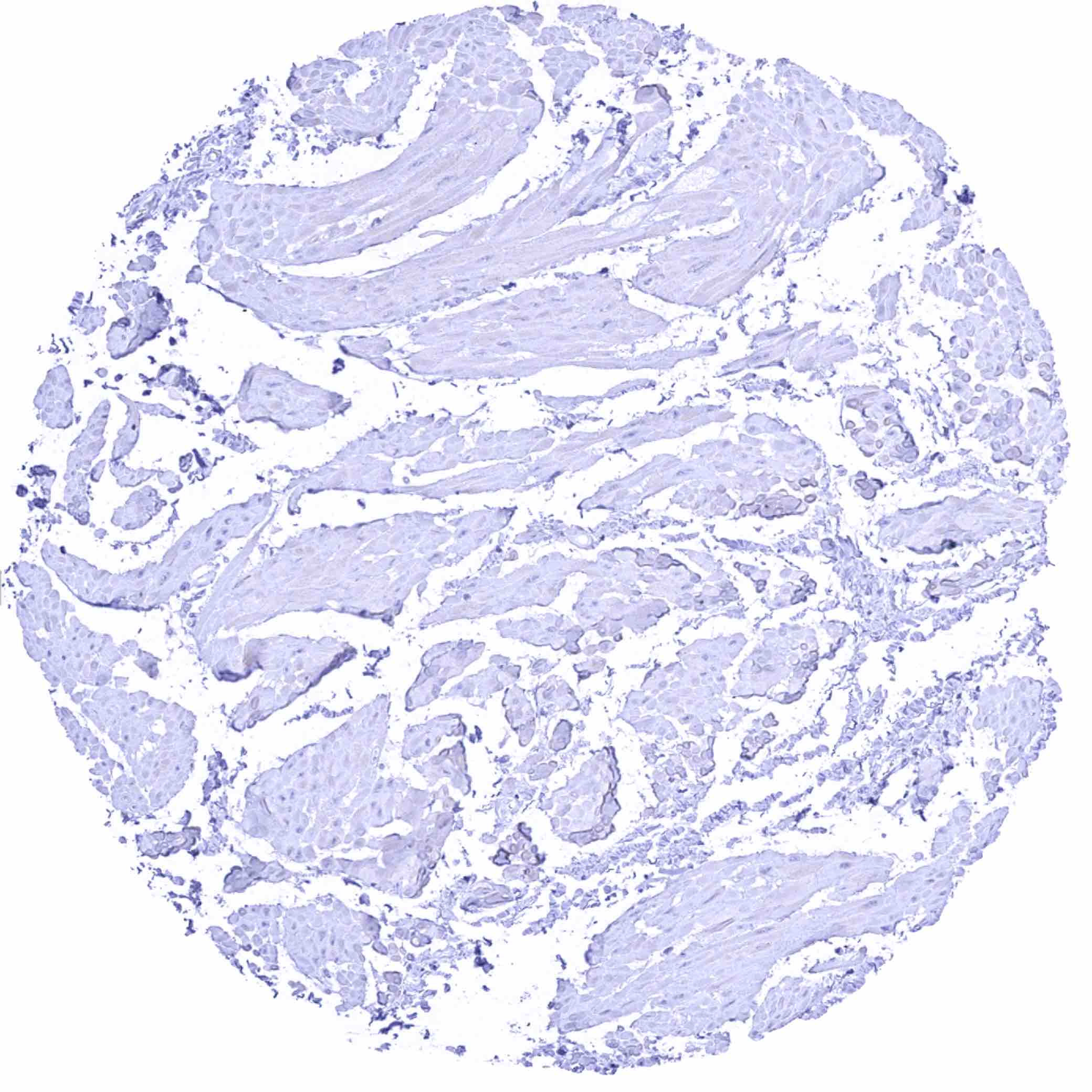
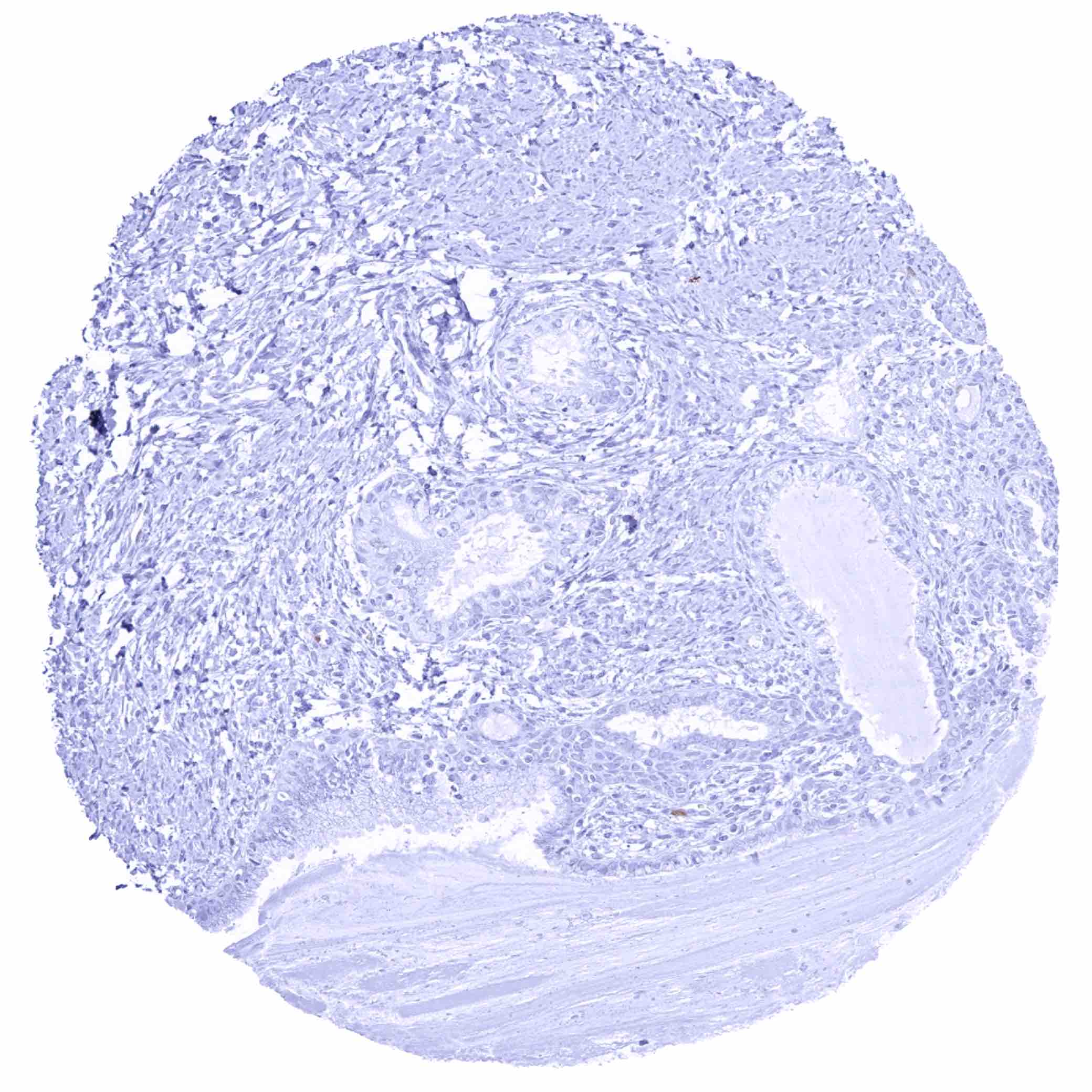
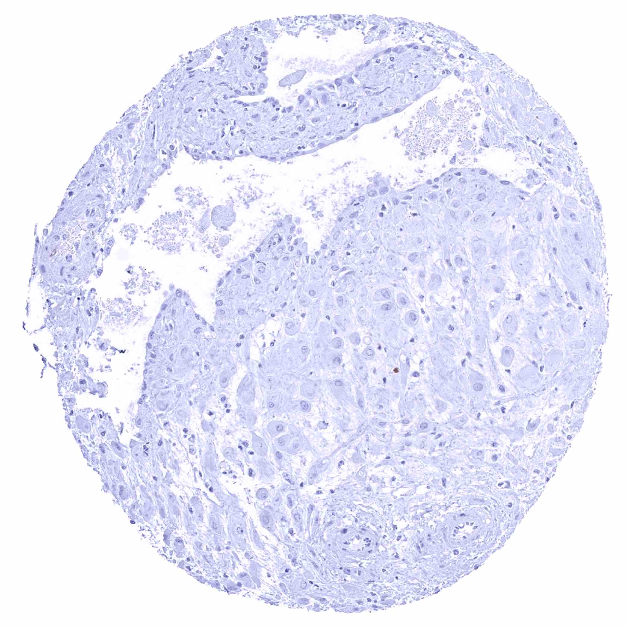
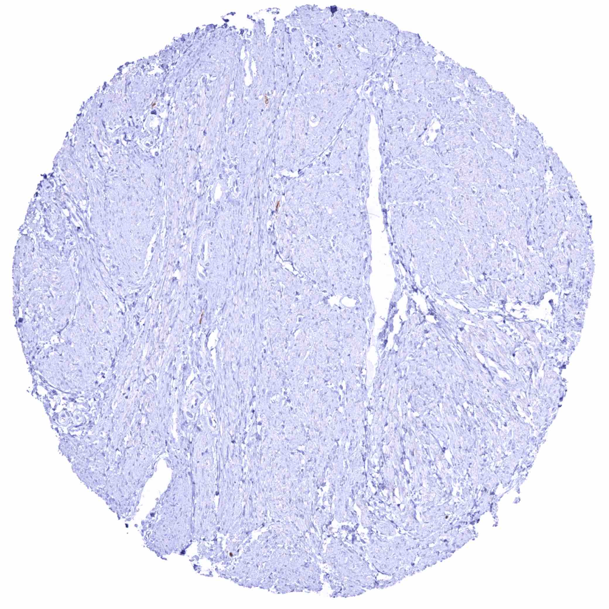
Thymidylate synthase antibody [HMV305] HistoMAXTM


Go to Cancer Tissue Gallery
  
 Go back to product page
  

Go to HistoMAX page


Adrenal gland

Aorta, media

Appendix, mucosa – Weak to moderate, nuclear and cytoplasmic TYMS staining of a fraction of crypt epithelial cells while staining is strong in many lymphocytic cells

Appendix, muscular wall


Bone marrow – Moderate to strong, nuclear and cytoplasmic TYMS staining of a large subset of hematopoetic cells

Breast – Strong, nuclear and cytoplasmic TYMS staining of a subset of luminal epithelial cells of breast glands

Bronchus, glands

Cerebellum (white matter)


Cerebellum, cortex (molecular layer, Purkinje cell layer, granule cell layer)

Cerebrum, grey matter

Cerebrum, white matter

Colon descendens, mucosa – Faint, nuclear and cytoplasmic TYMS staining of a fraction of crypt epithelial cells


Colon descendens, muscular wall

Duodenum, Brunner gland

Duodenum, mucosa – Weak, nuclear and cytoplasmic TYMS staining of a fraction of crypt epithelial cells

Epididymis, Caput


Epididymis, Cauda

Esophagus, squamous epithelium – Weak to moderate, predominantly nuclear TYMS staining of a subset of suprabasal squamous epithelial cells

Fallopian tube, mucosa

Fat


Gallbladder, epithelium – TYMS staining is lacking in this sample

Gallbladder, epithelium – Weak to moderate, nuclear and cytoplasmic TYMS staining of a subset of epithelial cells

Heart muscle

Ileum, mucosa – Weak to moderate, nuclear and cytoplasmic TYMS staining of a fraction of crypt epithelial cells


Kidney, cortex

Kidney, medulla

Liver

Lung


Lymph node – Strong TYMS staining of a fraction of lymphocytic cells

Ovary, corpus luteum – Moderate to strong, nuclear and cytoplasmic TYMS staining of some endothelial cells

Ovary, stroma

Pancreas – Weak to moderate, nuclear and cytoplasmic TYMS staining of very few epithelial cells


Parathyroid gland

Parathyroid gland

Parotid gland

Pituitary gland, posterior lobe


Pituitary gland, posterior lobe

Placenta (amnion and chorion)

Placenta (amnion and chorion)

Placenta, early – Variable, weak to strong, nuclear and cytoplasmic TYMS staining of few cells (cytotrophoblast, stromal)


Placenta, mature – TYMS staining is lacking in this sample

Placenta, mature – Variable, weak to strong, nuclear and cytoplasmic TYMS staining of few cells (trophoblast, endothelial)

Prostate

Rectum, mucosa – Weak to moderate, nuclear and cytoplasmic TYMS staining of a large subset of crypt epithelial cells


Seminal vesicle

Sinus paranasales

Skeletal muscle

Skin


Spleen – Moderate to strong TYMS staining of a fraction of lymphocytic cells

Stomach, antrum – Weak to moderate, nuclear and cytoplasmic TYMS staining of a fraction of epithelial cells

Stomach, corpus – Weak to moderate, nuclear and cytoplasmic TYMS staining of a fraction of epithelial cells

Testis – Weak to moderate, nuclear and cytoplasmic TYMS staining of spermatocytes


Thymus – Strong TYMS staining of a large fraction of lymphocytes

Thyroid gland

Tonsil – Variable, moderate to strong, nuclear and cytoplasmic TYMS staining of a fraction of lymphocytes, especially in a germinal centre.

Tonsil – Variable, moderate to strong, nuclear and cytoplasmic TYMS staining of a fraction of lymphocytes, especially in a germinal centre


Tonsil, surface epithelium – Weak to moderate, predominantly nuclear TYMS staining of a subset of suprabasal squamous epithelial cells

Urinary bladder, muscular wall

Urinary bladder, urothelium

Uterus, ectocervix


Uterus, endocervix

Uterus, endometrium (pregnancy)

Uterus, endometrium (proliferation)

Uterus, endometrium (secretion)


Uterus, myometrium


| TOP |