Normal Tissue Gallery- GTX04450

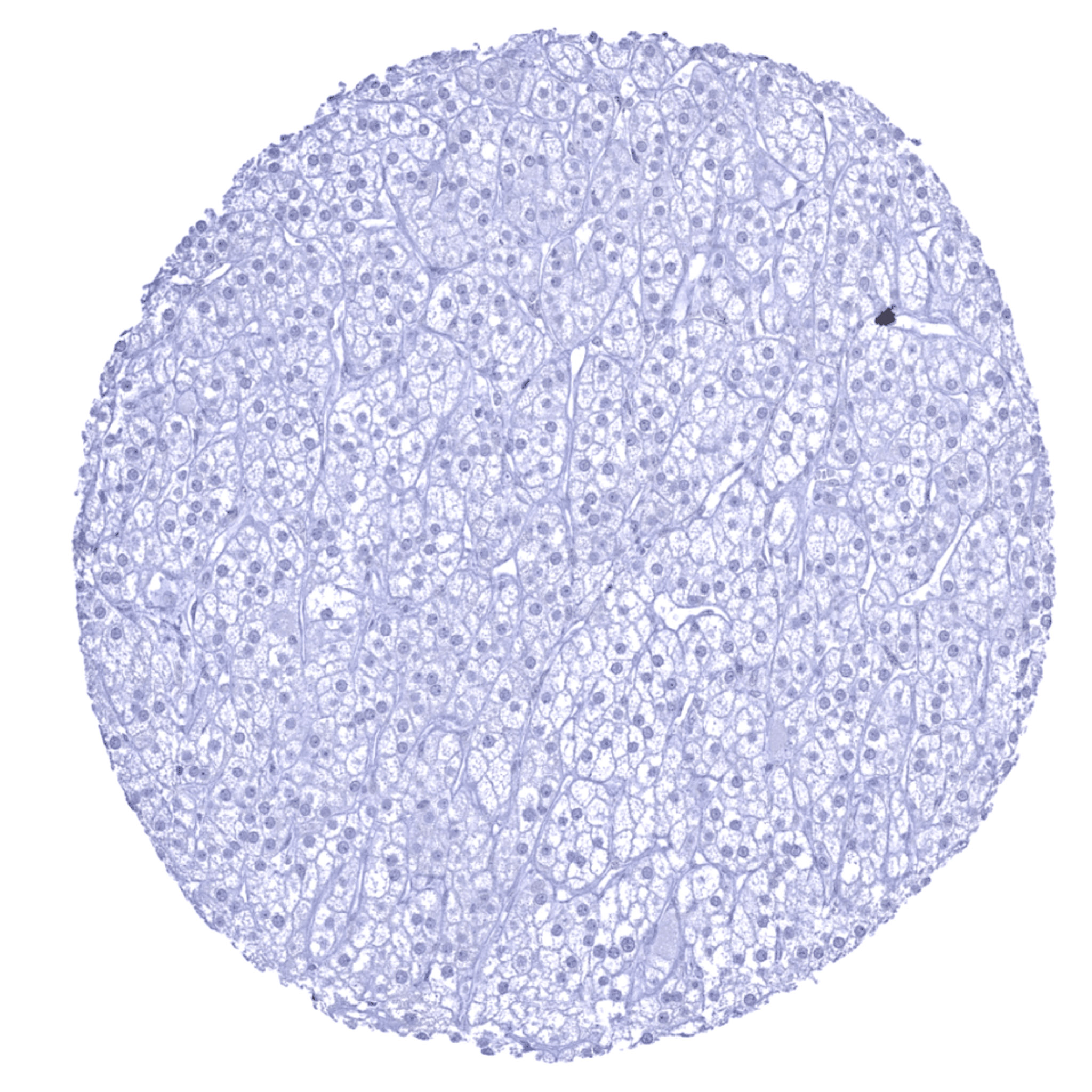
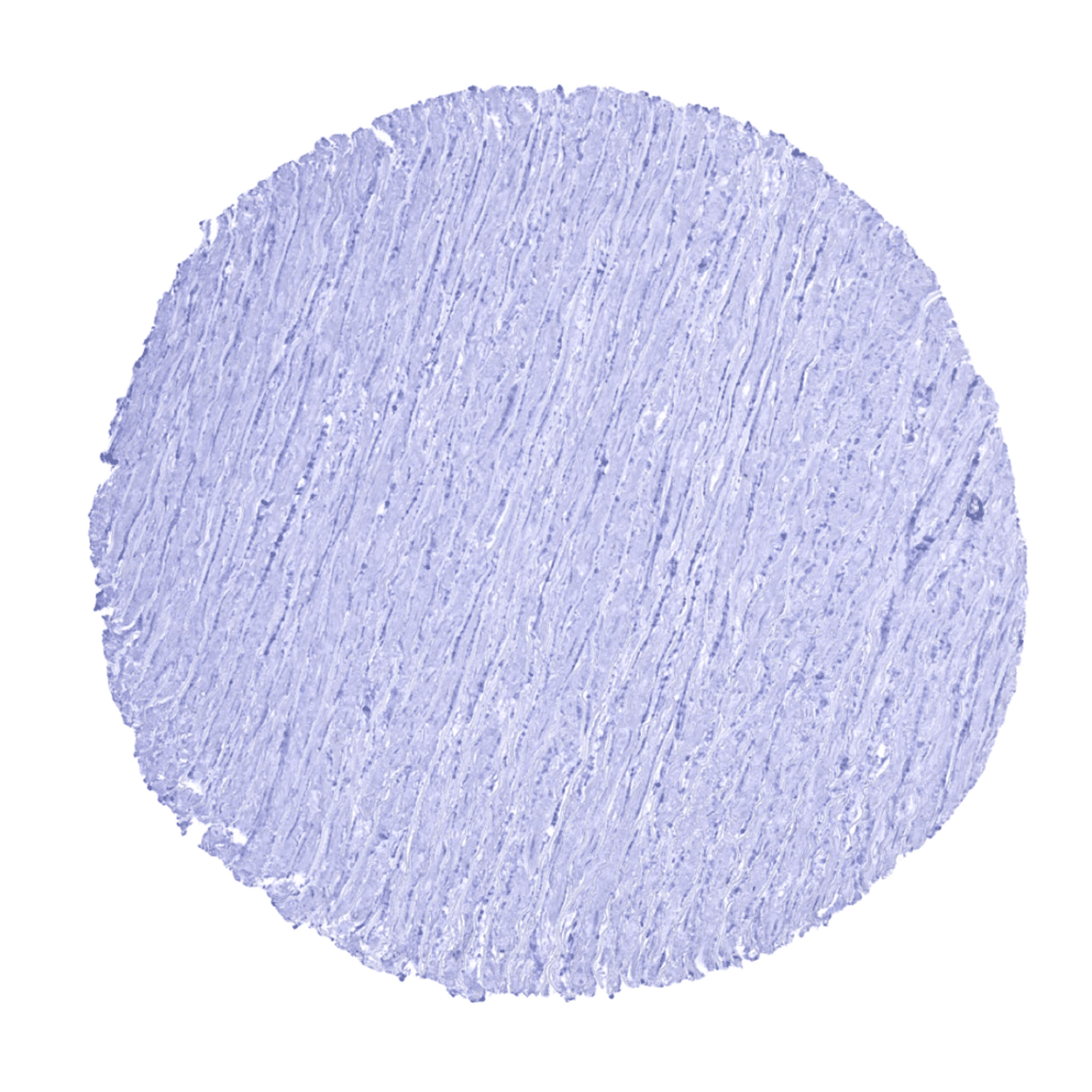
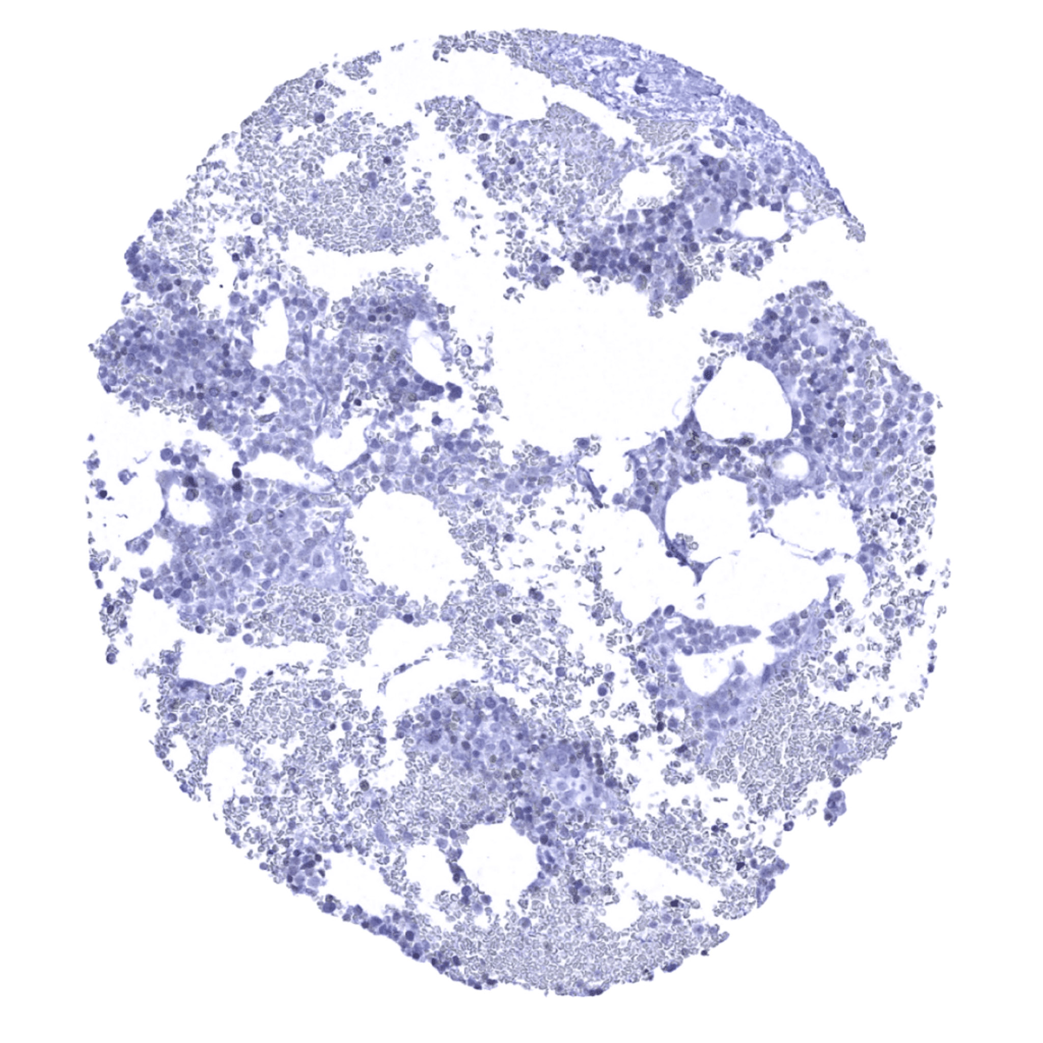
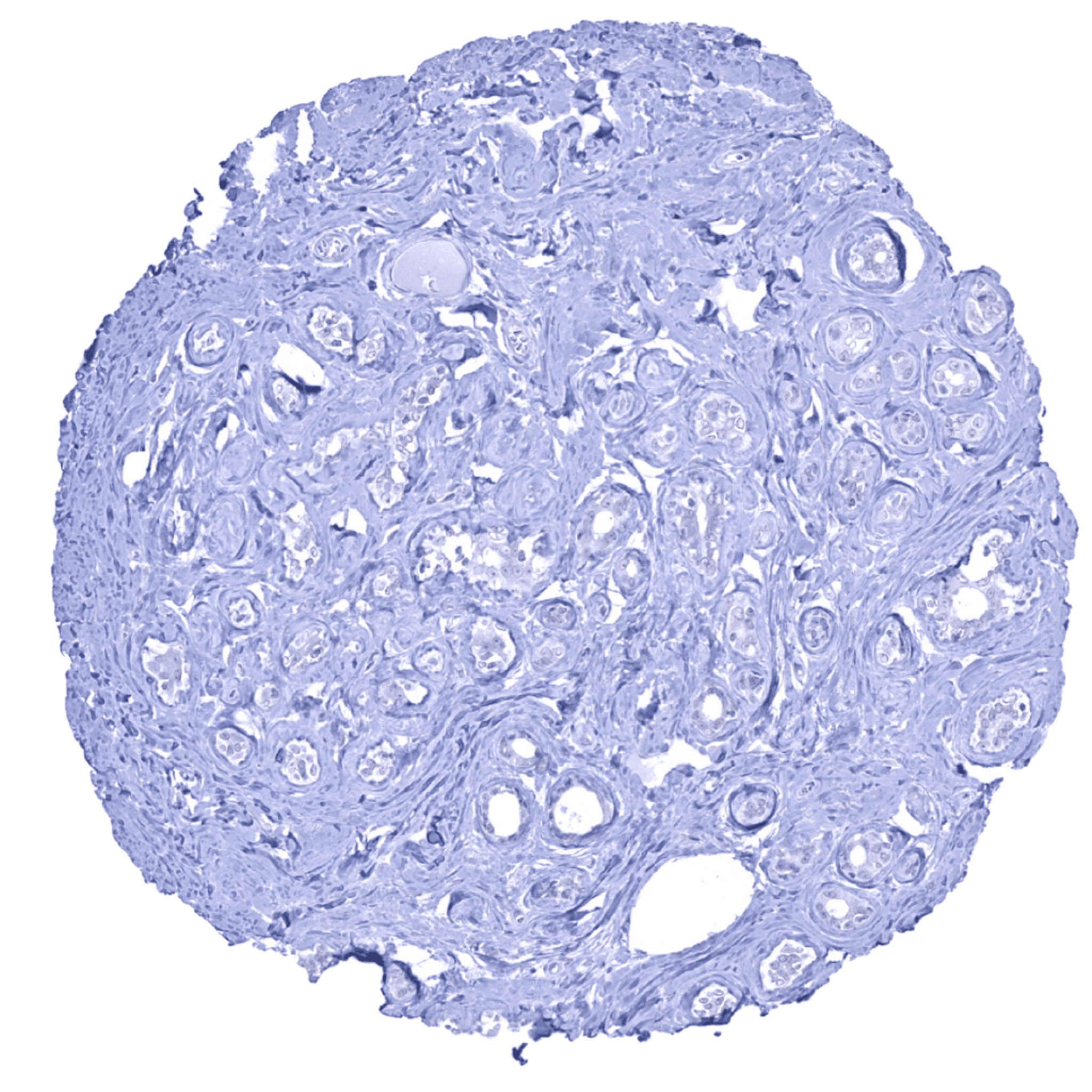
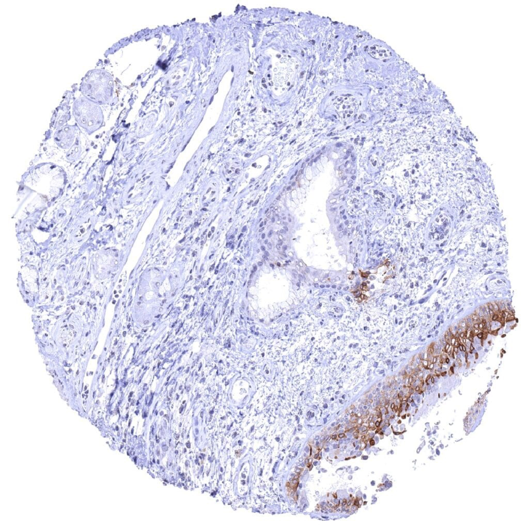
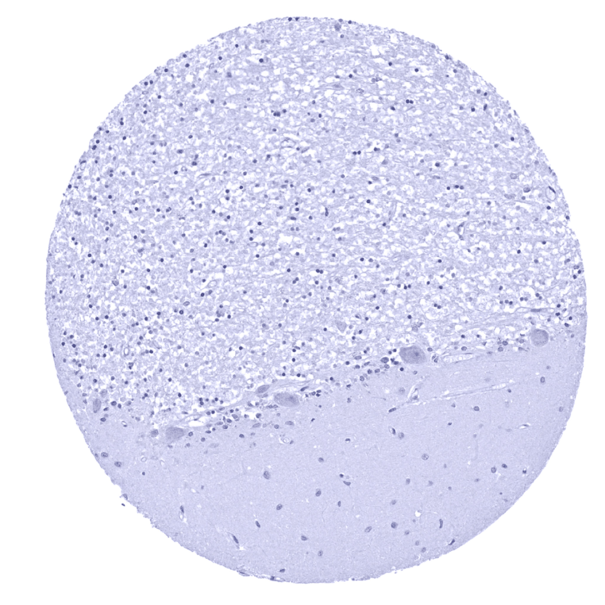
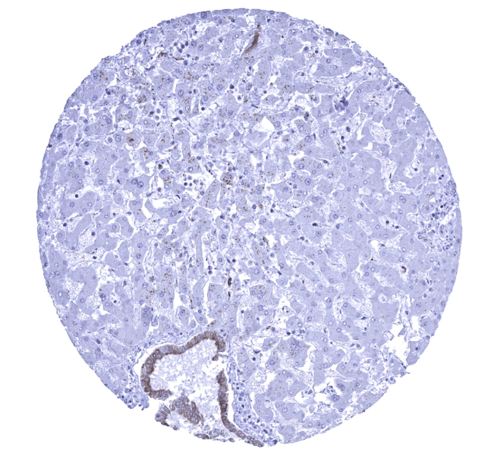
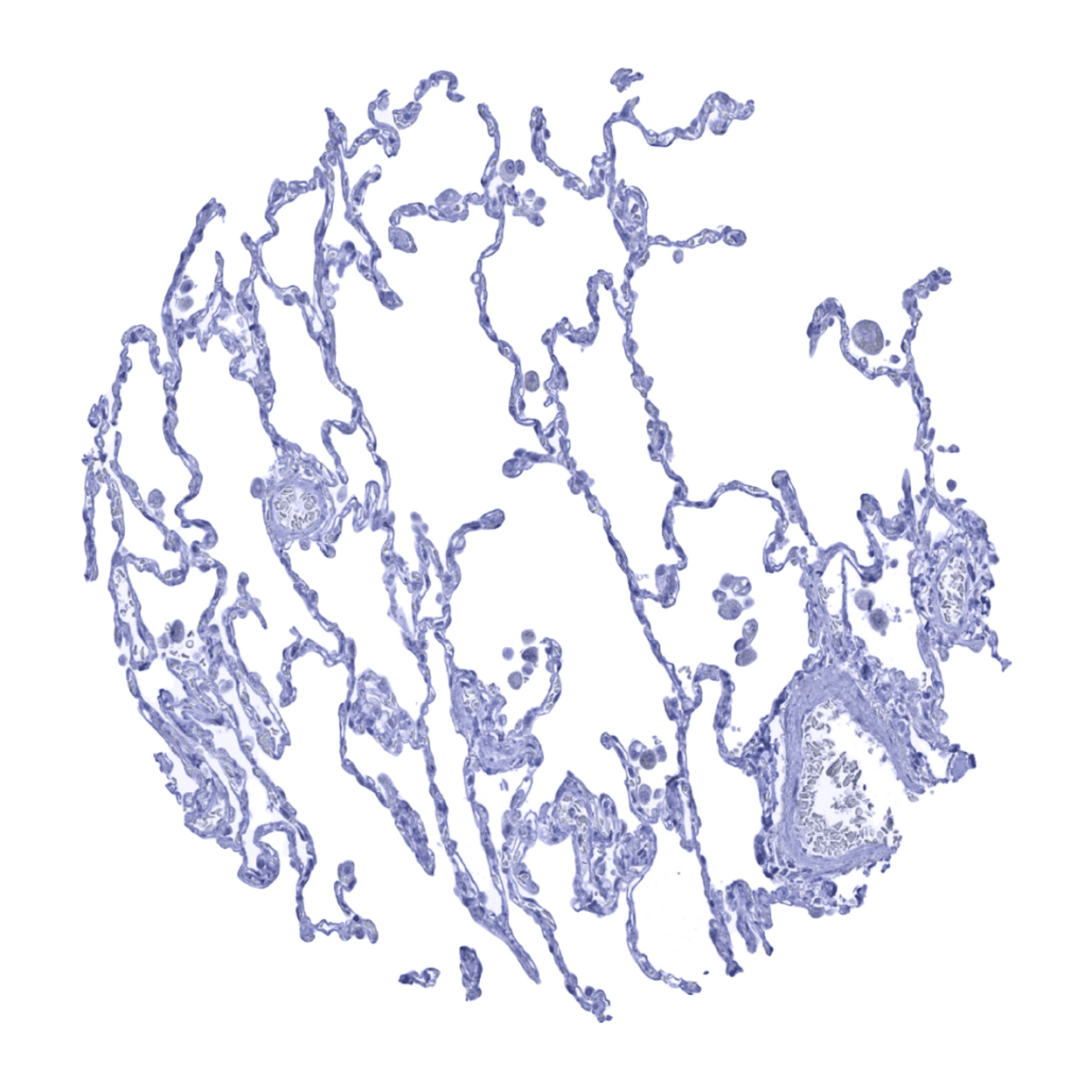
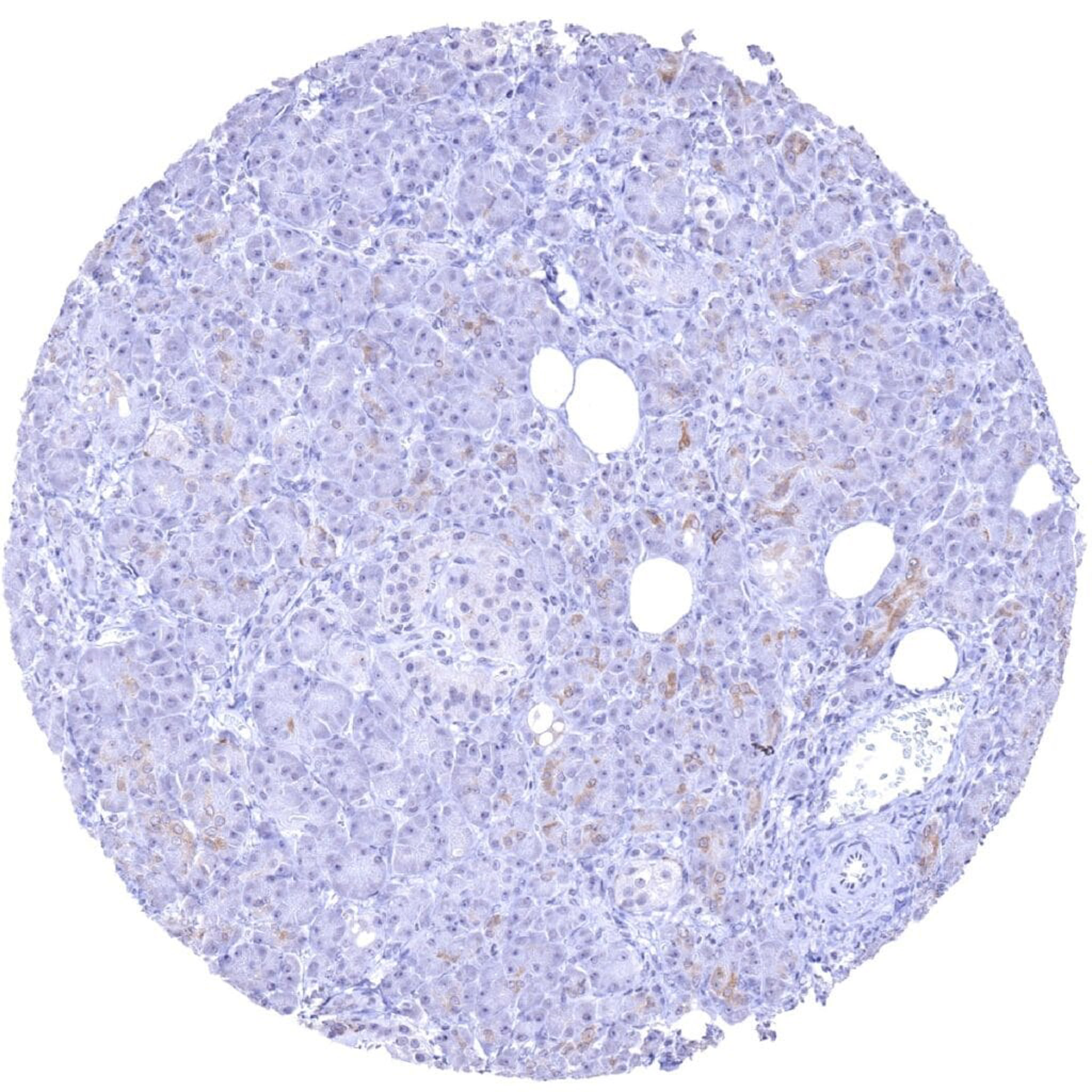
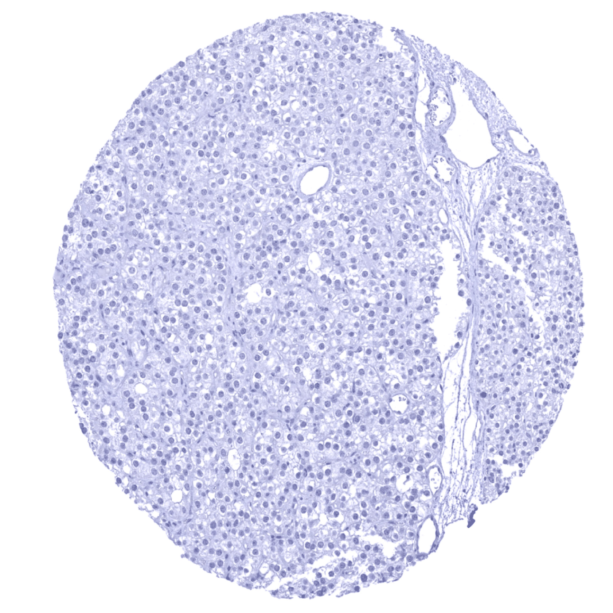
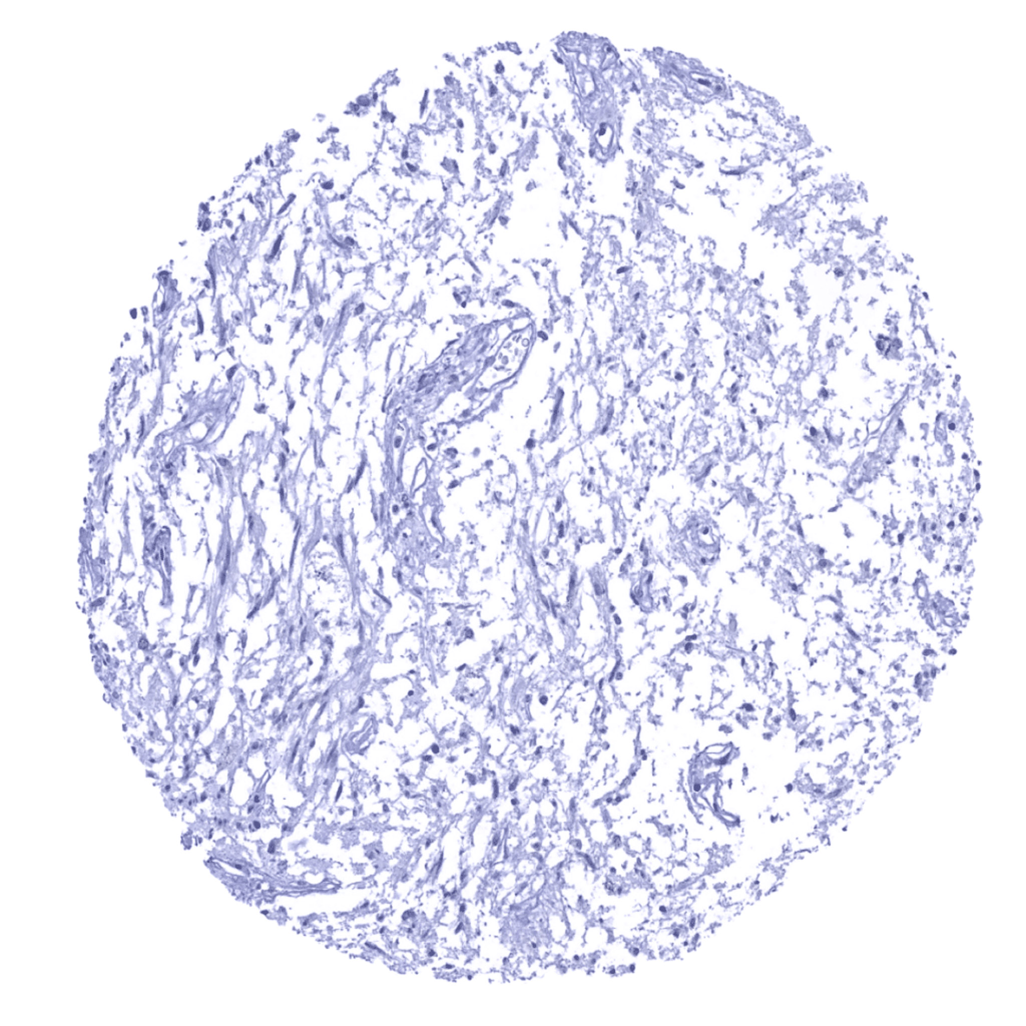
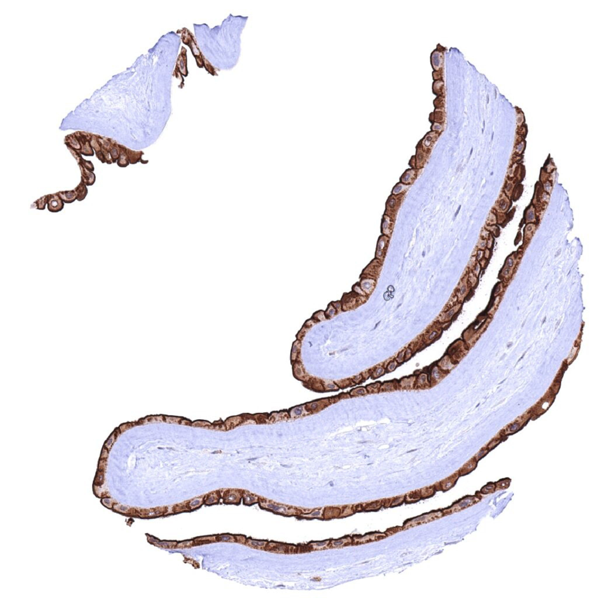
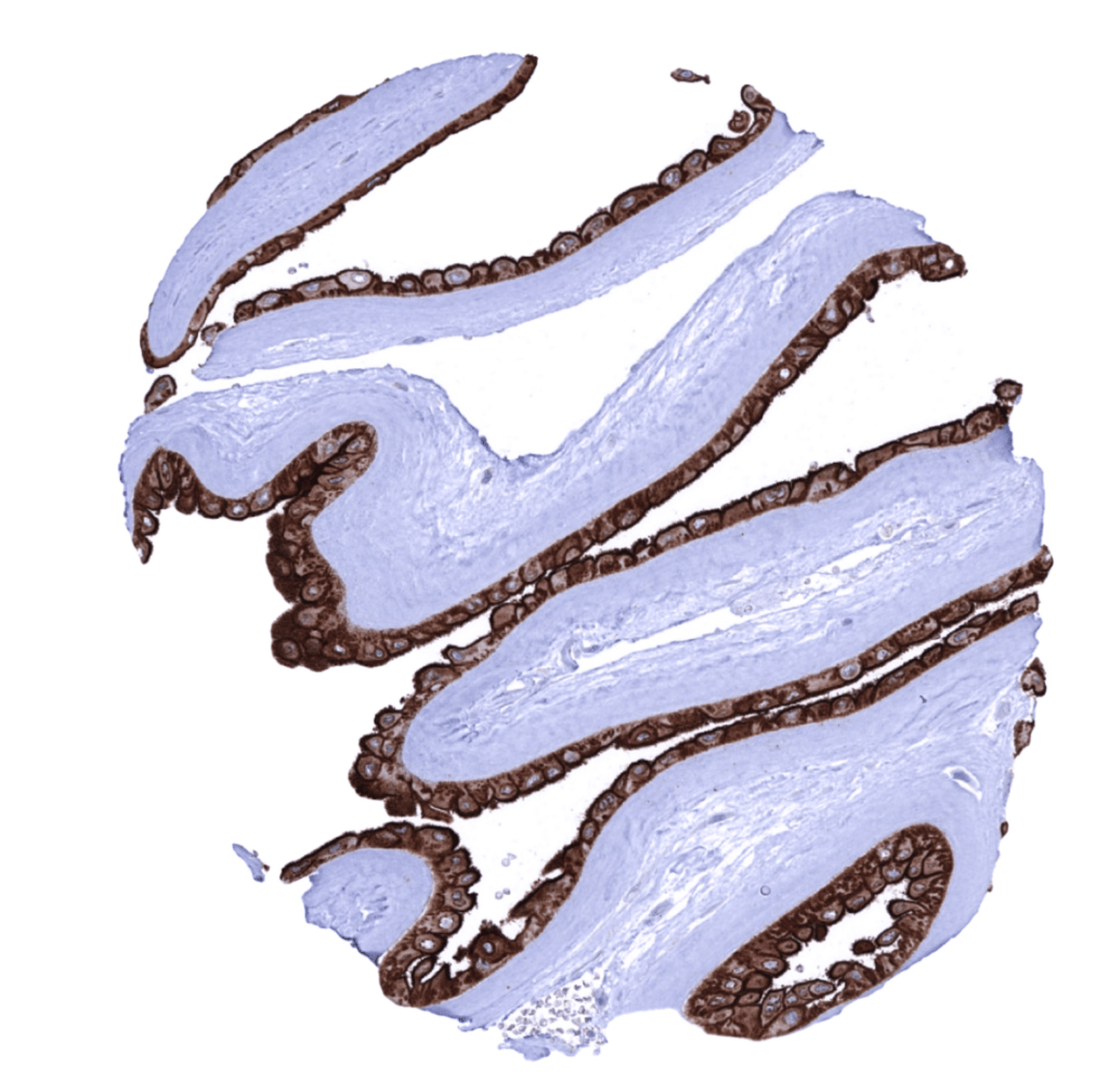
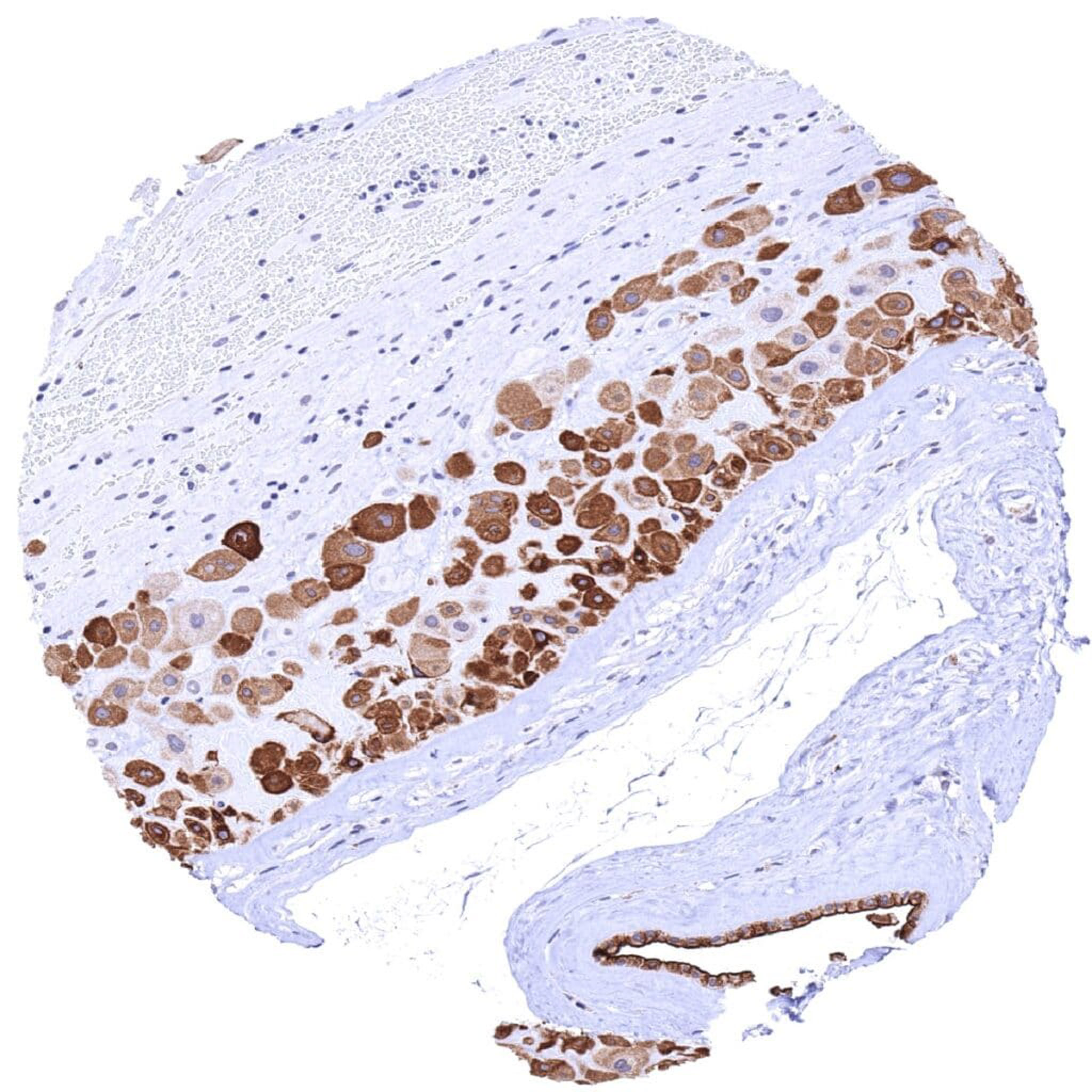
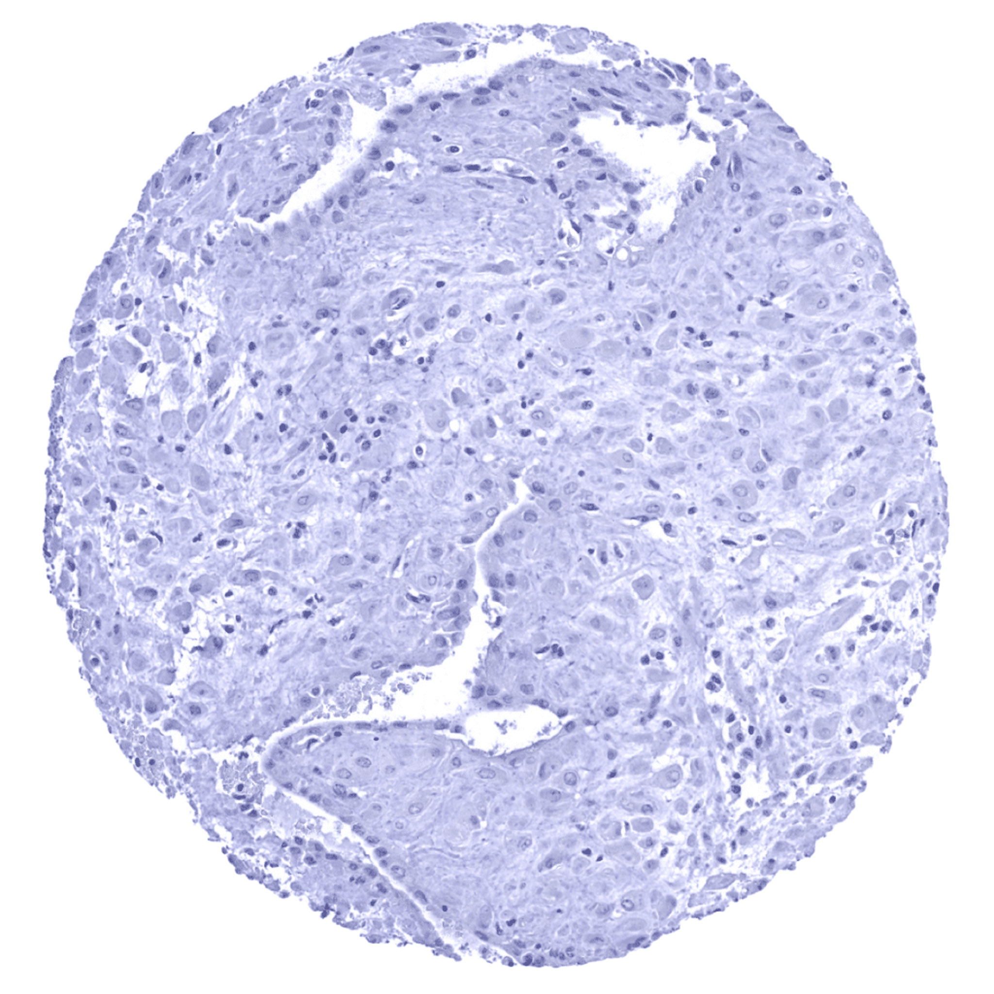
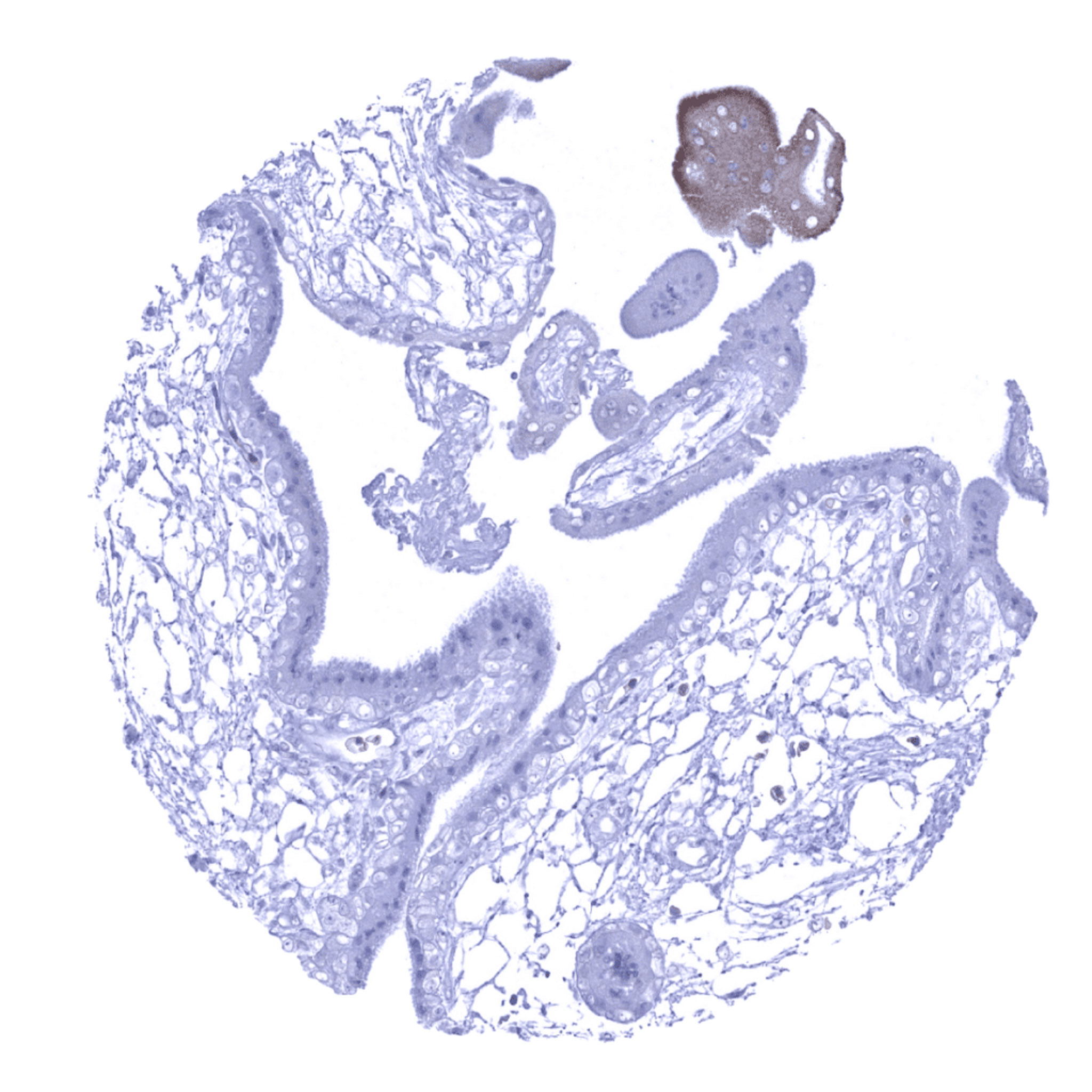
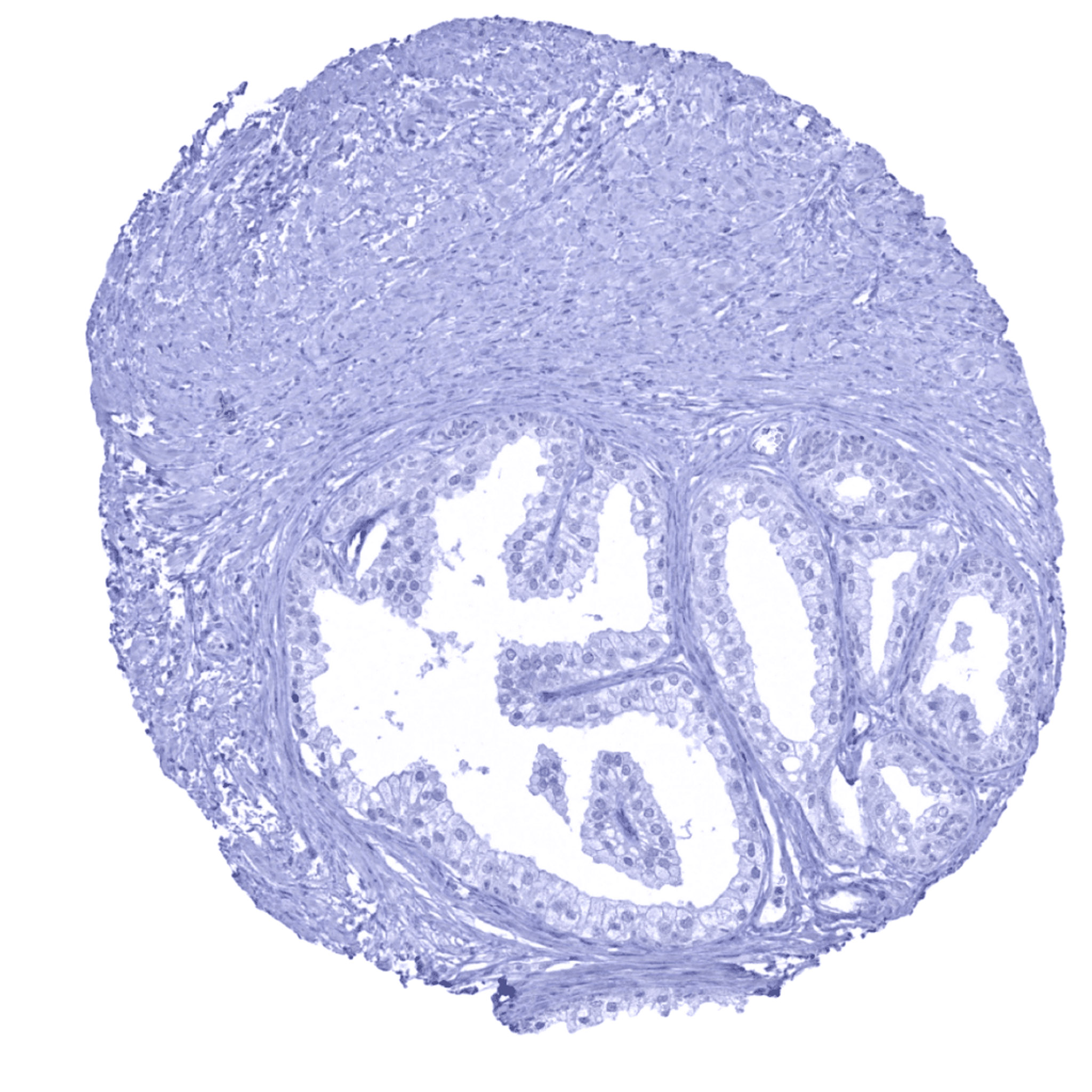
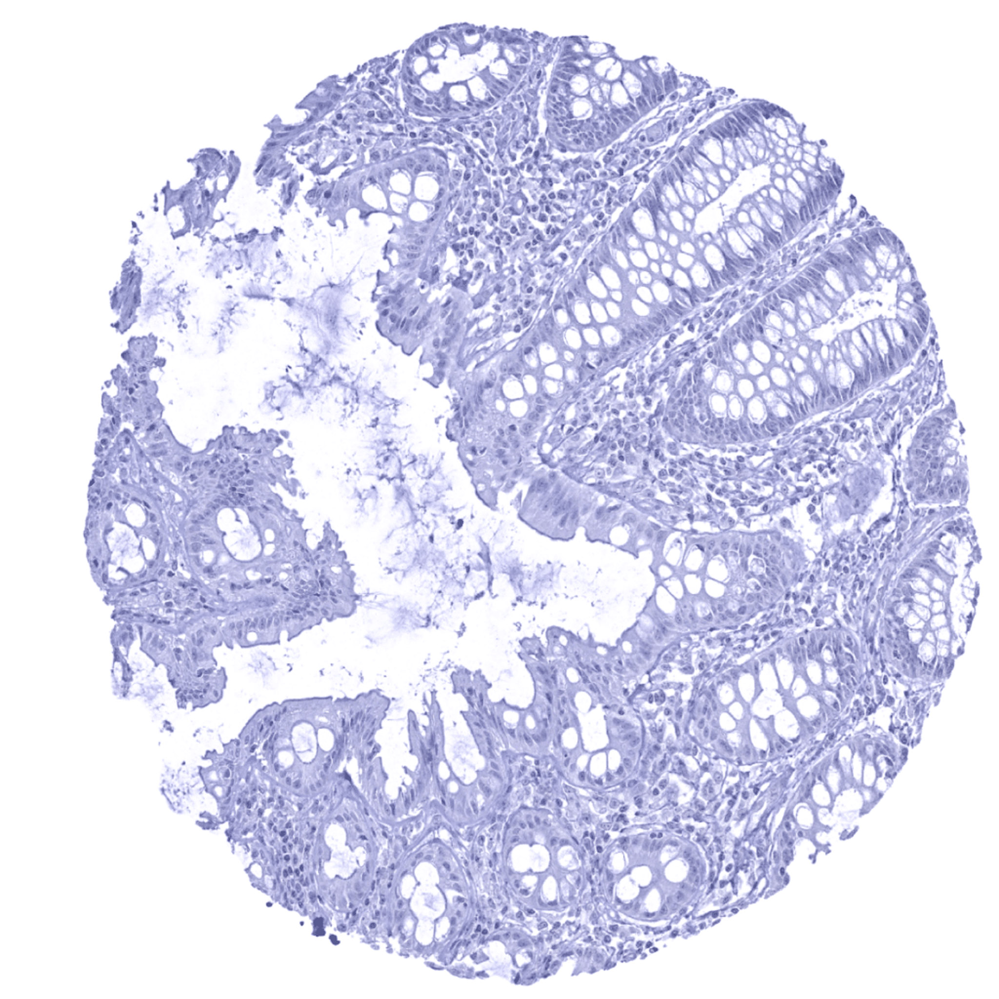
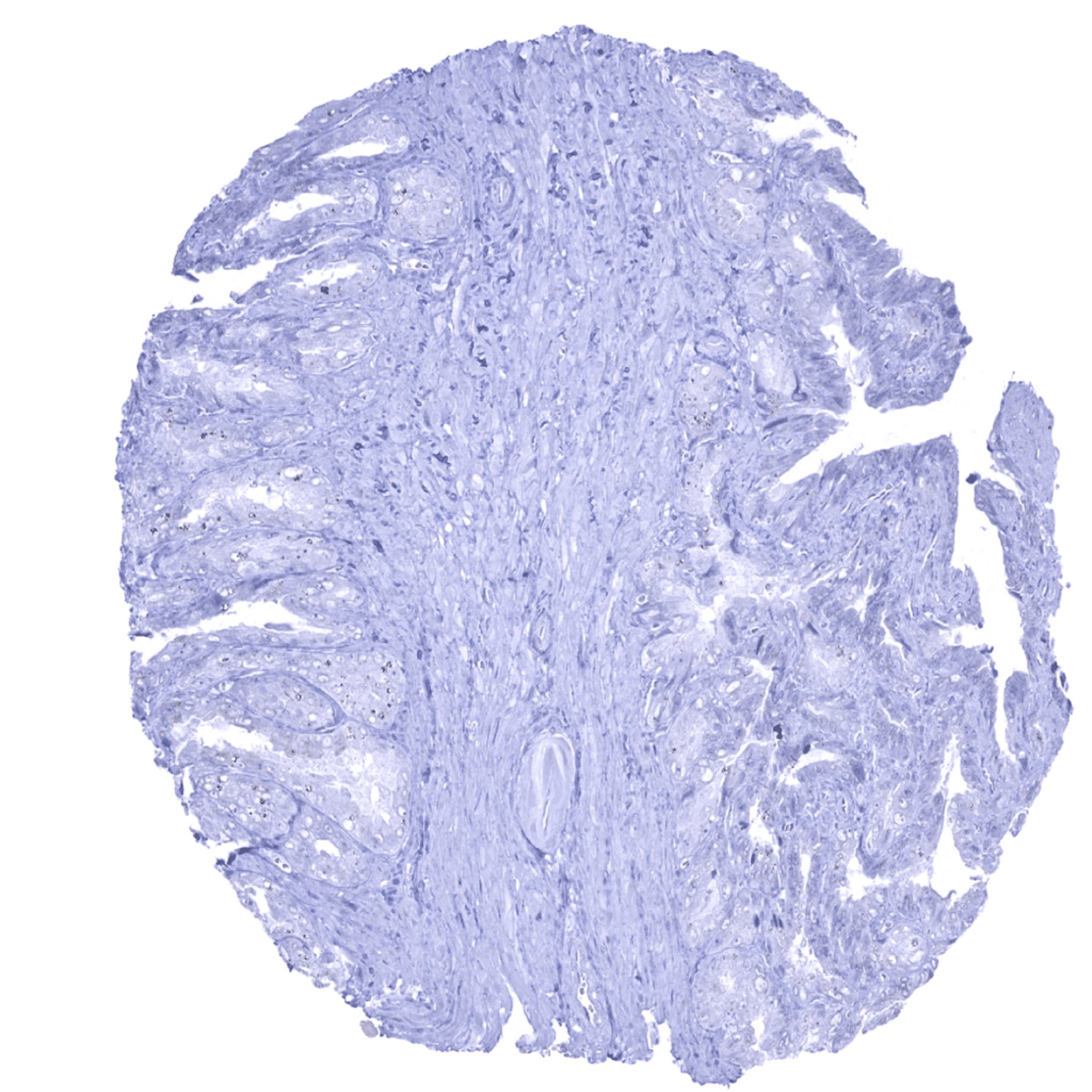
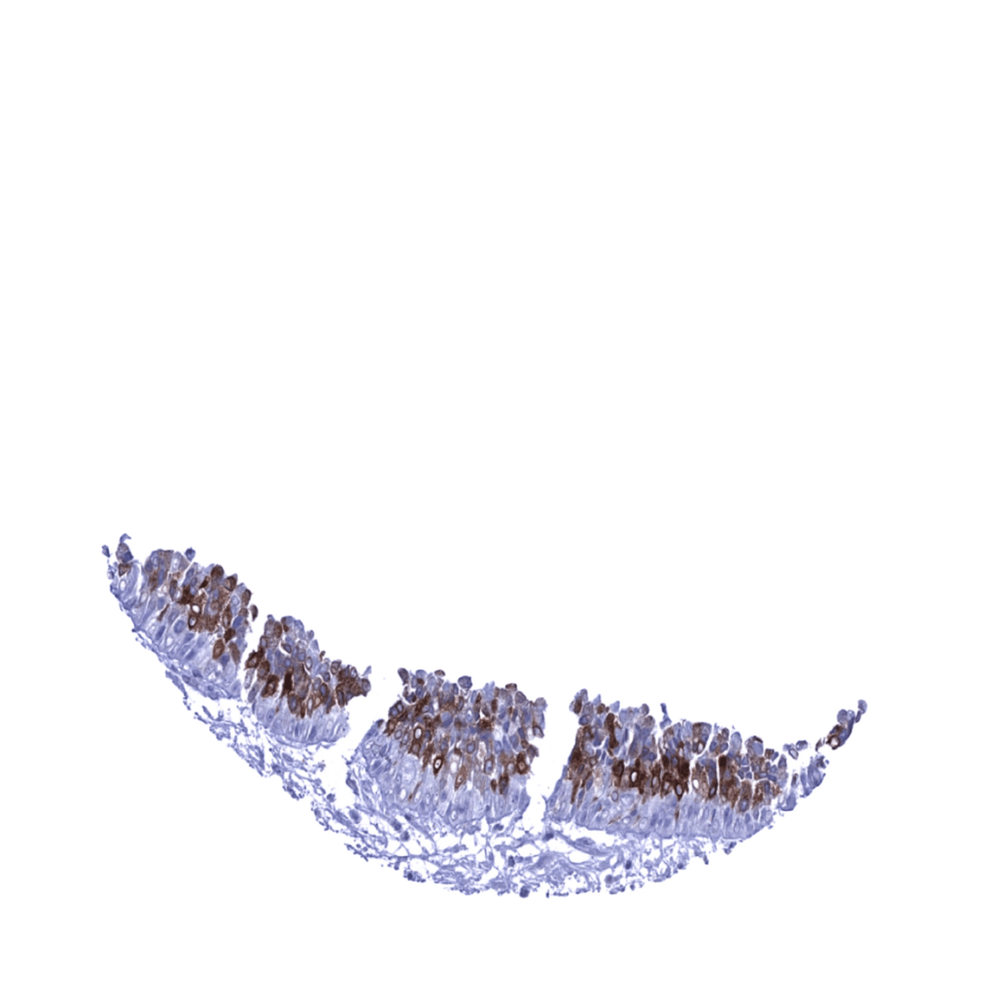
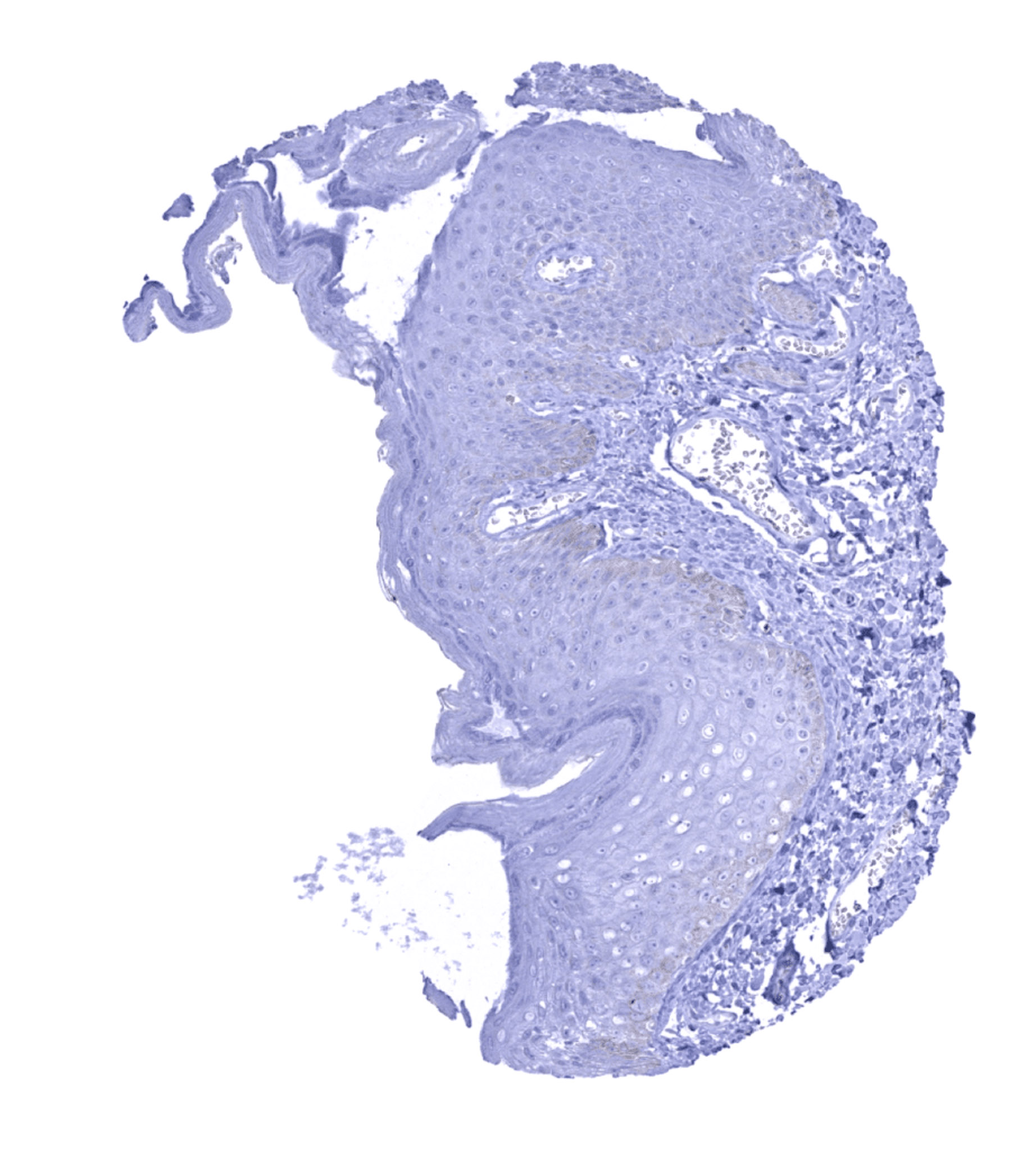
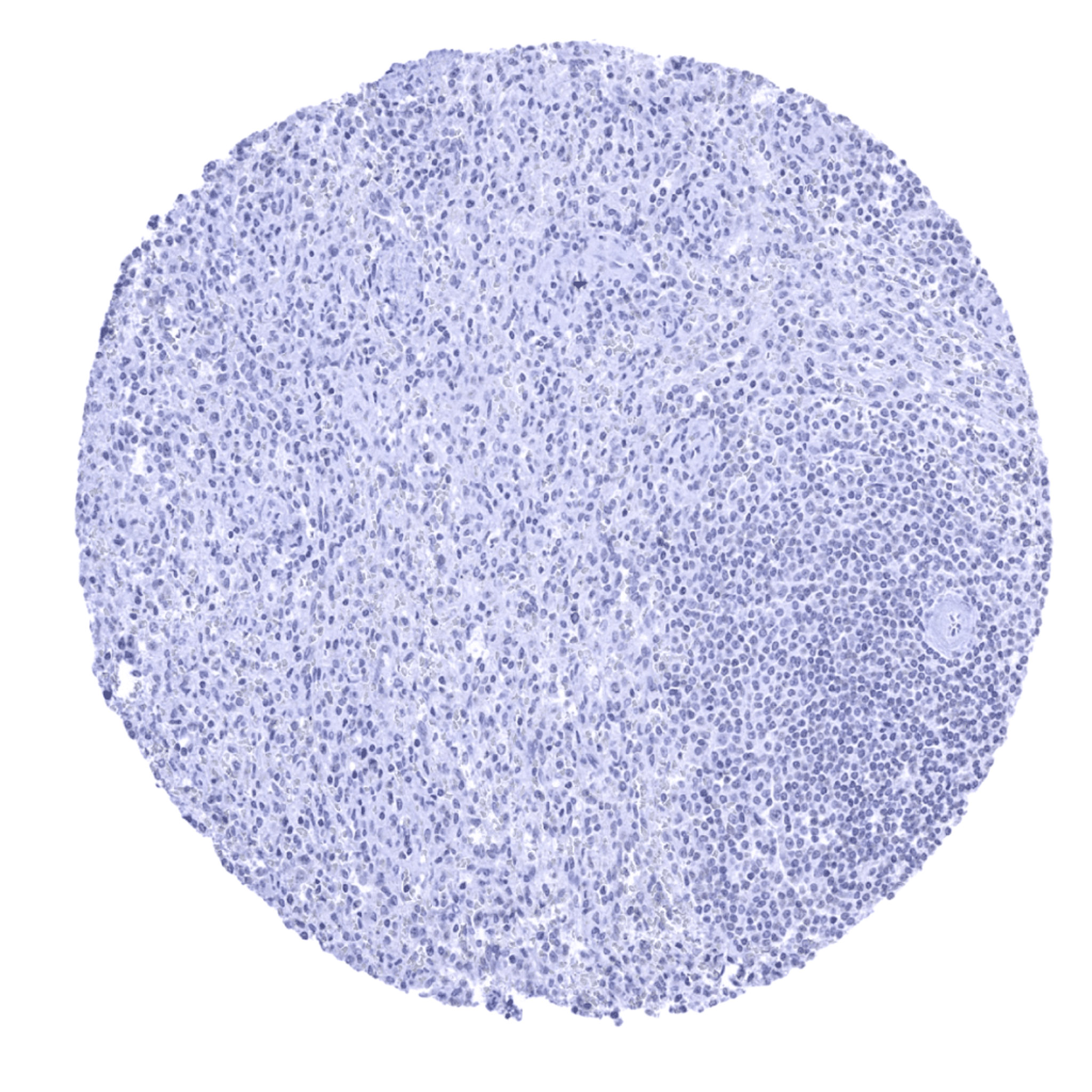
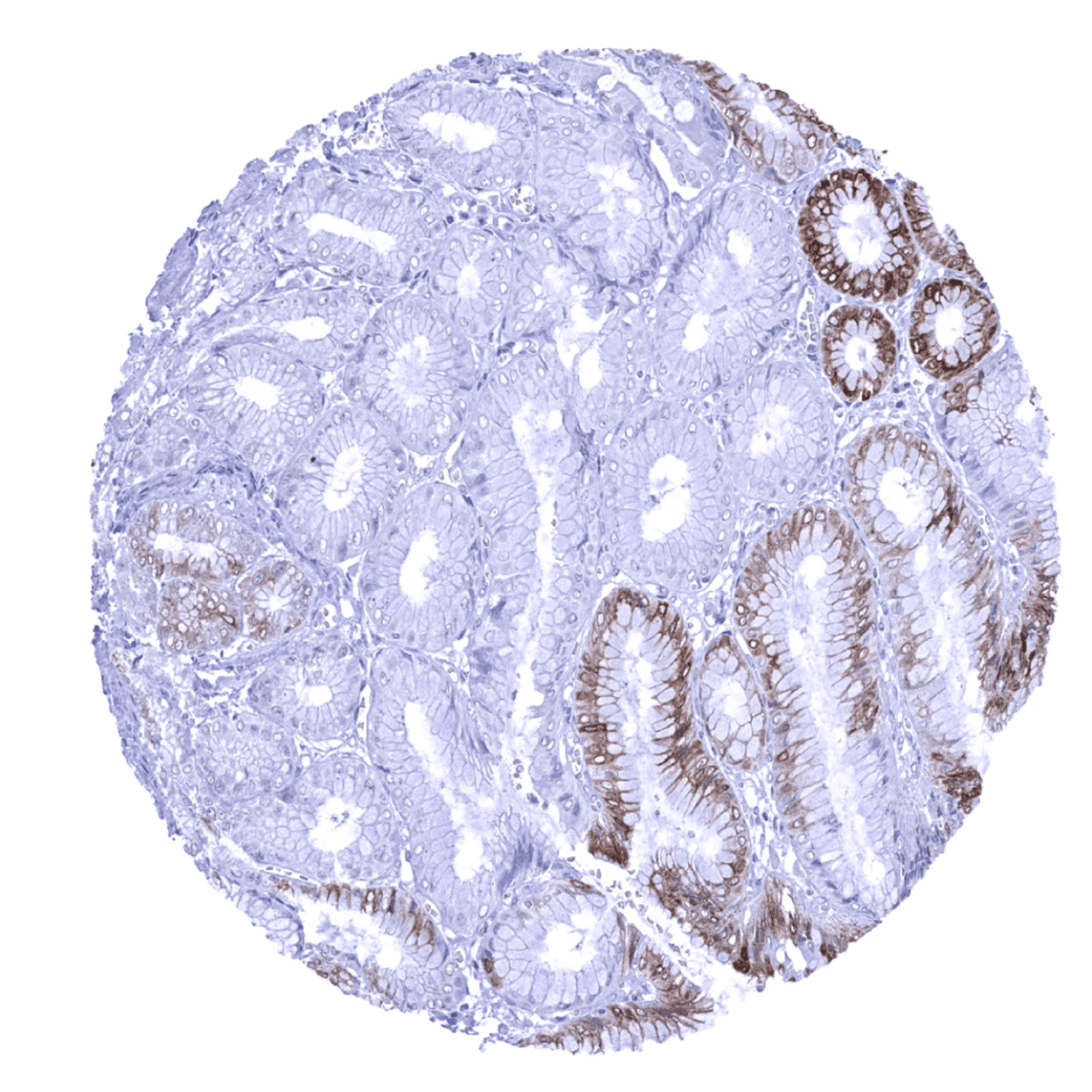
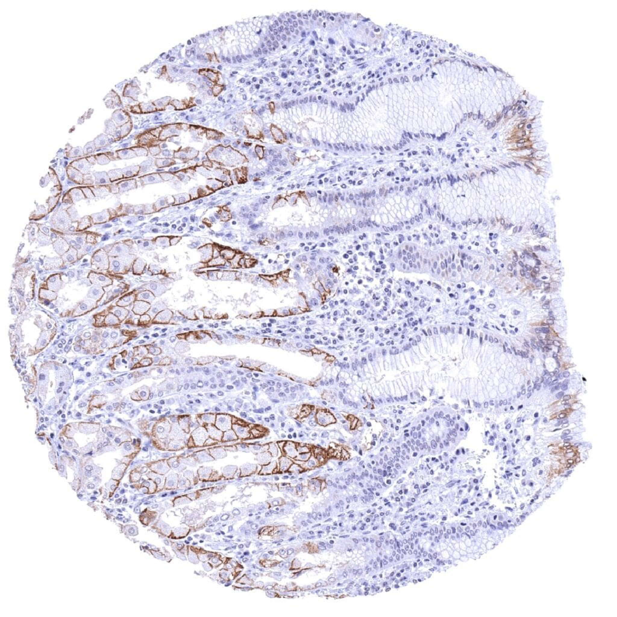
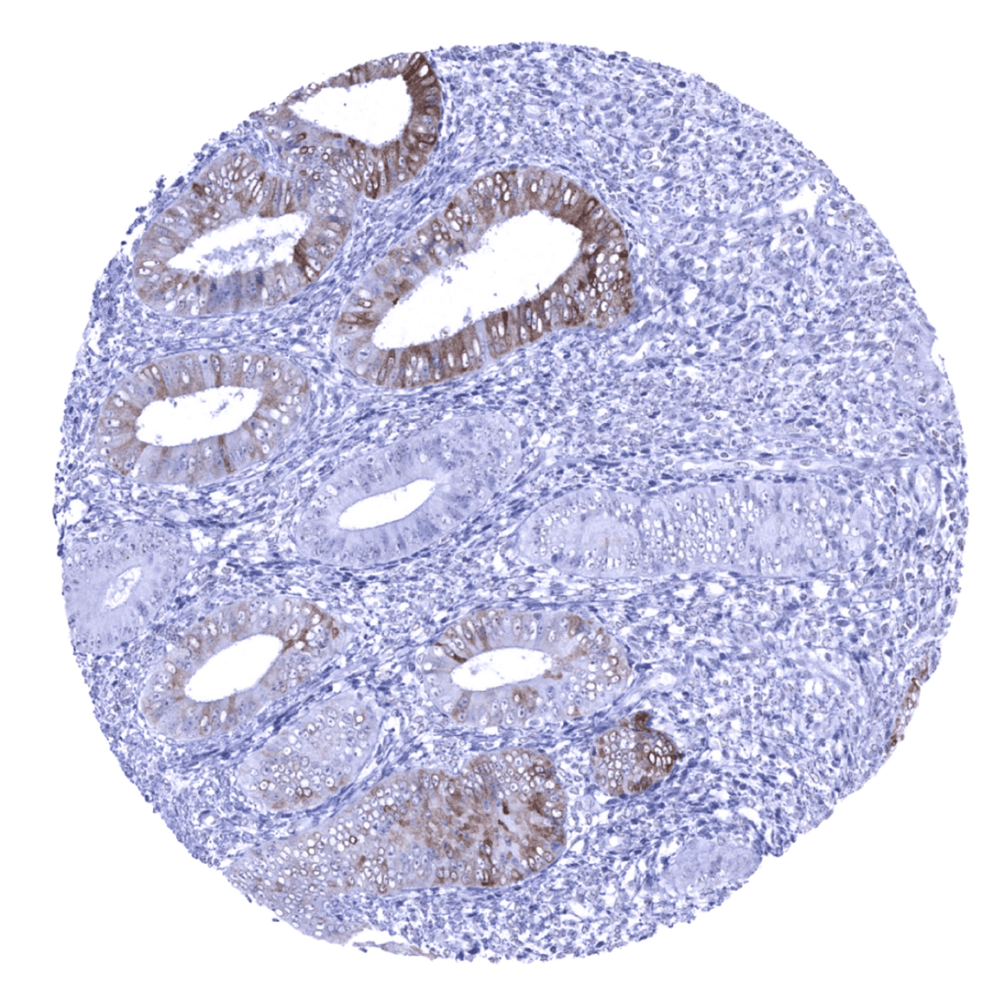
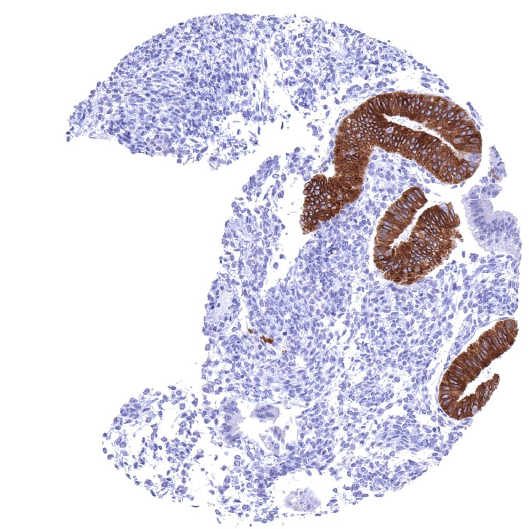
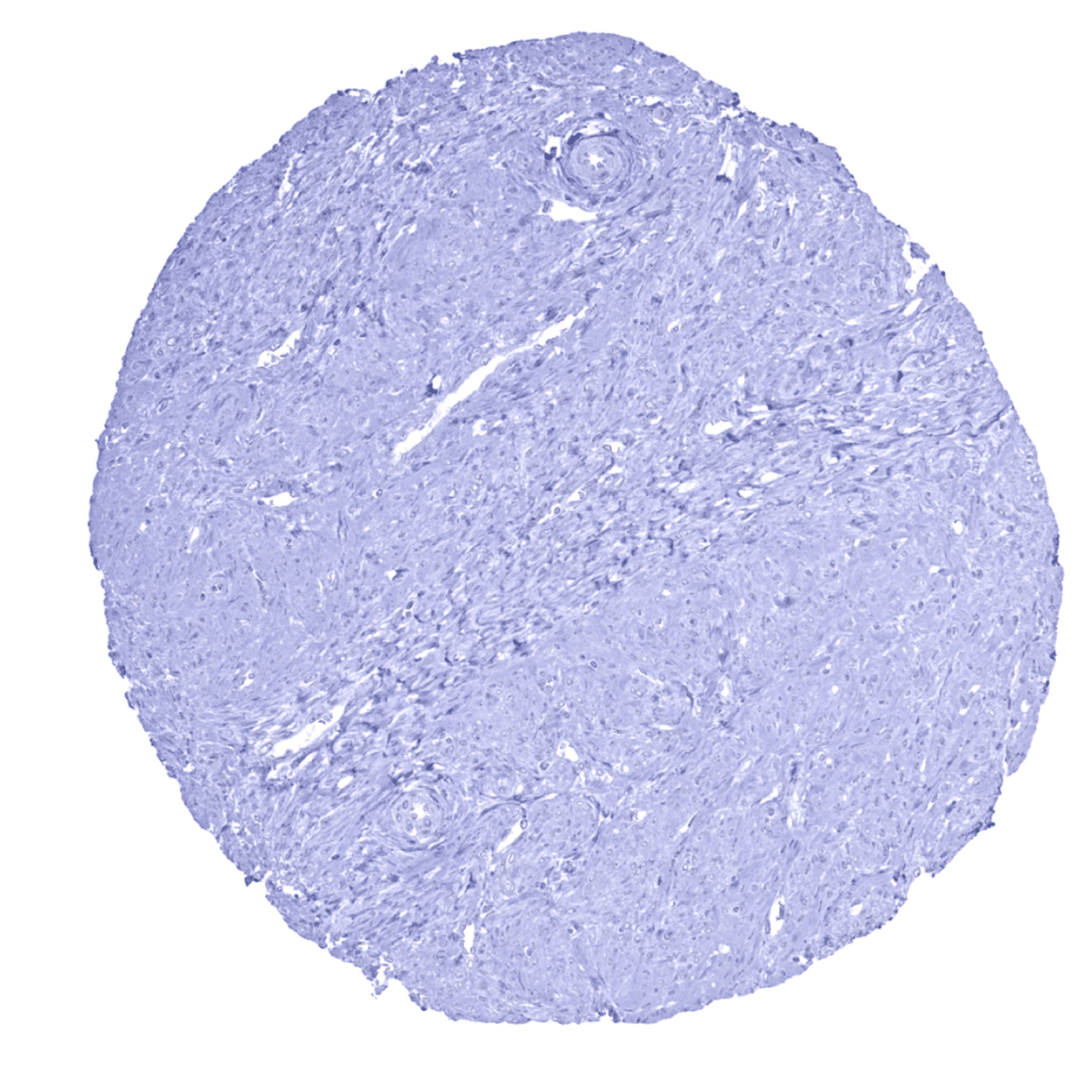
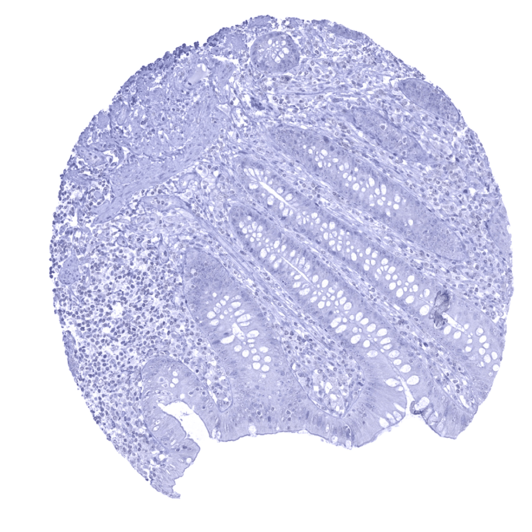
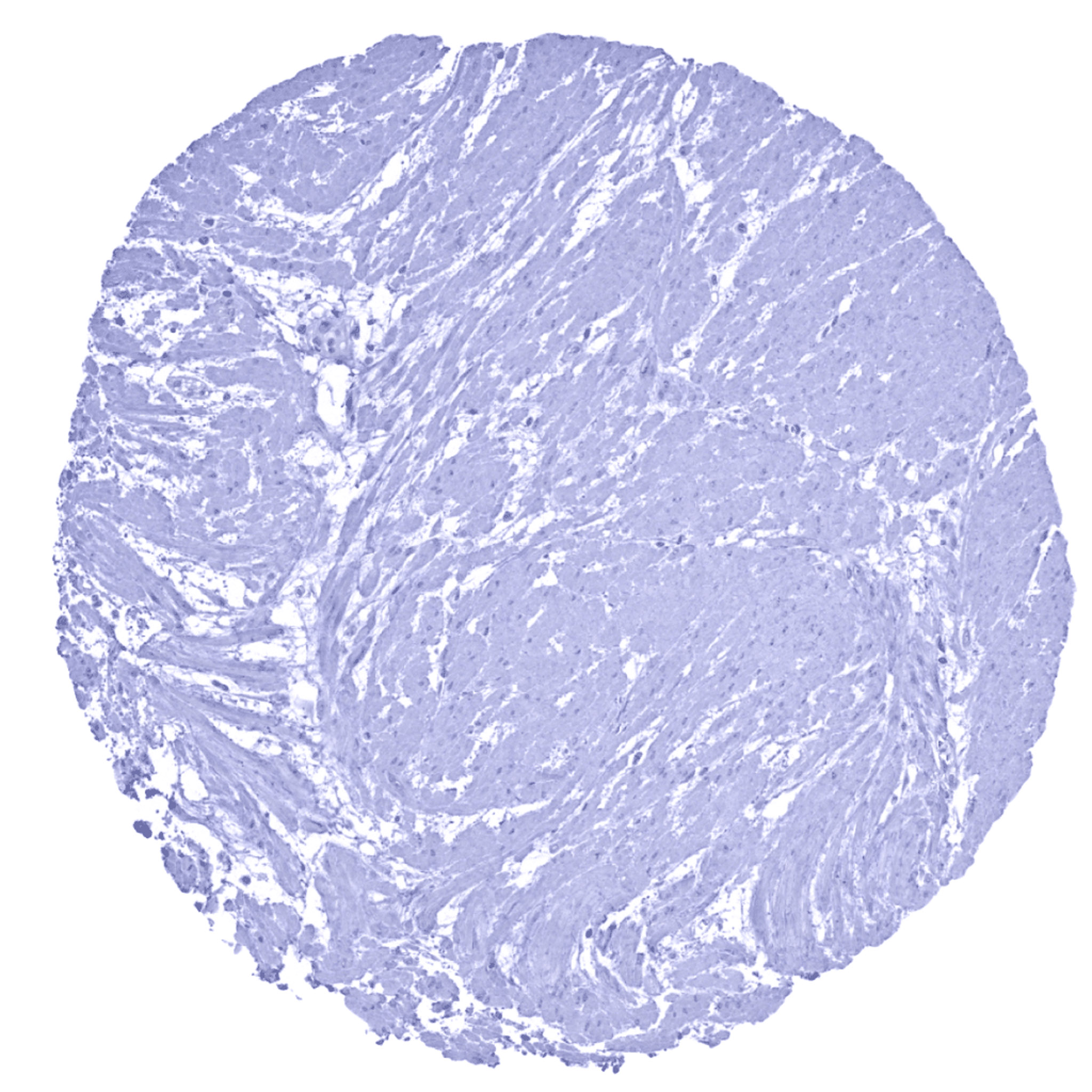
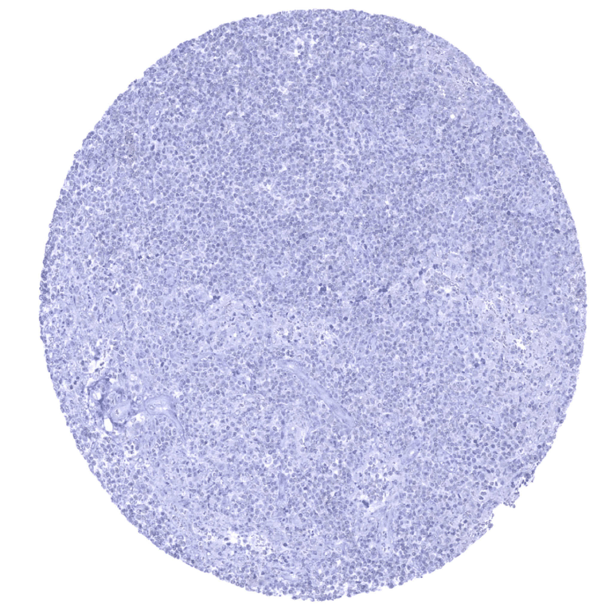
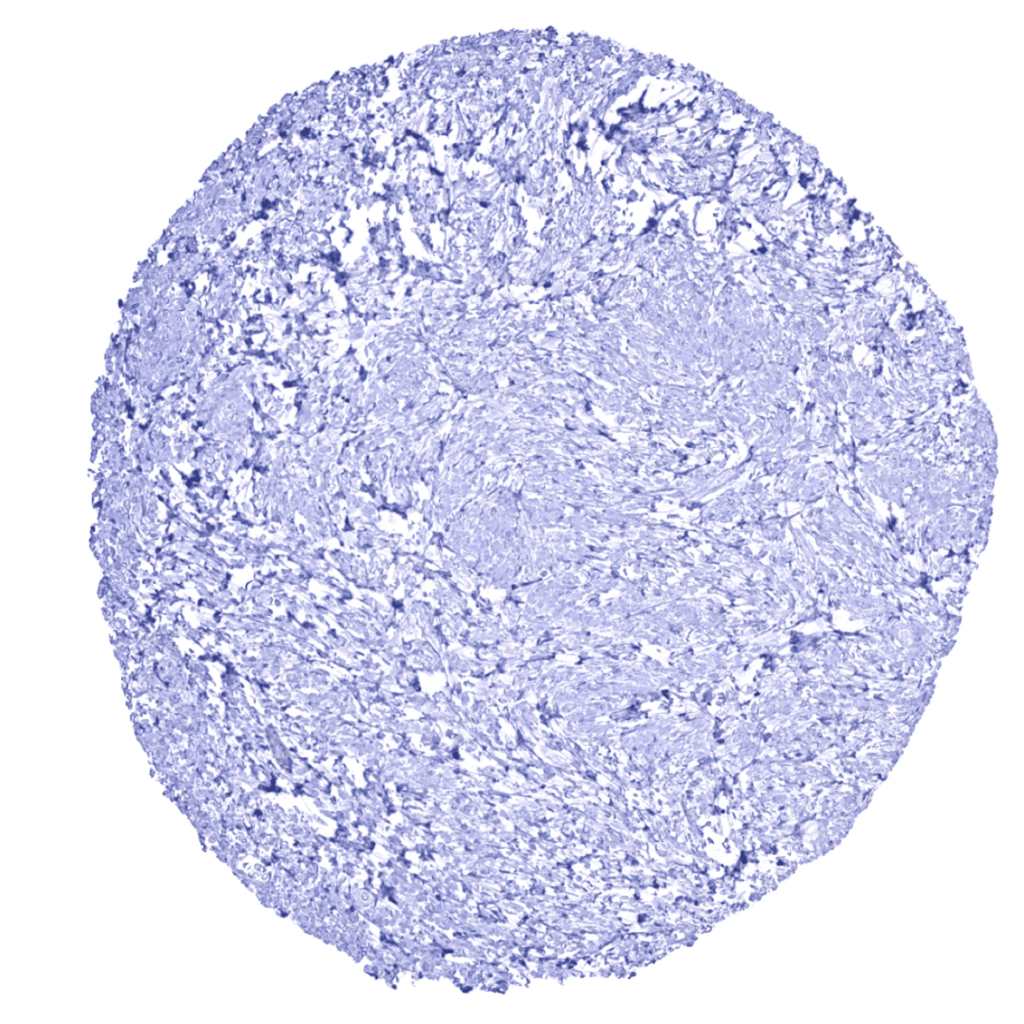
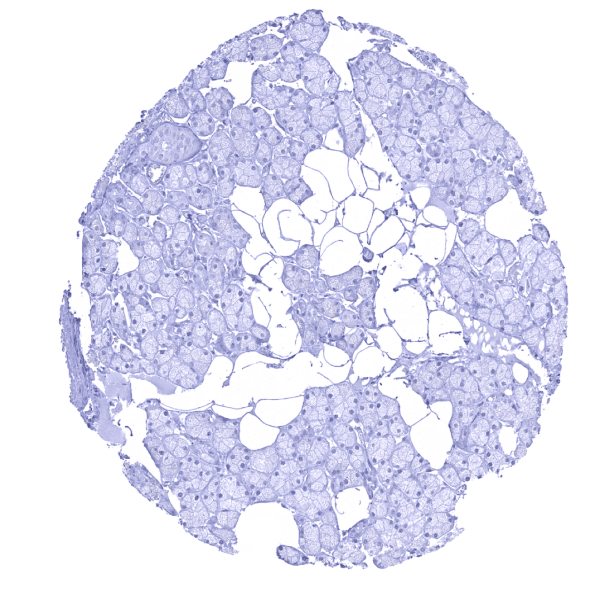
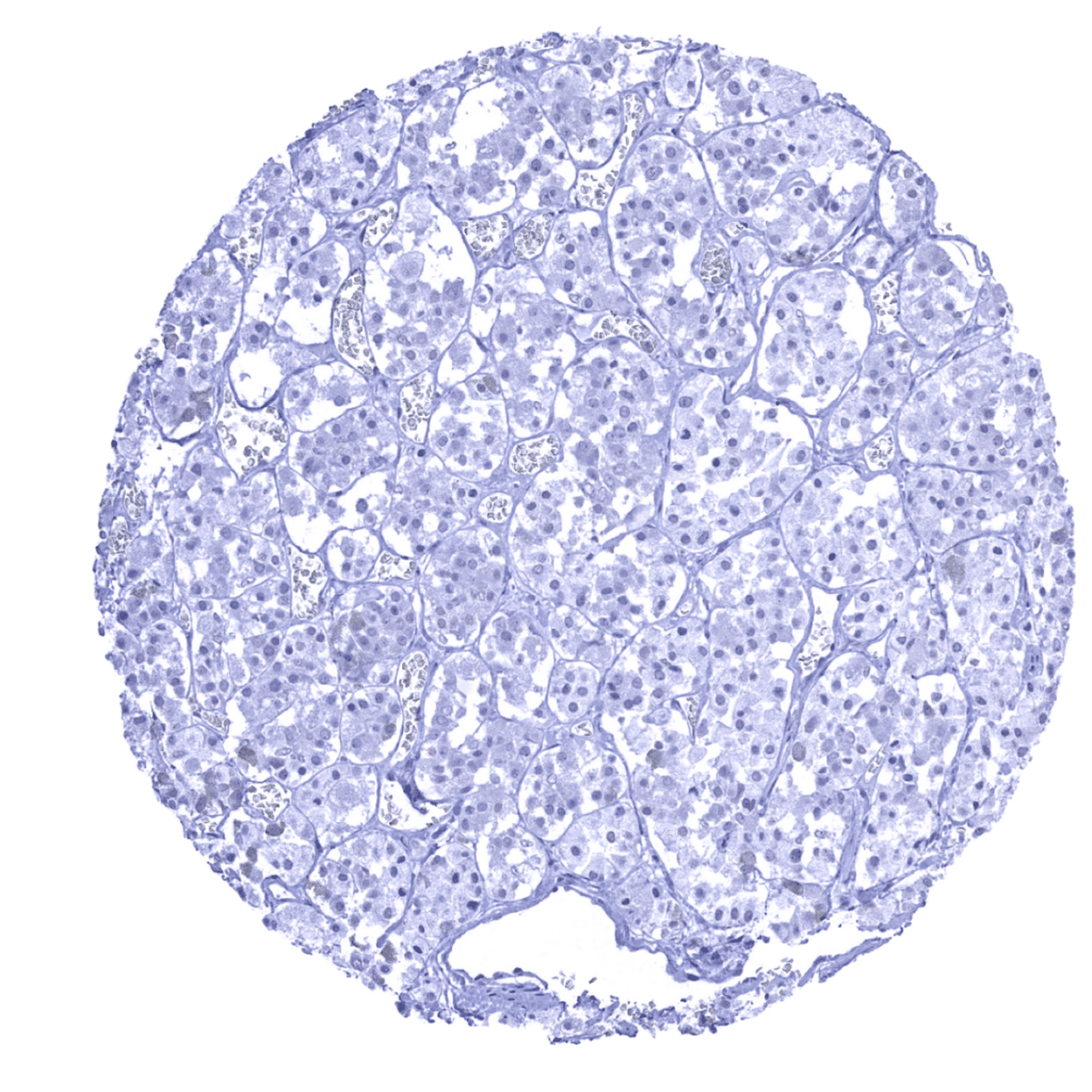
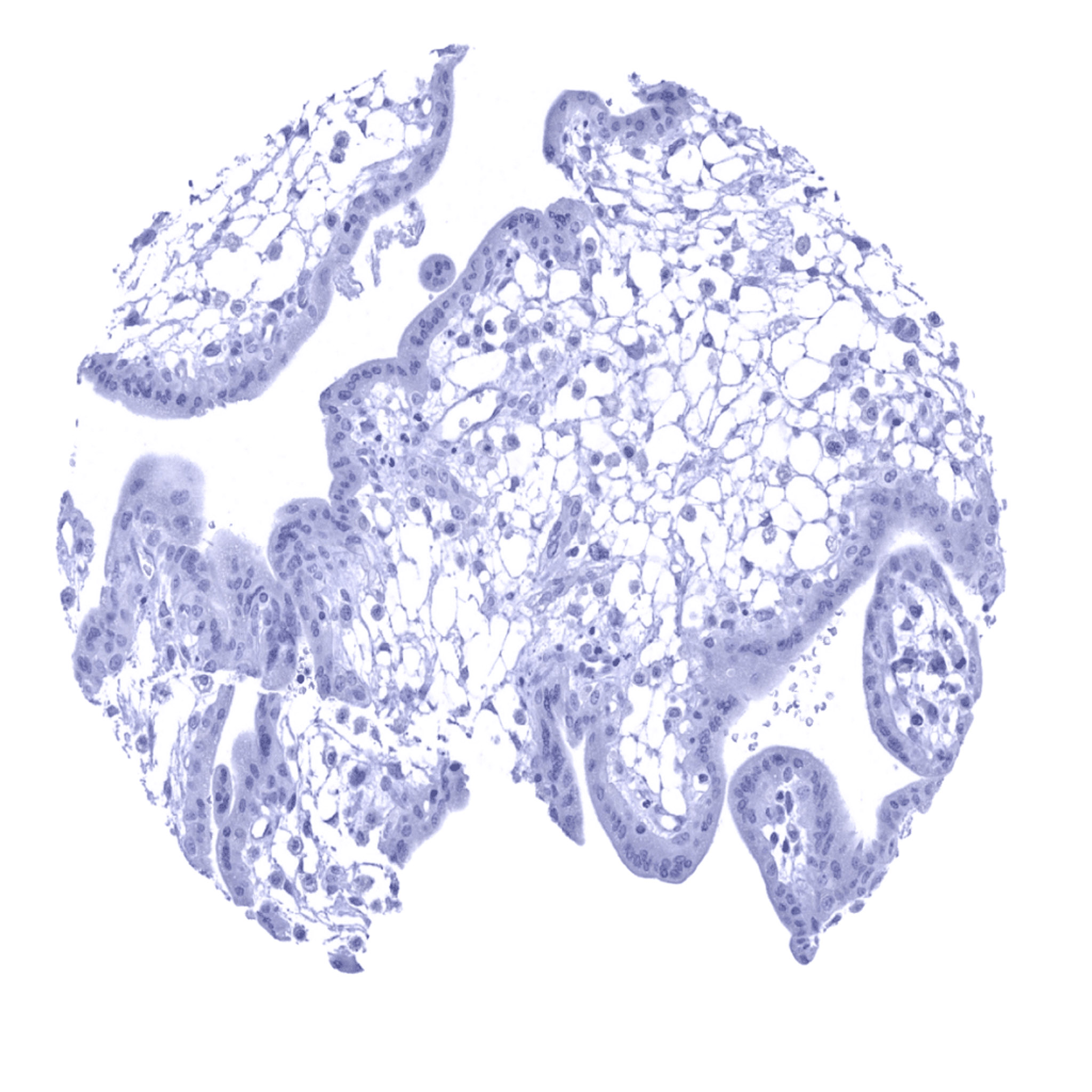
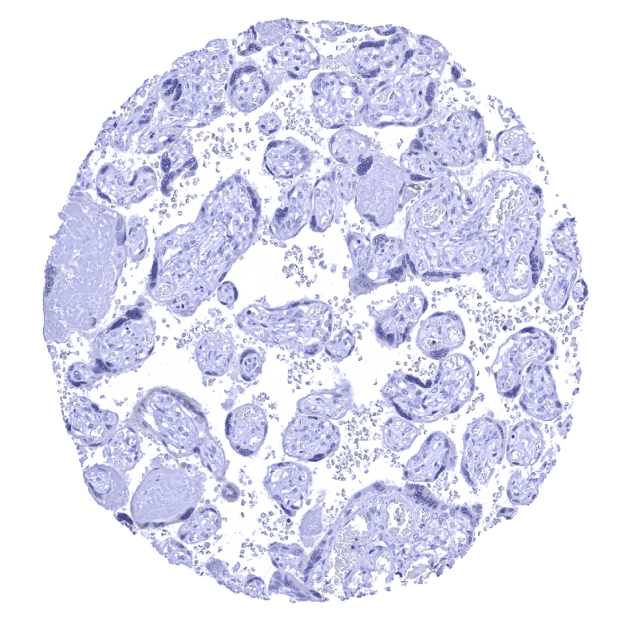
UPK1B antibody [MSVA-734M] HistoMAXTM


Go to Cancer Tissue Gallery
  
 Go back to product page

Go to HistoMAX page


Adrenal gland

Aorta, media

Appendix, mucosa

Appendix, muscular wall


Bone marrow

Breast

Bronchus, mucosa- Moderate to strong UPK1B staining of a fraction of cells

Cerebellum (molecular layer, Purkinje cell layer, granule cell layer, white matter)


Cerebellum (white matter)

Cerebrum, grey matter

Cerebrum, white matter

Colon descendens, mucosa


Colon descendens, muscular wall

Duodenum, Brunner gland

Duodenum, mucosa

Epididymis


Esophagus, squamous epithelium – Scattered UKP1b positive cells can be seen in the non-keratinizing squamous epithelium of the esophagus

Fallopian tube, mucosa – A weak to moderate UPK1B immunostaining occurs in a fraction of epithelial cells of the fallopian tube.

Fat

Gallbladder, epithelium – A weak to moderate cytoplasmic and membranous UPK1B immunostaining is seen in the gallbladder epithelium.


Heart

Ileum, mucosa

Kidney, cortex- UPK1B staining is largely absent. One cell of the parietal cell layer of the capsule of Bowman in the kidney stains positive

Kidney, medulla


Kidney, pelvis (urothelium) – Most urothelial cells in the renal pelvis show a strong UPK1B immunostaining

Liver – A moderate cytoplasmic and membranous UPK1B immunostaining is seen in intrahepatic bile ducts.

Lung

Lymph node


Ovary, stroma

Pancreas – A weak to moderate UPK1B staining occurs in intercalated ducts

Parathyroid gland

Parotid gland


Pituitary gland, anterior lobe

Pituitary gland, posterior lobe

Placenta (amnion)- Strong UPK1B staining of all amnion cells

Placenta (amnion) – Strong UPK1B immunostaining in amnion cells of the placenta.


Placenta (chorion)- Strong cytoplasmic and membranous UPK1B staining of chorion cells

Pregnant uterus (decidua)

Placenta, early – A weak to moderate UPK1B immunostaining can be found in a fraction of syncytiotrophoblast cells of the placenta.

Placenta, early


Placenta, mature

Prostate

Rectum, mucosa

Seminal vesicle


Sinus paranasales – A fraction of respiratory epithelium cells (mostly goblet cells) show a moderate to strong UPK1B immunostaining.

Skin – UPK1B immunostaining is absent in keratinizing squamous epithelium of the skin.

Spleen

Stomach, antrum – A moderate UPK1B immunostaining is seen in the surface epithelial cell layer of the stomach.


Stomach, corpus- Membranous UPK1B staining of surface epithelial and parietal cells

Testis

Thymus – Some elements of corpuscles of Hassall's are UPK1B positive

Thyroid gland


Tongue, muscle

Tonsil – Some layers of tonsil crypt epithelium show a moderate to strong UPK1B immunostaining.

Tonsil surface – Strong UPK1B staining in suprabasal epithelial cells

Tonsil surface – Absence of UPK1B staining


Urinary bladder, muscular wall

Urinary bladder, urothelium – Strong UPK1B staining in suprabasal urothelial cells

Uterus, ectocervix – UPK1B staining is lacking

Uterus, endocervix – In this sample, endocervical epithelial cells show a weak to moderate UPK1B positivity.


Uterus, endocervix

Uterus, endometrium (proliferation) – A weak to moderate UPK1B immunostaining is seen in a fraction of endometrial glands.

Uterus, endometrium – Strong UPK1B staining of some glands while others are completely negative

Uterus, myometrium


| TOP |