Normal Tissue Gallery- GTX640564

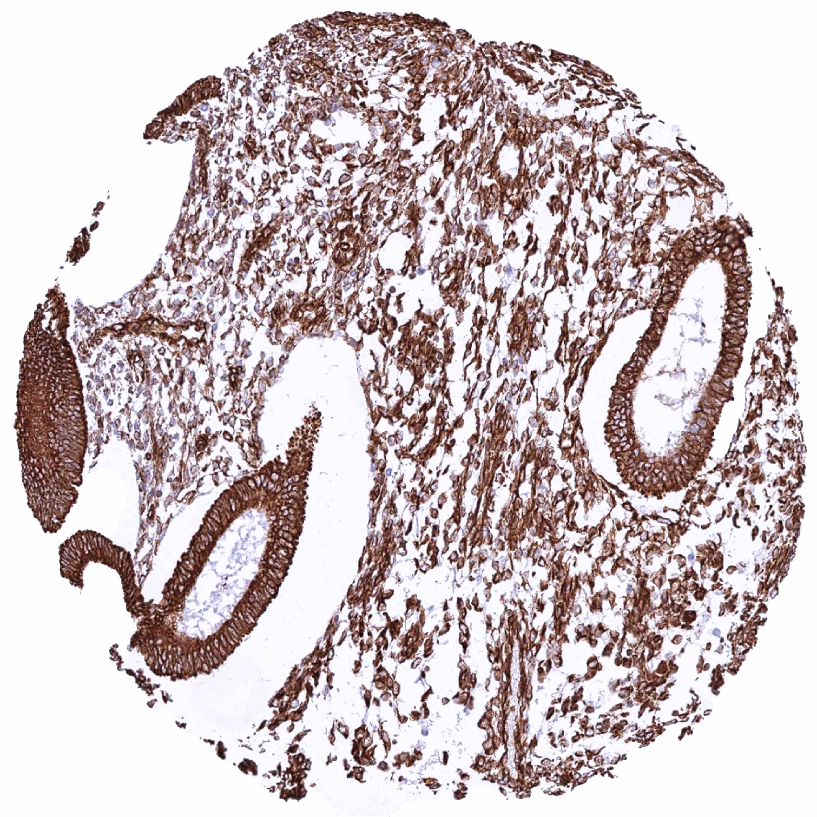
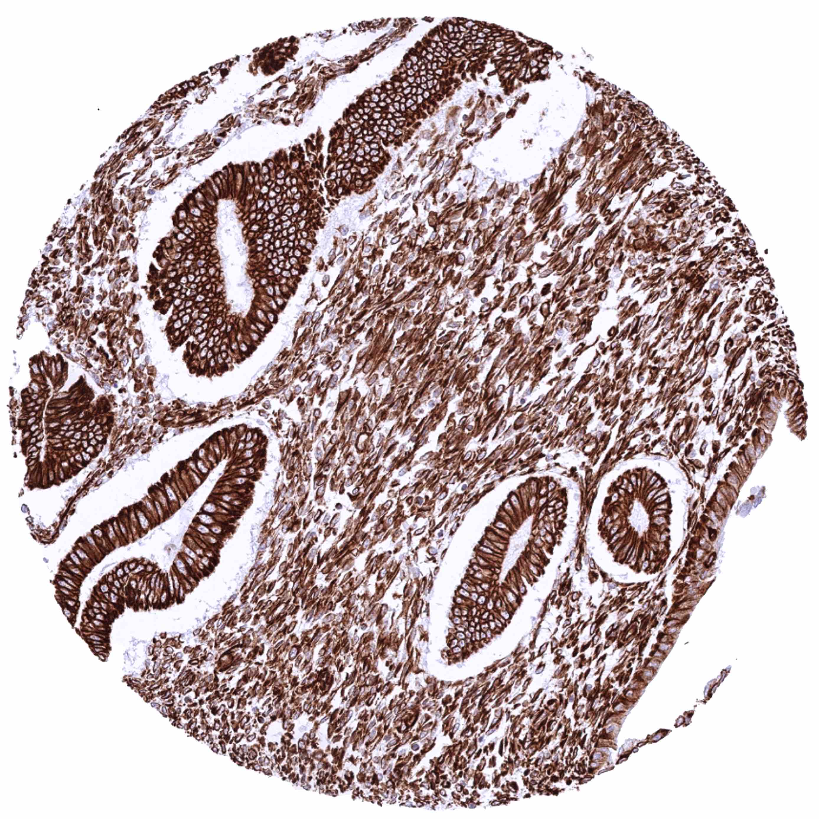
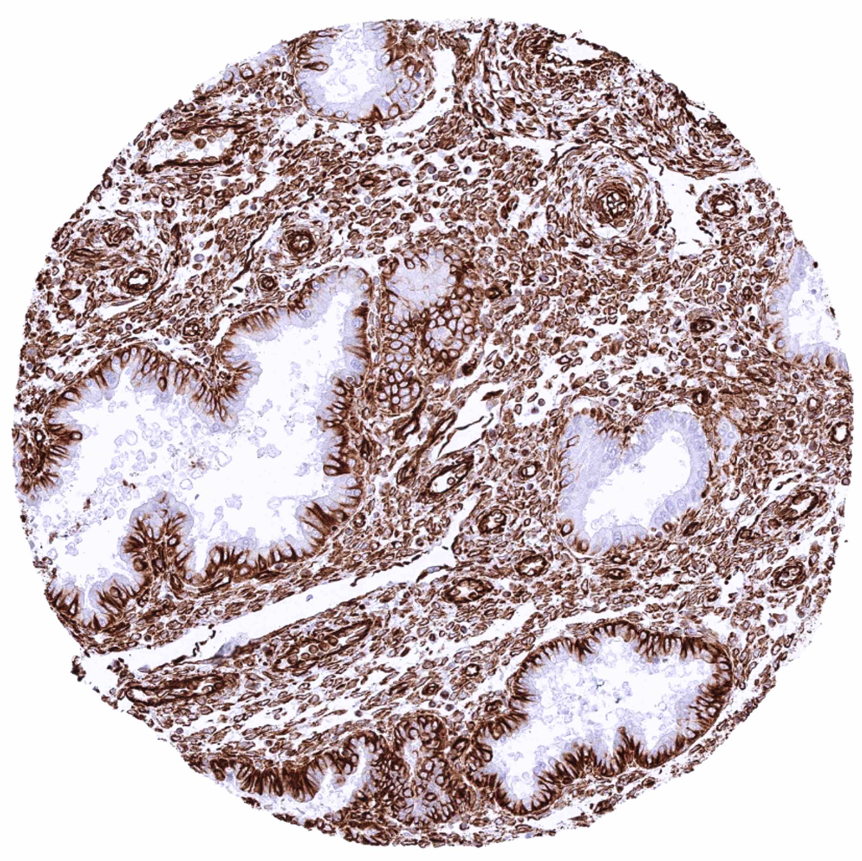
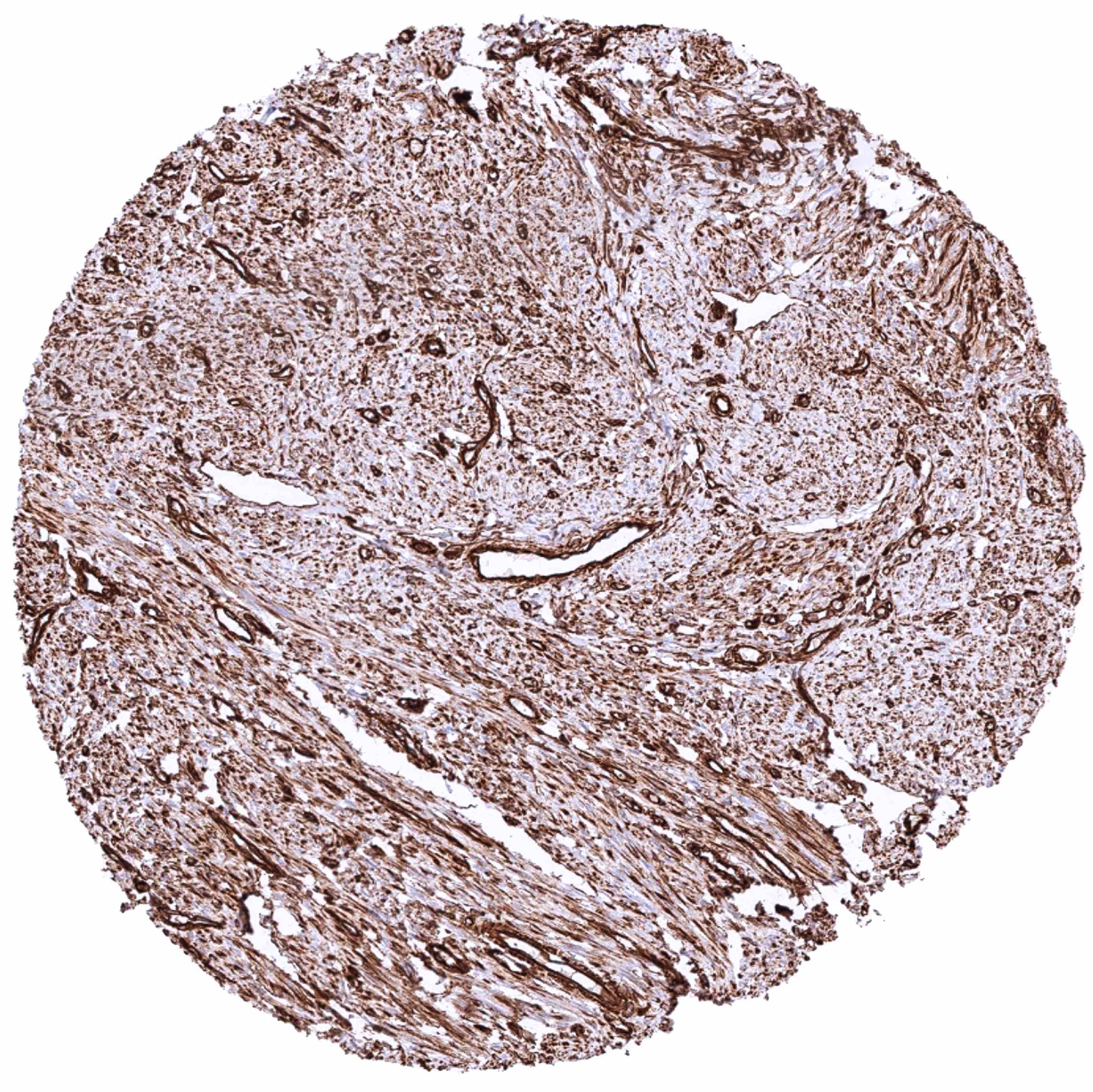
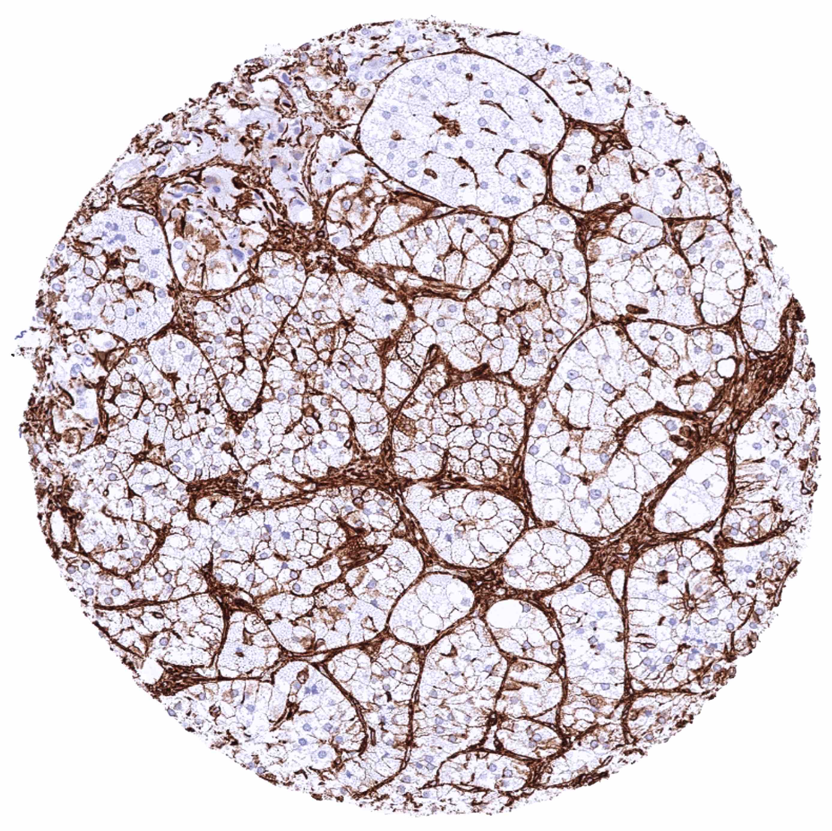
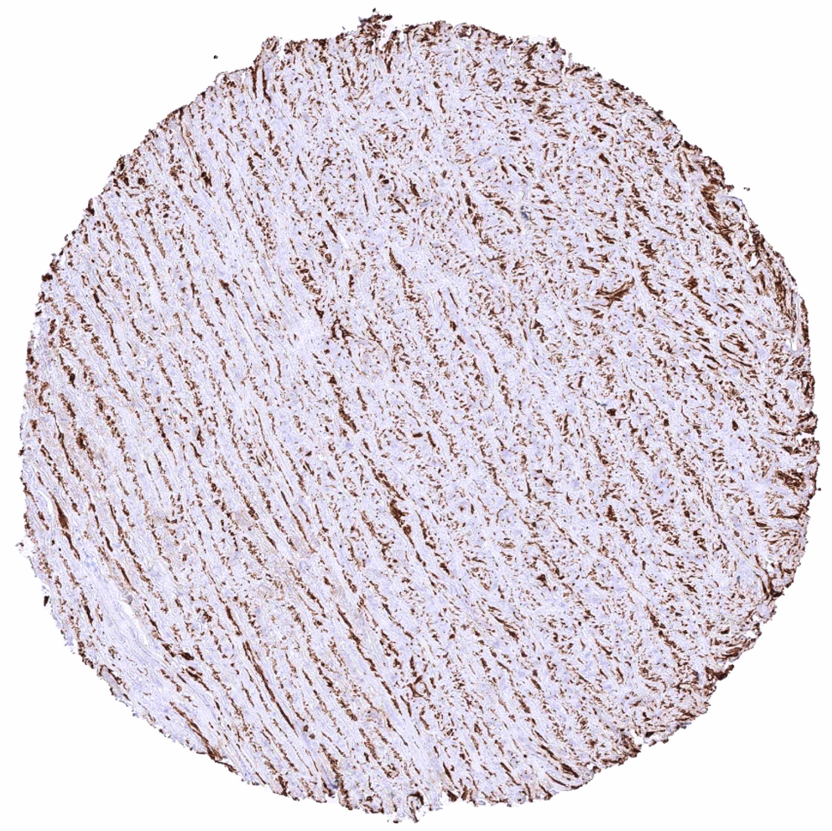
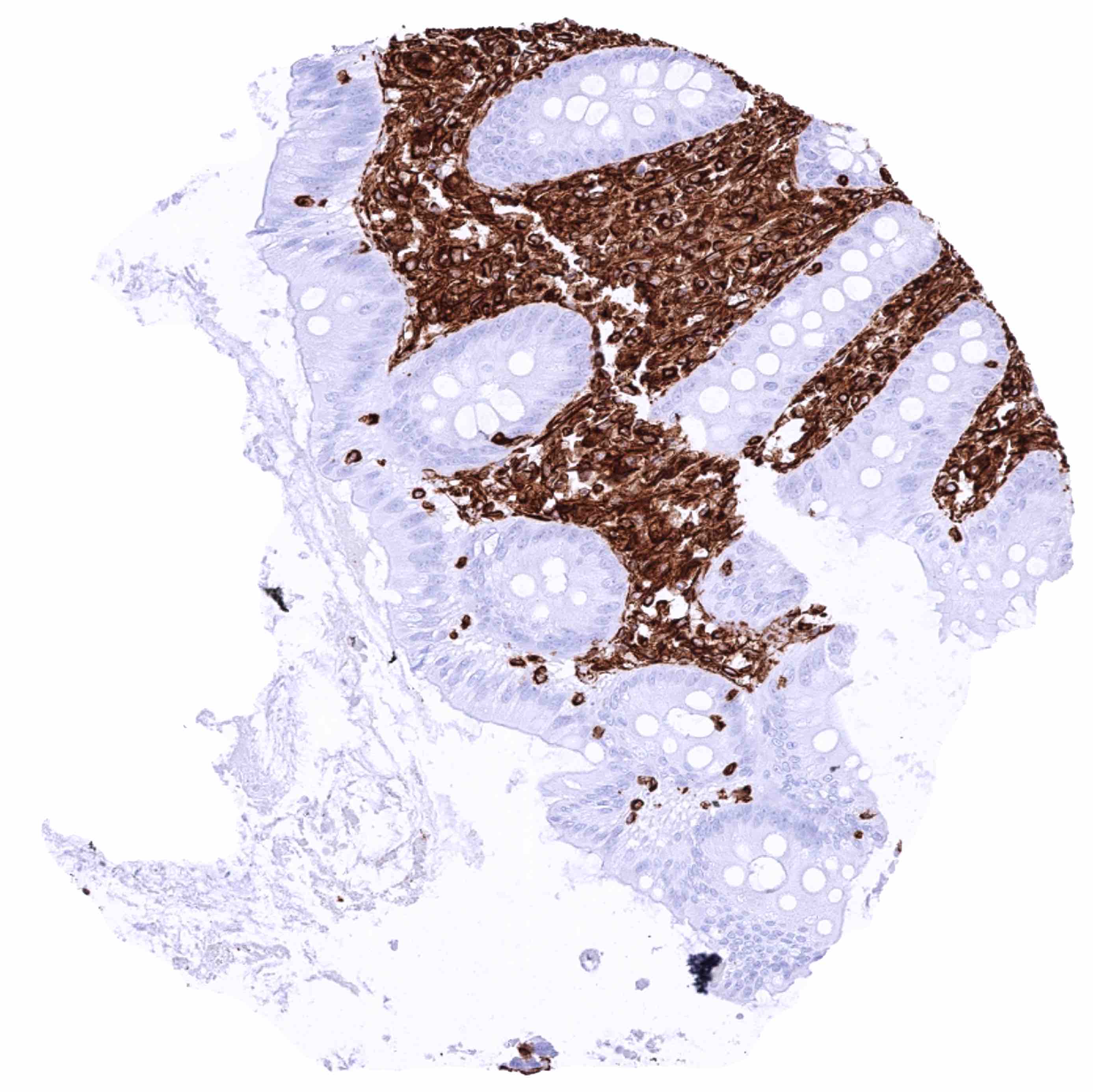
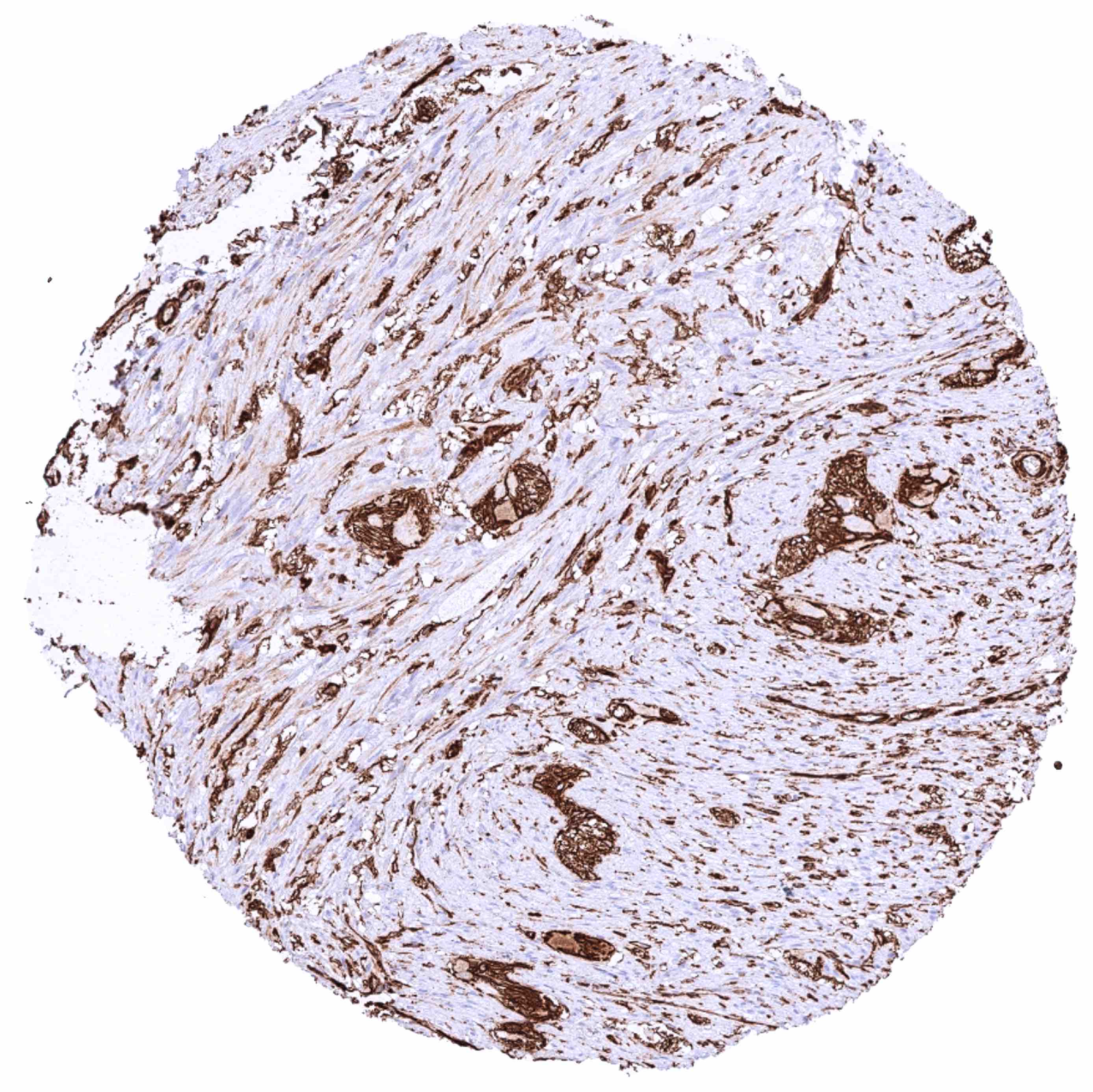
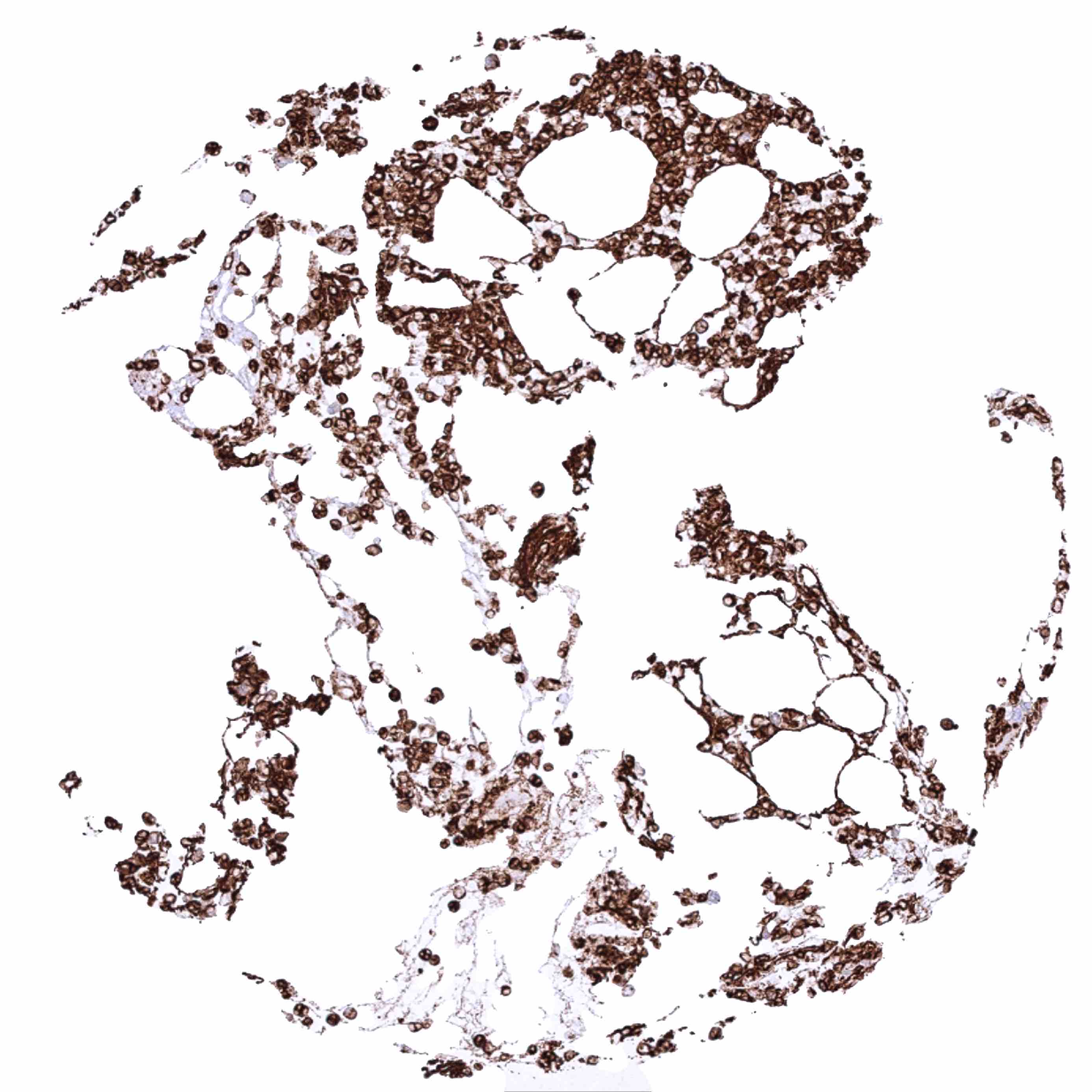
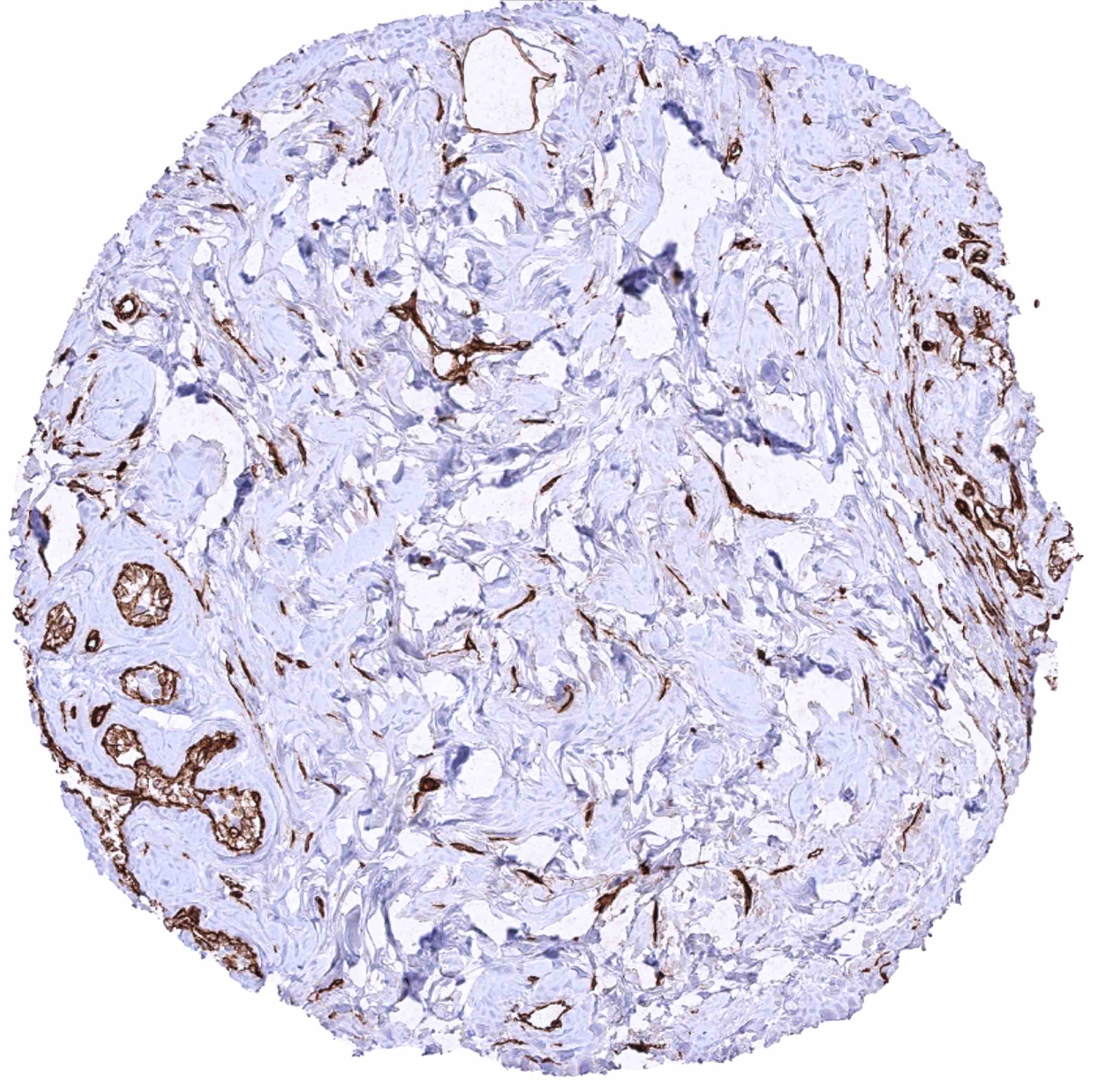
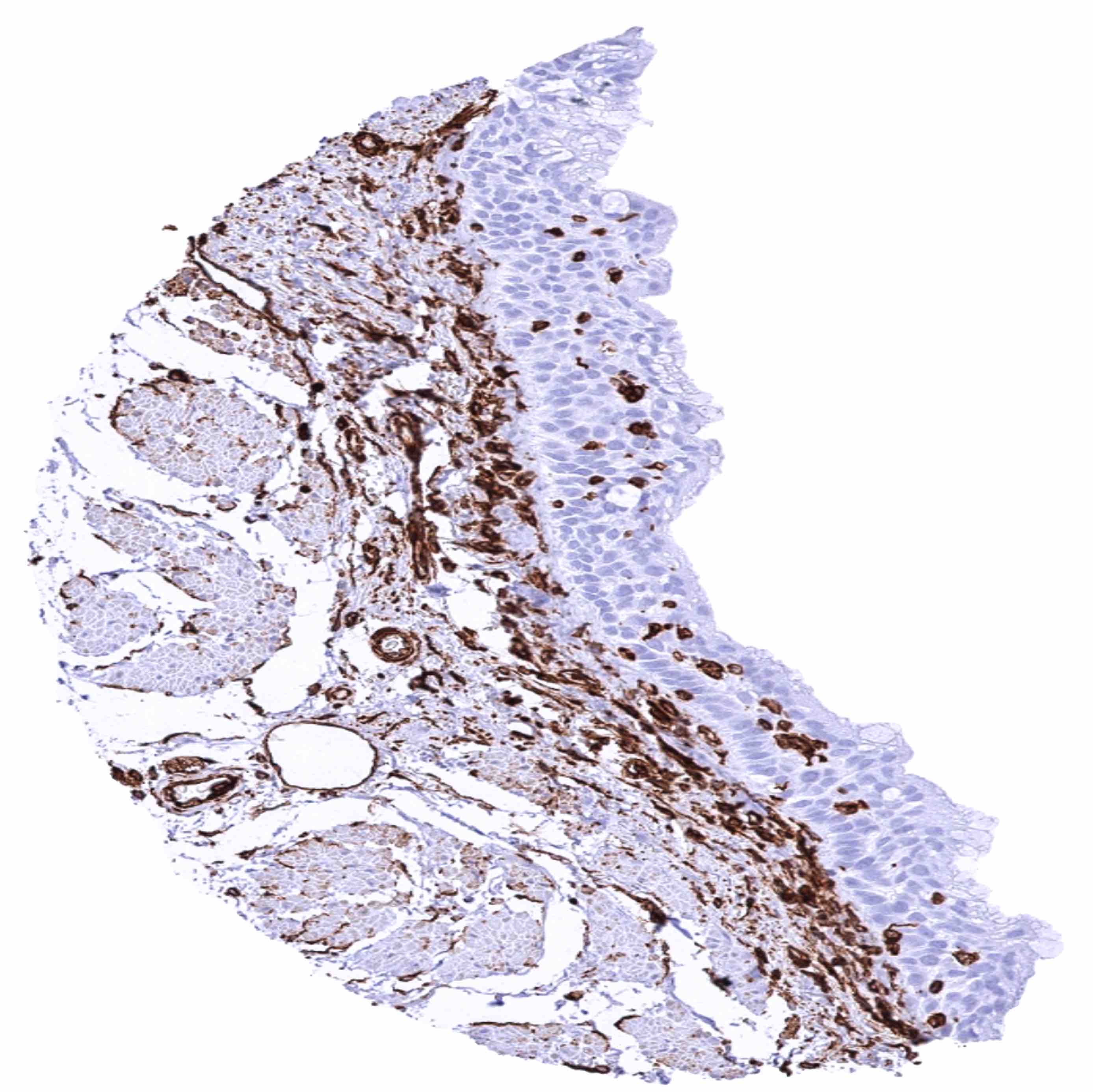
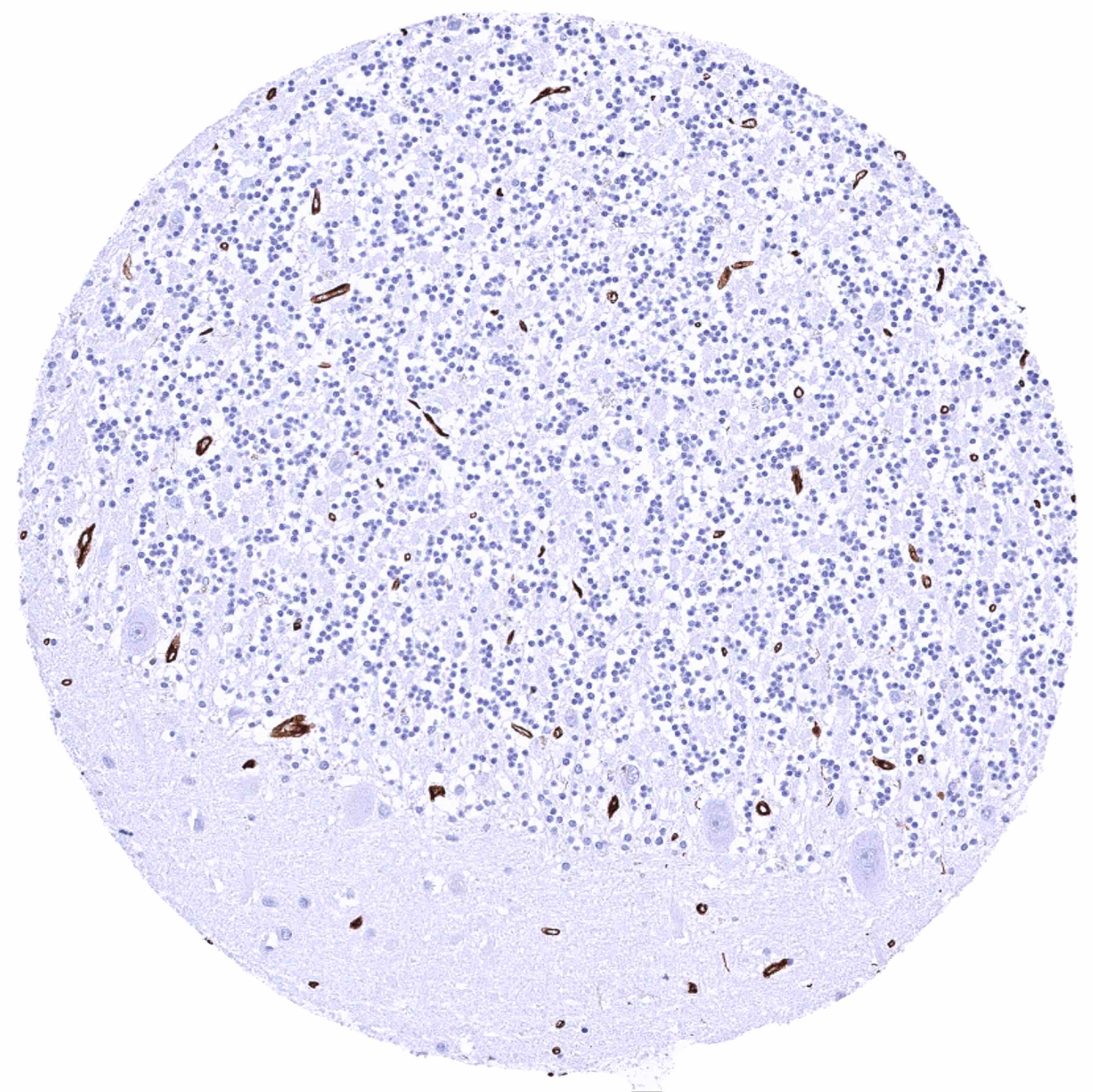
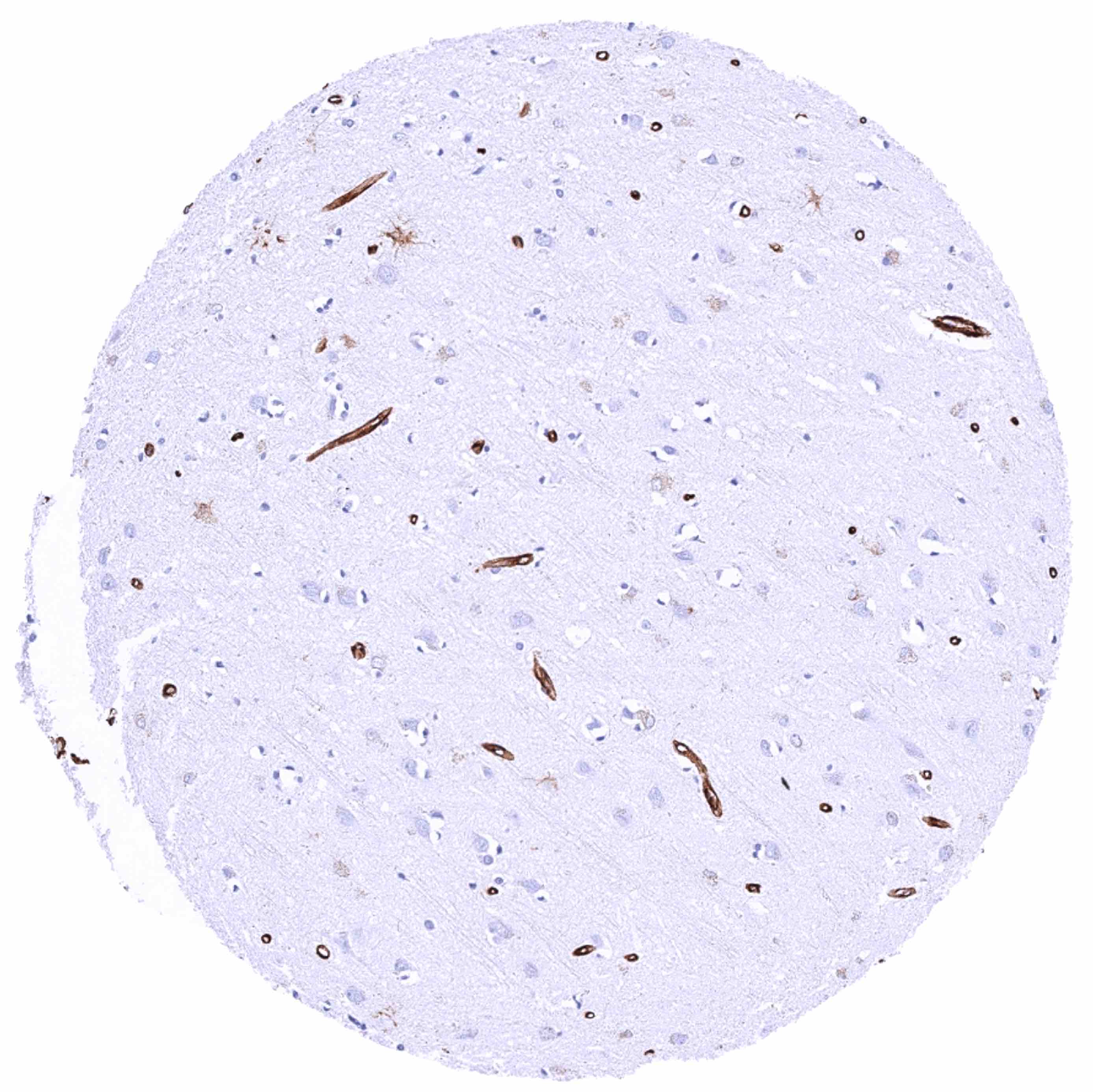
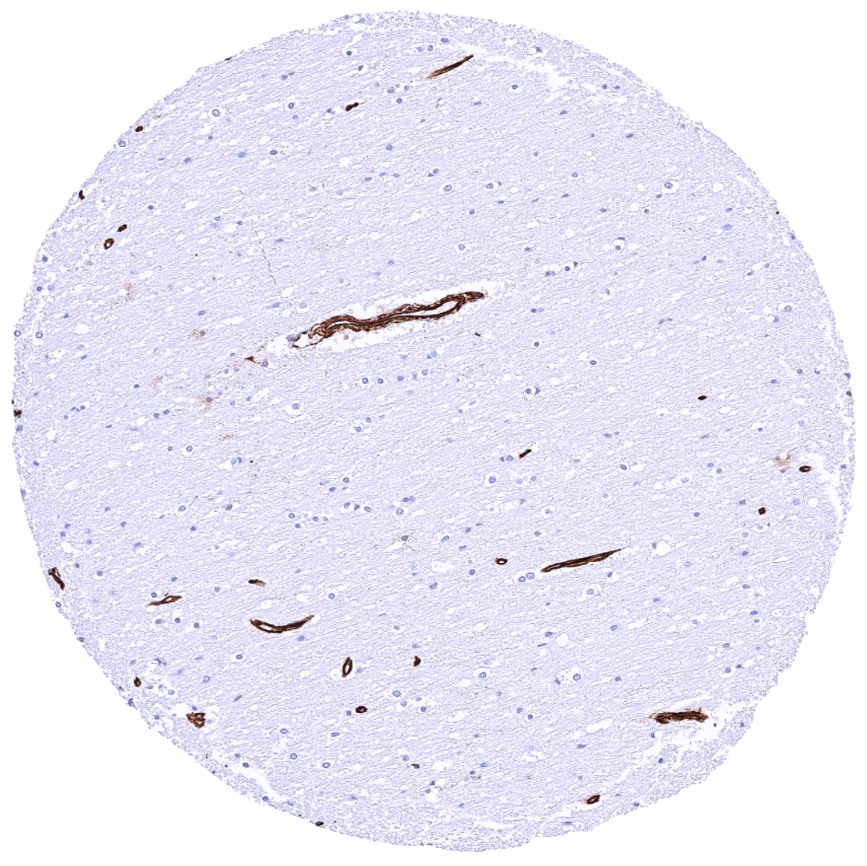
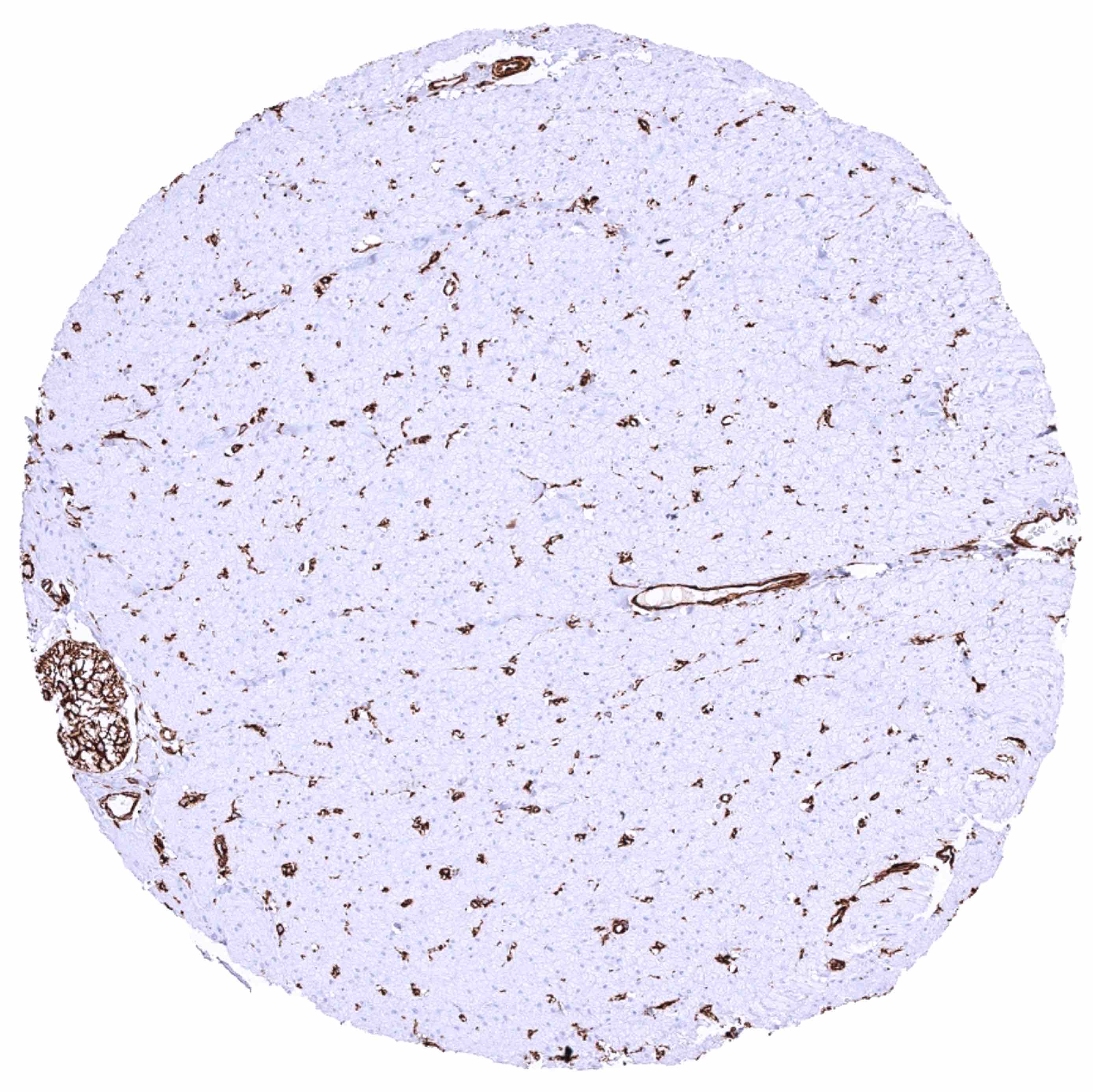
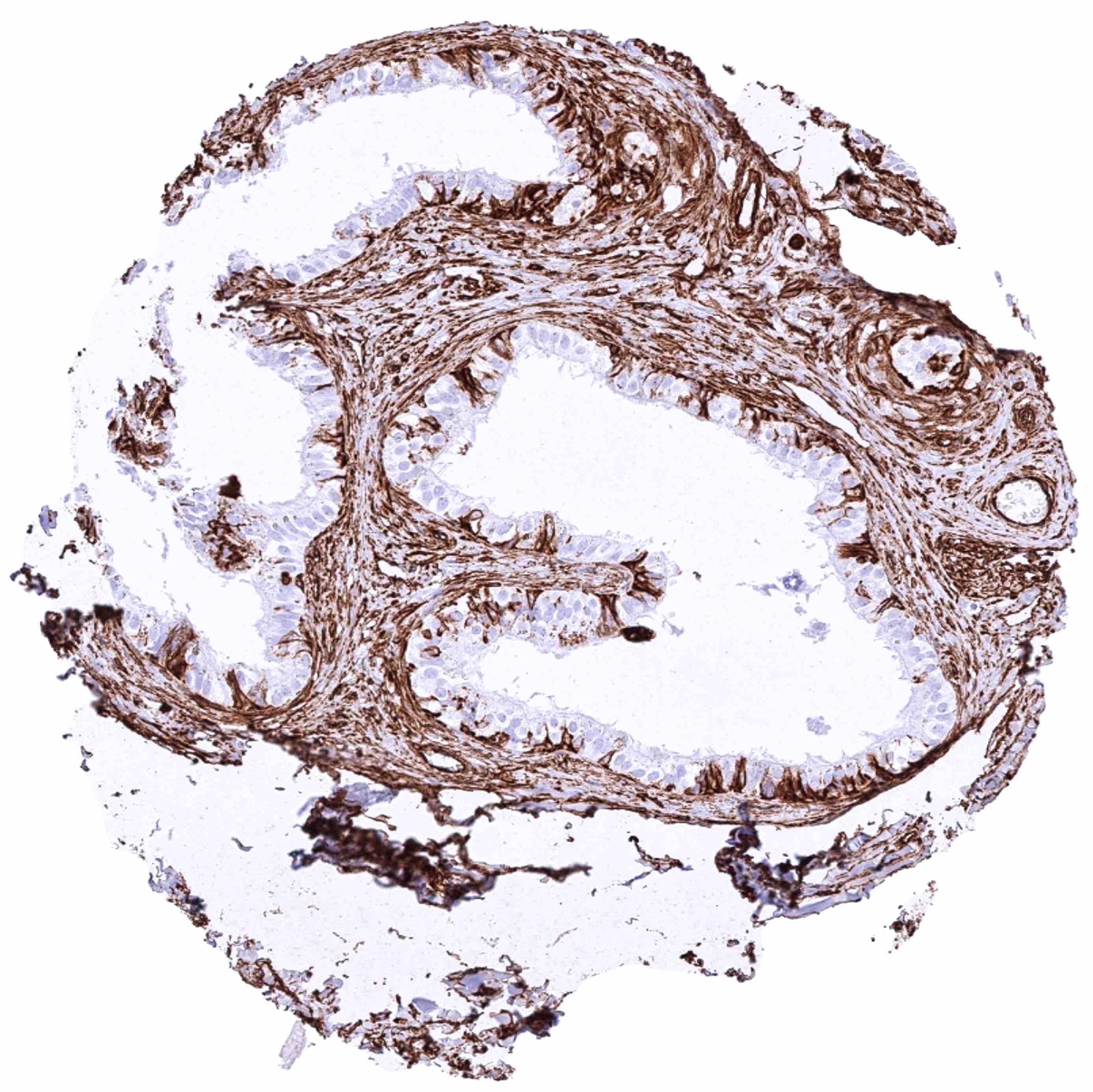
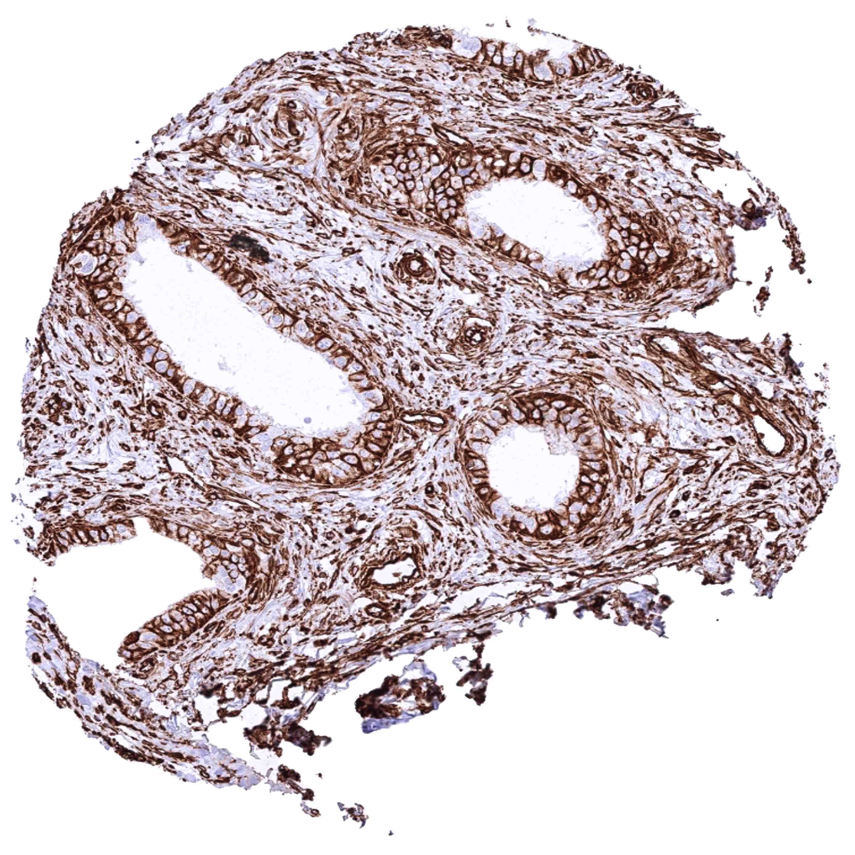
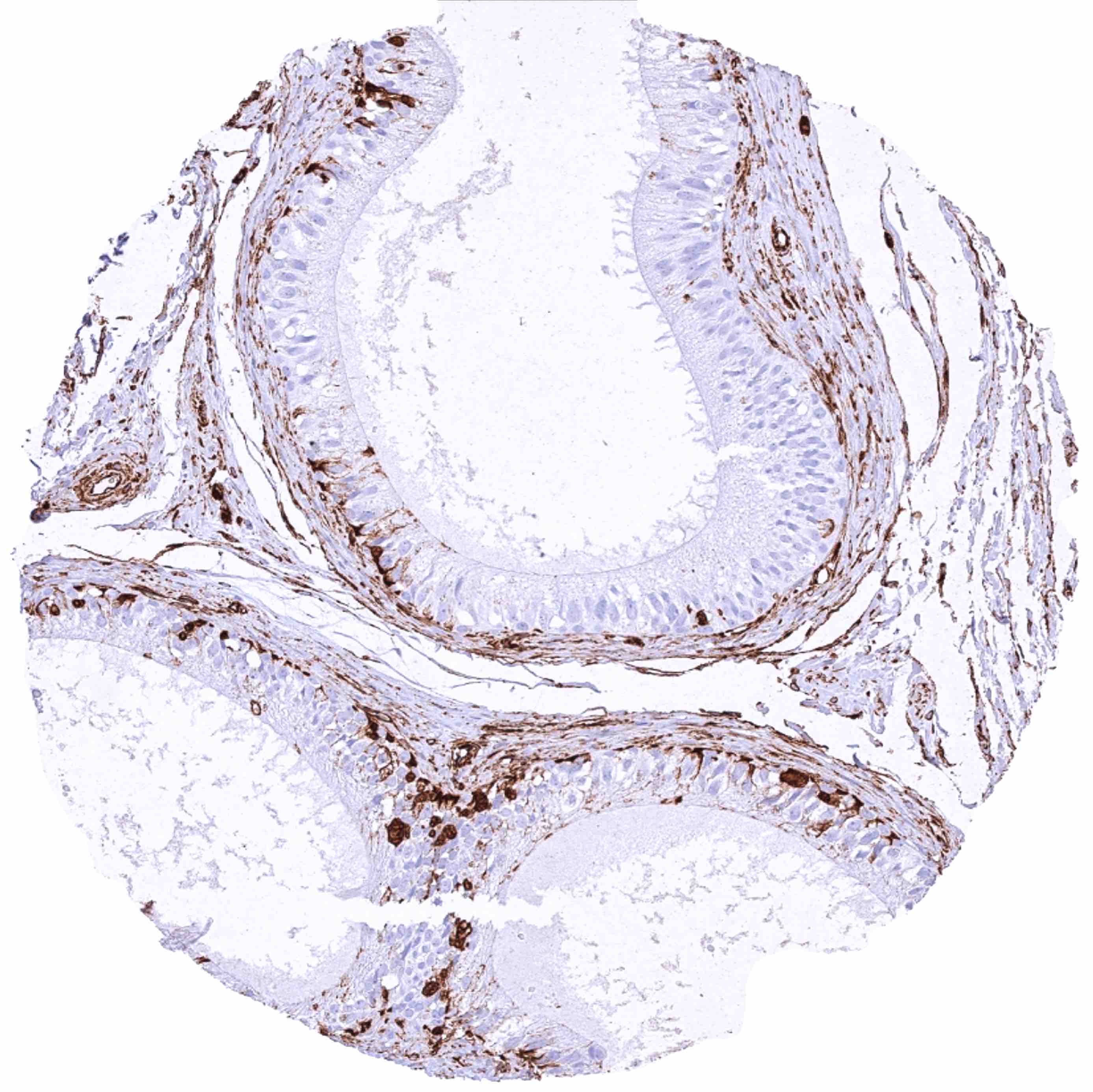
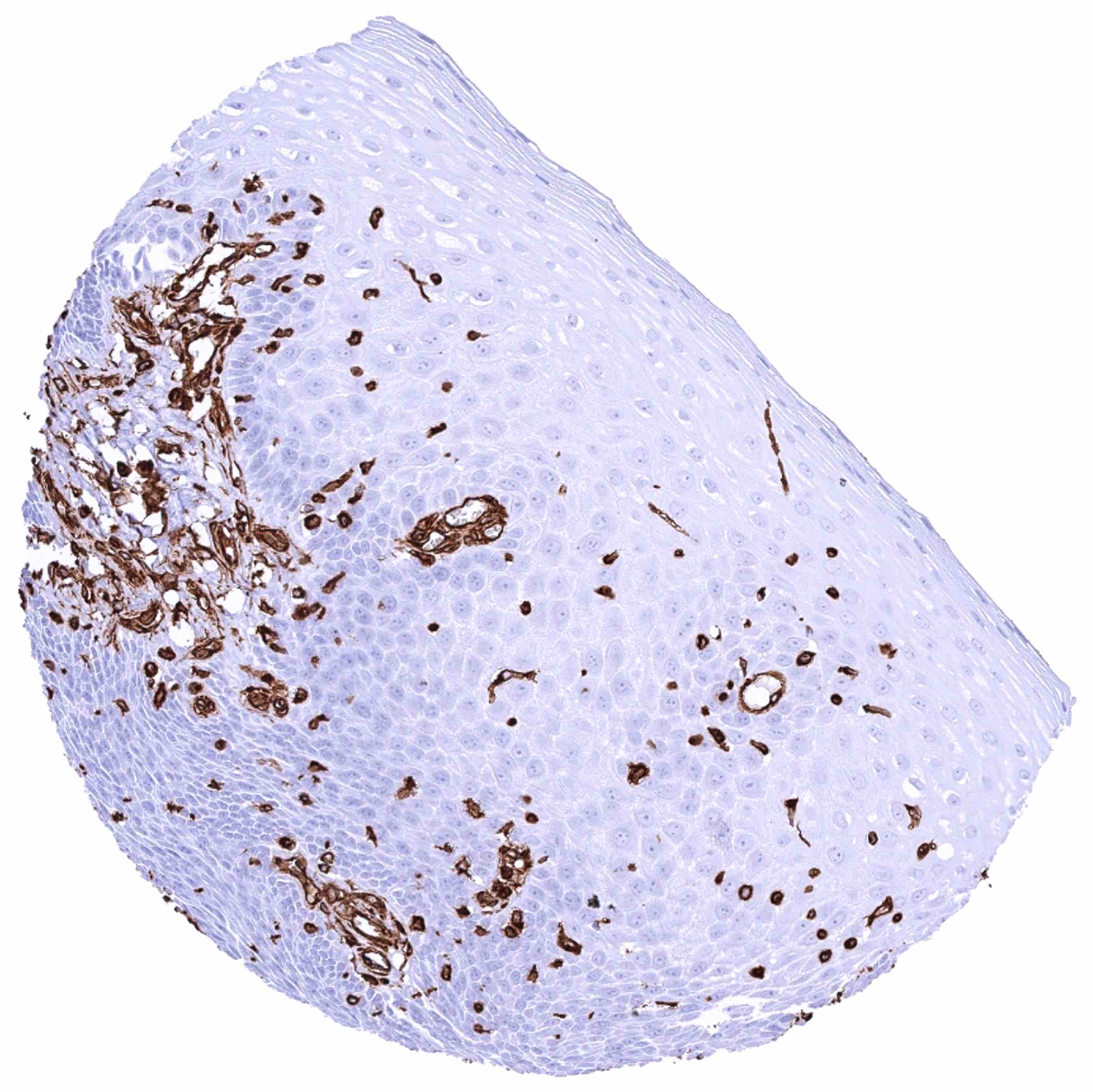
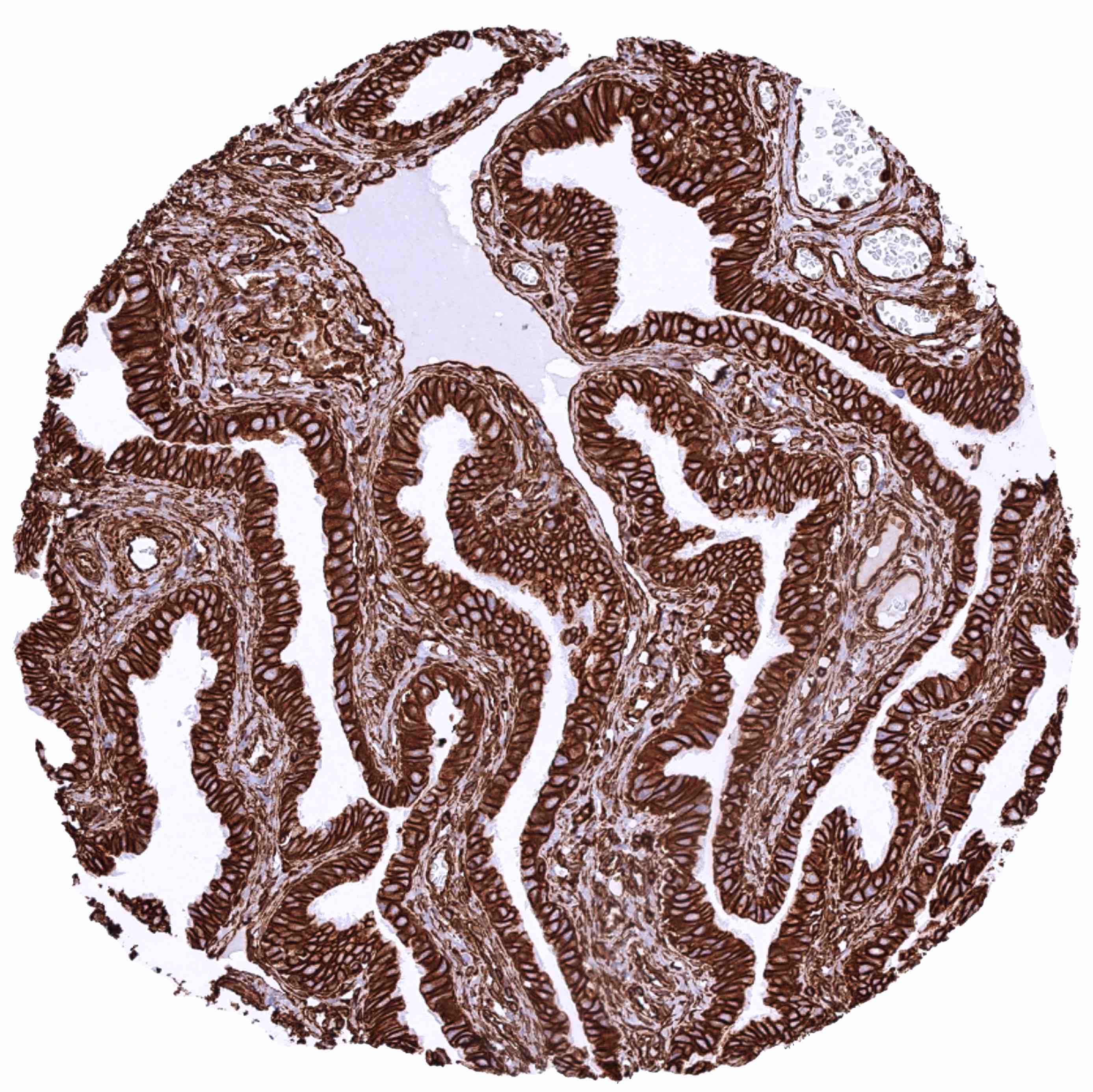
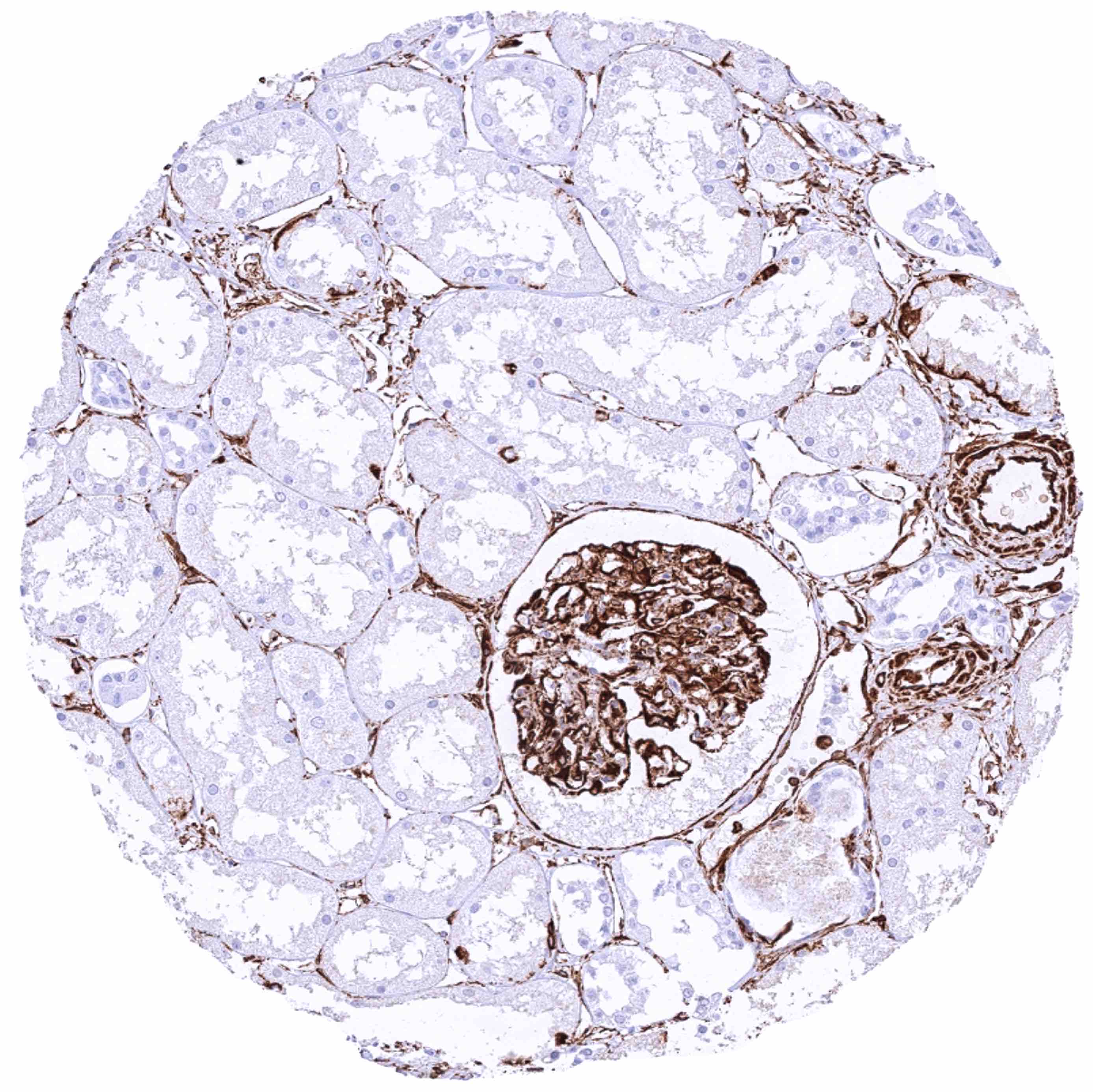
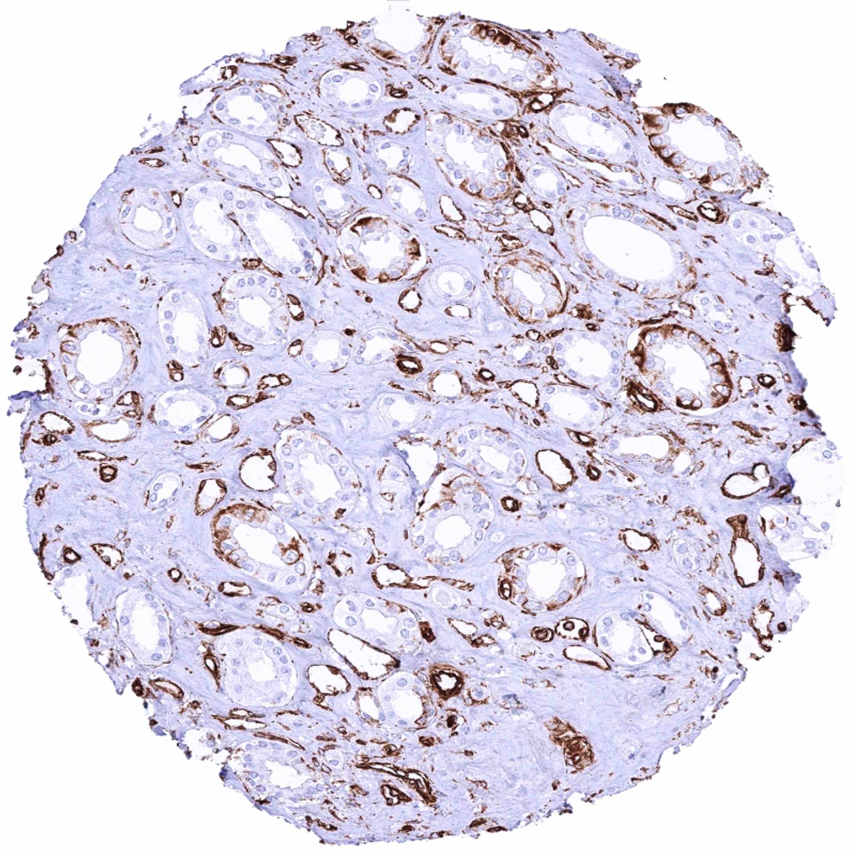
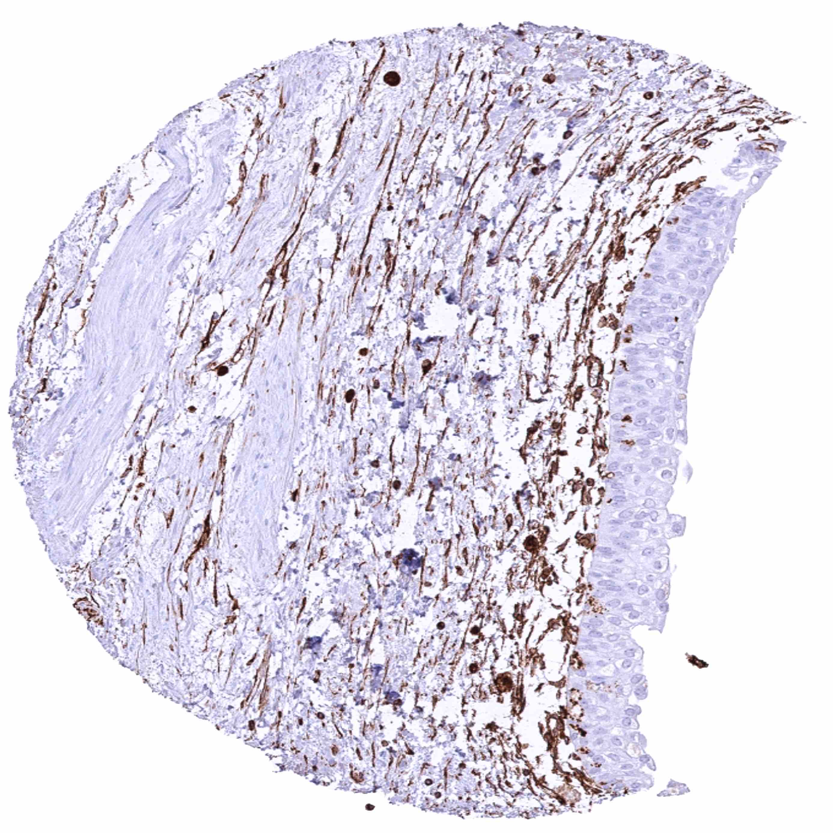
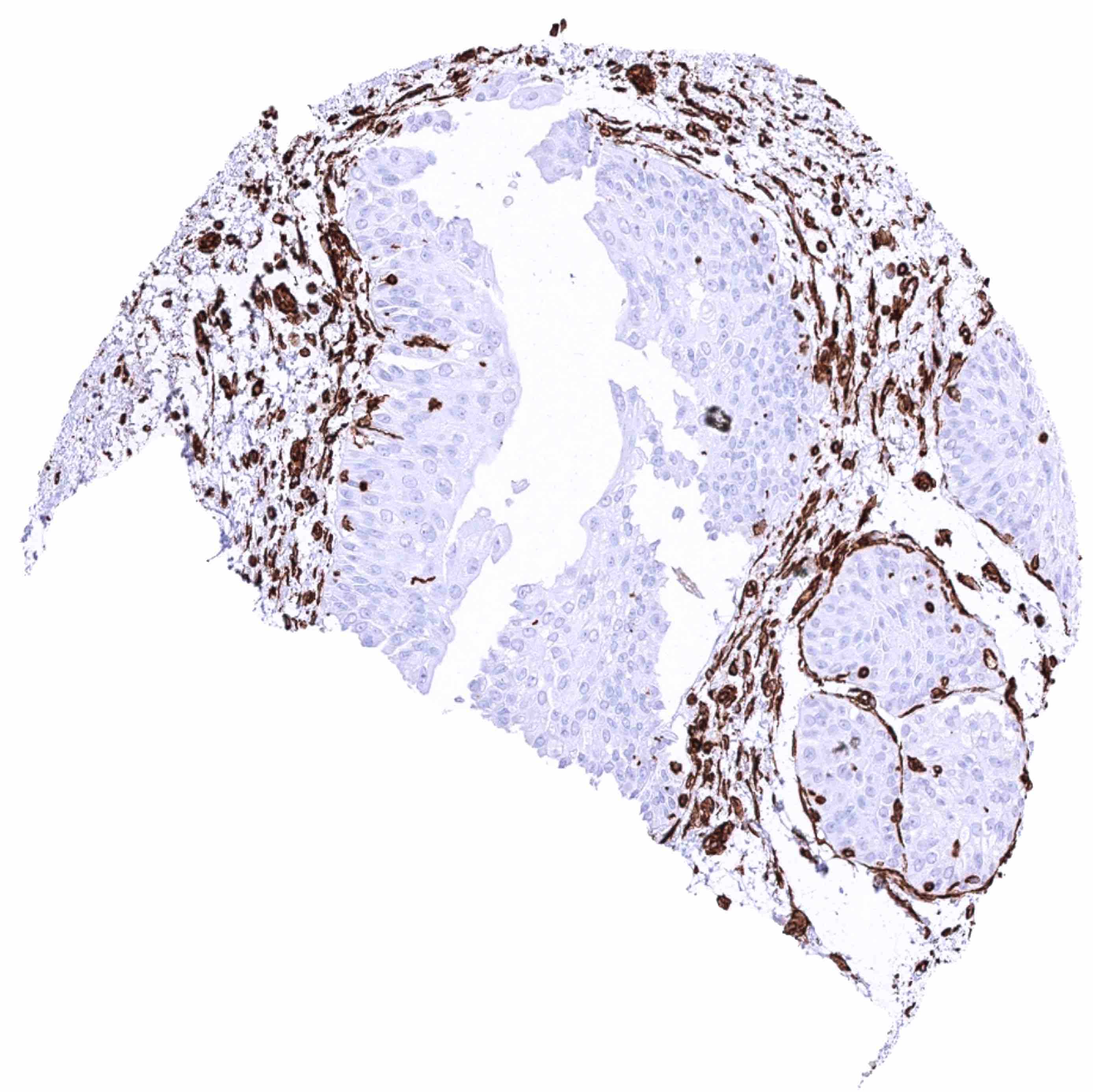
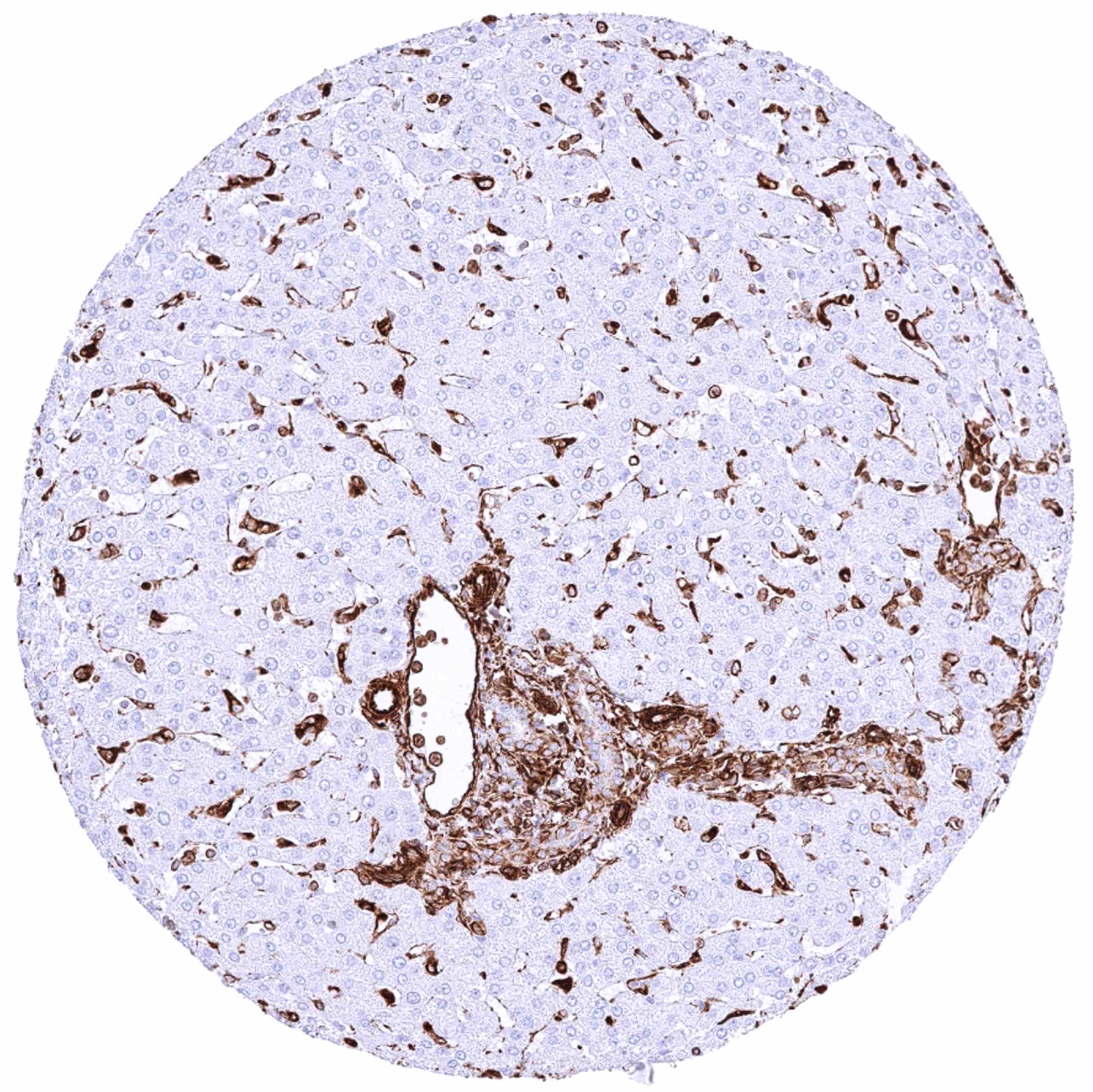
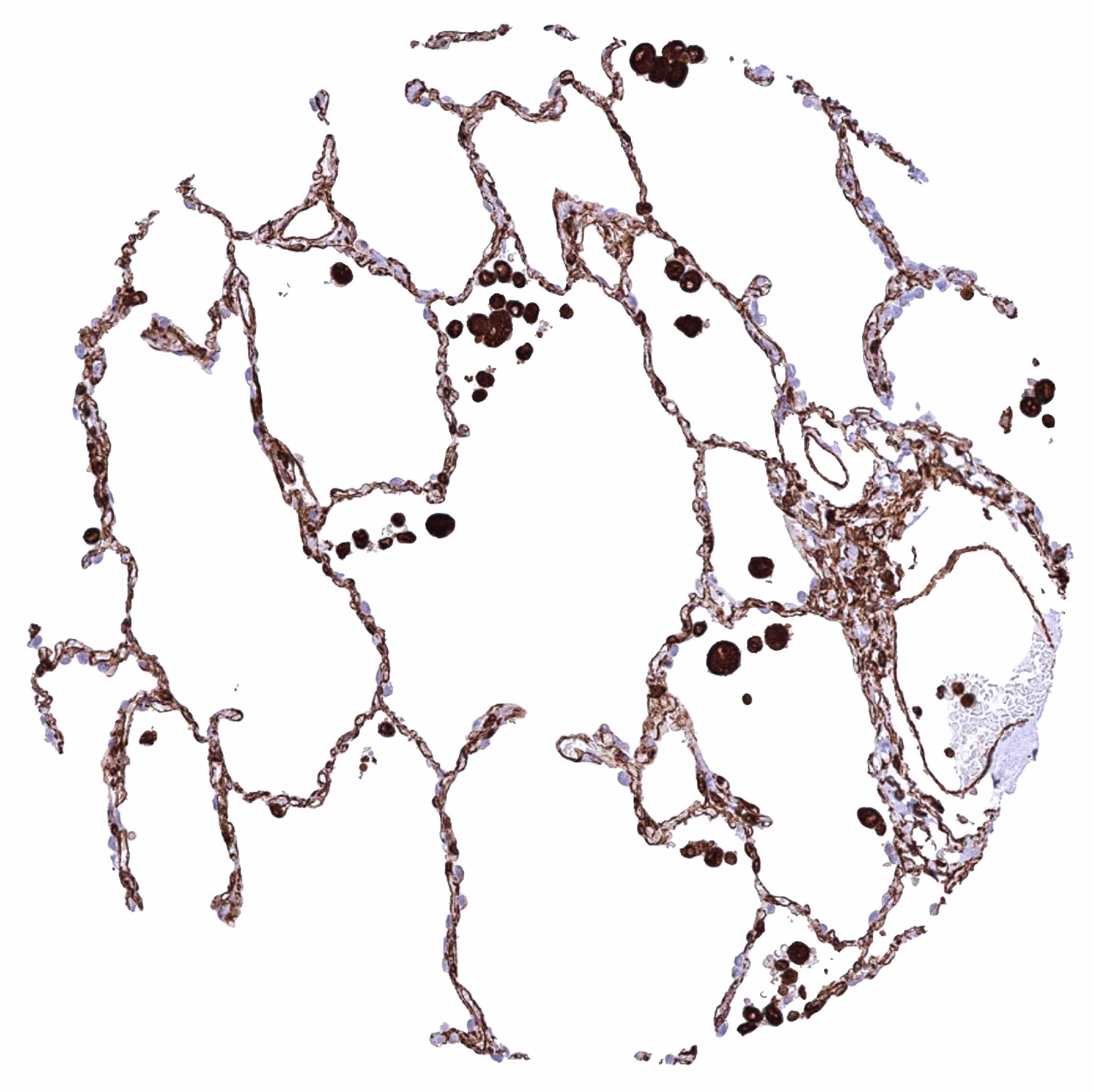
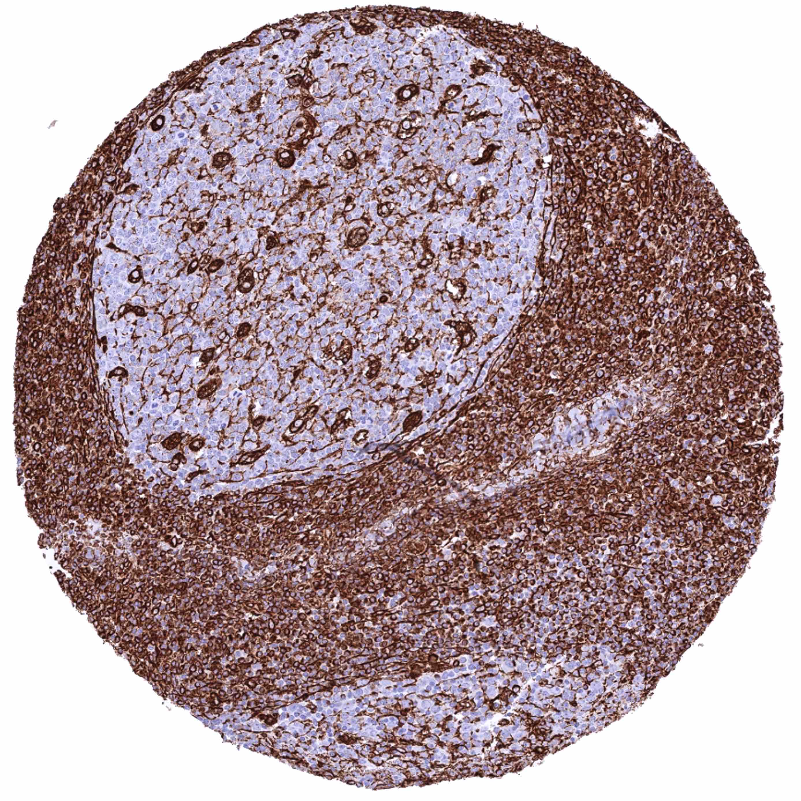
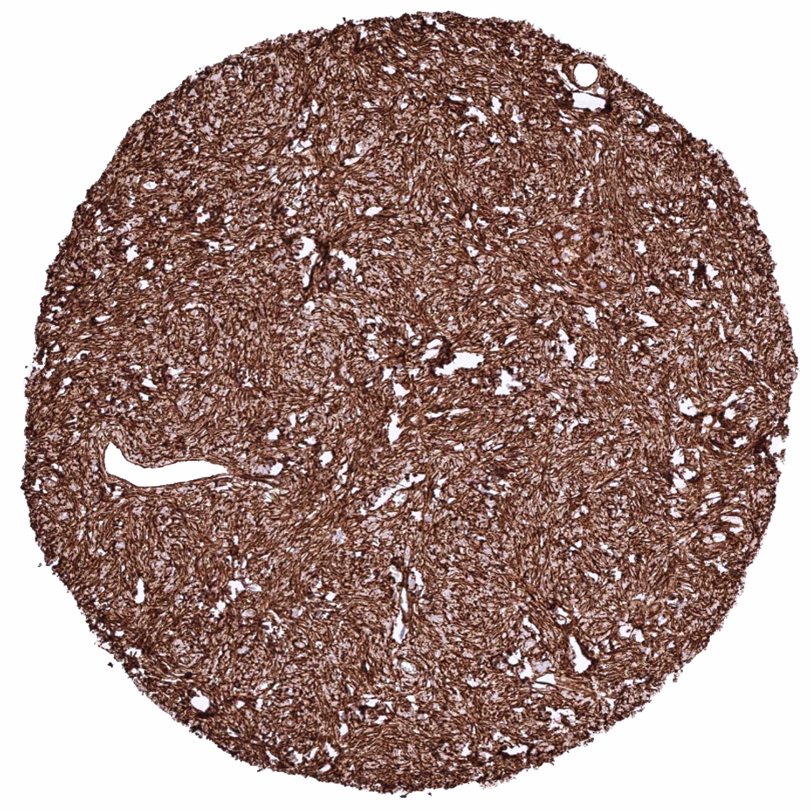
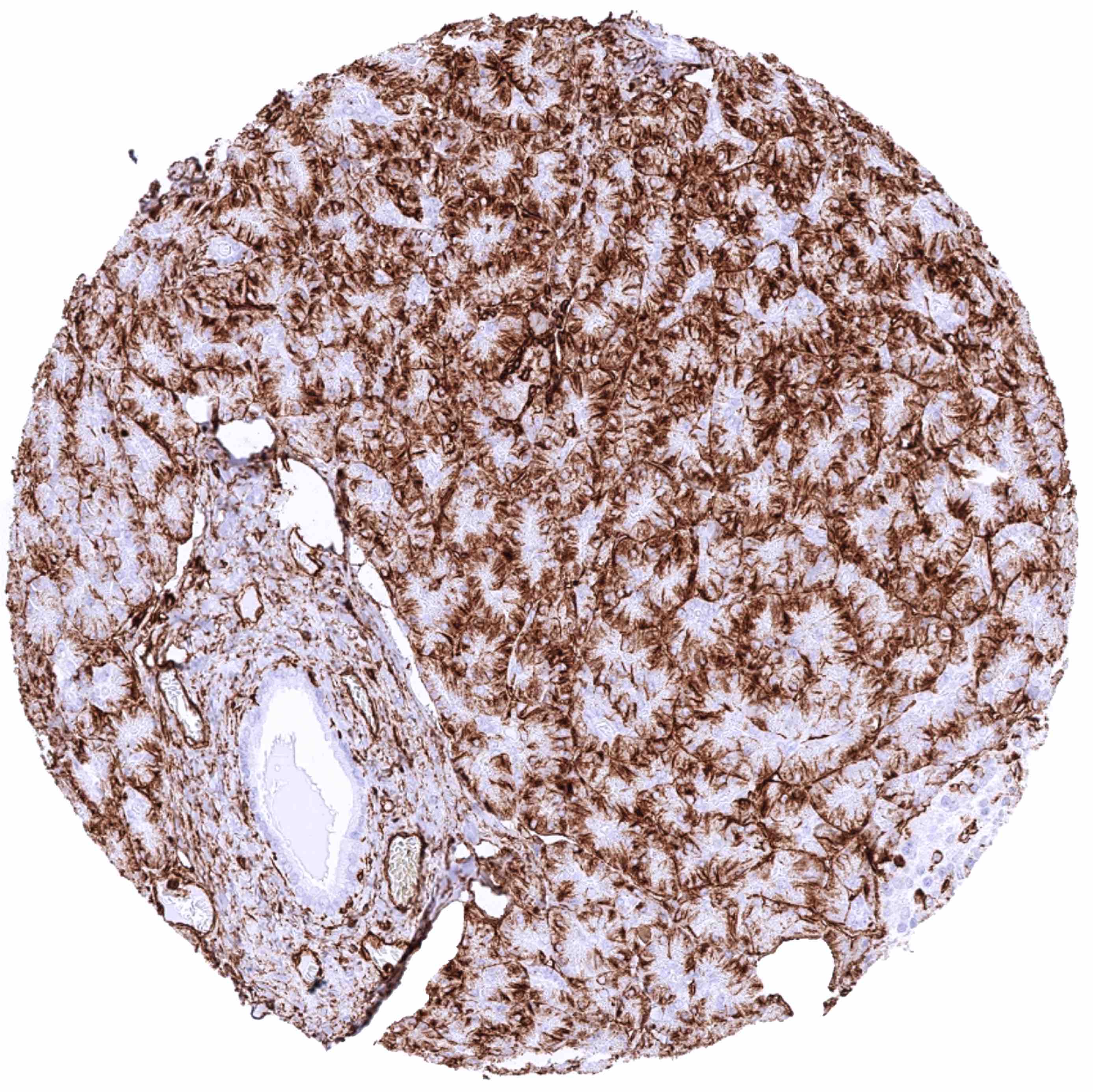
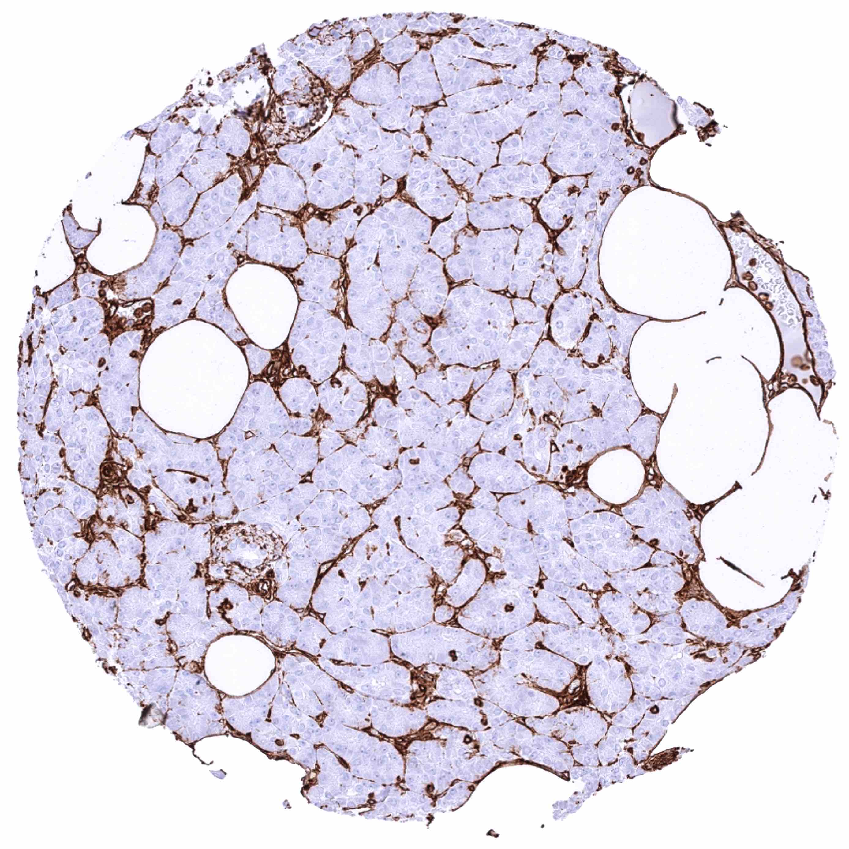
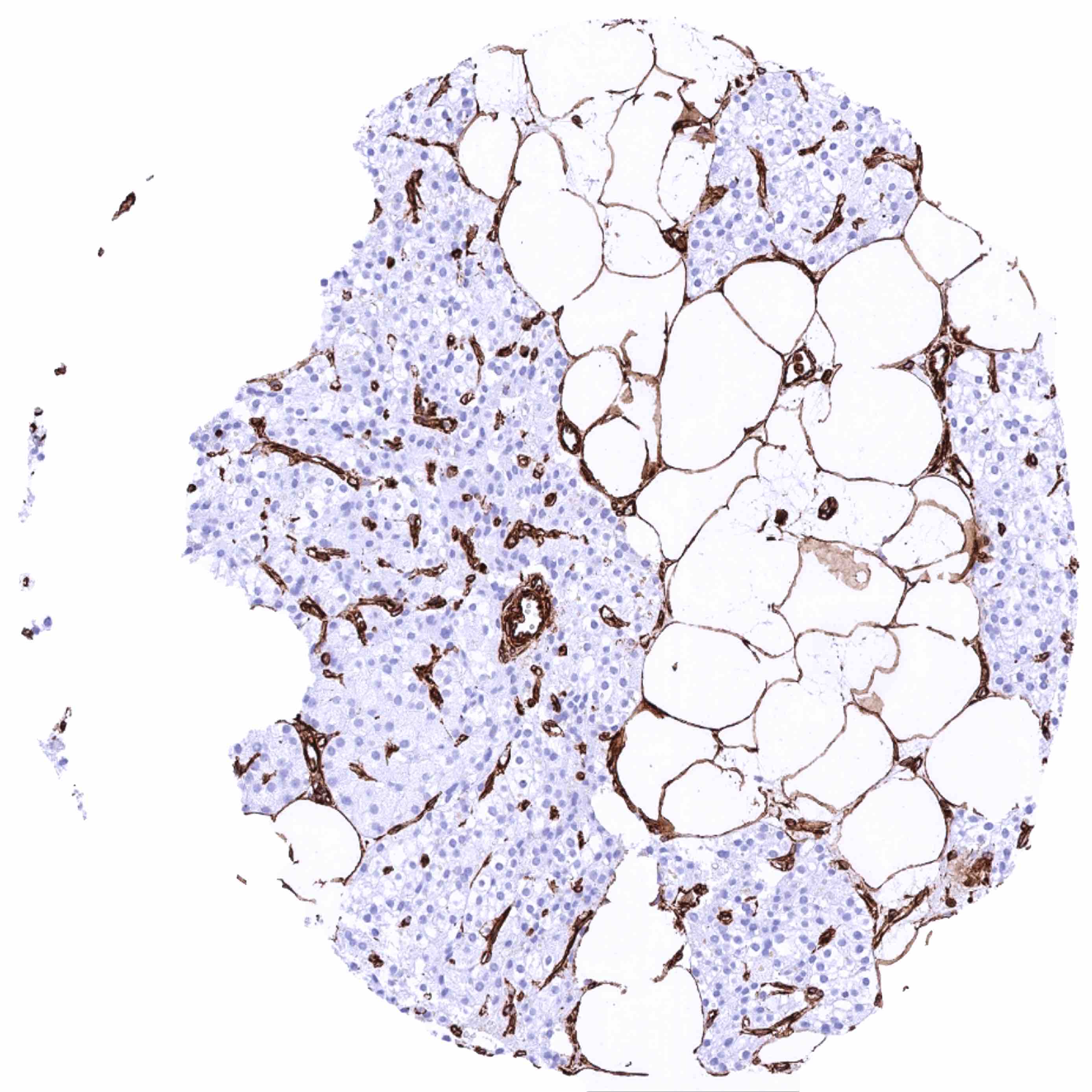
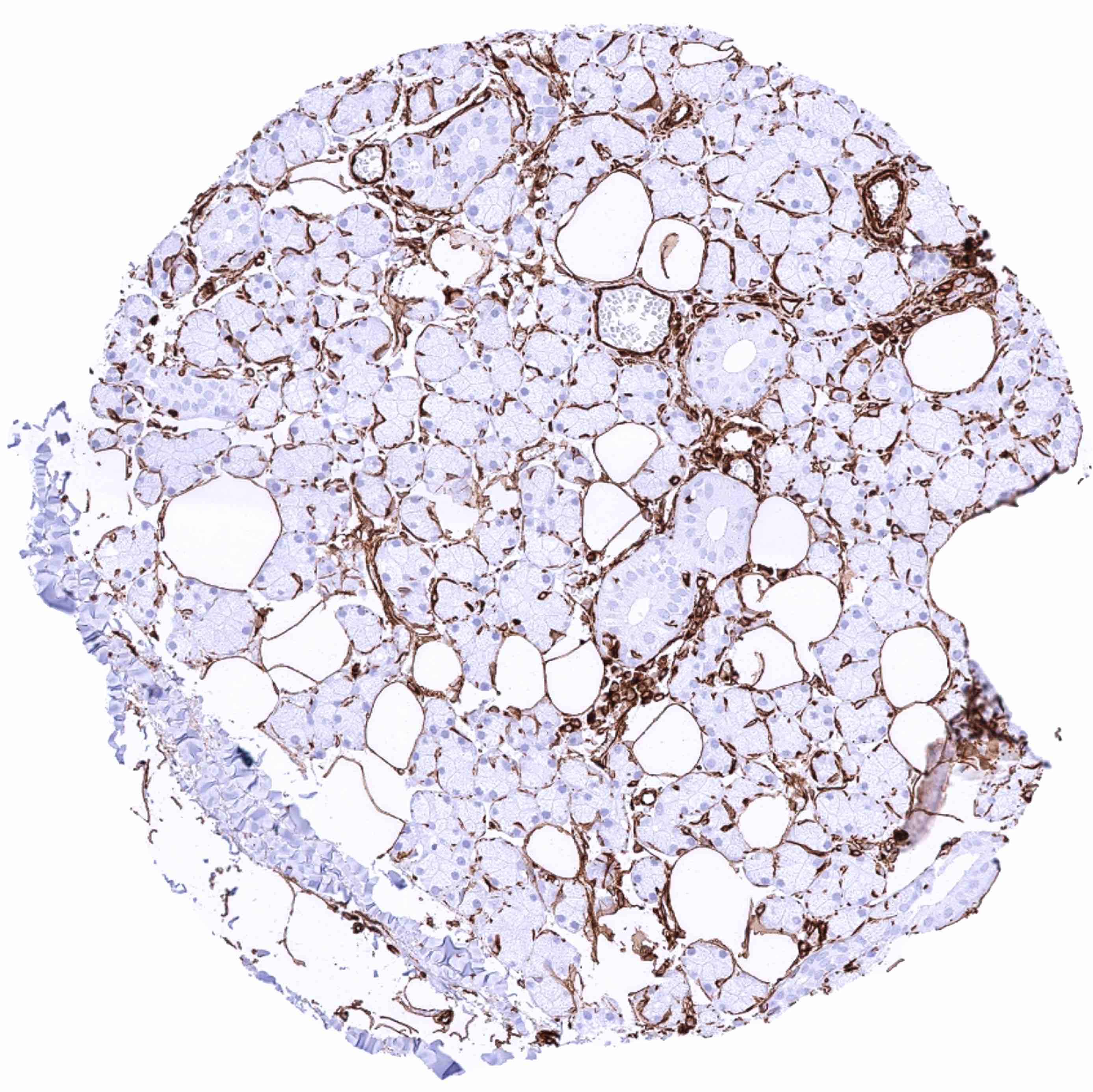
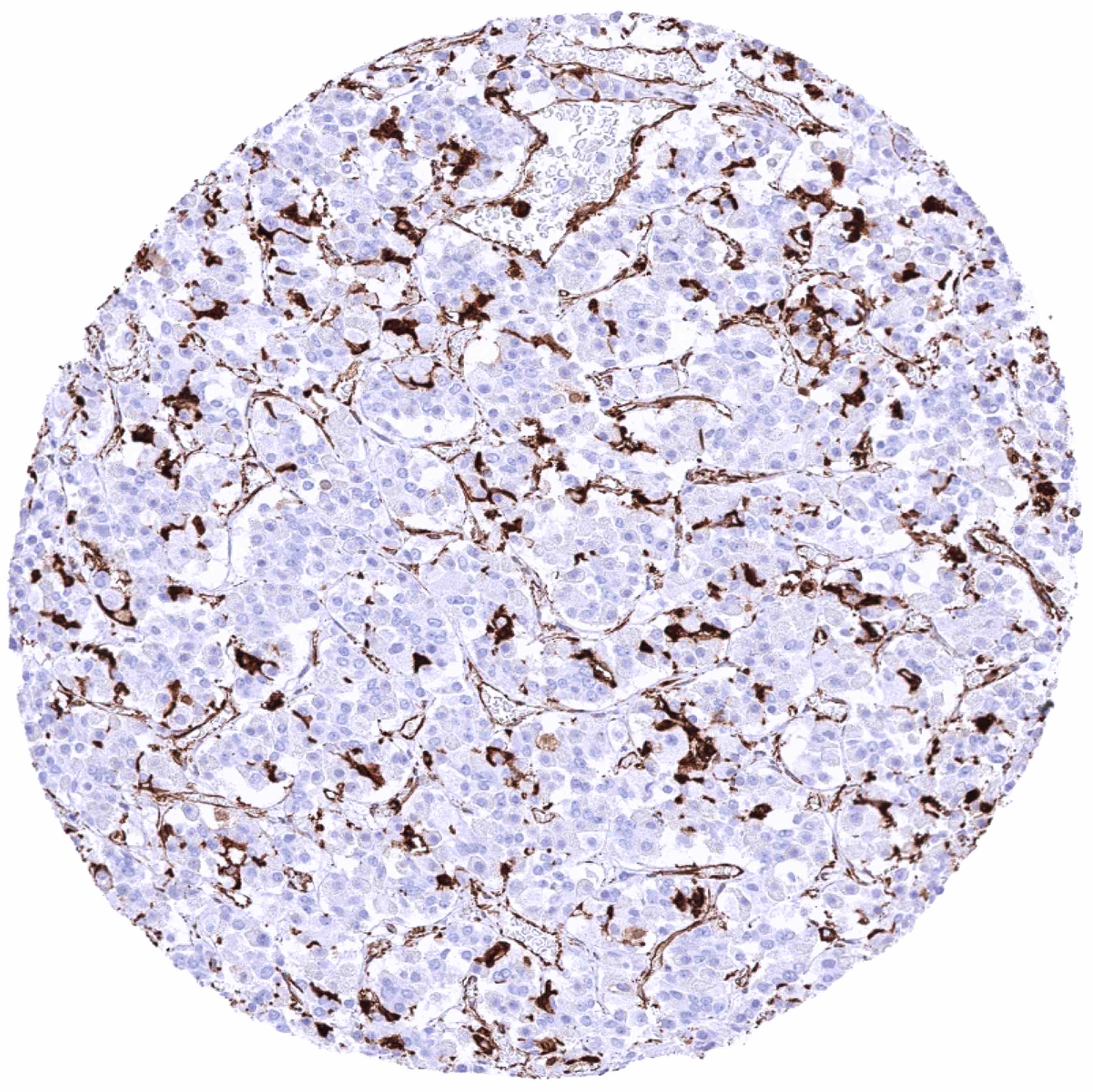
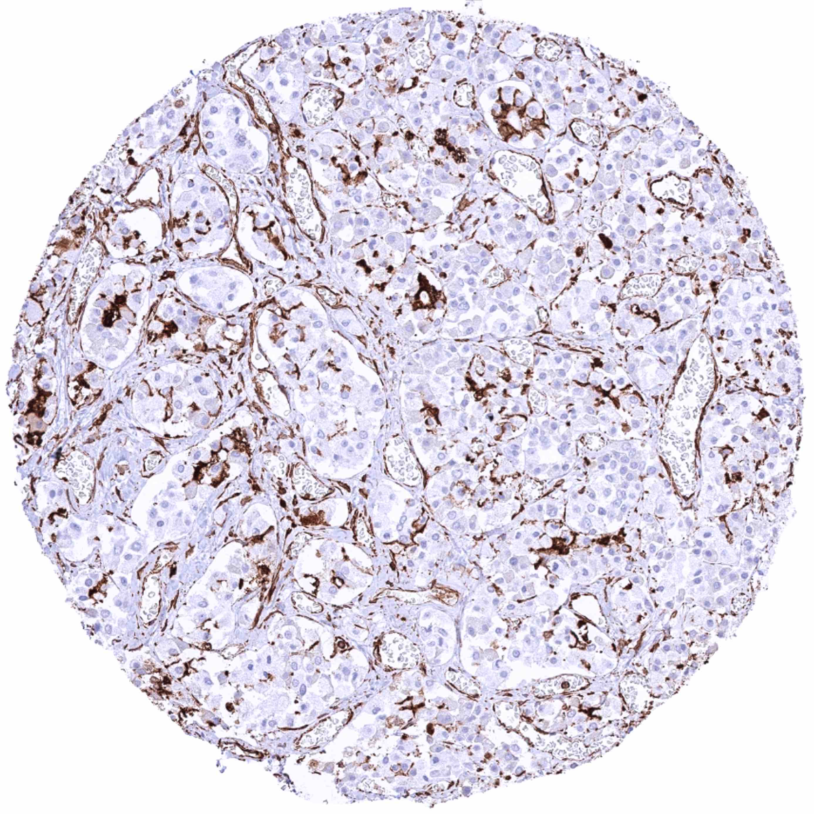
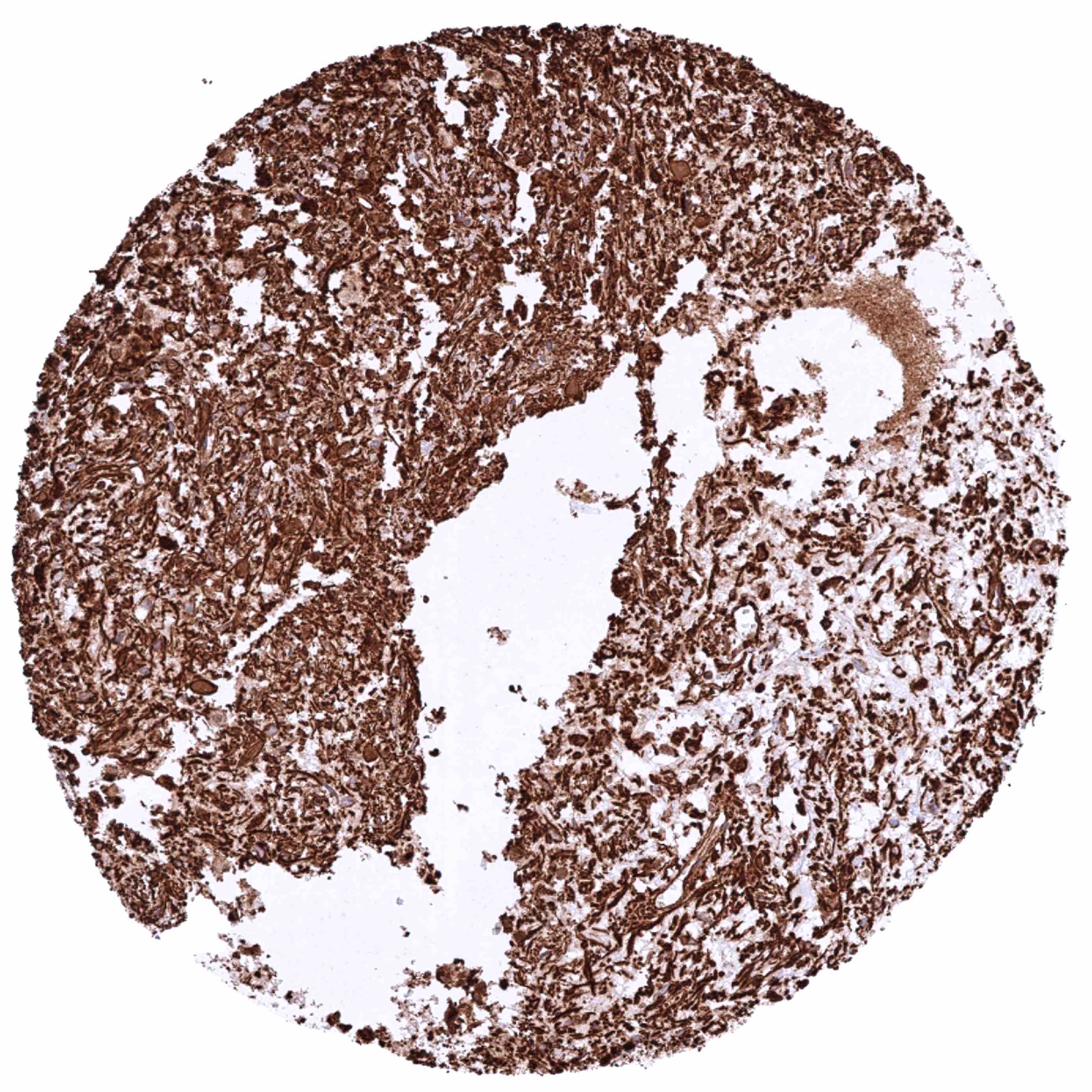
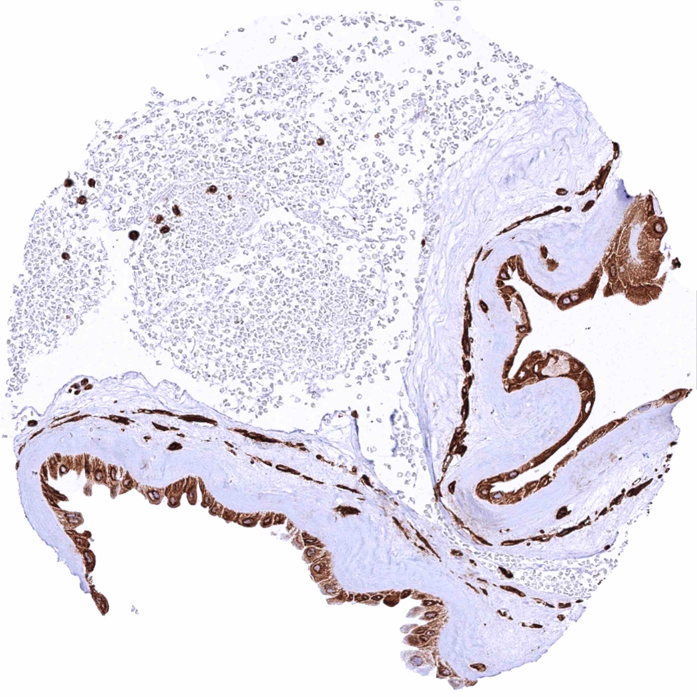
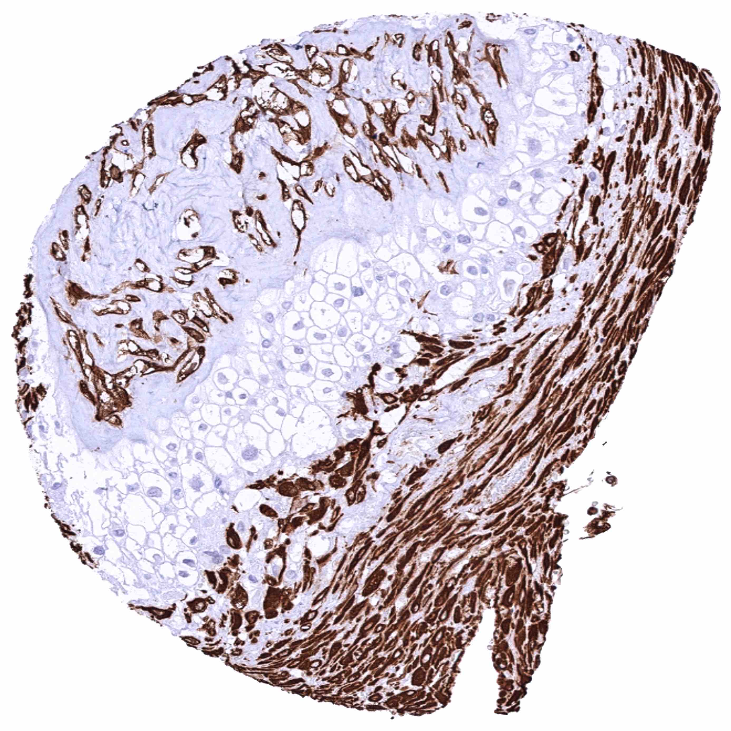
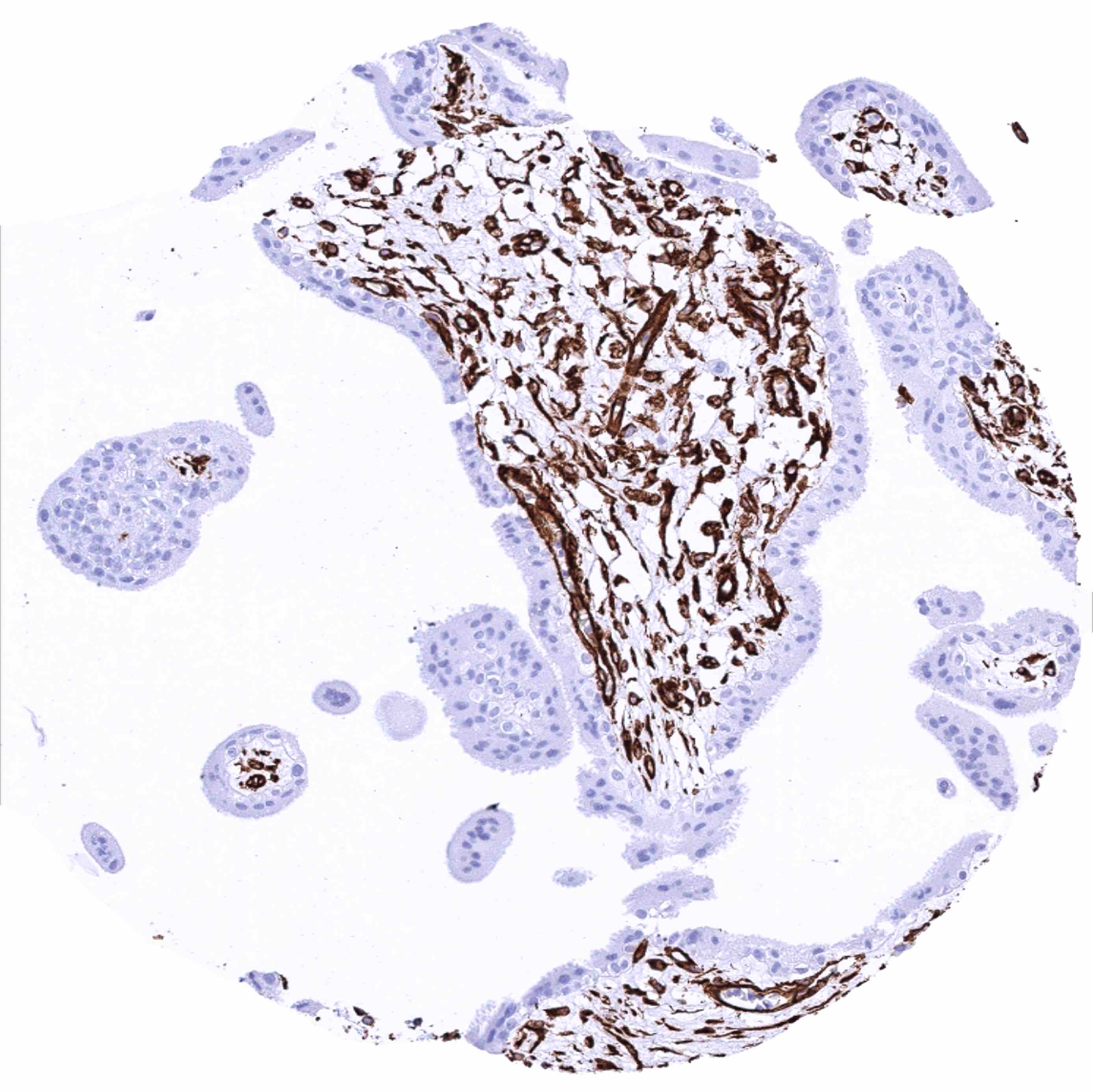
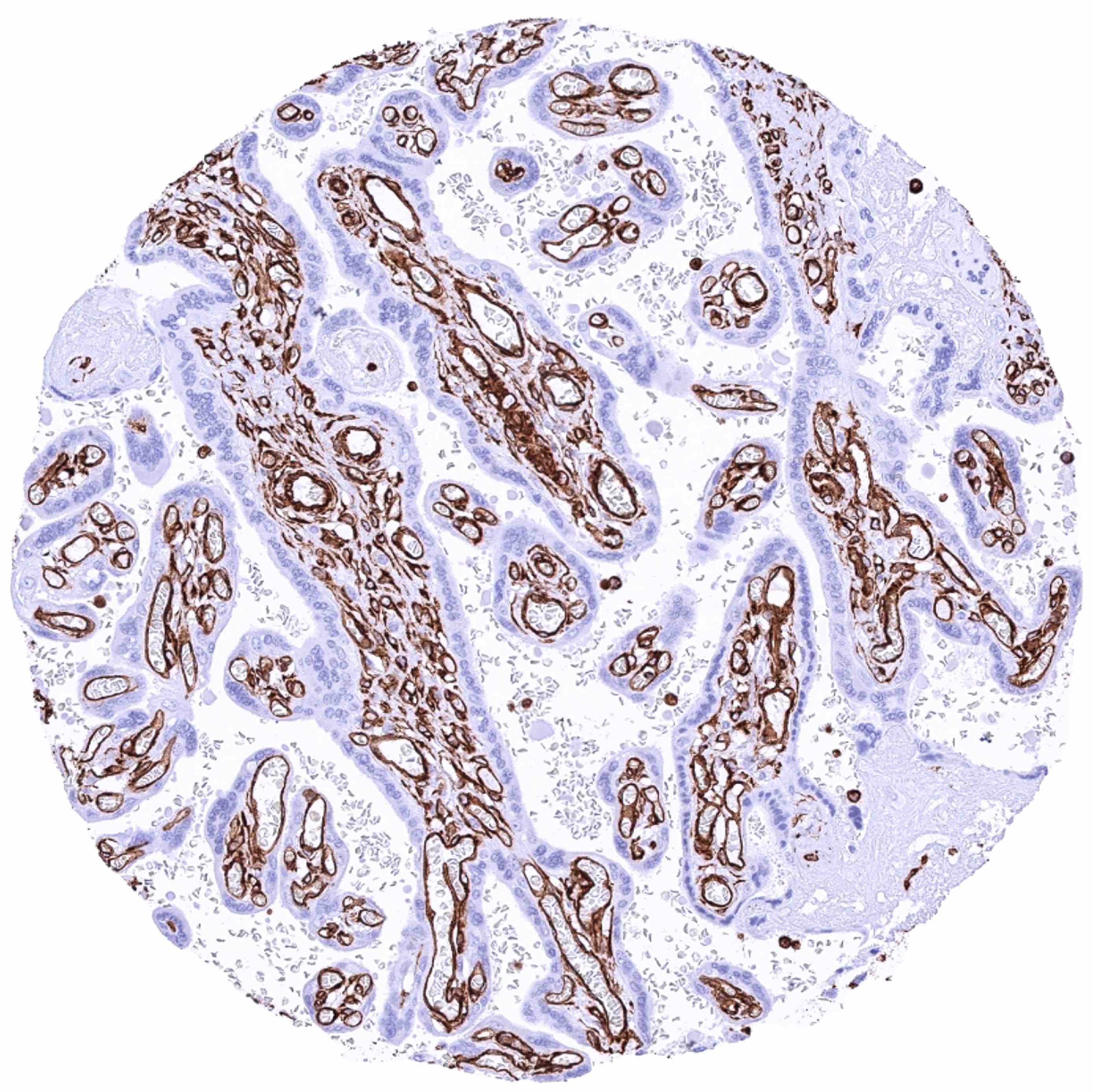
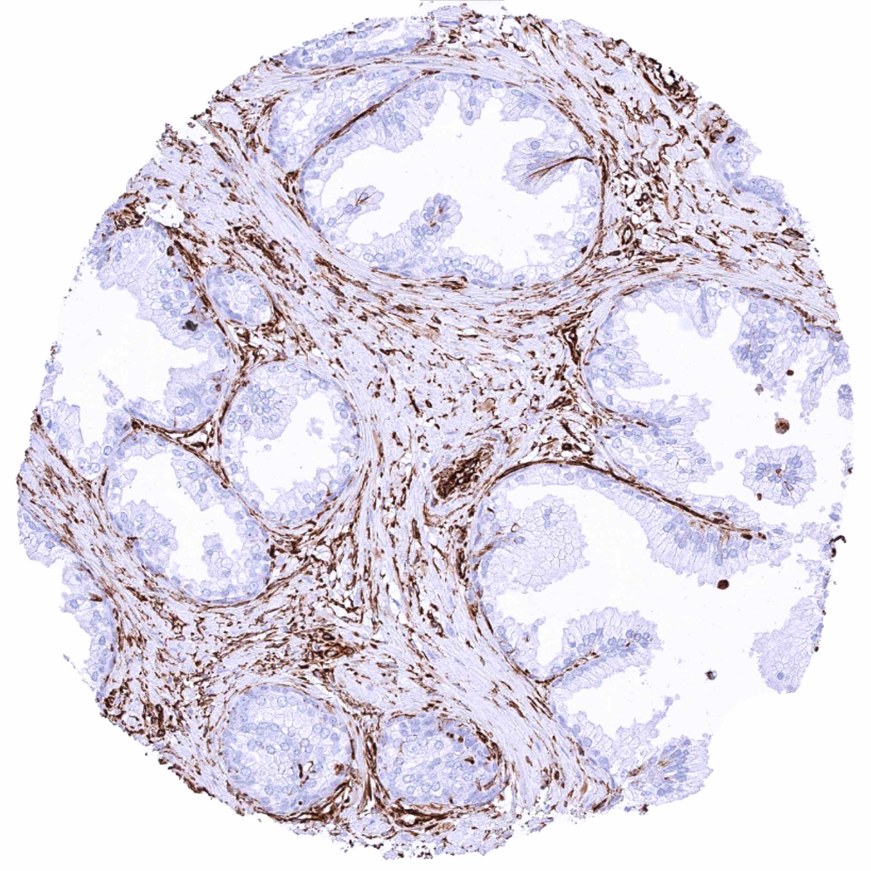
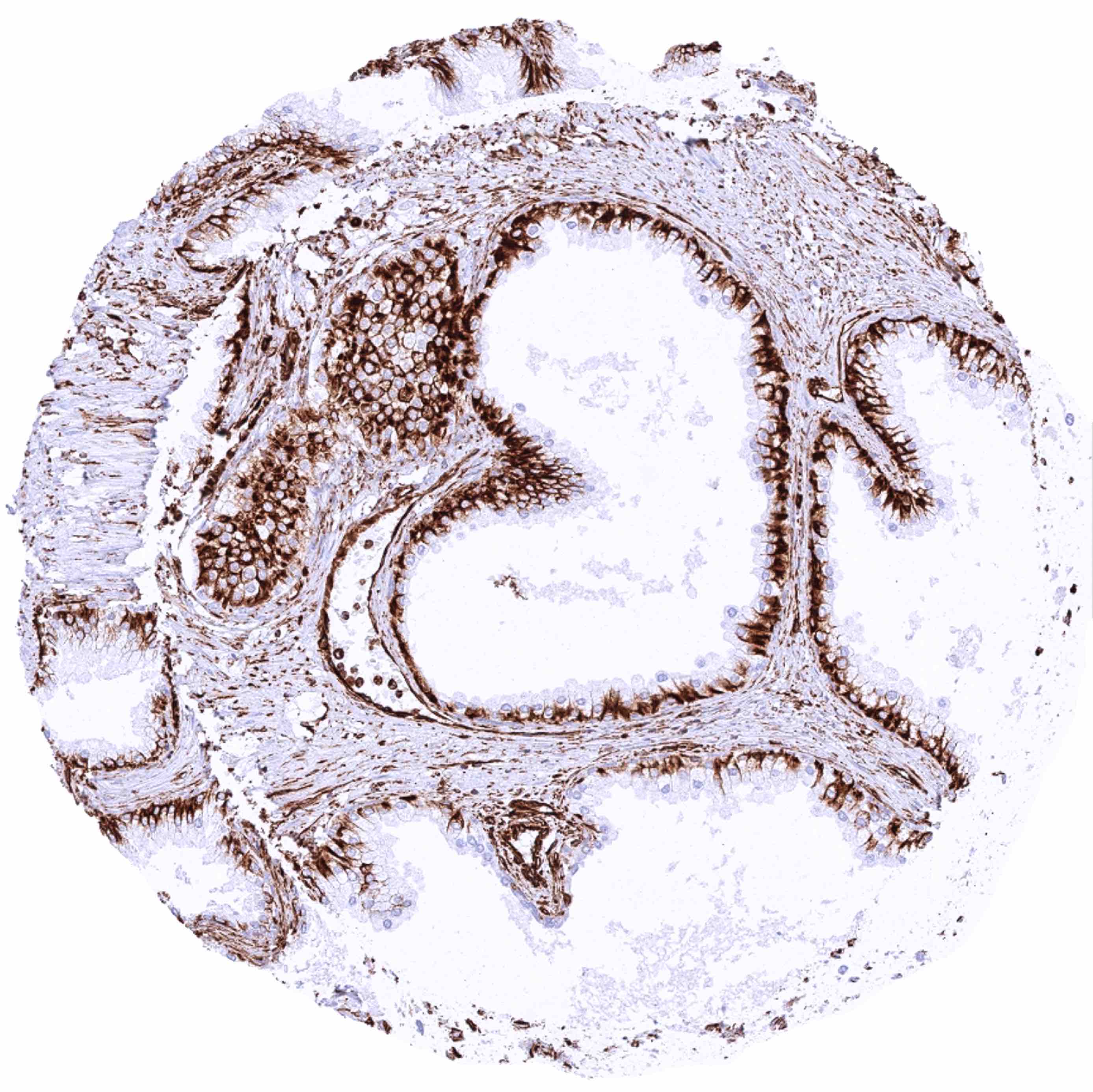
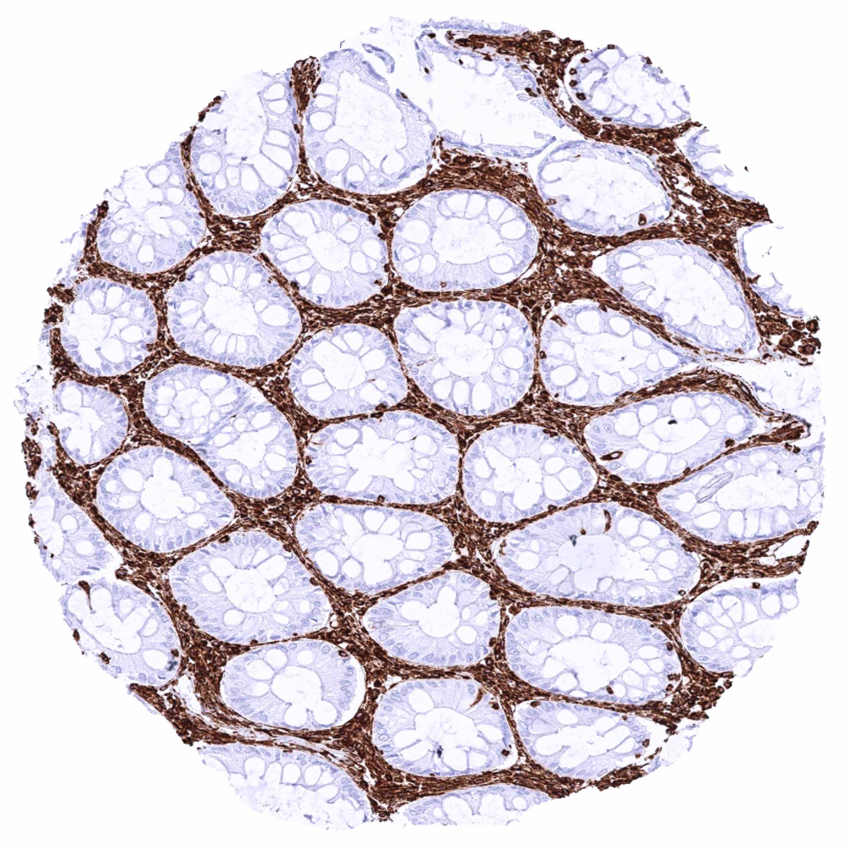
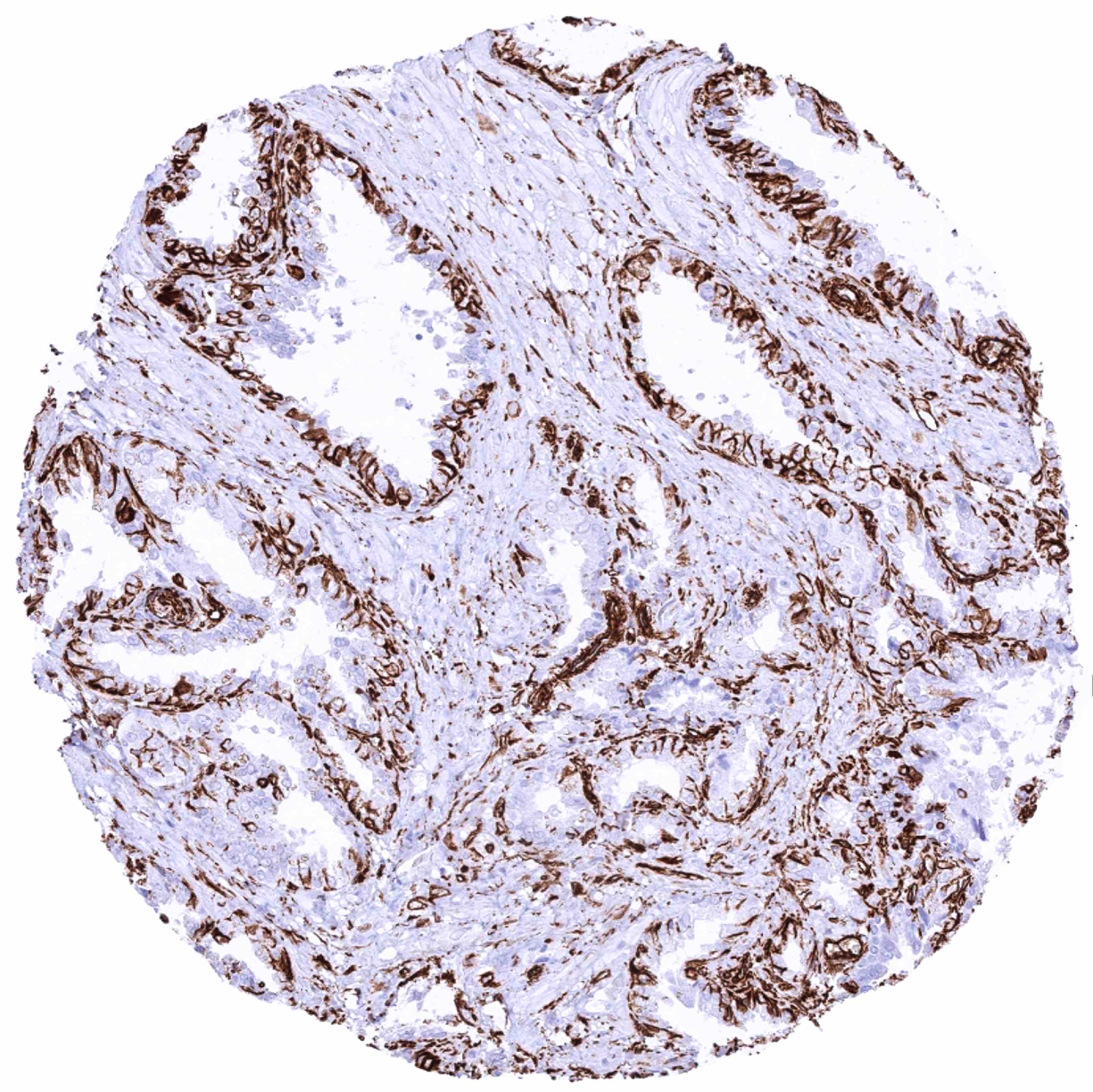
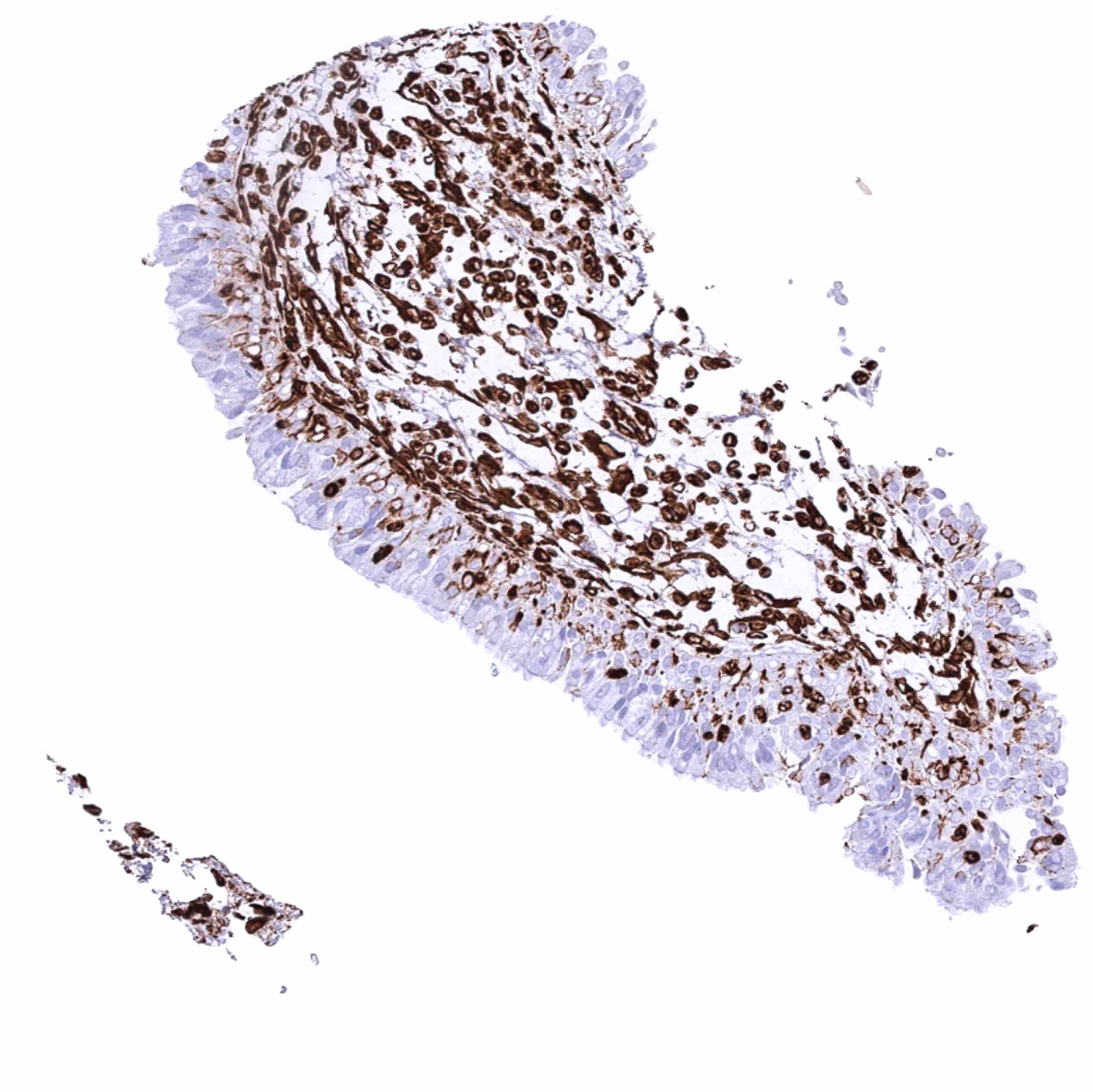
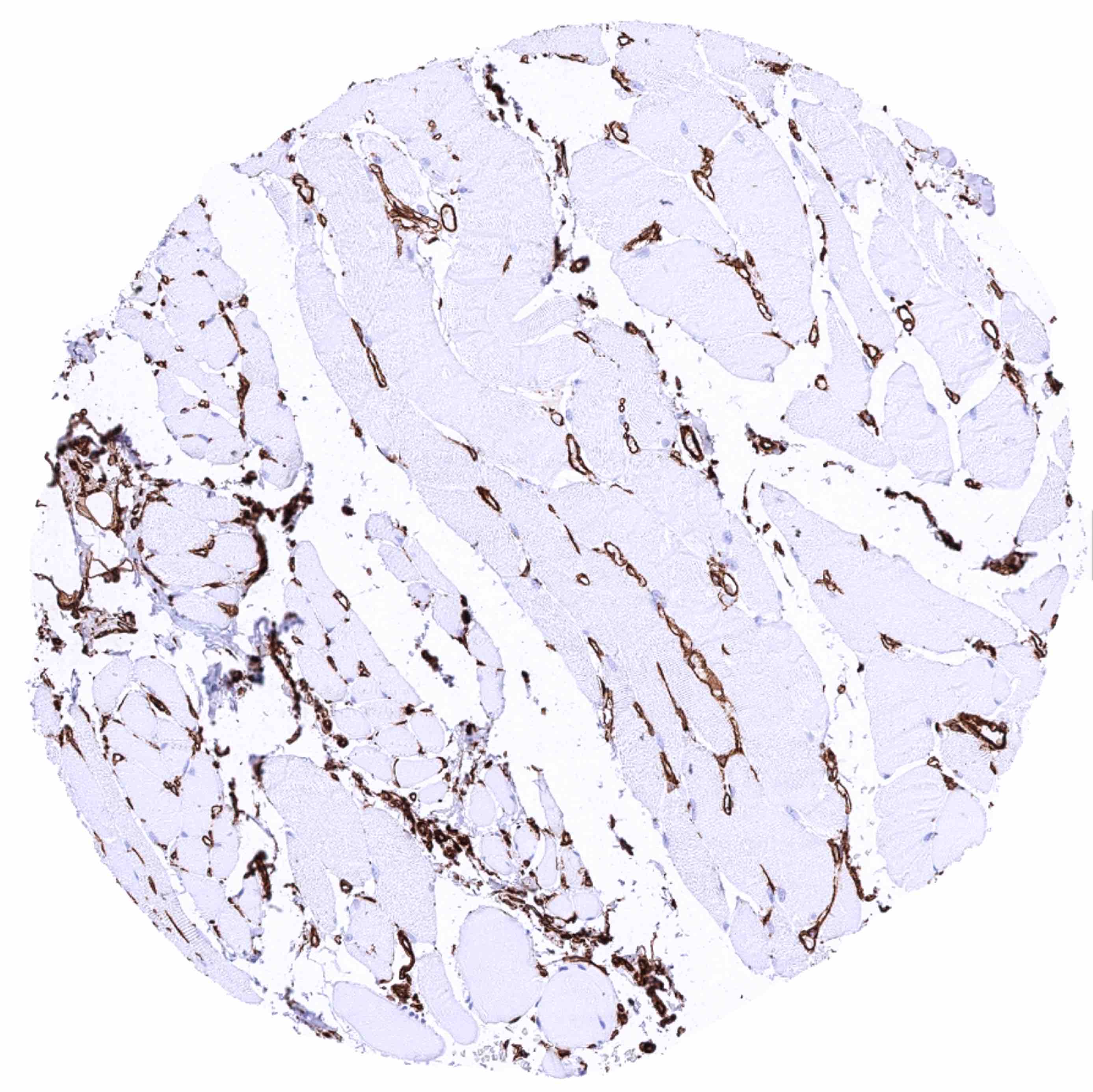
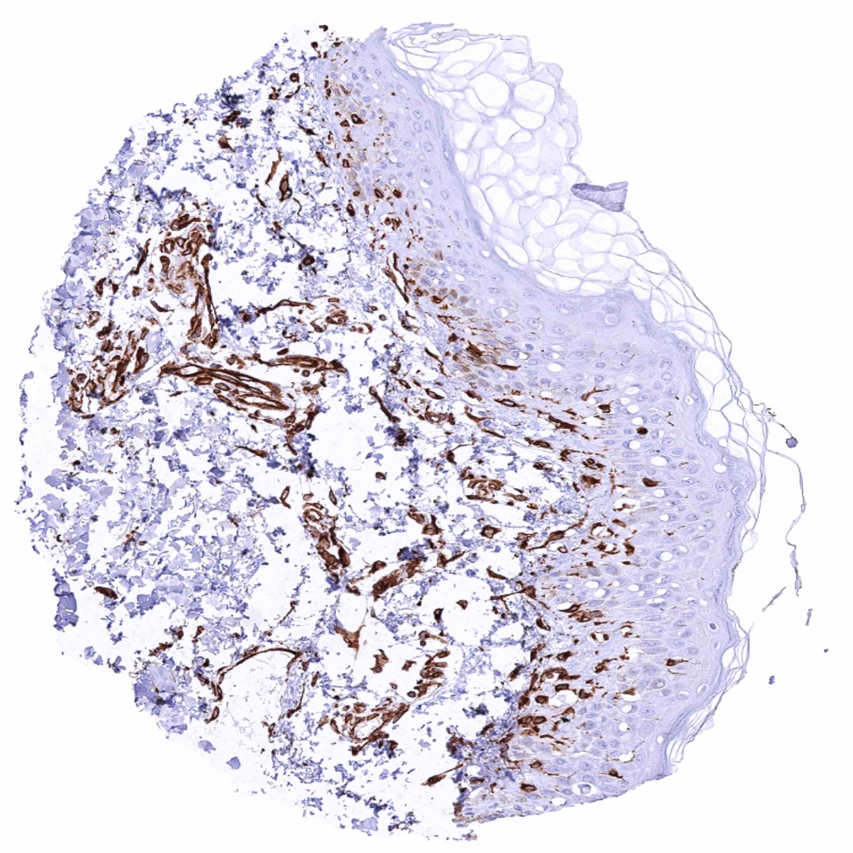
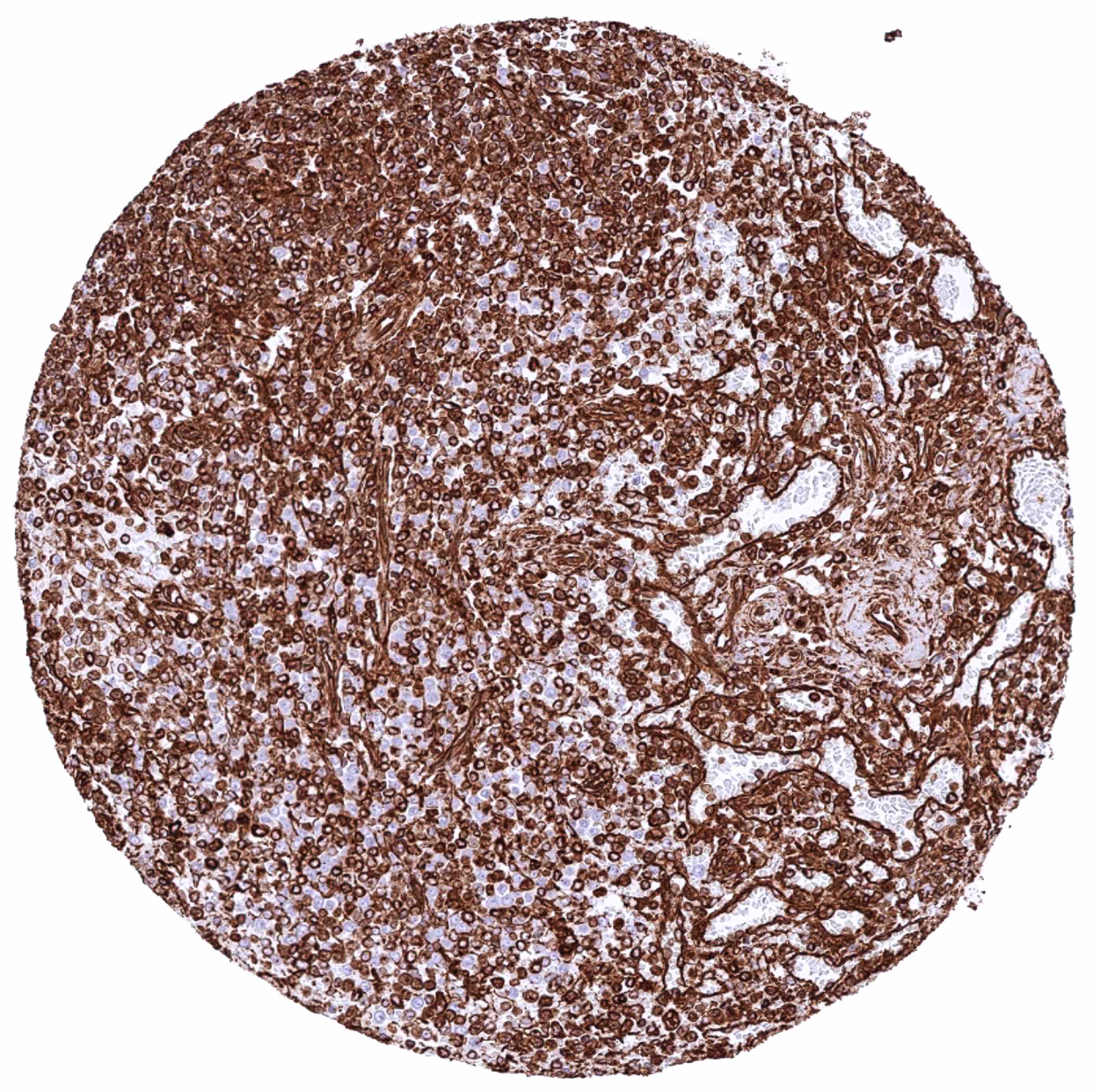
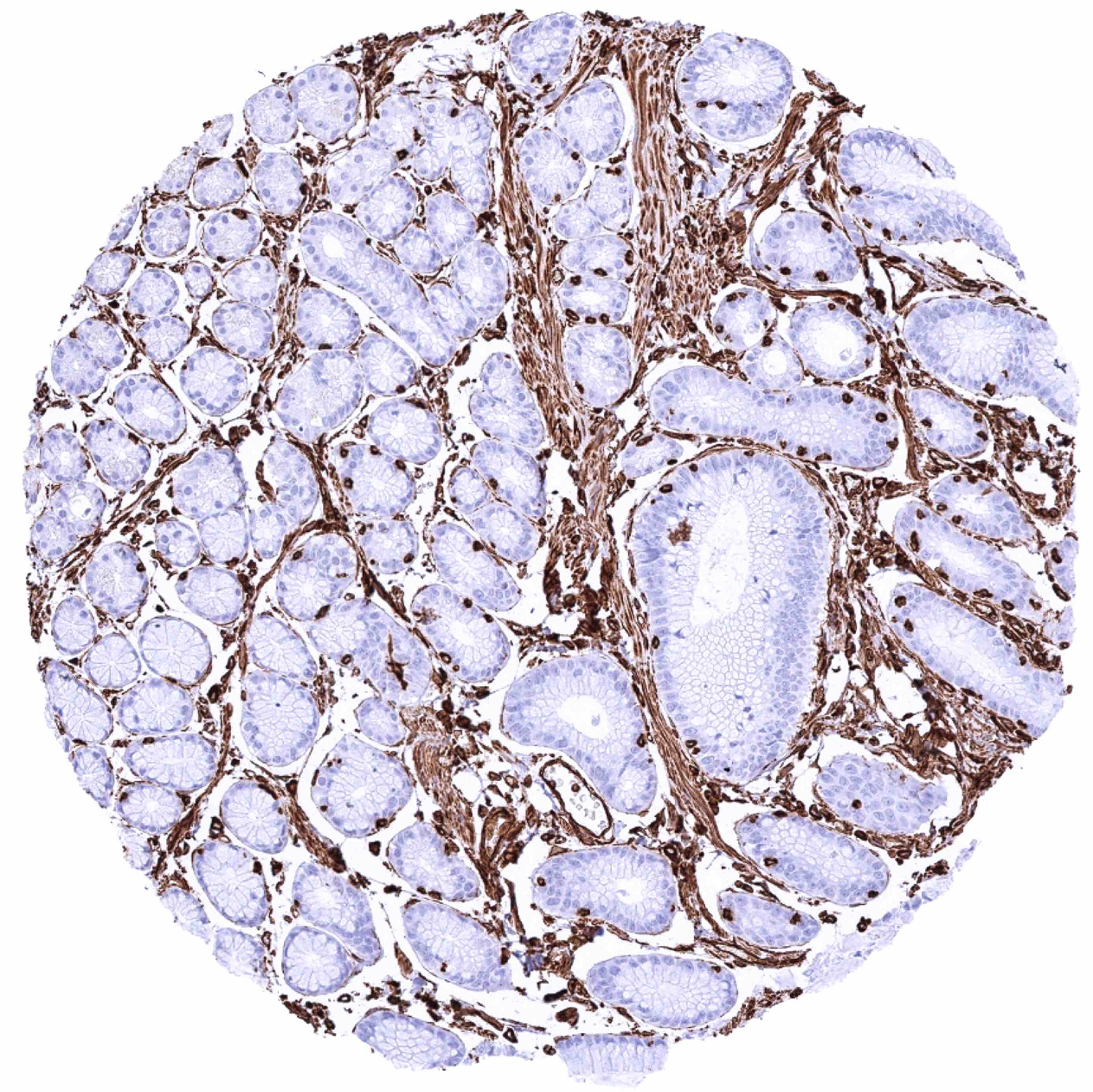
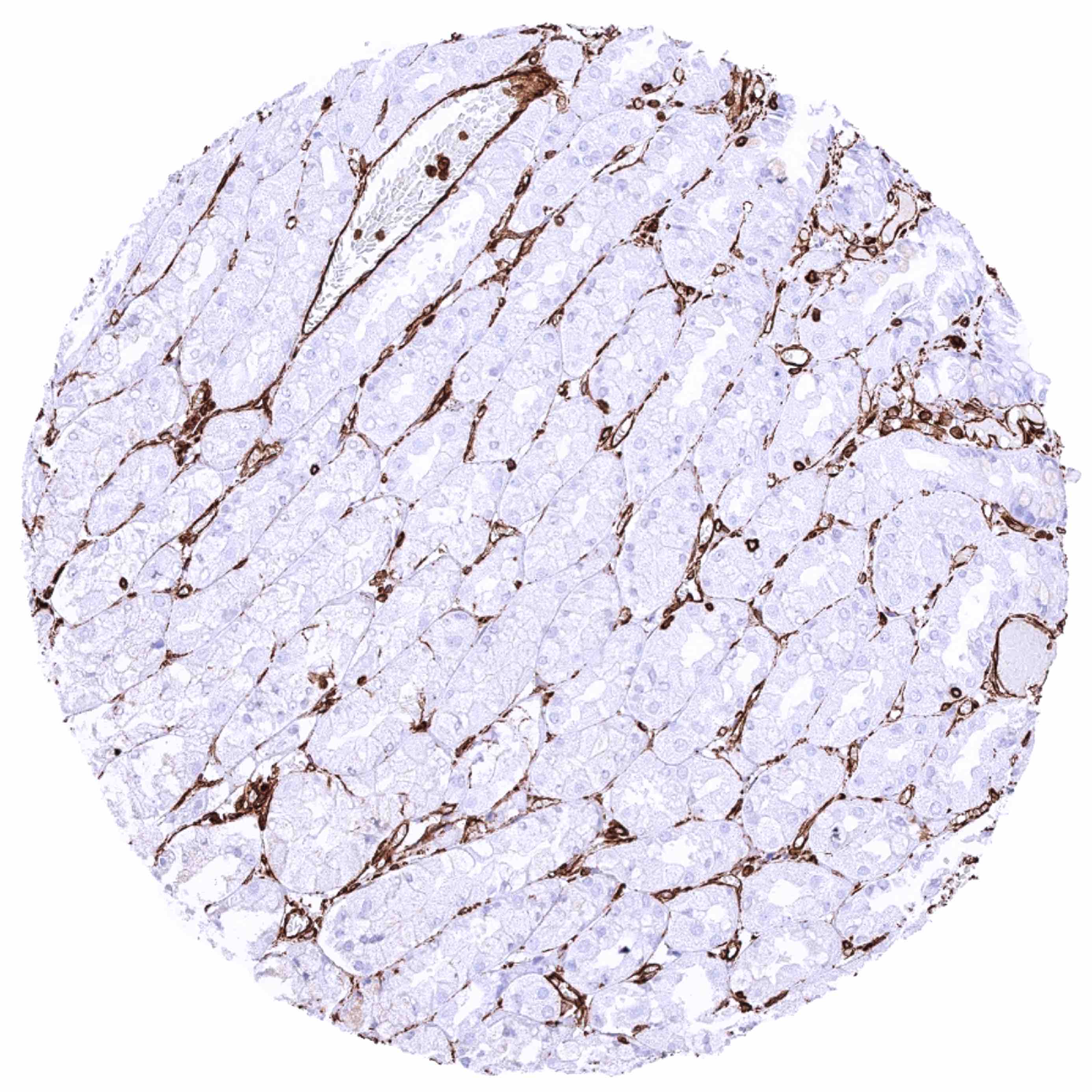
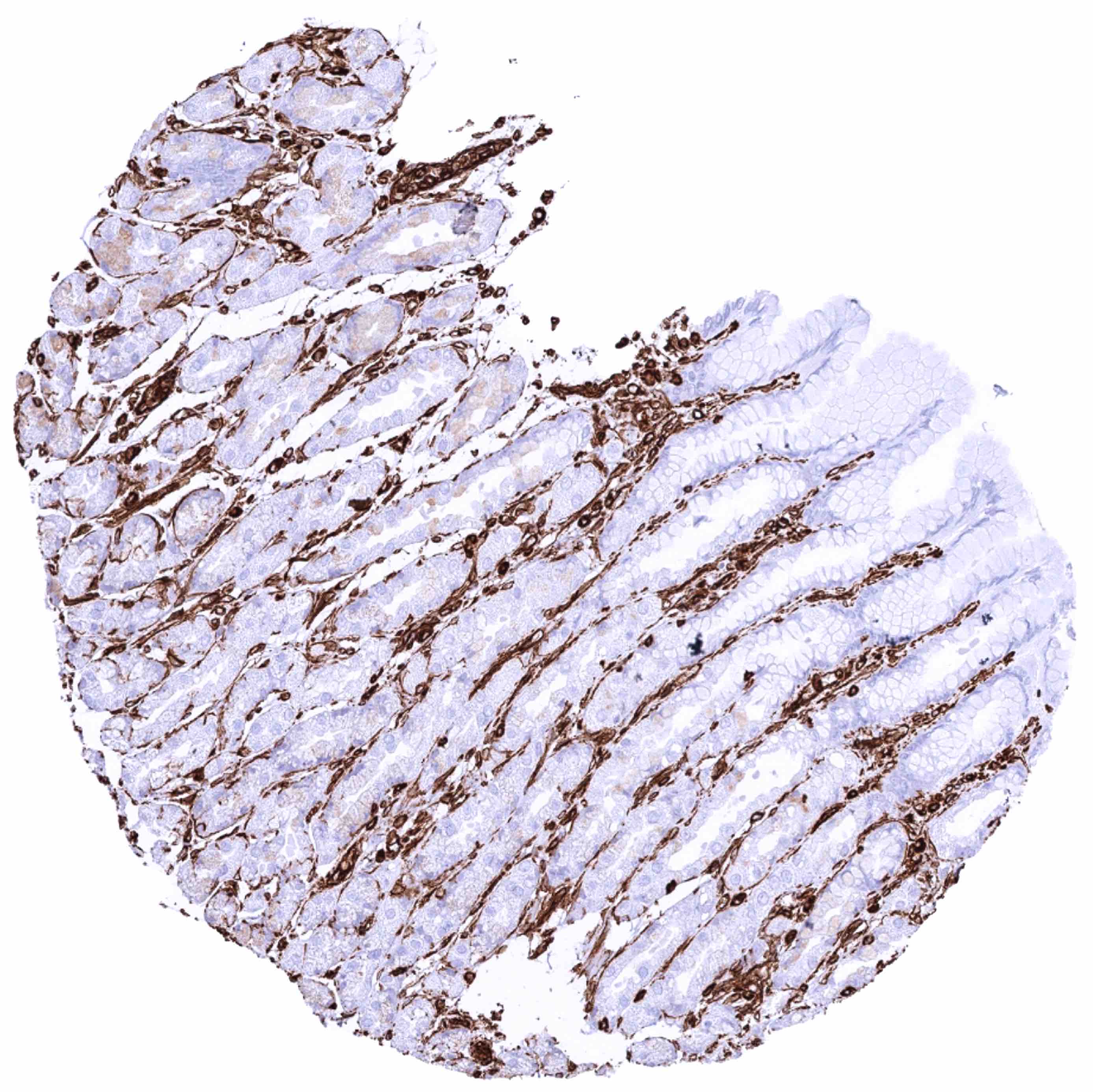
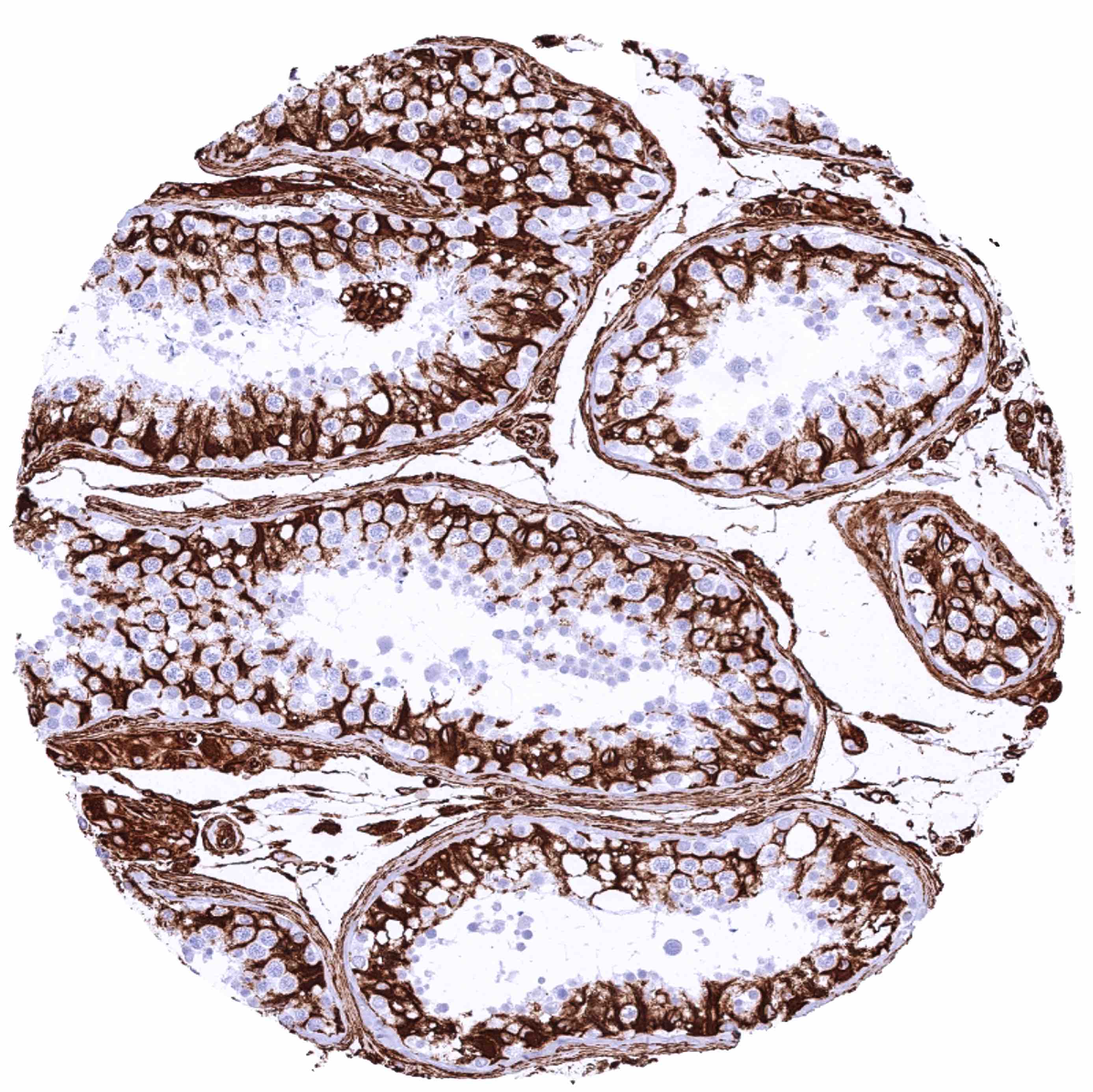
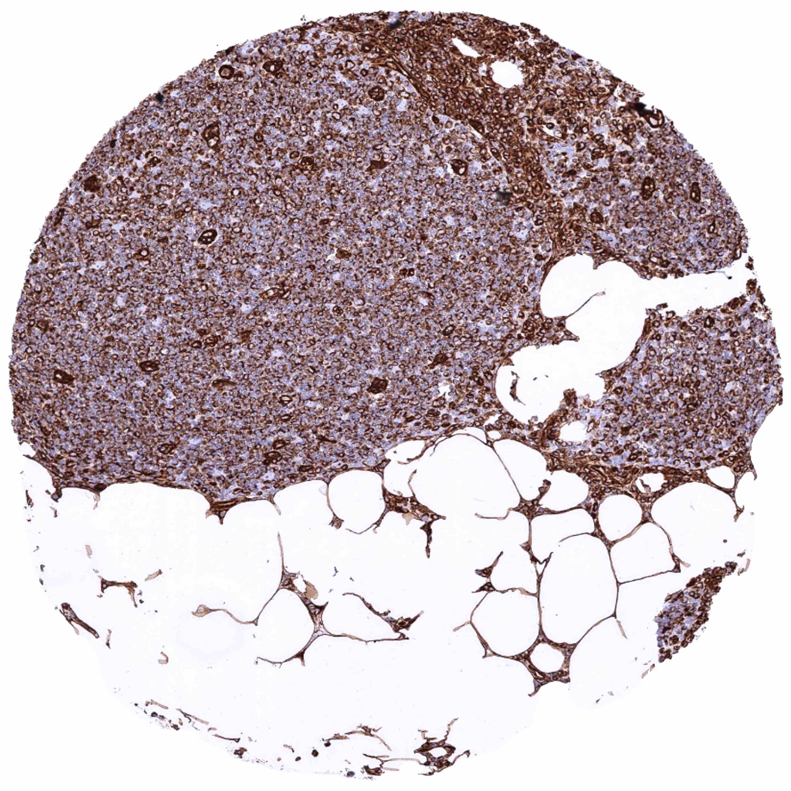
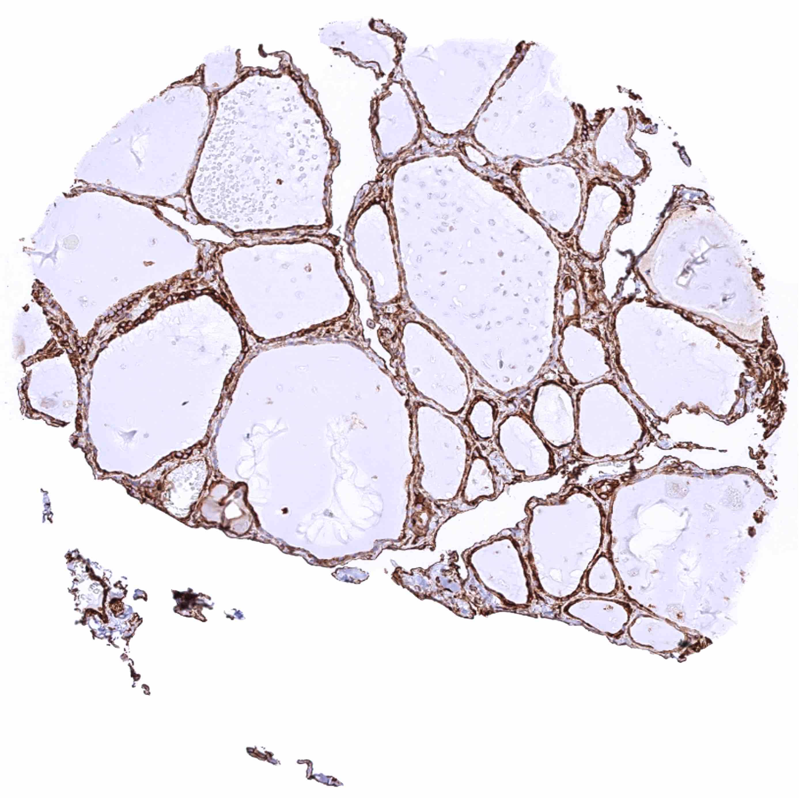
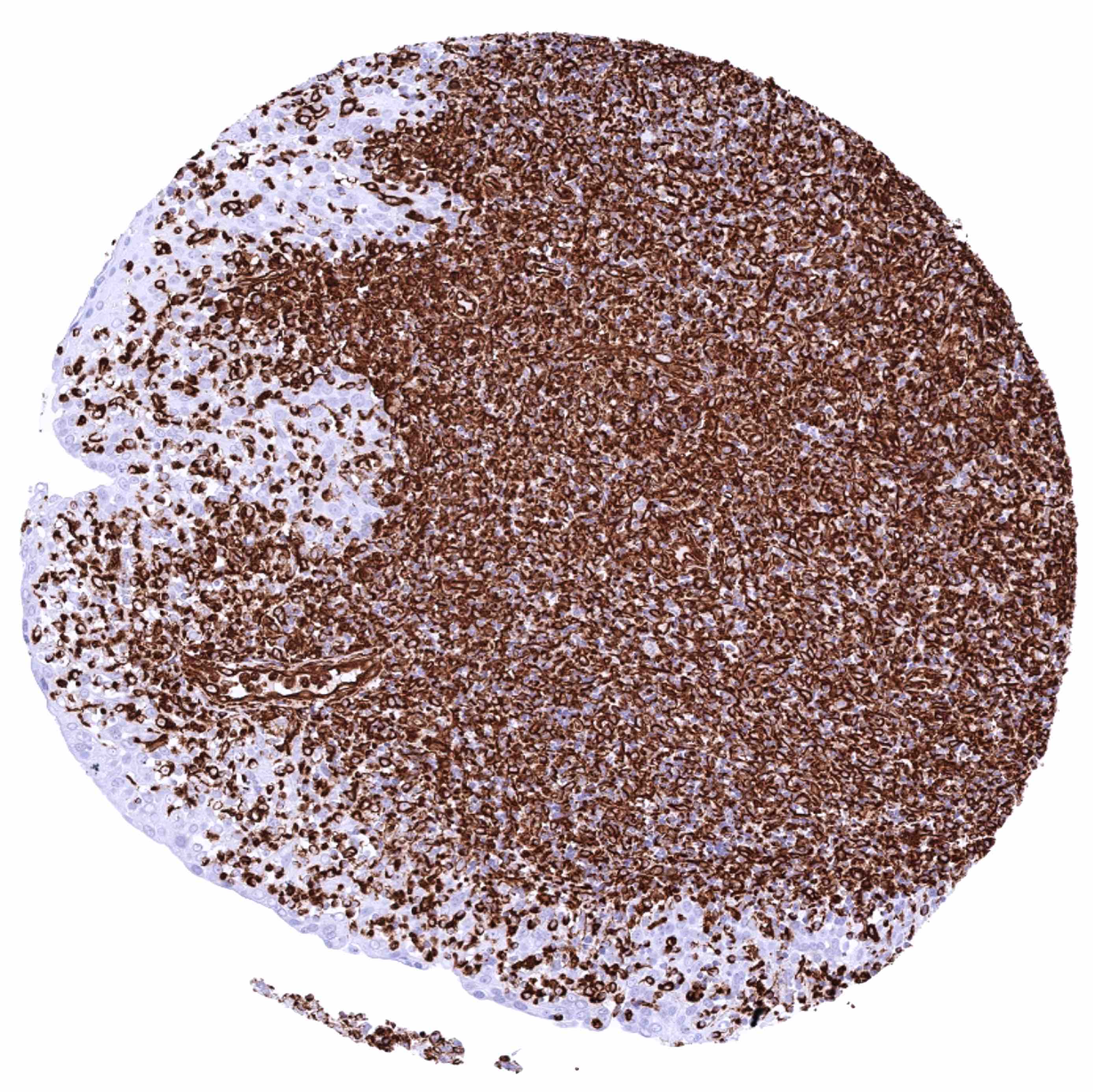
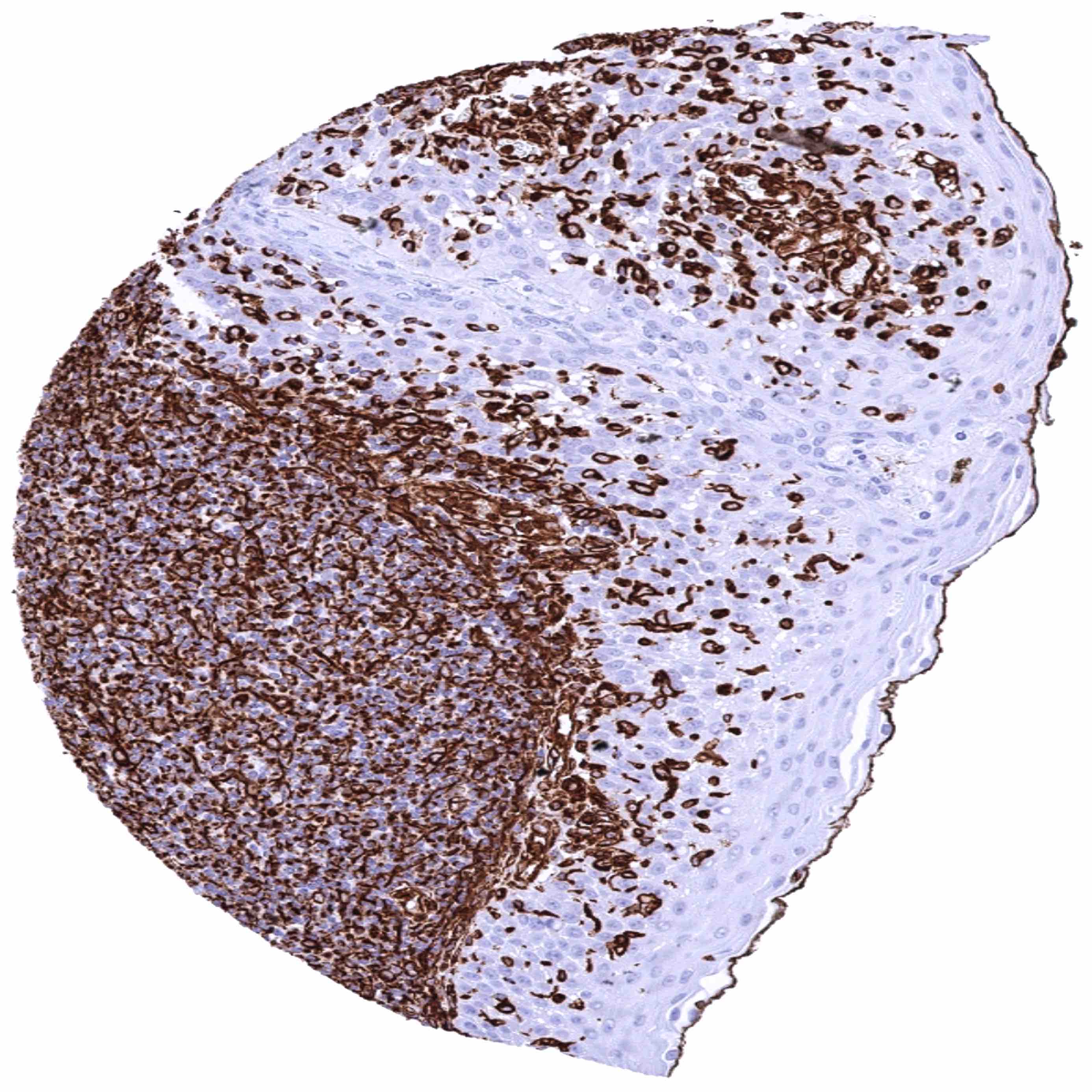
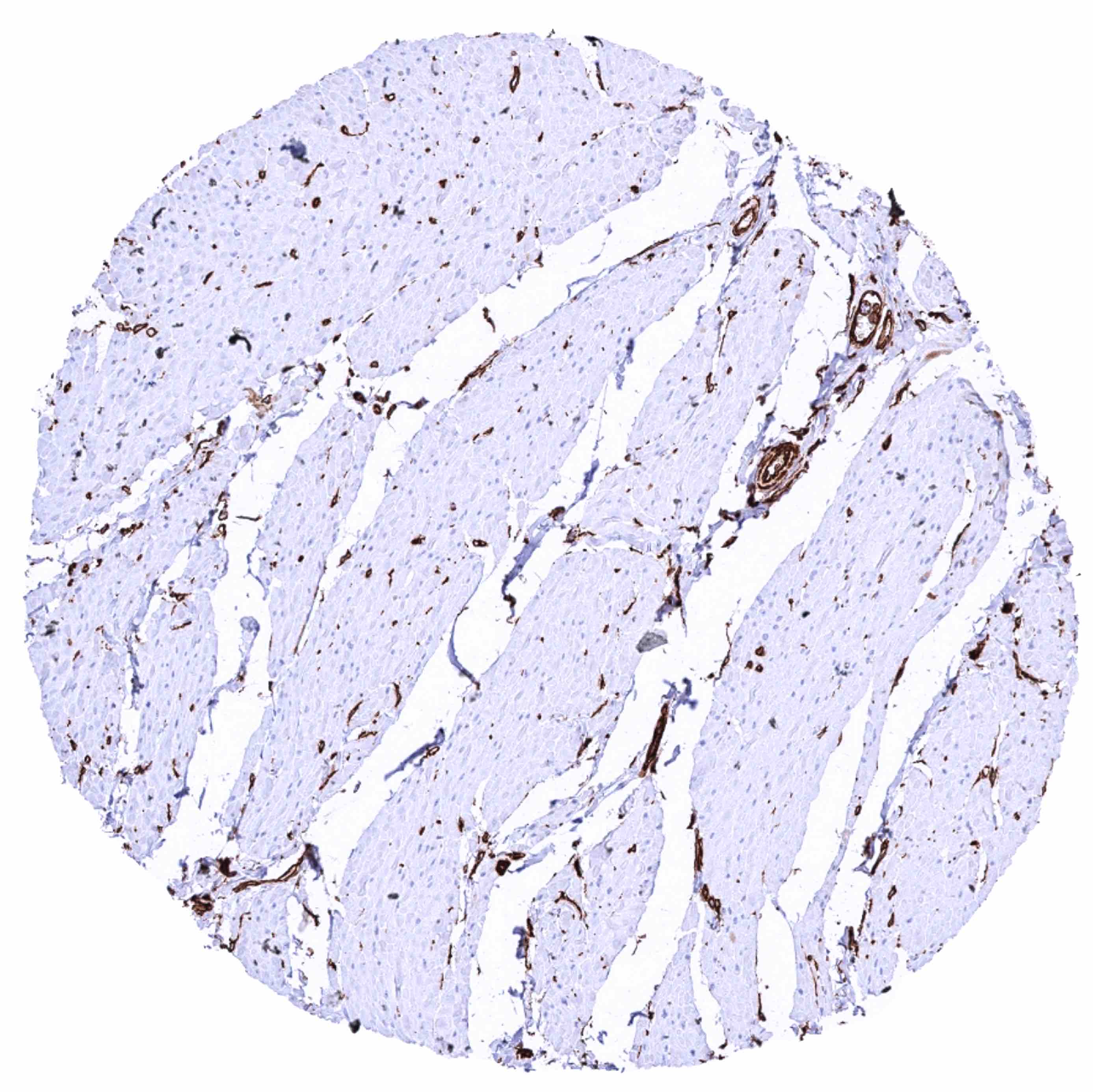
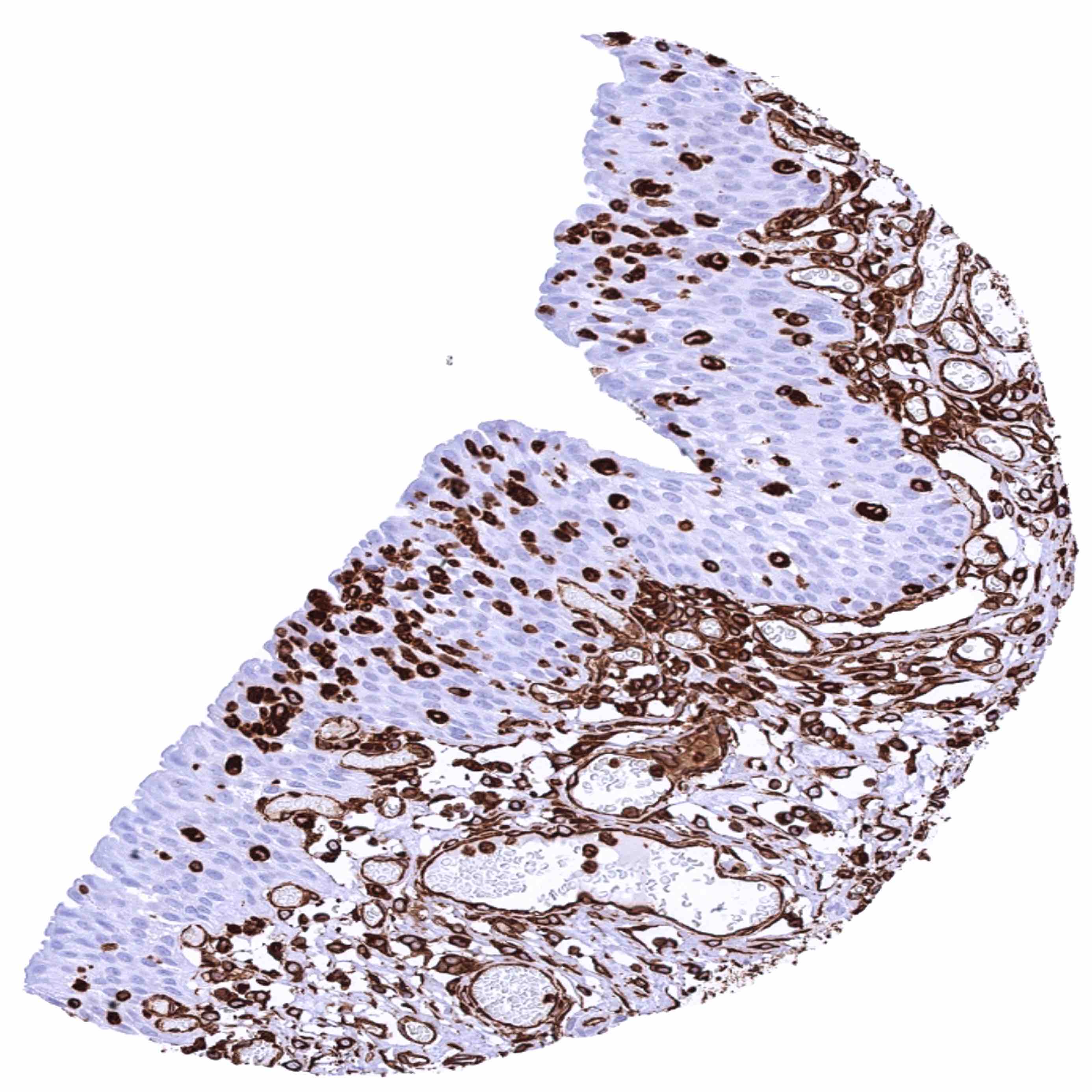
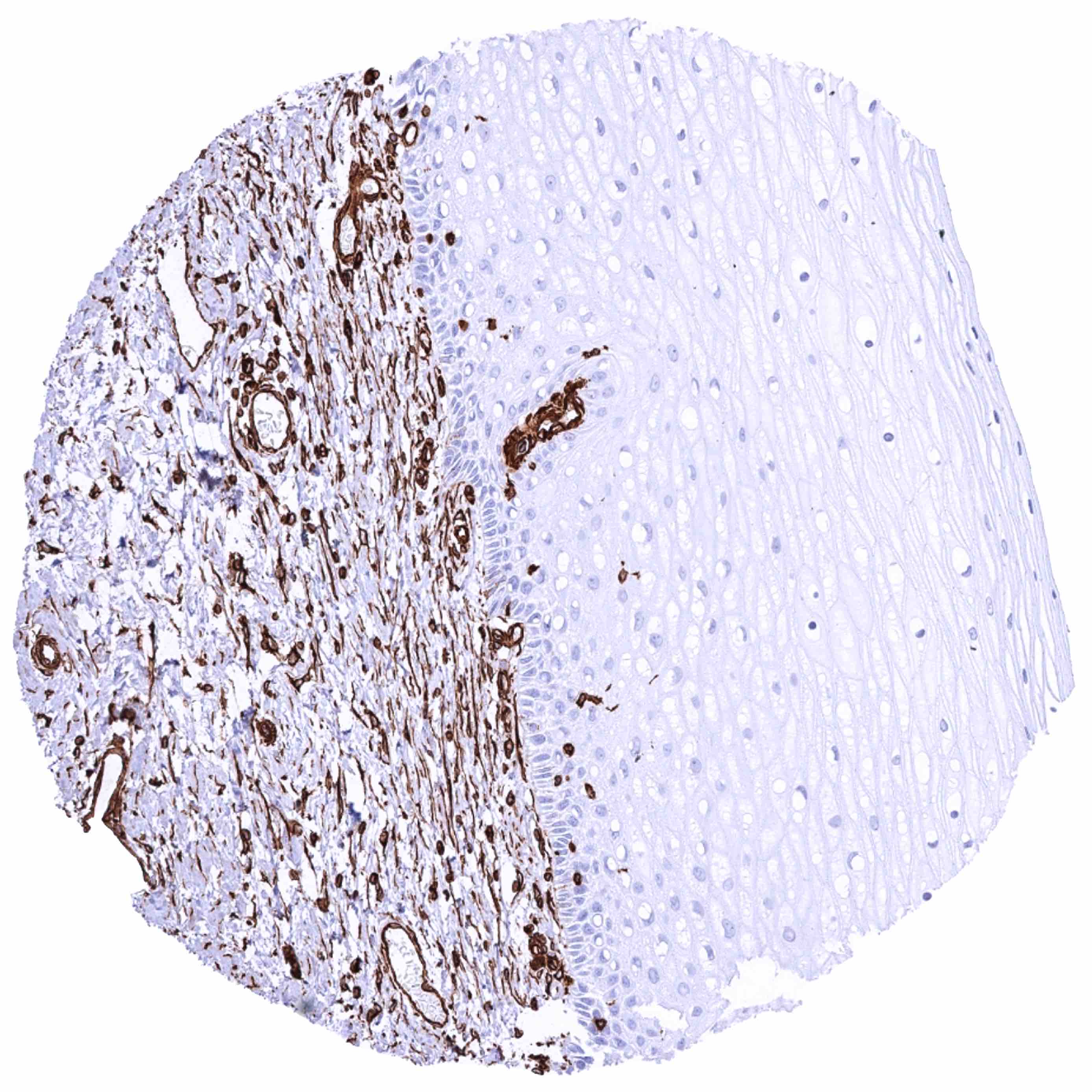
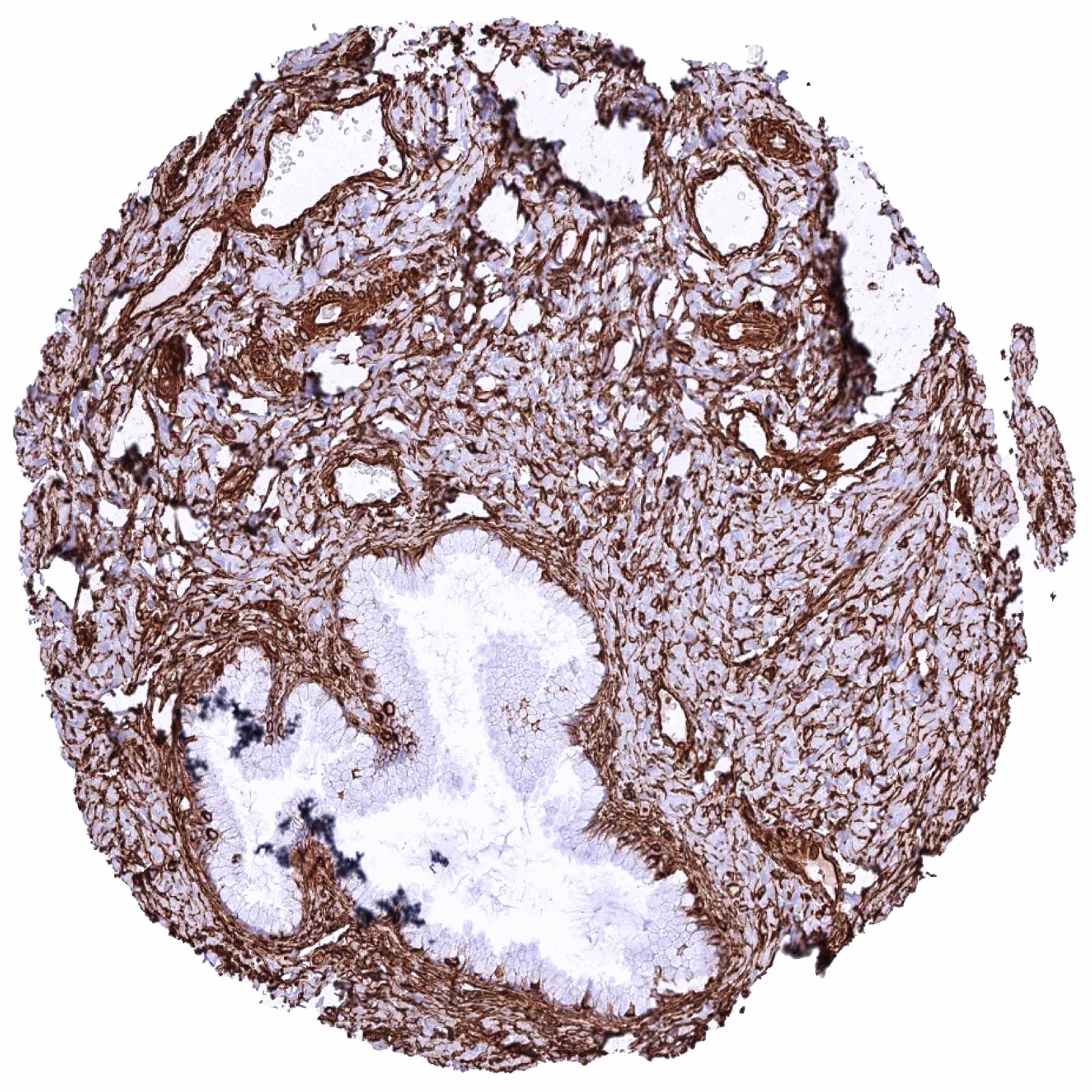
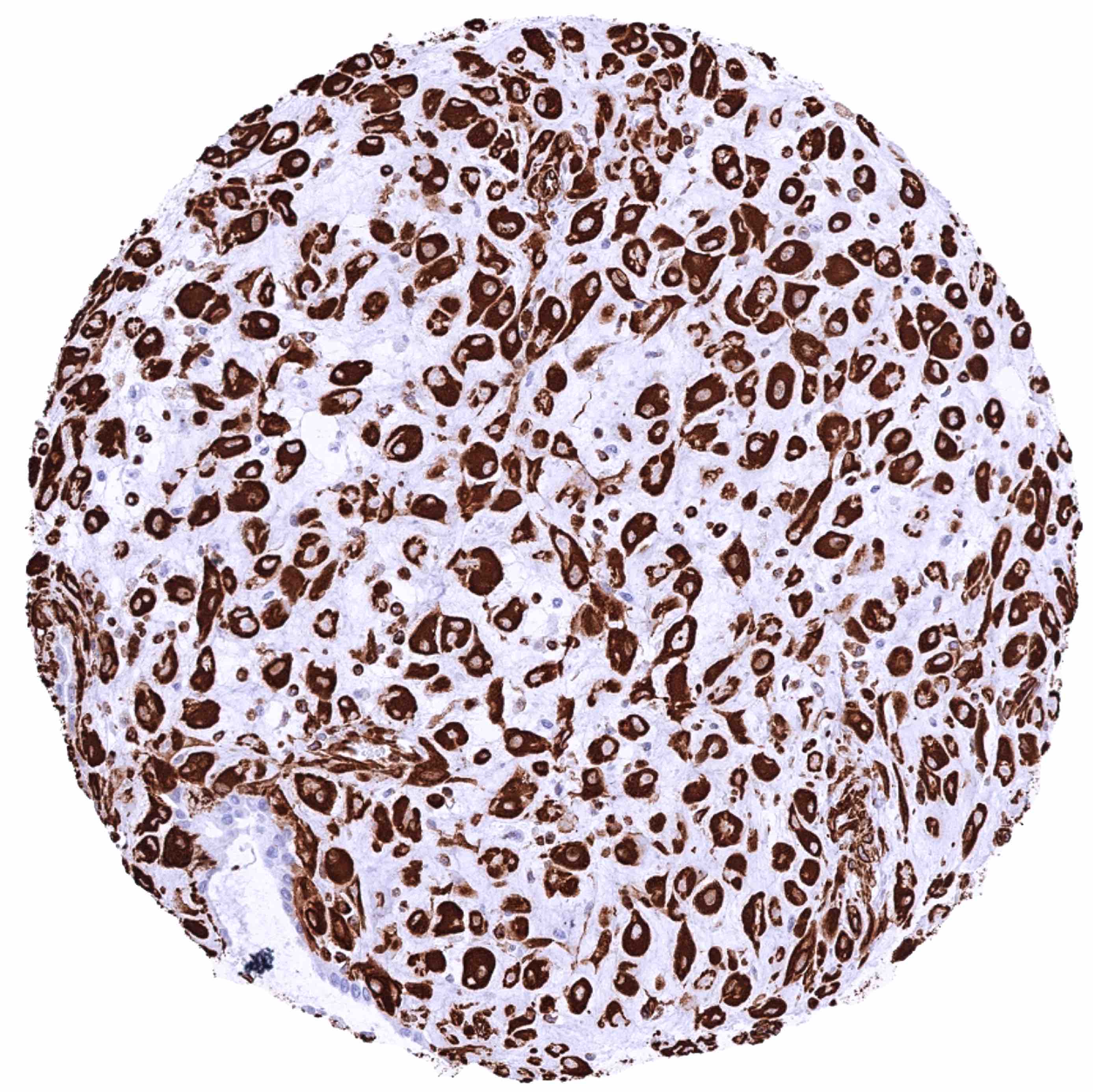
Vimentin antibody [HMV332] HistoMAXTM


Go to Cancer Tissue Gallery Go back to product page Go to HistoMAX page


Adrenal gland – Membranous vimentin staining of variable intensity of cortical cells
Aorta, media
Appendix, mucosa
Appendix, muscular wall
Bone marrow – Strong vimentin immunostaining of virtually all cells
Breast – Strong vimentin staining of myoepithelial-basal cells while luminal cells are negative
Bronchus, mucosa – Strong vimentin staining also occurs in intra-epithelial inflamatory cells
Cerebellum (white matter)
Cerebellum, cortex (molecular layer, purkinje cell layer, granule cell layer)
Cerebrum, grey matter – Strong vimentin staining of glial cells and endothelial cells of vessels
Cerebrum, white matter
Colon descendens, mucosa
Colon descendens, muscular wall
Duodenum, mucosa – Strong membranous vimentin positivity of mesenchymal cells and of lymphocytes. Epithelial cells are completely negative
Epididymis (Cauda) – Distinct membranous vimentin staining of a subset of epithelial cells
Epididymis (Cauda) – Strong membranous vimentin staining of a large subset of epithelial cells in this sample
Epididymis (Corpus) – Strong basolateral membranous vimentin staining of few basal cells
Esophagus, squamous epithelium – Strong vimentin staining occurs in intra-epithelial inflammatory cells
Fallopian tube, mucosa – Strong membranous vimentin staining of epithelial and stromal cells
Fat
Gallbladder, epithelium
Heart muscle
Ileum, mucosa
Kidney, cortex – Strong vimentin staining of all cell types of glomeruli and Bowman capsule as well as of muscular and endothelial cells in vessels. Epithelial cells are vimentin negative in this sample
Kidney, medulla – Few epithelial cells of collecting ducts show a moderate to strong basolateral membranous vimentin staining
Kidney, pelvis, urothelium (2)
Kidney, pelvis, urothelium
Liver – Strong staining of all Kupffer cells while sinusoidal endothelial cells show only a moderate staining intensity
Lung – Vimentin staining is particularly strong in alveolar macrophages
Lymph node – Strong vimentin staining of germinal centre dendritic cells-macrophages and of interfollicular lymphocytes
Ovary, stroma – Intense vimentin staining of all stromal cells
Pancreas – Acinar epithelial cells display a strong basolateral membranous (and cytoplasmic) vimentin staining. Moderate to strong vimentin staining of vascular smooth muscle cells
Pancreas – Epithelial cells do not show any vimentin staining in this sample
Parathyroid gland
Parotid gland – Distinct vimentin staining of fat cells, myoepithelial cells, stroma and blood vessels
Pituitary gland, anterior lobe – Strong vimentin staining of few epithelial cells in this sample
Pituitary gland, posterior lobe – Strong vimentin staining of pituicytes and fibres
Placenta (amnion) – Strong vimentin staining of amnion cells
Placenta (chorion) – Chorion cells are vimentin negative
Placenta, early – Strong vimentin staining ofvessels and stromal cells while trophoblastic cells are strictly negative
Placenta, mature – Strong vimentin staining of vessels and stromal cells while trophoblastic cells are strictly negative
Prostate – Epithelial cells do not display vimentin staining in this sample.
Prostate – Moderate to strong basolateral membranous vimentin staining of most basal cells
Prostate, mucosa
Seminal vesicle – Moderate to strong basolateral membranous vimentin staining of most basal cells
Sinus paranasales – Strong vimentin staining also occurs in intra-epithelial inflamatory cells
Skeletal muscle, tongue – Strong vimentin staining of blood vessels
Skin – Strong vimentin staining of intra-epidermal Langerhans cells
Spleen
Stomach, antrum
Stomach, corpus (2)
Stomach, corpus
Testis – Sertoli and Leydig cells do show strong membranous vimentin positivity
Thymus – Vimentin staining is stronger in medullary than in cortical lymphocytes
Thyroid gland – Strong vimentin staining of follicular epithelial cells and of stromal cells
Tonsil – Strong vimentin staining also occurs in intra-epithelial inflammatory cells
Tonsil – Strong vimentin staining of germinal centre dendritic cells and of interfollicular lymphocytes
Tonsil, surface epithelium
Urinary bladder, muscular wall
Urinary bladder, urothelium – Strong vimentin staining occurs in intra-urothelial inflammatory cells
Uterus, ectocervix – Vimentin staining occurs in intra-epithelial Langerhans cells
Uterus, endocervix – Strong vimentin staining of stroma cells while all epithelial cells are negative
Uterus, endometrium (pregnancy) – Strong membranous vimentin staining of decidua cells while epithelial cells are negative
Uterus, endometrium (proliferation) – Strong membranous vimentin staining of epithelial and stromal cells
Uterus, endometrium (secretion) – Strong membranous vimentin staining of epithelial and stromal cells
Uterus, endometrium (secretion) – Strong vimentin staining of stromal cells. A subset of epithelial cells display a strong basolateral membranous vimentin staining in this sample
Uterus, myometrium – Strong vimentin staining of all smooth mucular cells

| TOP |