Normal Tissue Gallery- GTX04458

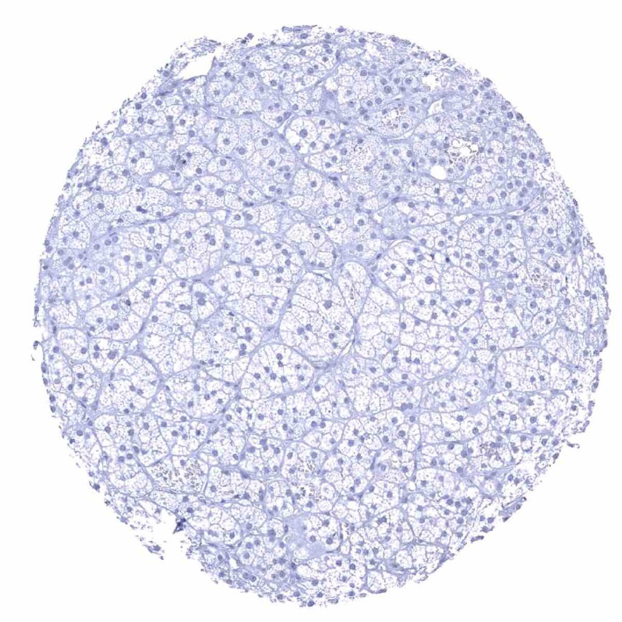
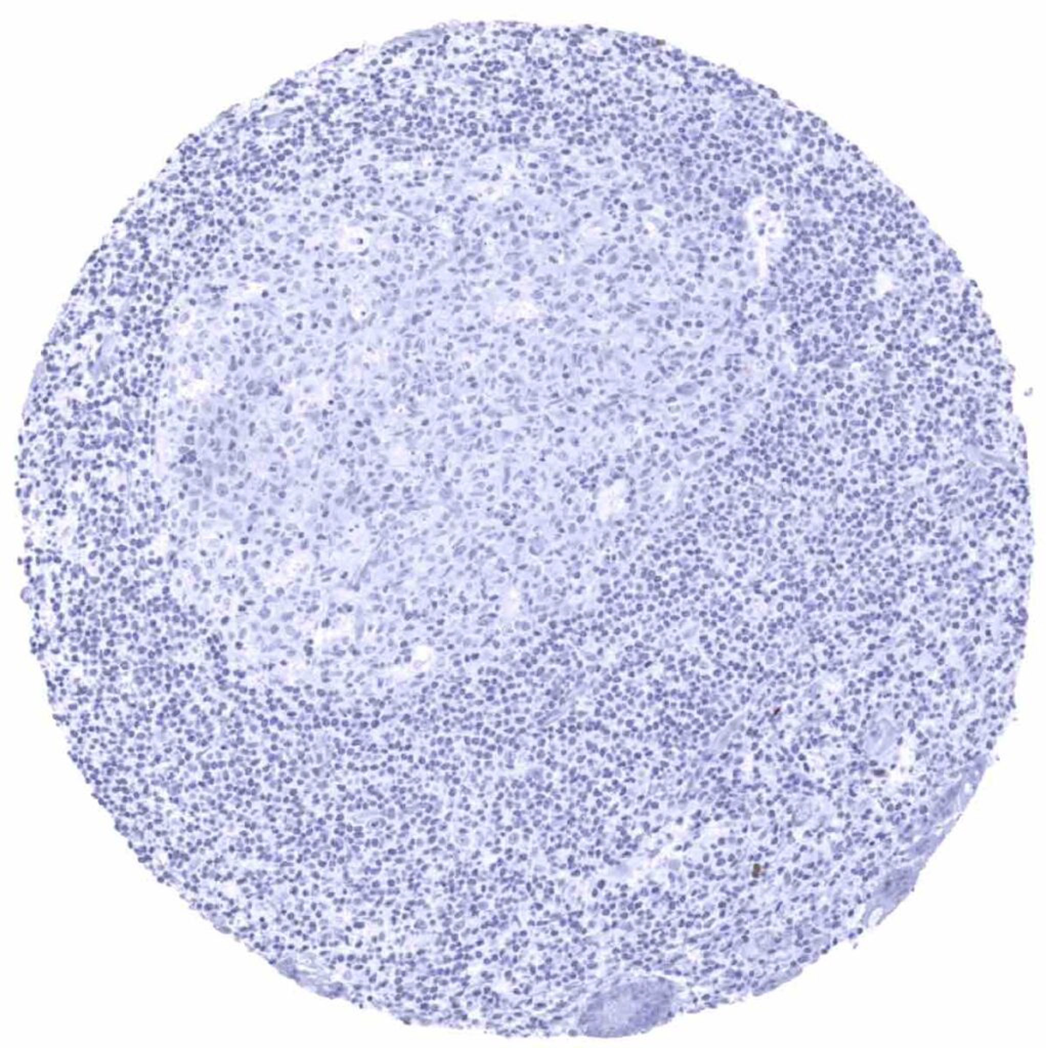
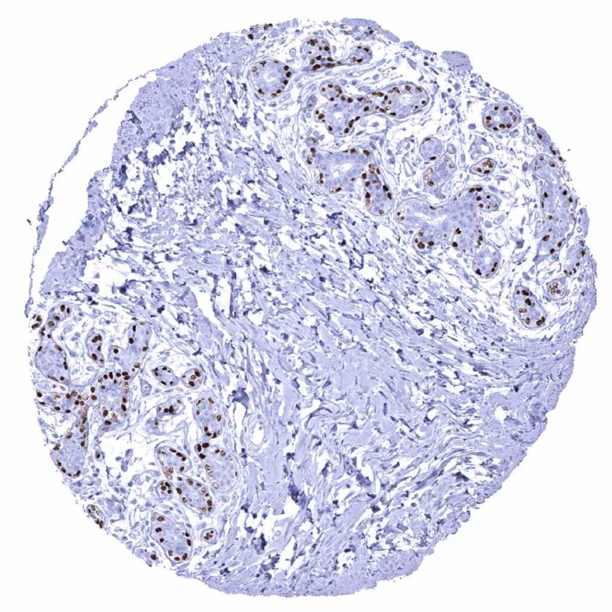
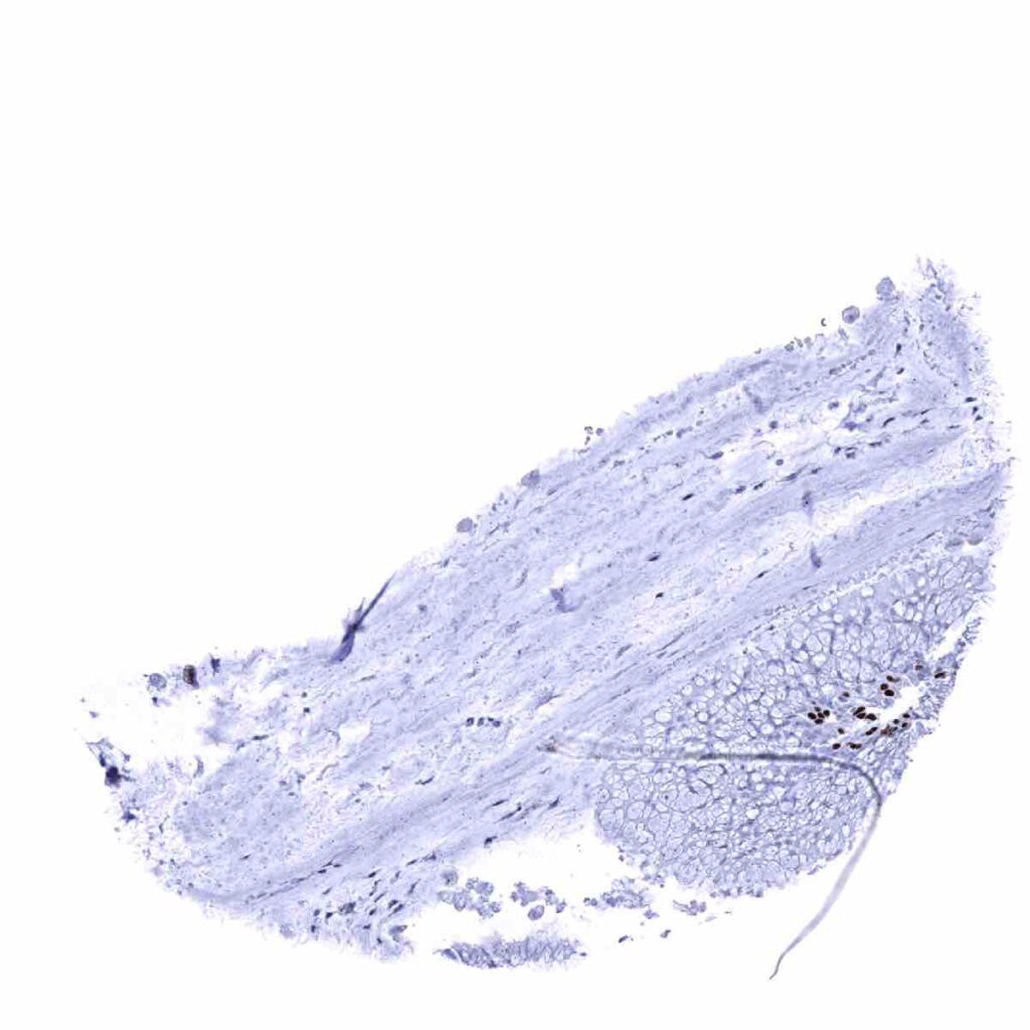
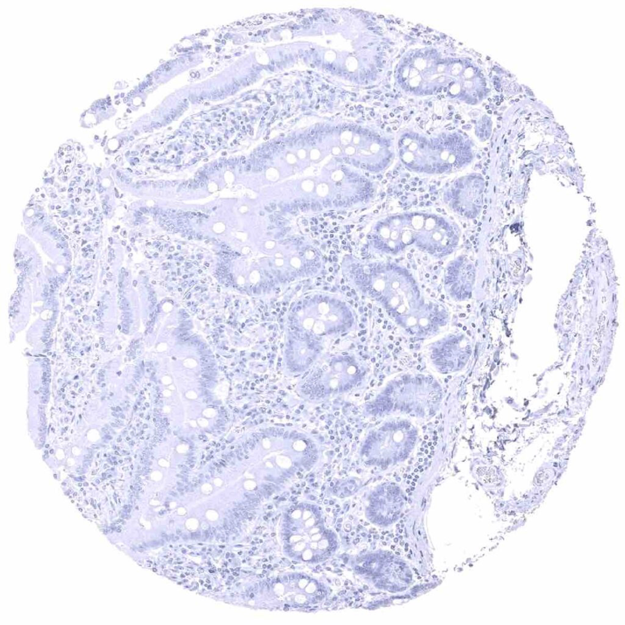
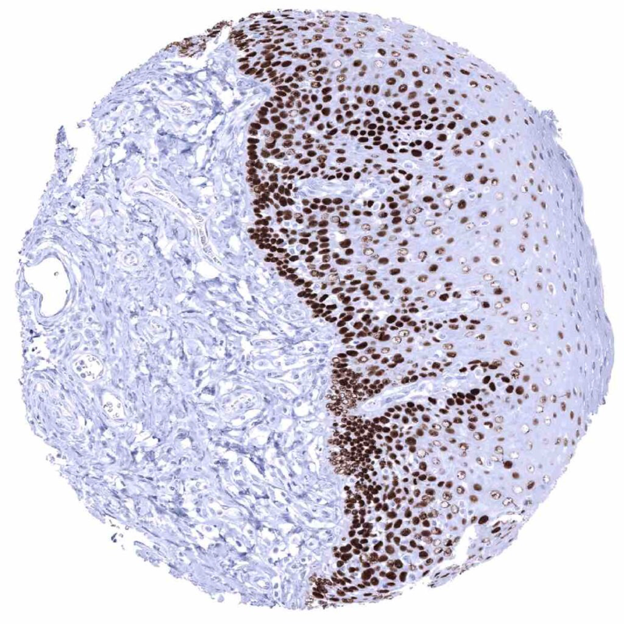
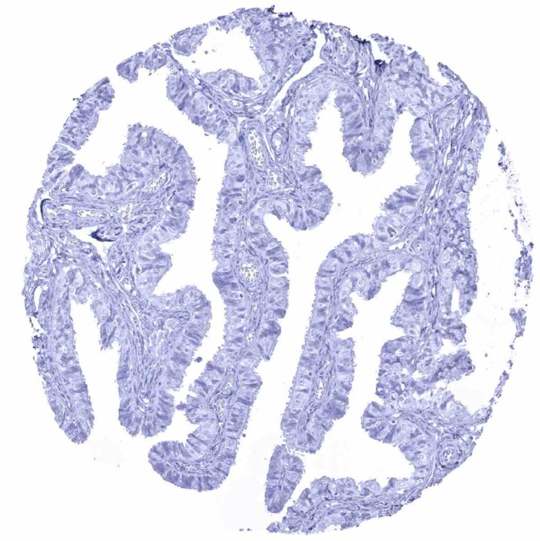
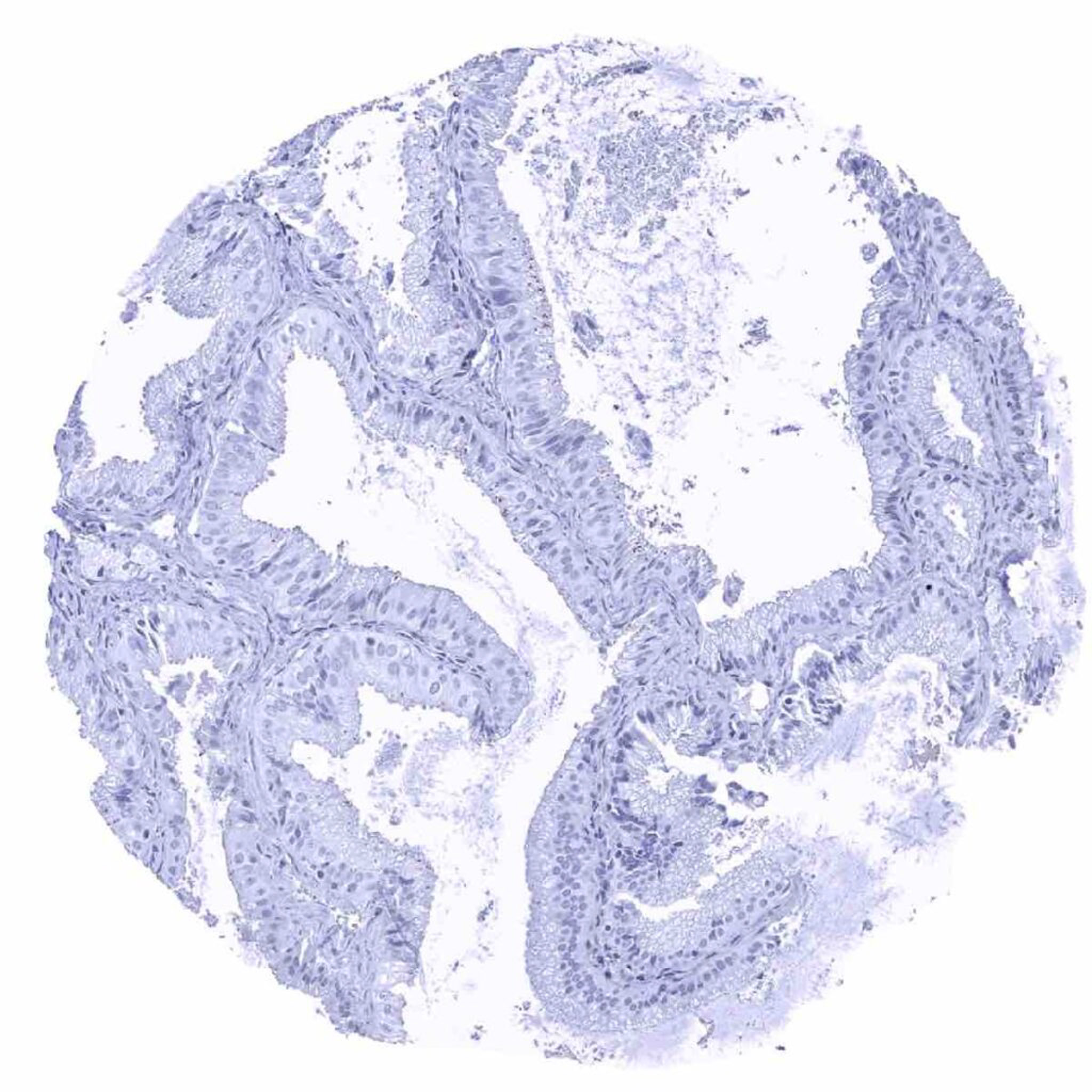
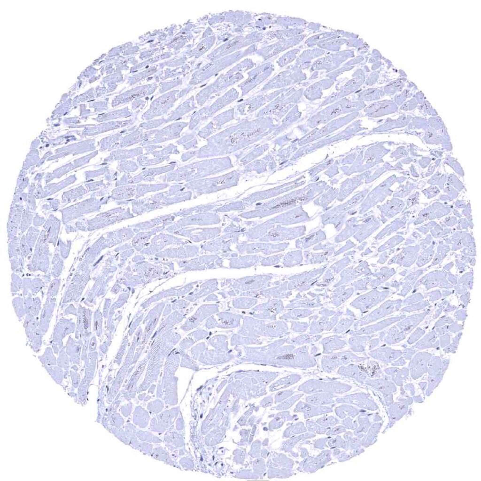
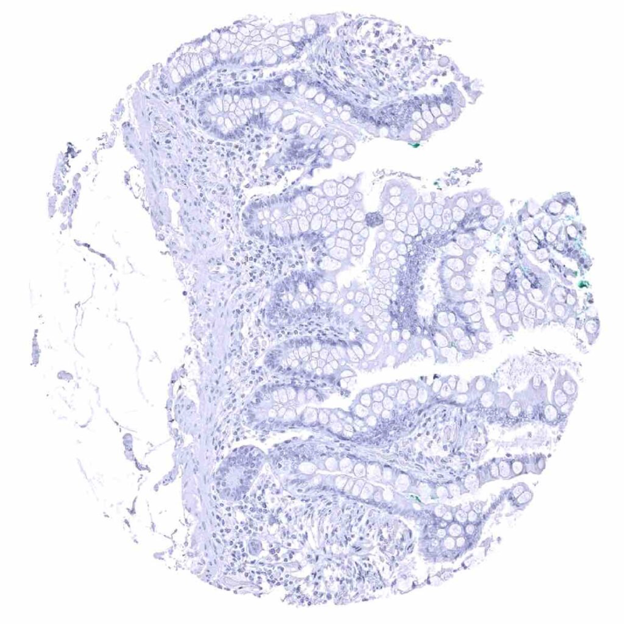
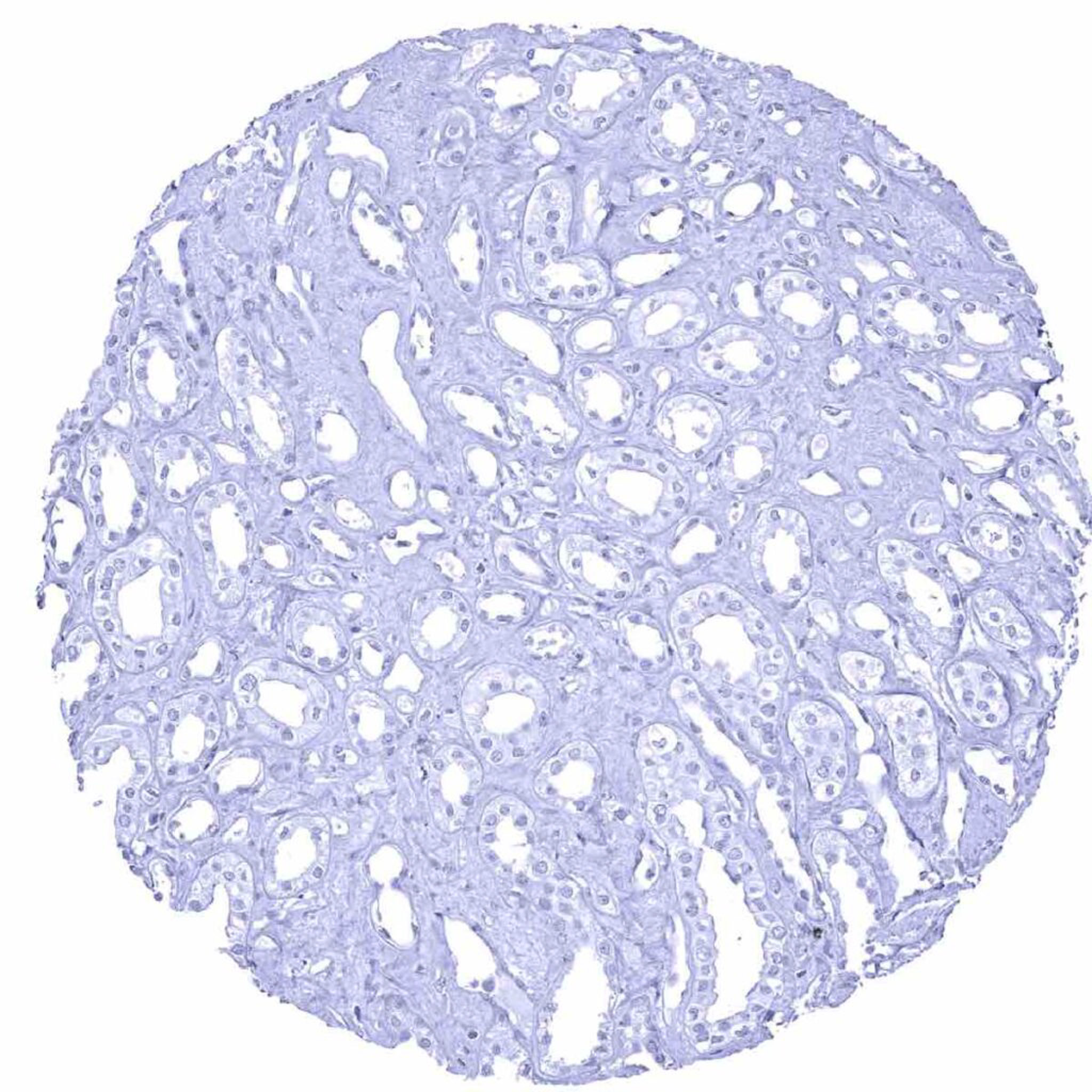
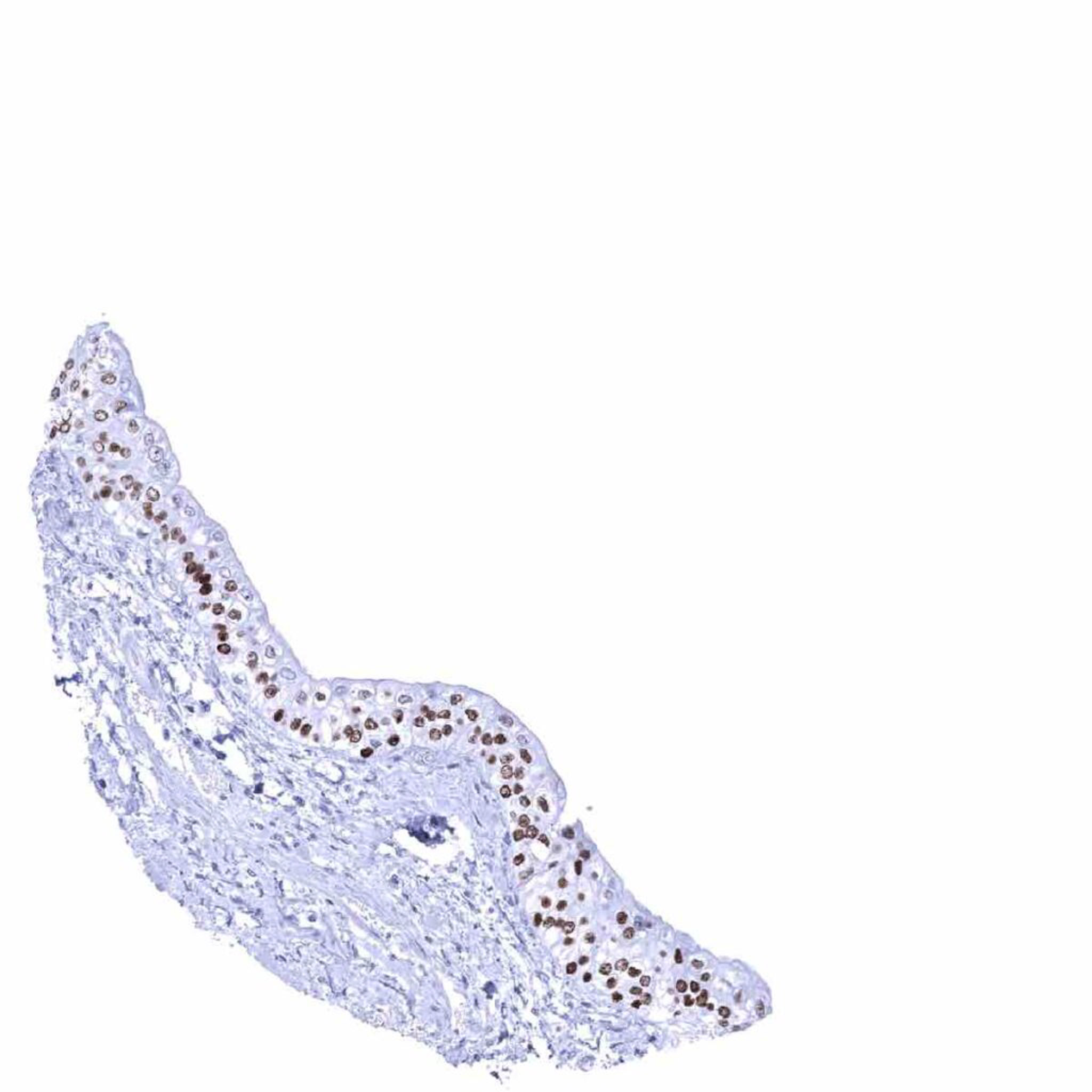
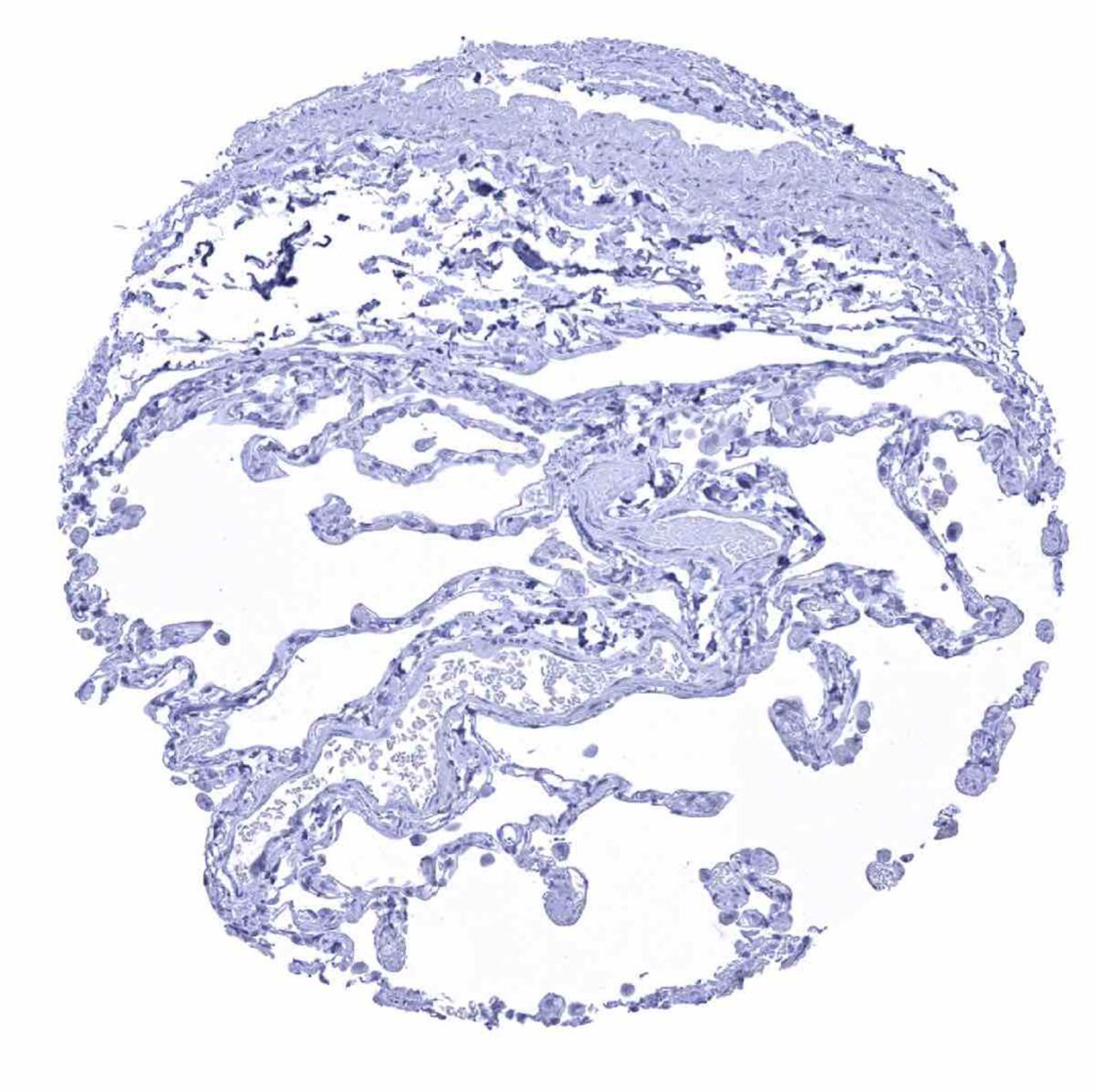
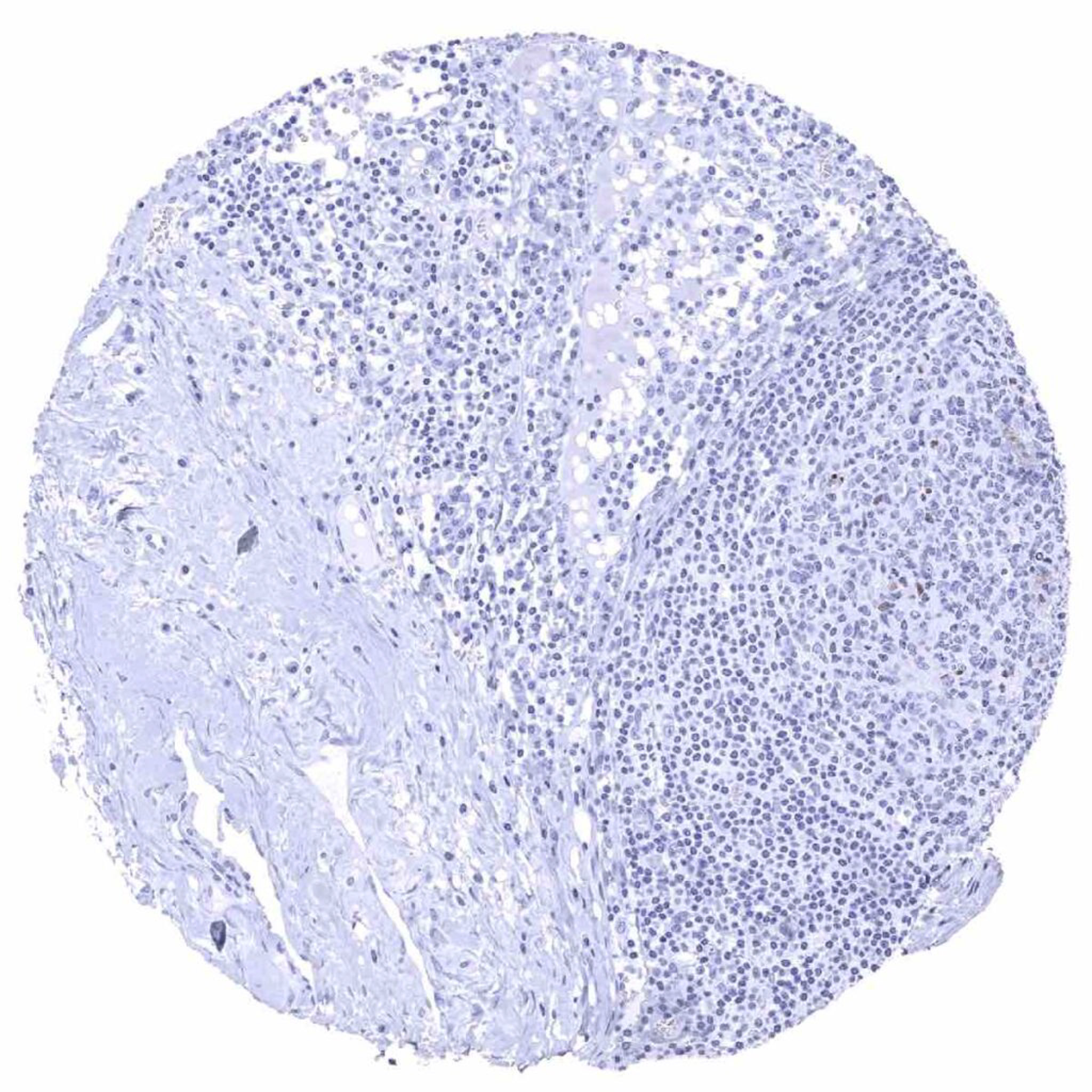
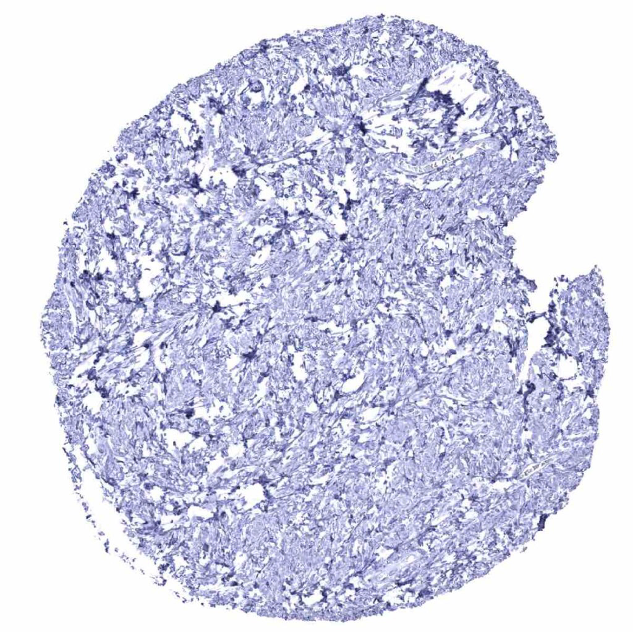
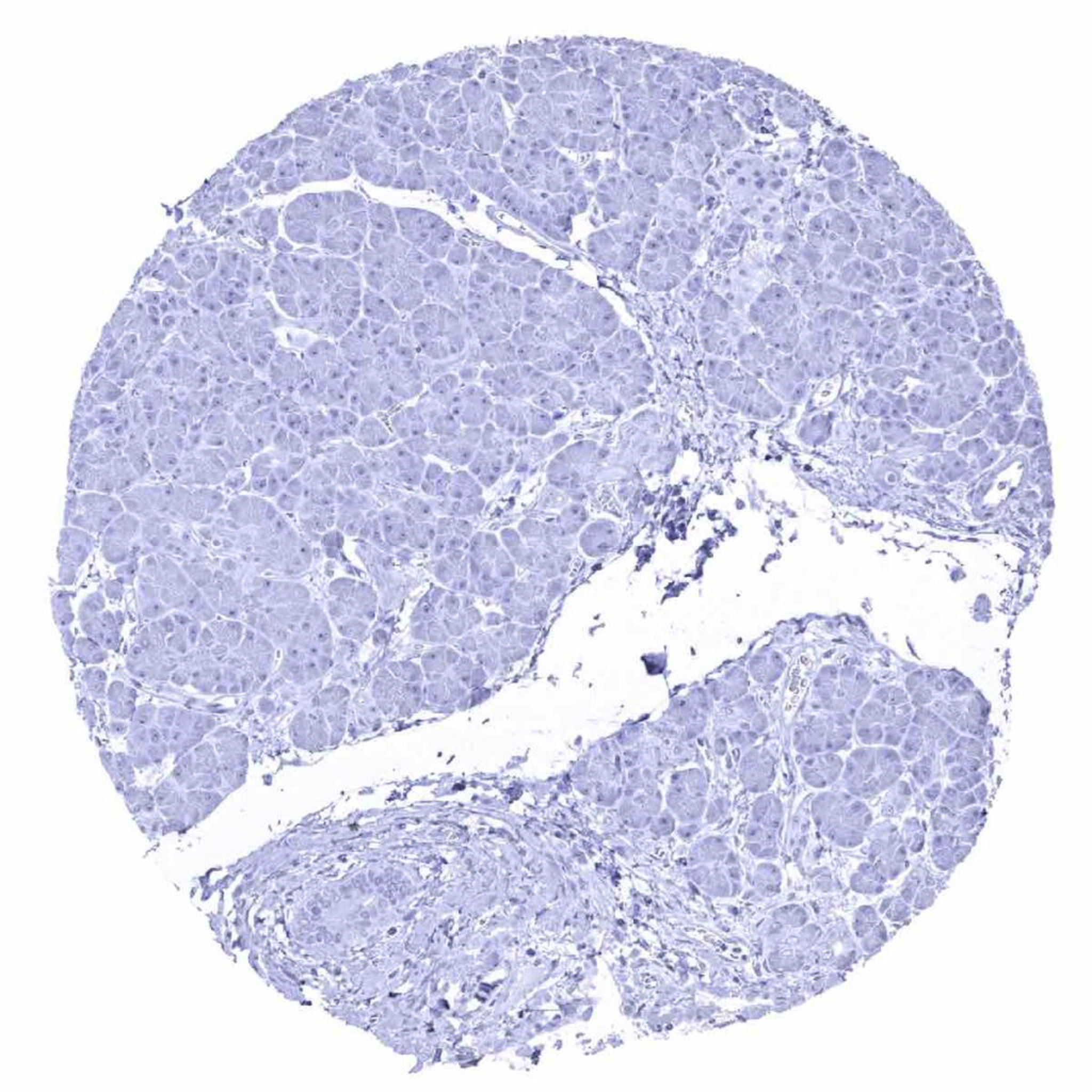
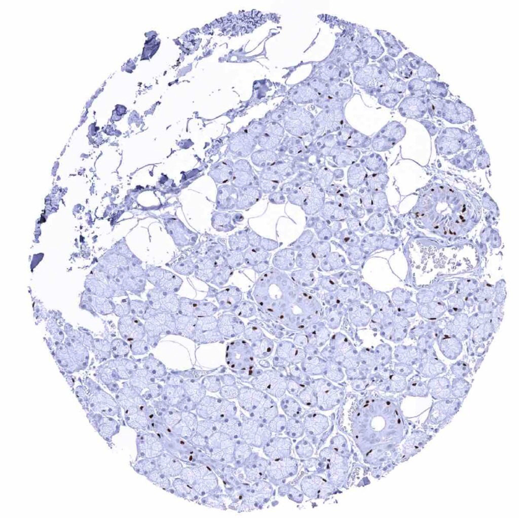
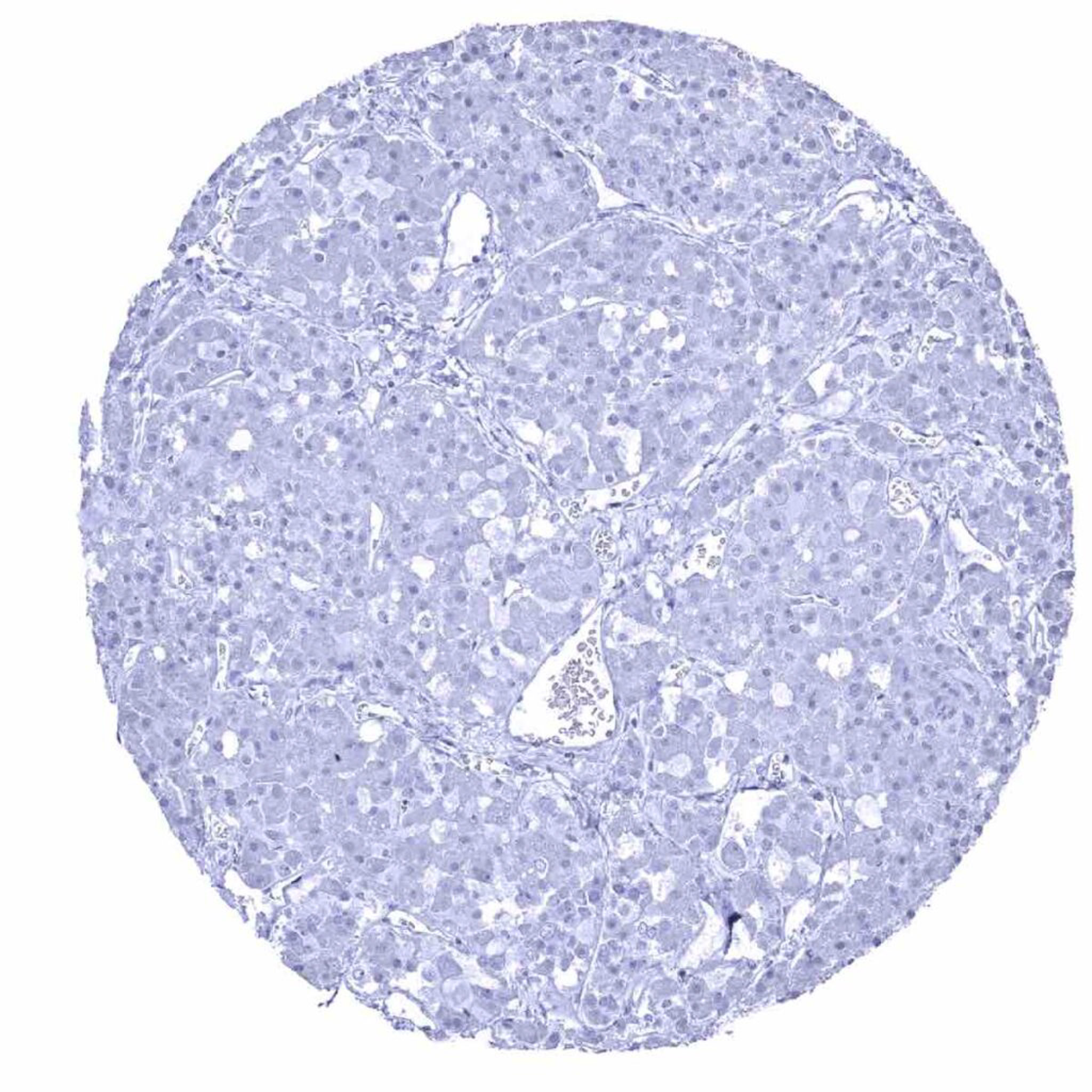
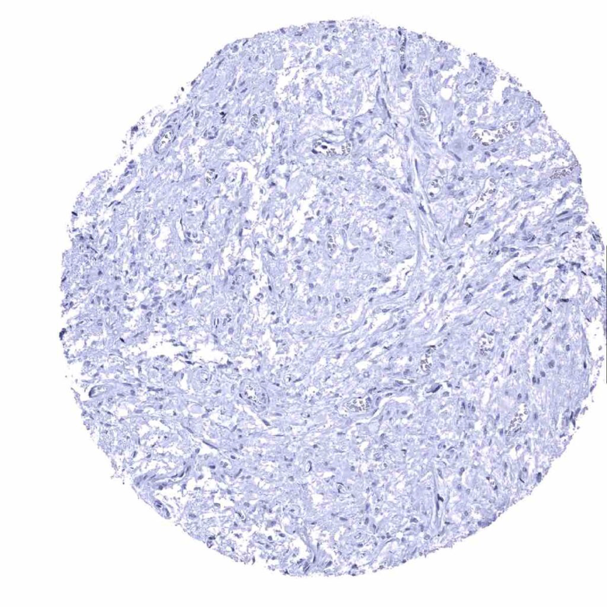
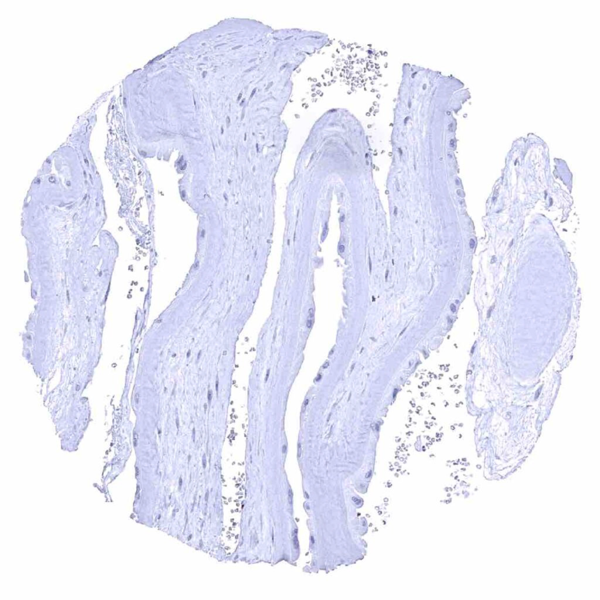
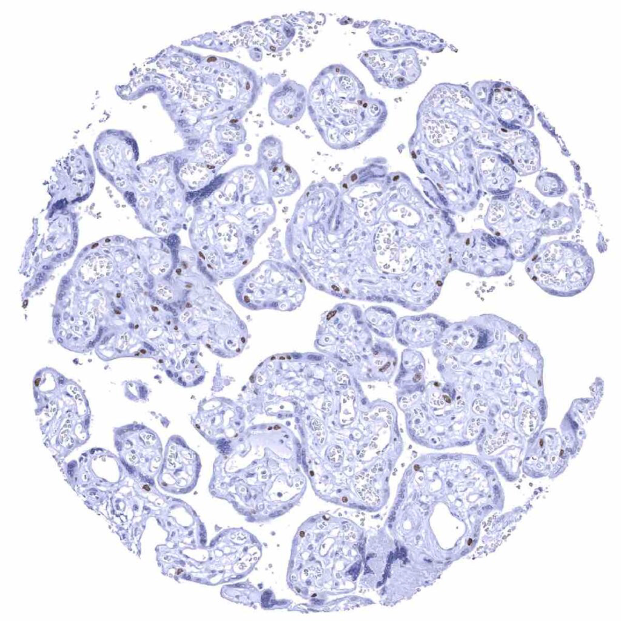
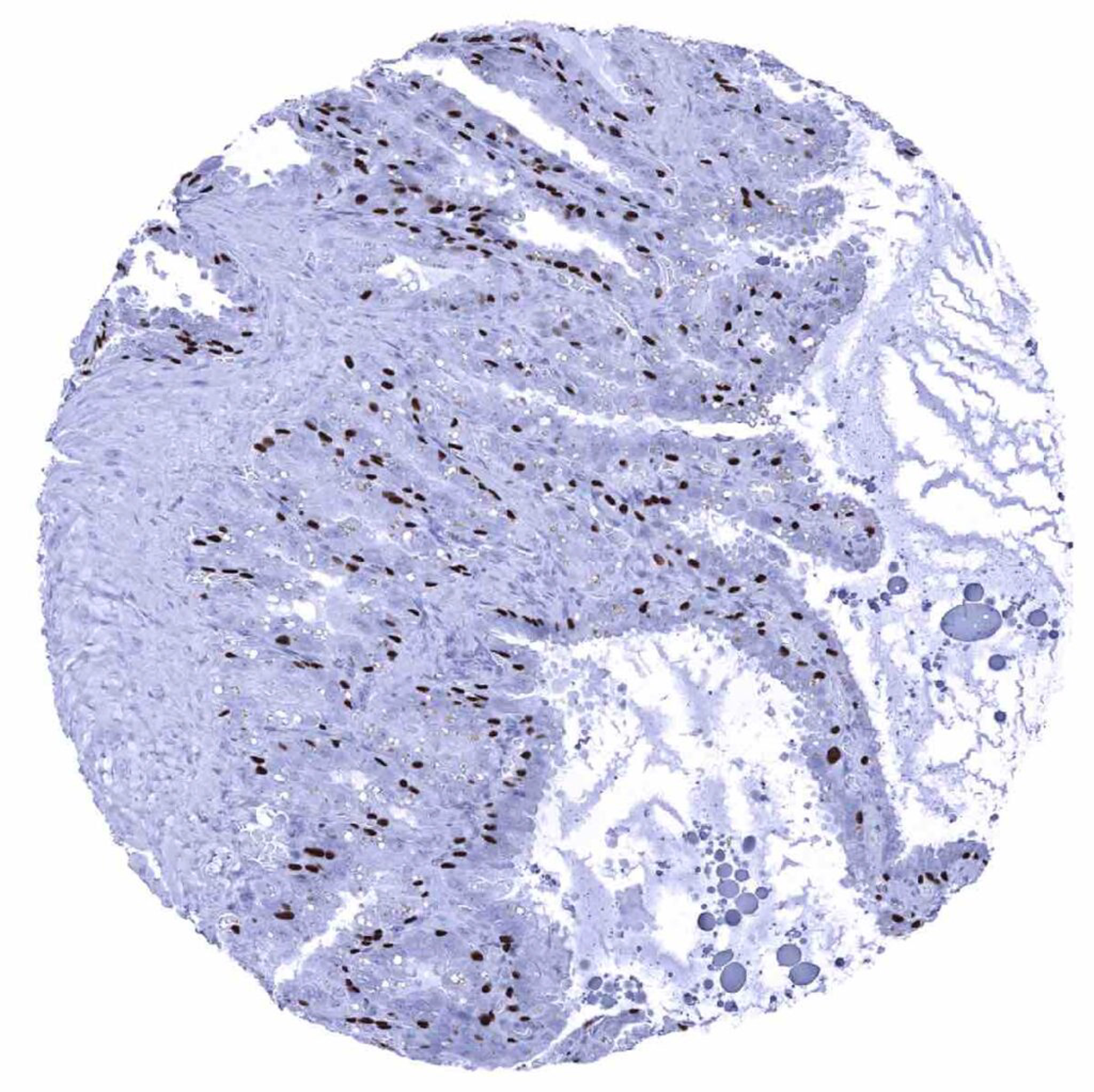
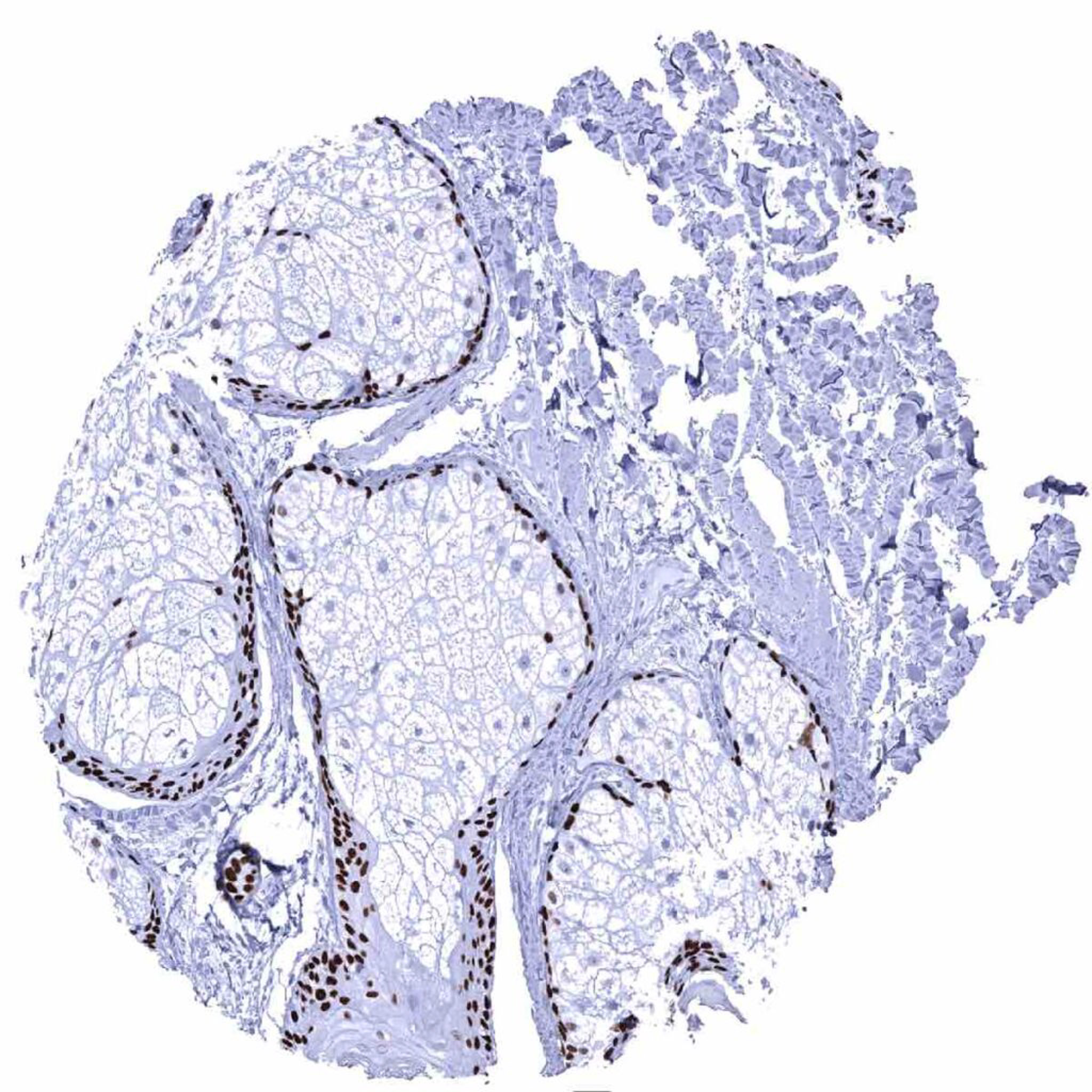
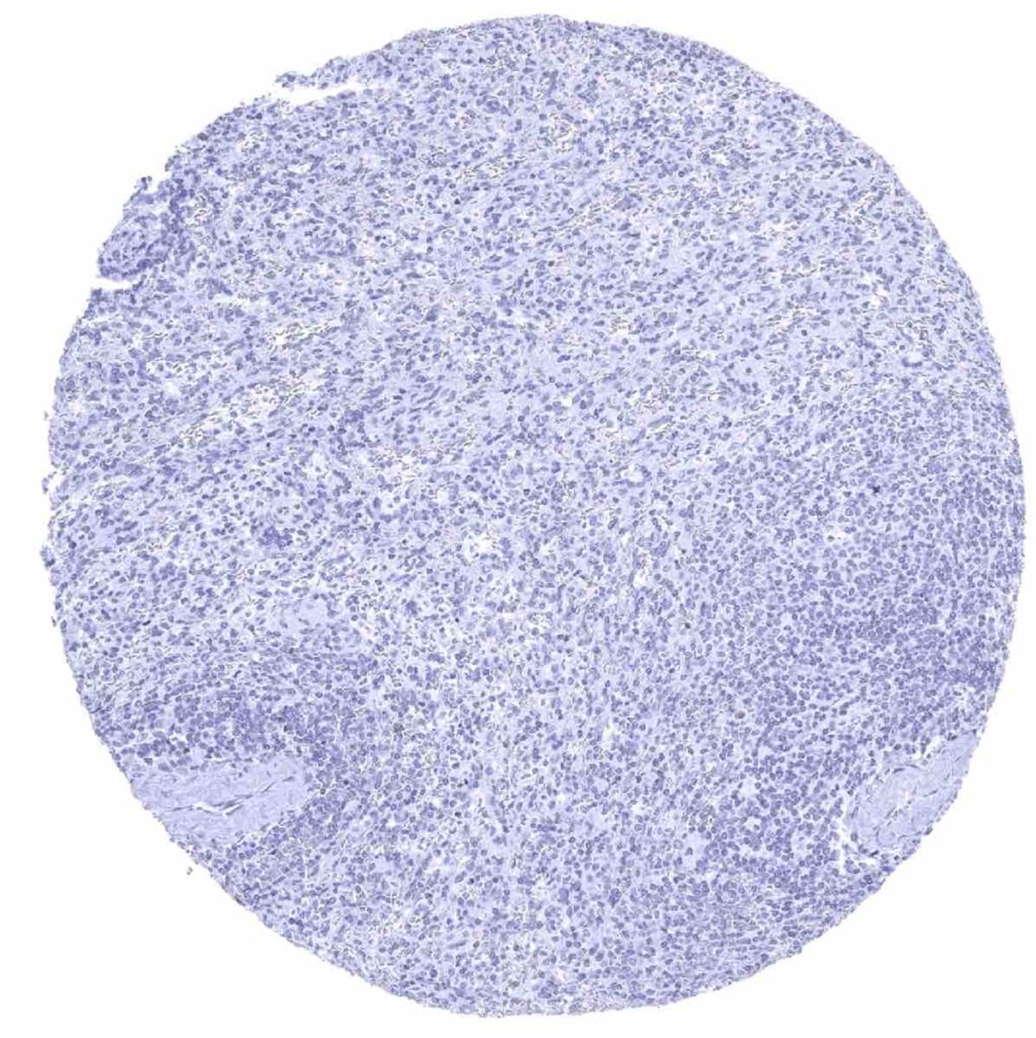
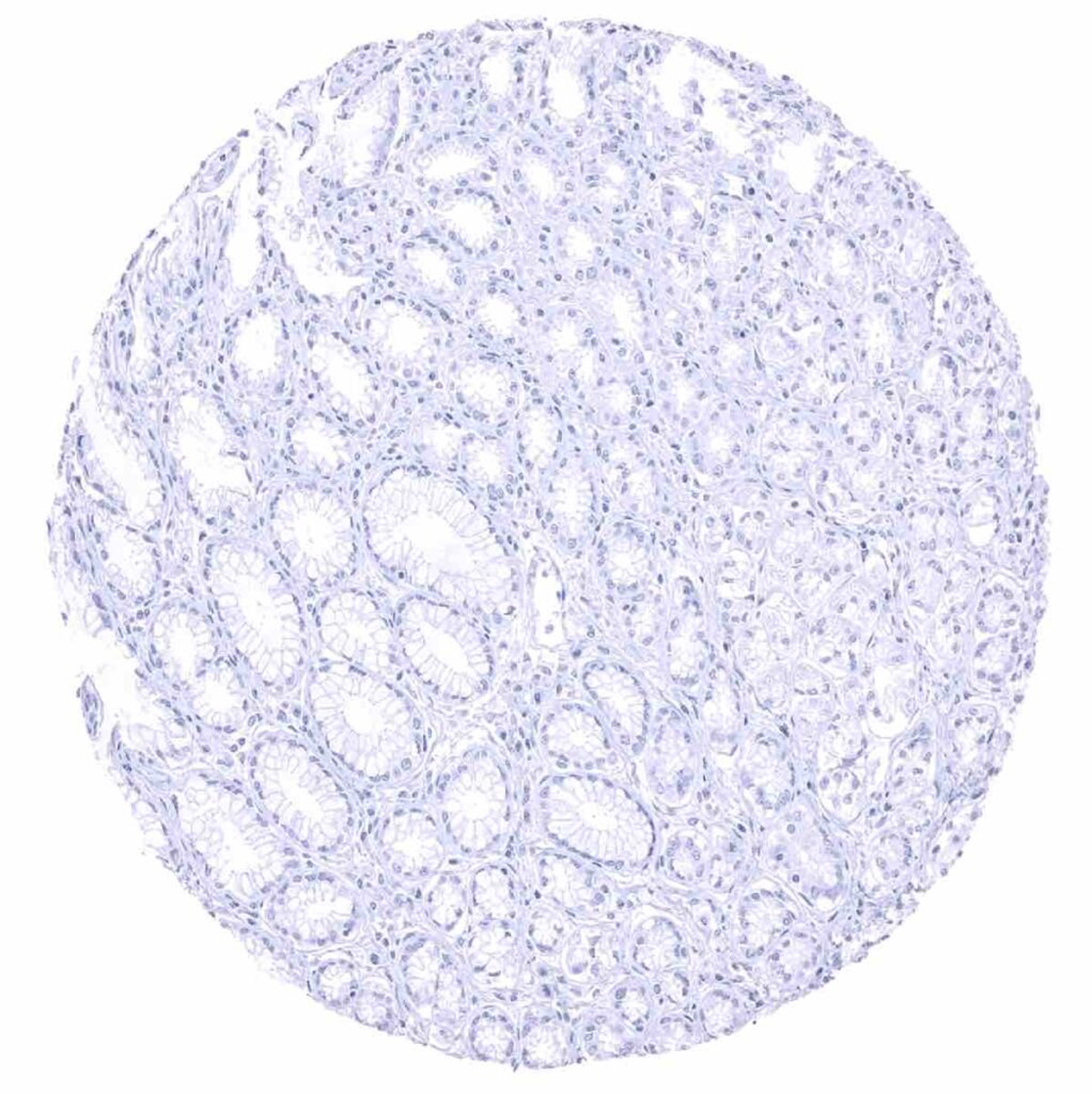
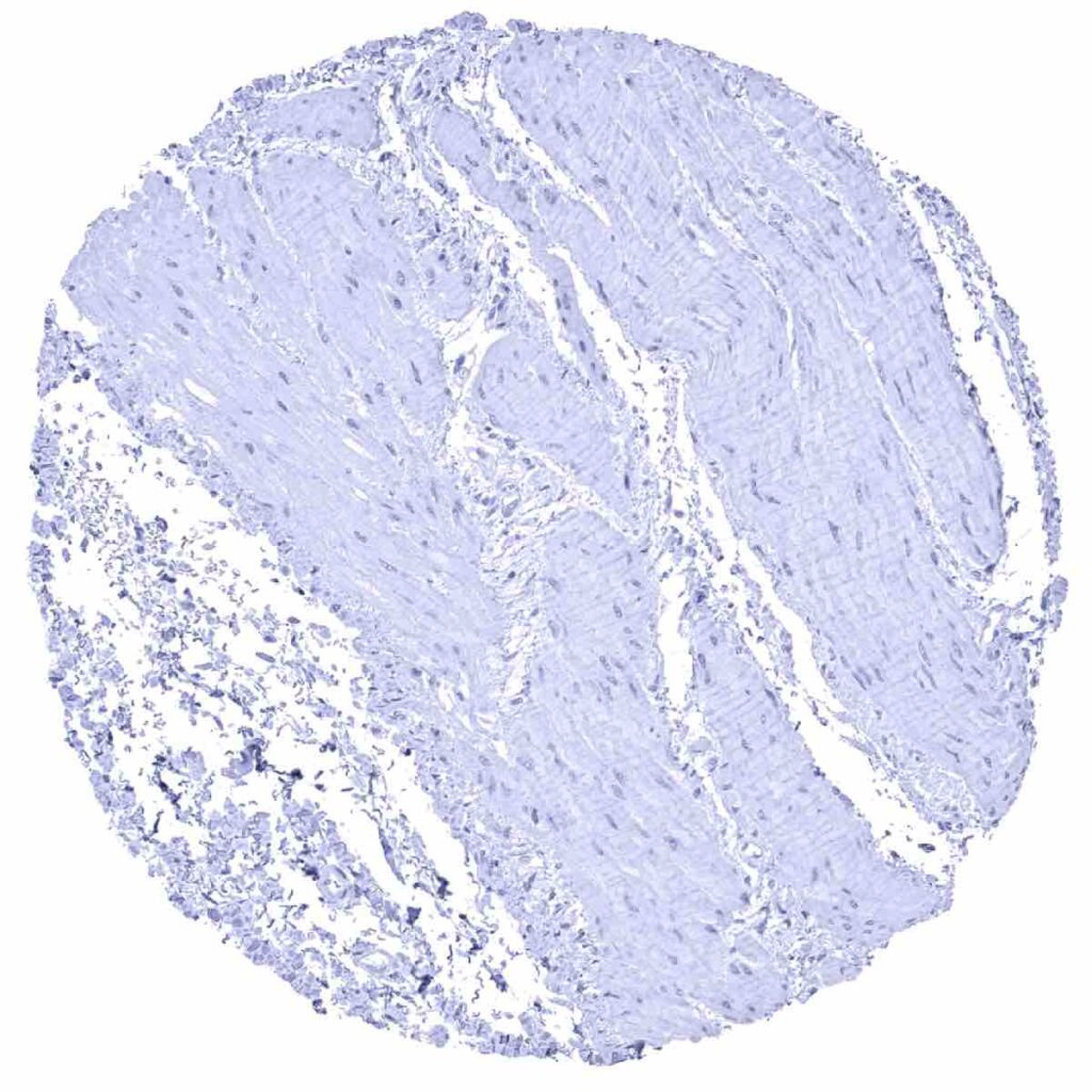
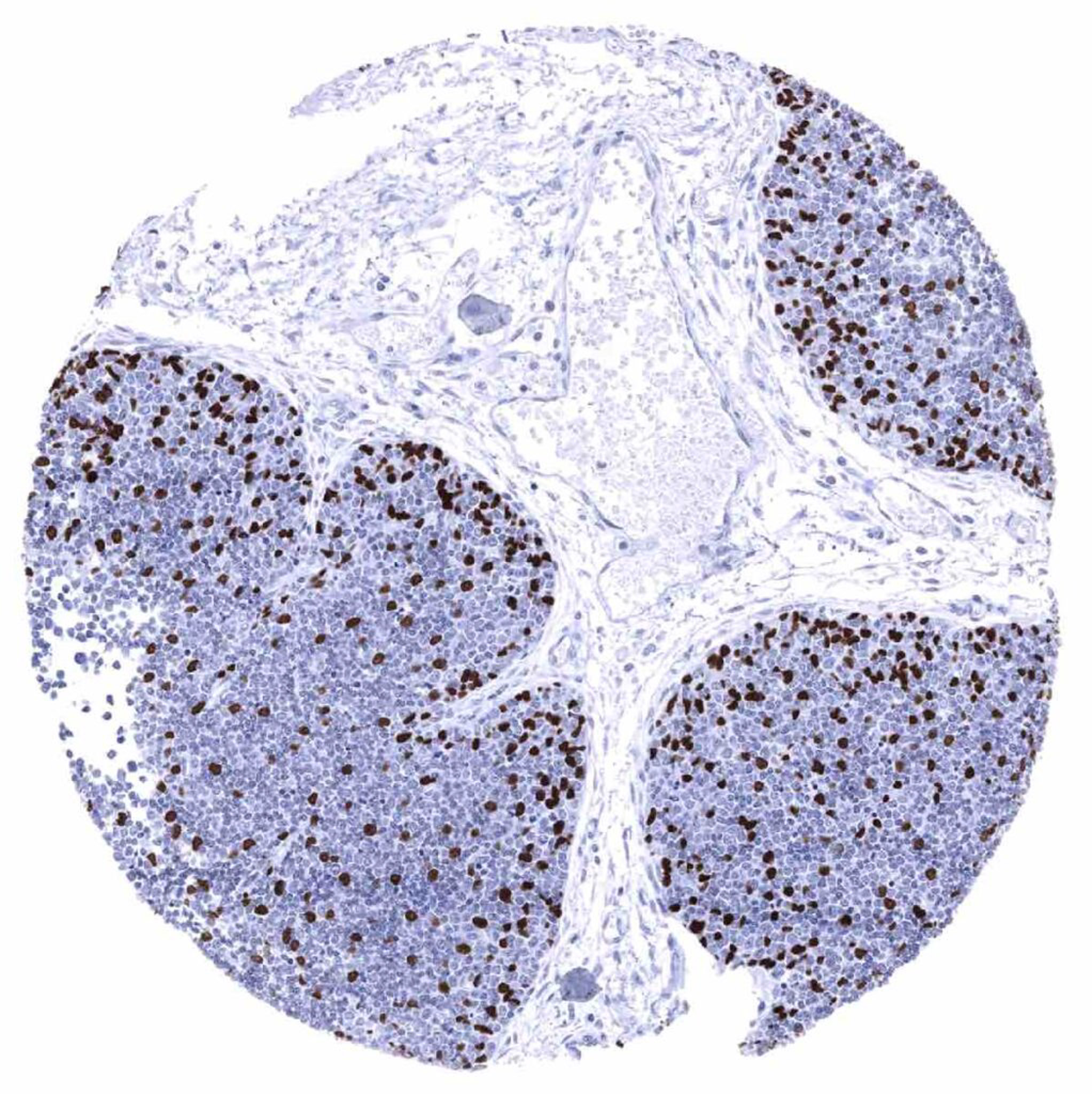
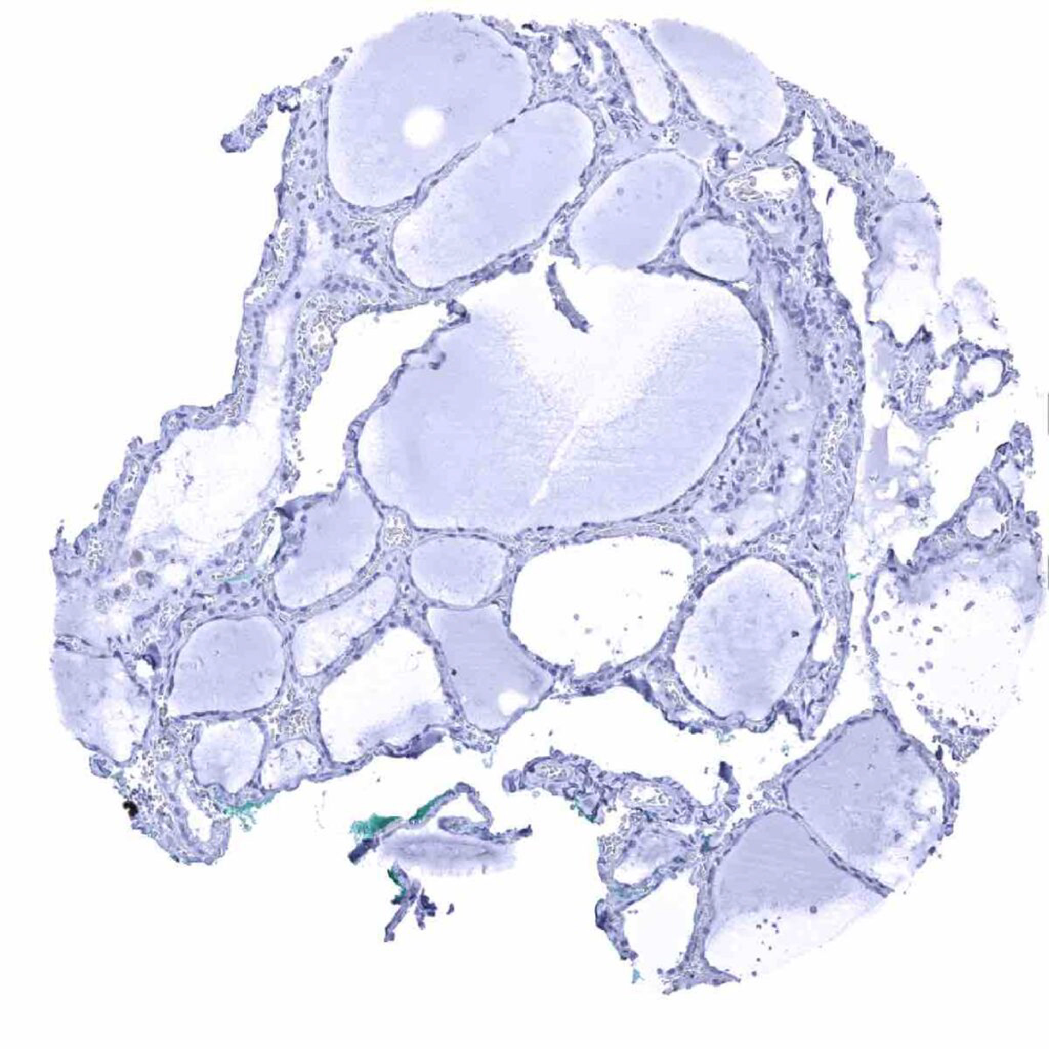
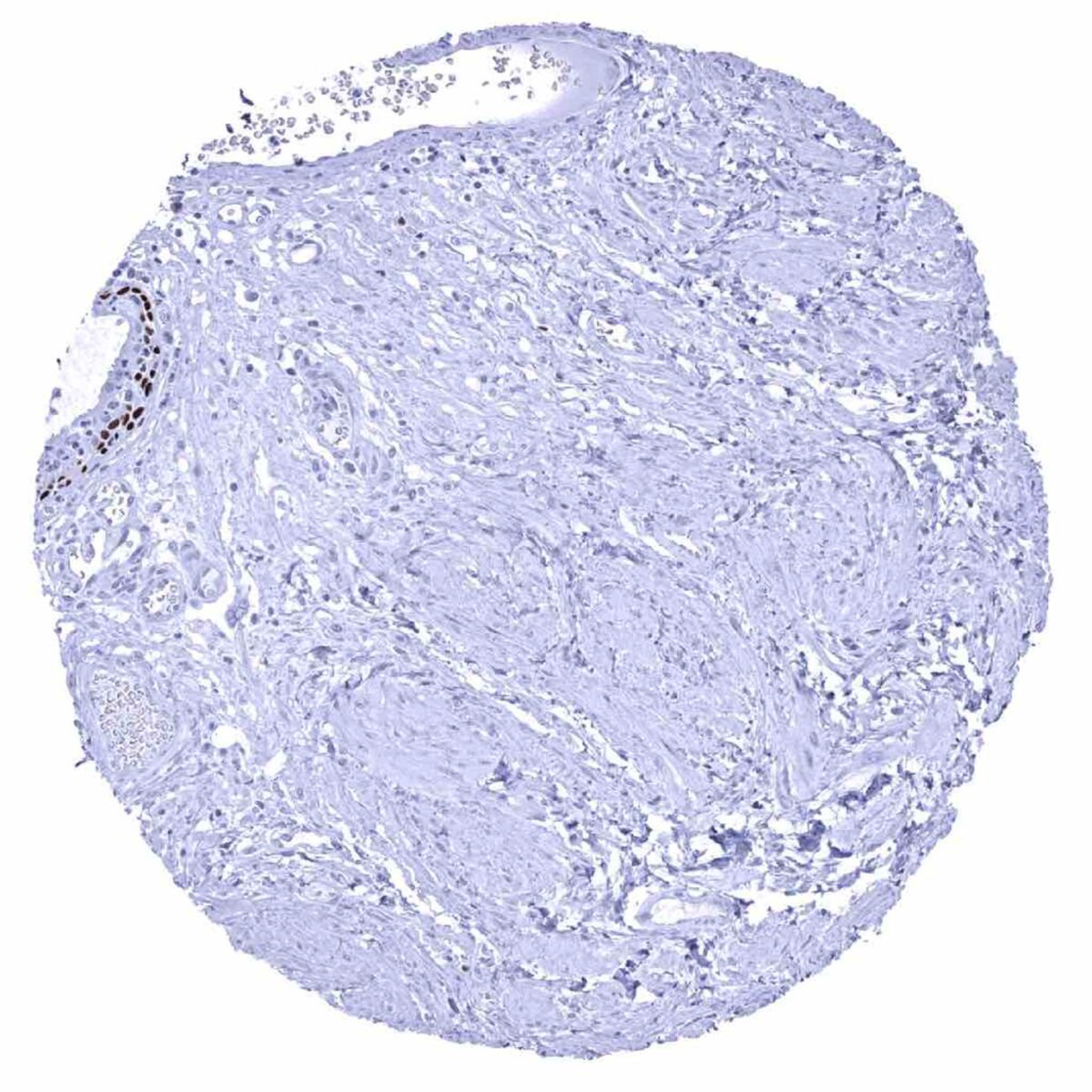
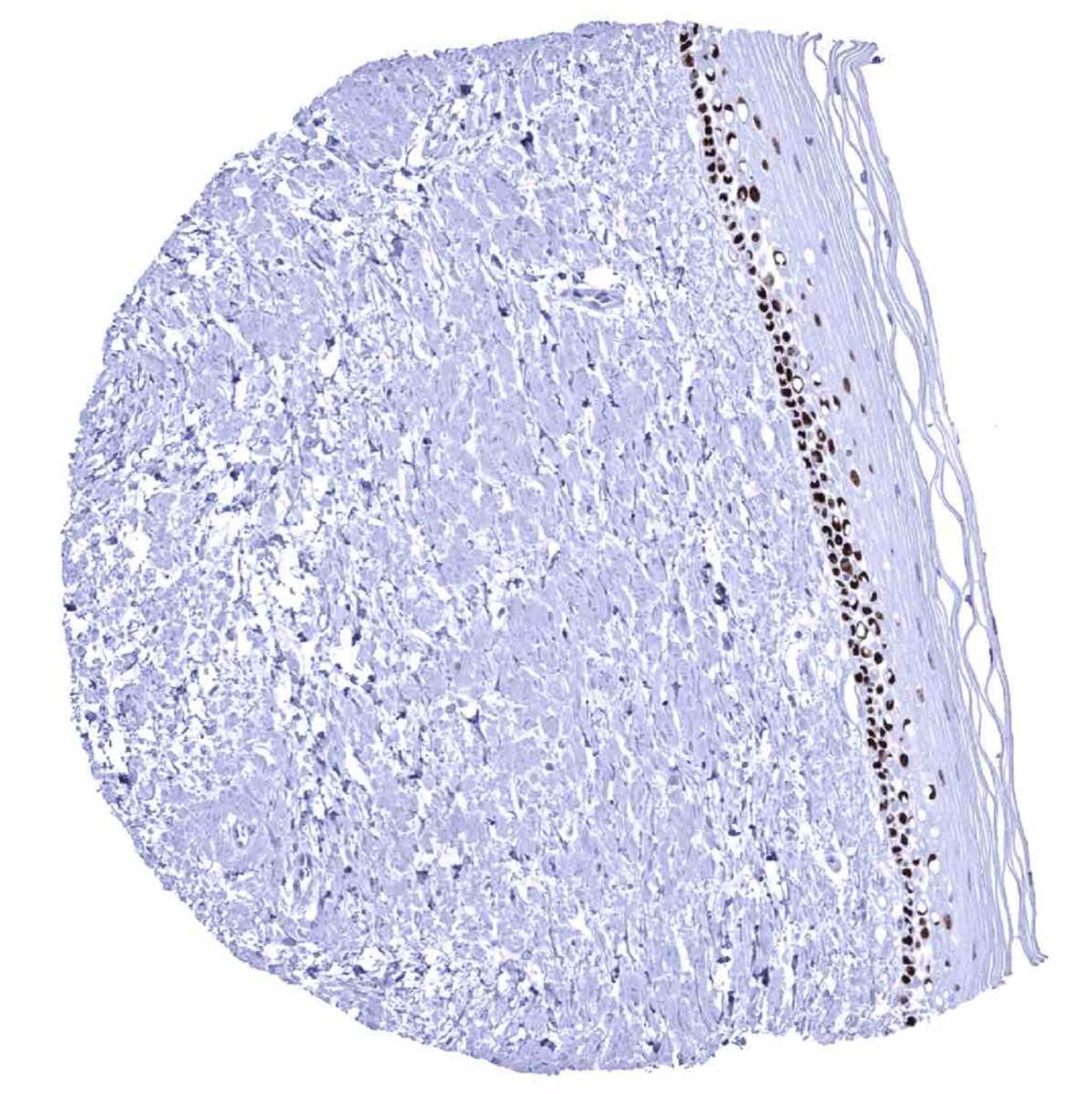
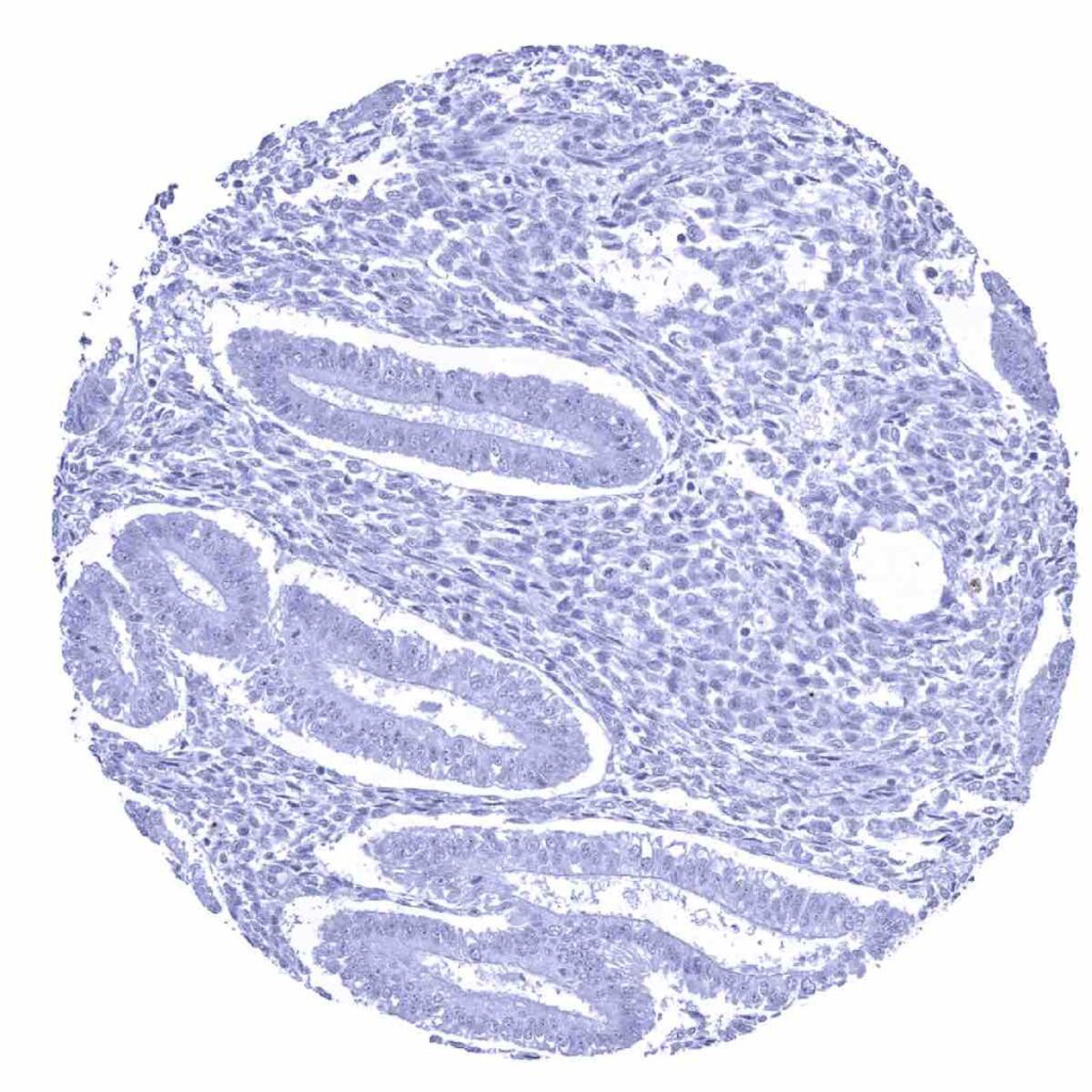
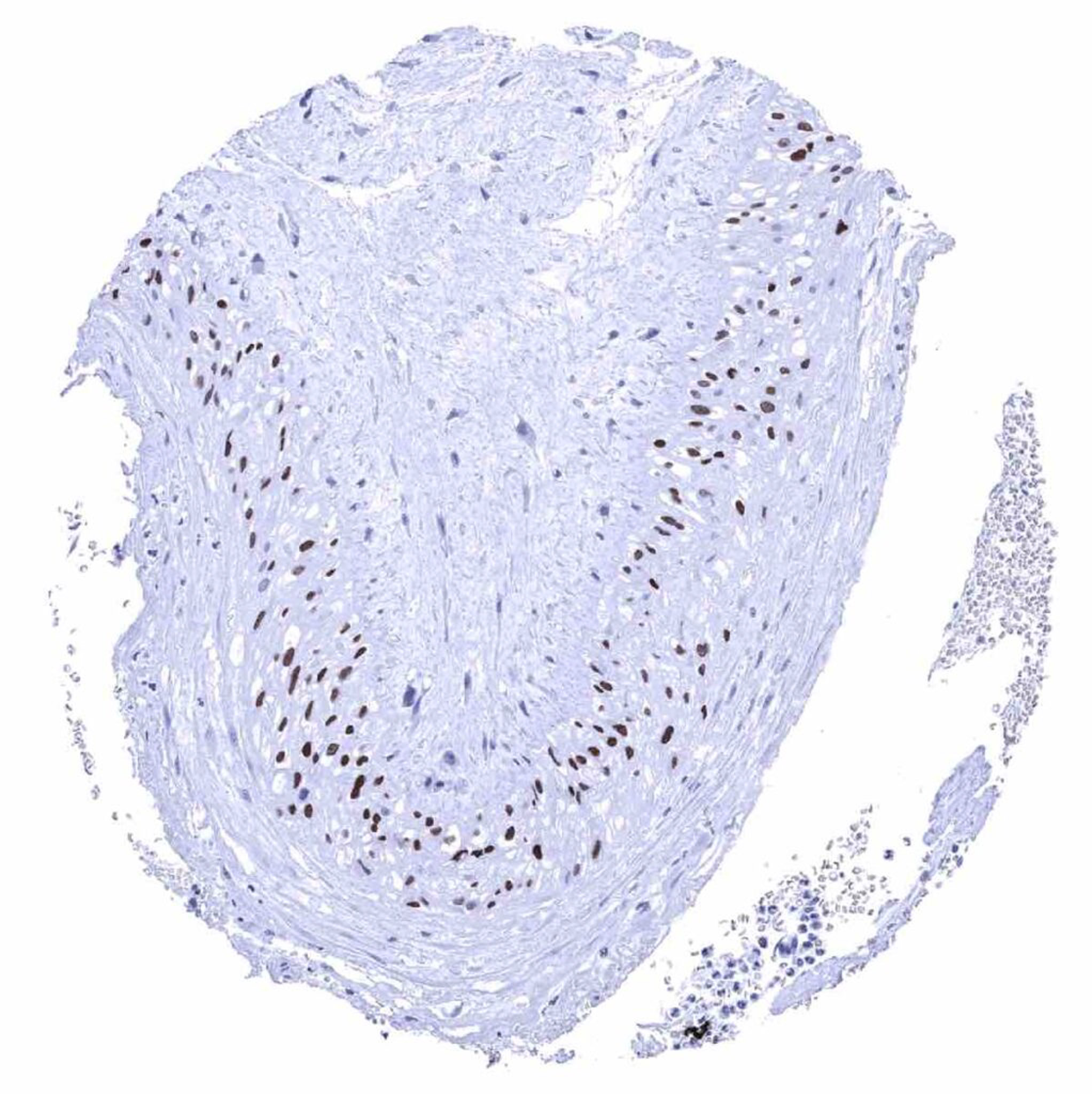
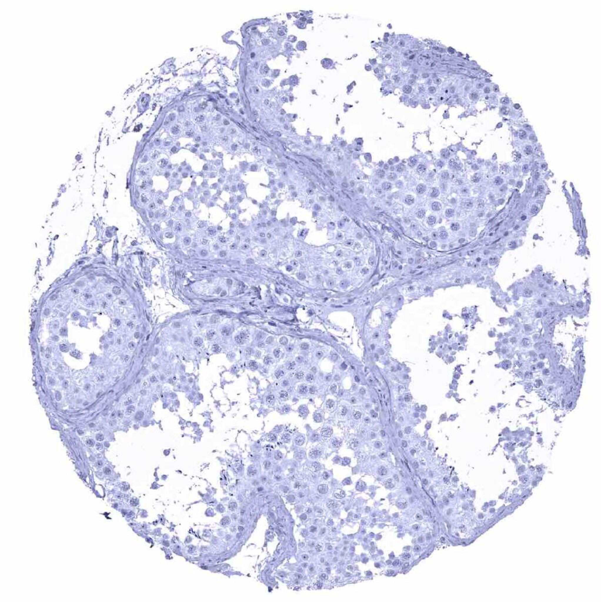
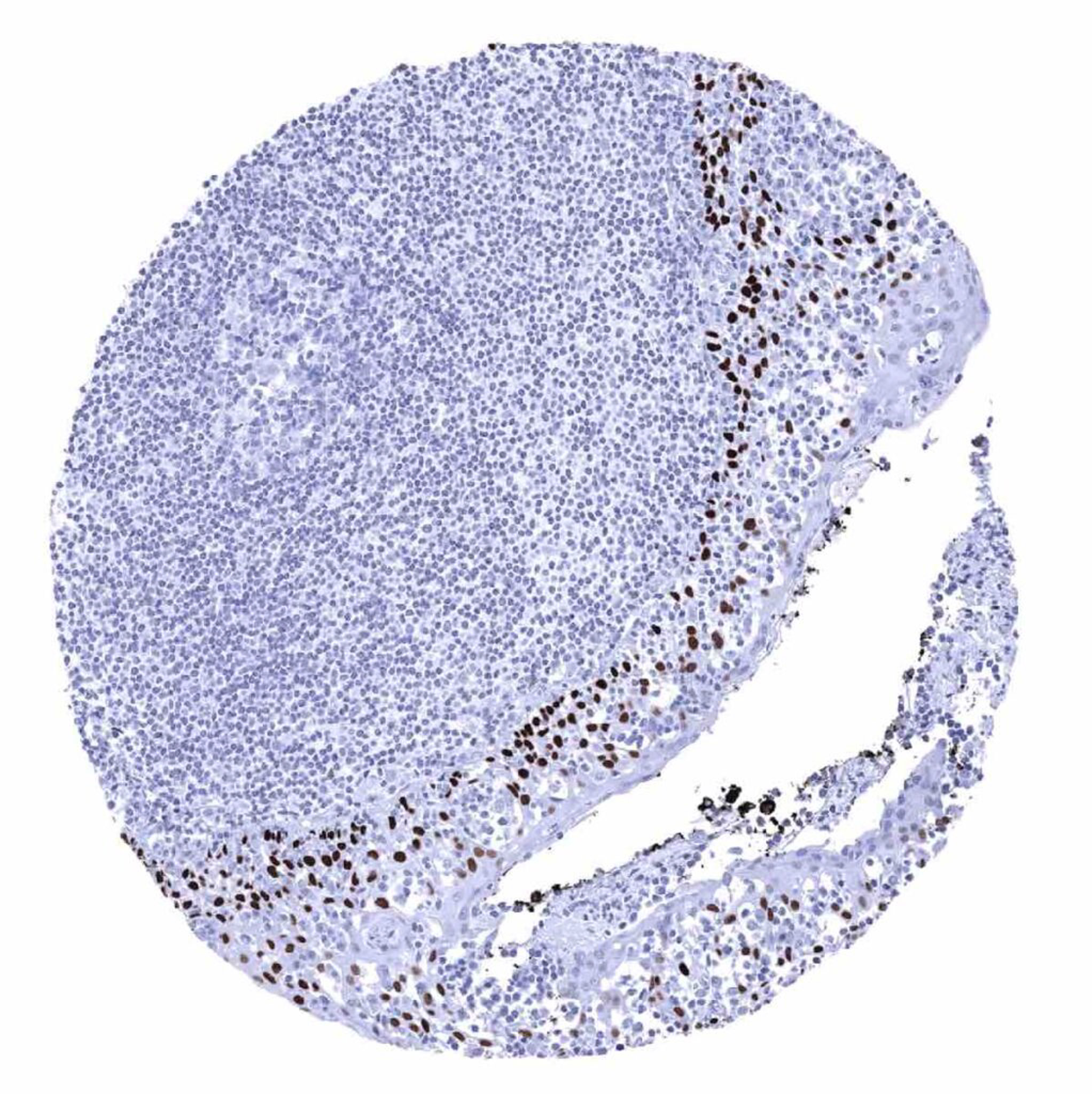
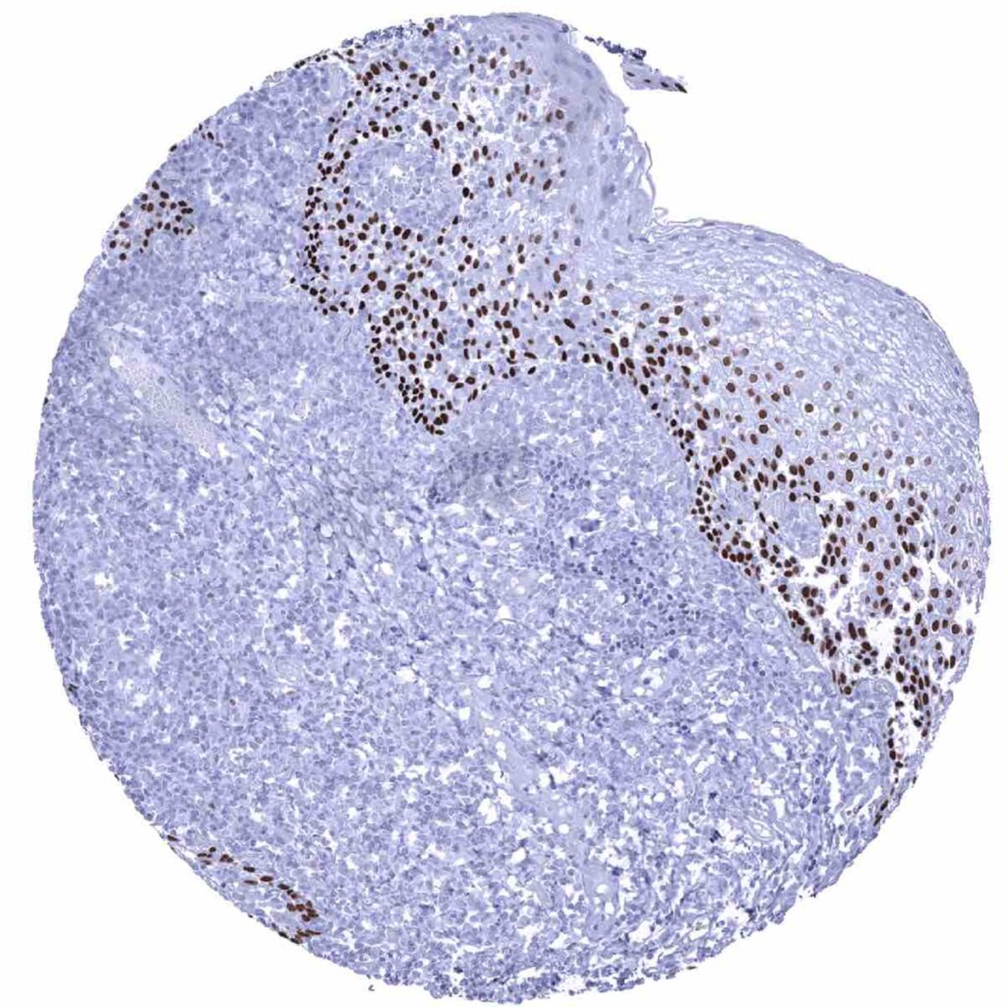
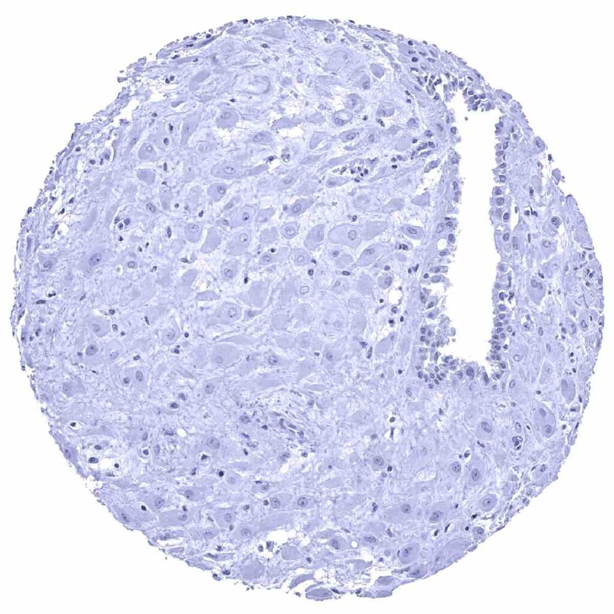
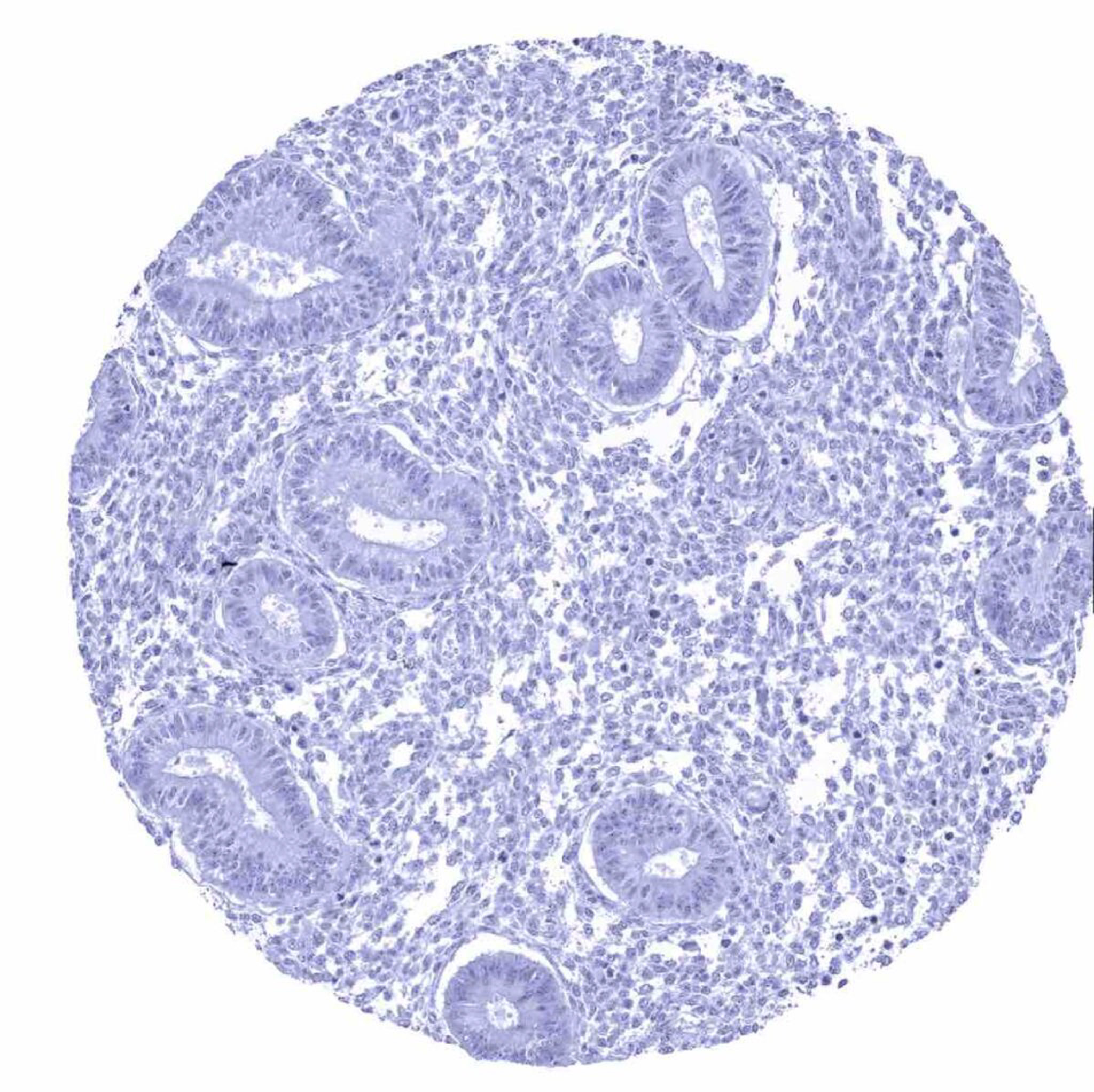
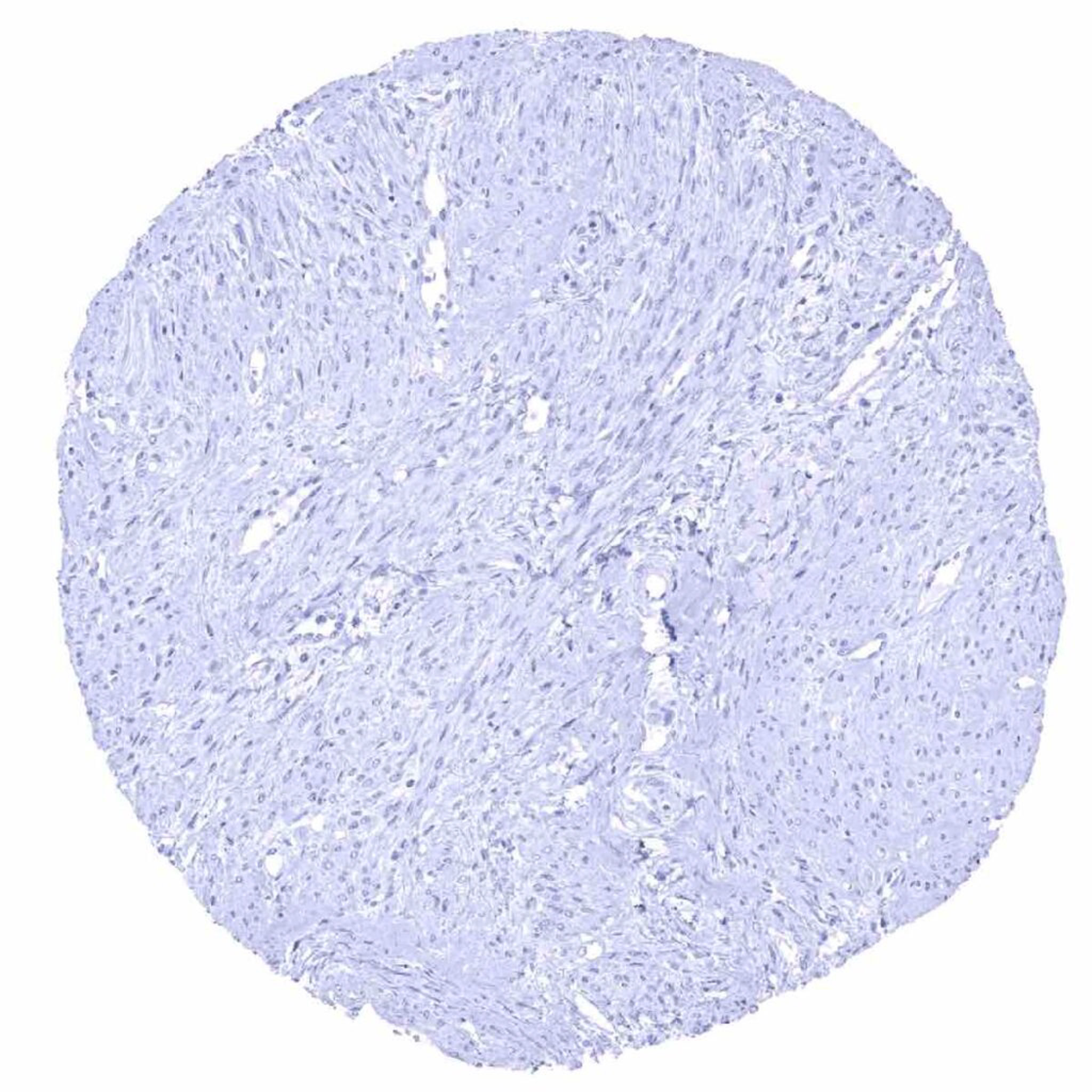
p63 antibody [MSVA-063R] HistoMAXTM


a
Go to Cancer Tissue Gallery
  
 Go back to product page
  

Go to HistoMAX page


Adrenal gland

Aorta, media

Appendix, mucosa – Very few lymphocytes show a weak but distinct p63 staining

Appendix, muscular wall


Bone marrow

Breast – Marked nuclear p63 staining in myoepithelial cells

Bronchus, glands – Nuclear p63 positivity can be observed in basal cells

Cerebellum (molecular, Purkinje cell, and granule cell layers)


Cerebellum (white matter)

Cerebrum, grey matter

Cerebrum, white matter

Colon descendens, mucosa


Colon descendens, muscular wall

Duodenum, Brunner gland

Duodenum, mucosa

Epididymis – Strong nuclear p63 positivity in basal cells


Esophagus, squamous epithelium – p63 immunostaining is strongest in the more basally located cells of the esophageal squamous epithelium

Fallopian tube, mucosa

Fat

Gallbladder, epithelium


Heart muscle

Ileum, mucosa

Kidney, cortex

Kidney, medulla


Kidney, pelvis (urothelium) – A moderate to strong p63 staining is predominantly seen in the lower half of the urothelium

Kidney, pelvis (urothelium) – A moderate to strong p63 staining predominates in the lower half of the urothelium

Liver

Lung


Lymph node – Few lymphocytes, especially in the germinal centre, show a weak but distinct nuclear p63 positivity

Ovary, stroma

Pancreas

Parathyroid gland


Parotid gland – Strong nuclear p63 positivity in myoepithelial cells

Pituitary gland, anterior lobe

Pituitary gland, posterior lobe

Placenta (amnion and chorion) – A moderate to strong nuclear p63 staining occurs in chorion cells


Placenta (amnion and chorion)

Placenta, early – A strong nuclear p63 staining occurs in cytotrophoblast cells

Placenta, mature – A moderate to strong nuclear p63 positivity is seen in cytotrophoblast cells

Prostate – Strong nuclear p63 positivity in basal cells


Rectum, mucosa

Seminal vesicle – Strong nuclear p63 positivity in basal cells_png

Skin – A positive p63 immunostaining is seen in virtually all squamous epithelial cells of the skin

Skin – A strong p63 immunostaining occurs in peripheric germinative cells of sebaceous glands


Spleen

Stomach, antrum

Stomach, corpus

Stomach, muscular wall


Testis

Thymus – A strong nuclear p63 staining is seen in thymus epithelial cells

Thymus – Thymus epithelial cells exhibit a strong nuclear p63 staining

Thyroid gland


Tonsil – p63 staining is strongest in basally located cells of the squamous epithelium of tonsil crypts

Tonsil, surface epithelium – A moderate to strong p63 staining is seen in most squamous epithelial cells

Urinary bladder, muscular wall – A positive p63 staining is seen in a small fragment of urothelium

Uterus, ectocervix – A moderate to strong p63 staining is predominantly seen in the lower half of the squamous epithelium of the ectocervix


Uterus, endocervix

Uterus, endometrium (pregnancy)

Uterus, endometrium (proliferation)

Uterus, endometrium (secretion)


Uterus, myometrium


| TOP |