Country / Location Selection
Search suggestions
MCM3 antibody [MSVA-503M] HistoMAX
Go to HistoMAX page
Adrenal gland- Adrenocortical adenoma showing only few MCM3 positive tumor cells.
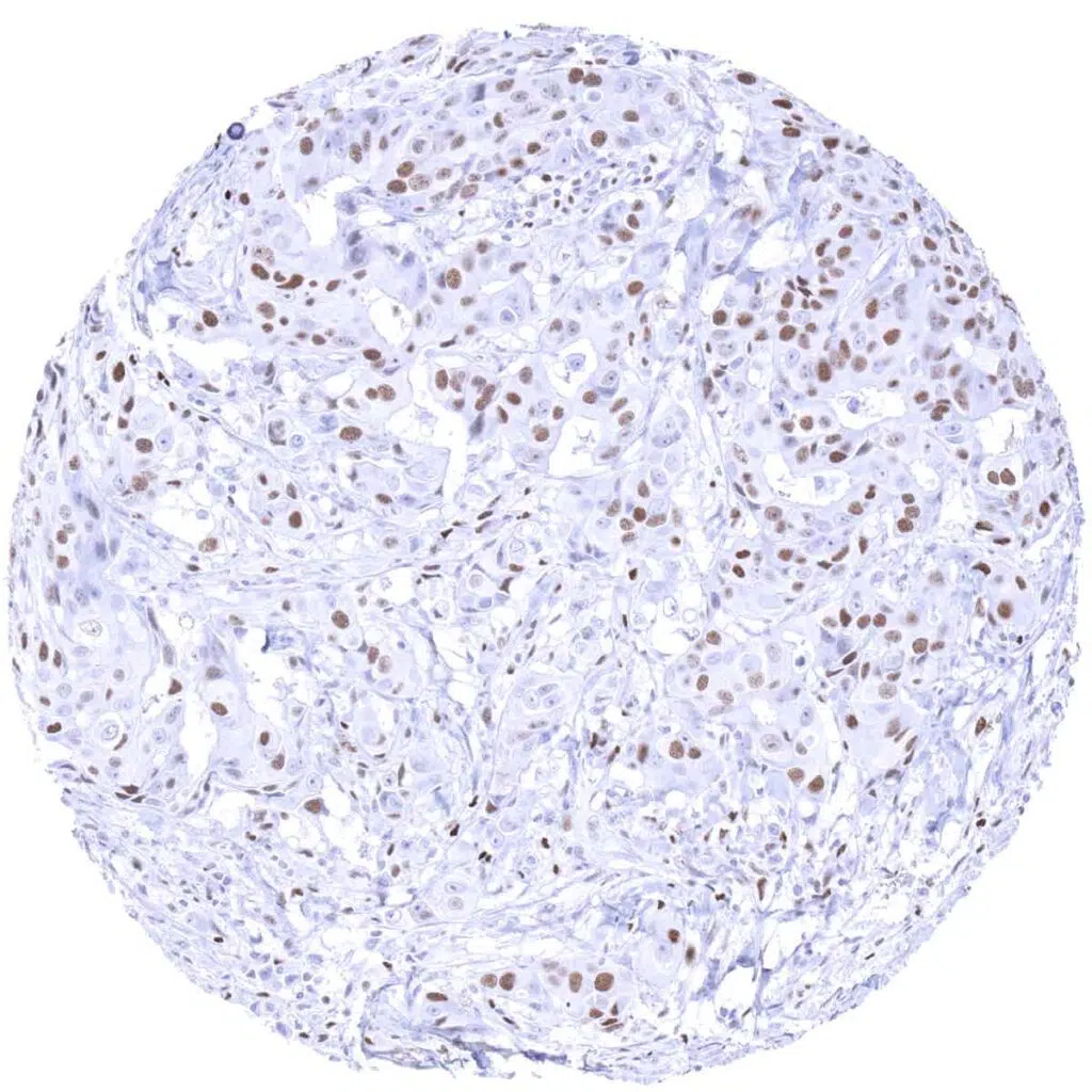
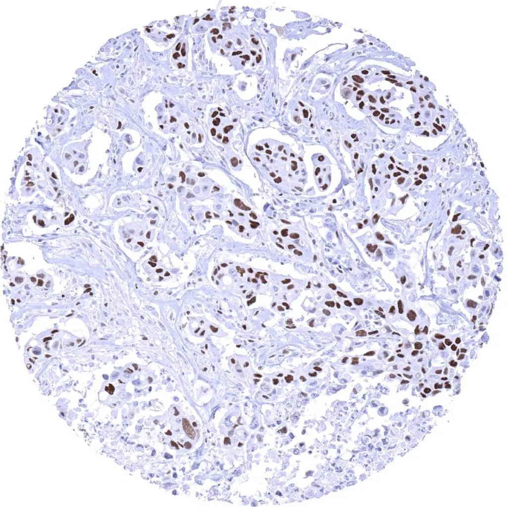
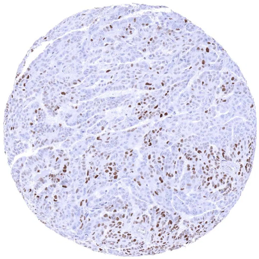
Breast- Invasive breast cancer of no special type (NST) with moderate to strong MCM3 positivity of most tumor cells.
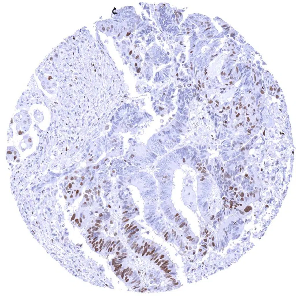
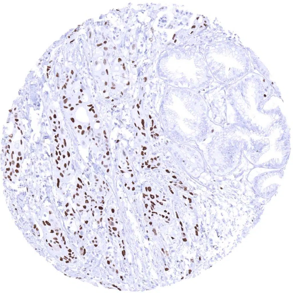
Colon- Colorectal adenocarcinoma with focal accumulation of MCM3 positive tumor cells.
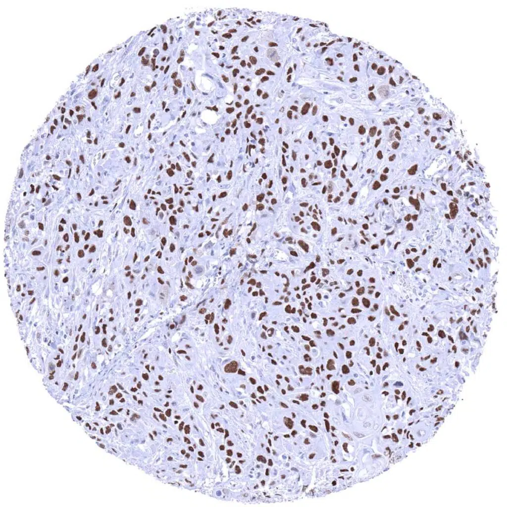
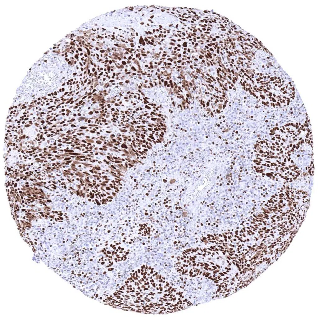
Esophagus- Squamous cell carcinoma showing strong nuclear MCM3 staining of tumor cells.
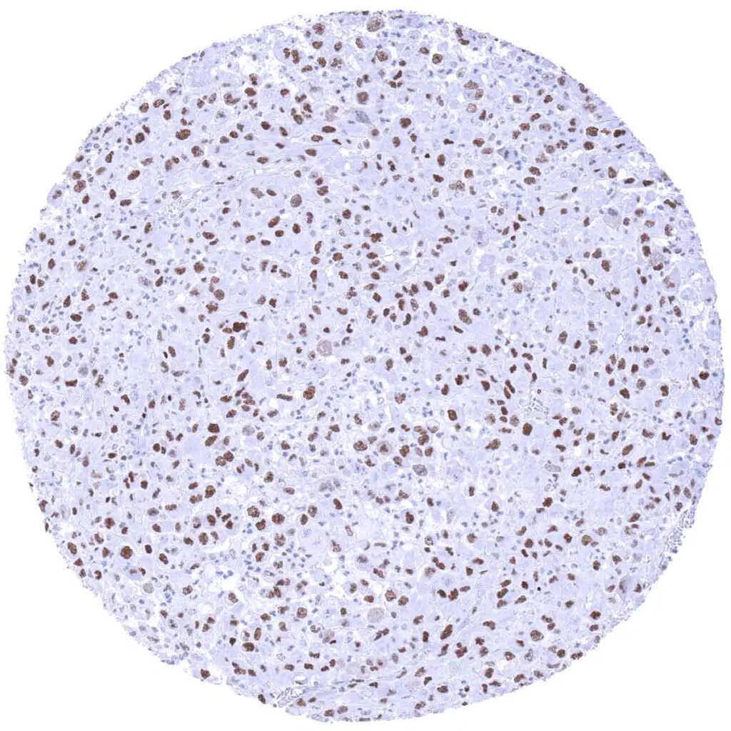
Kidney- Clear cell renal cell carcinoma containing few MCM3 positive tumor cells.
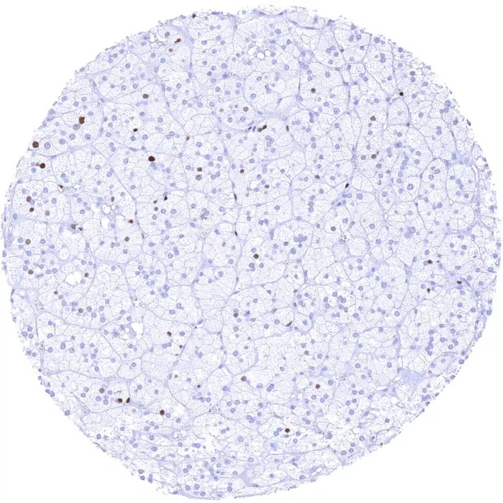
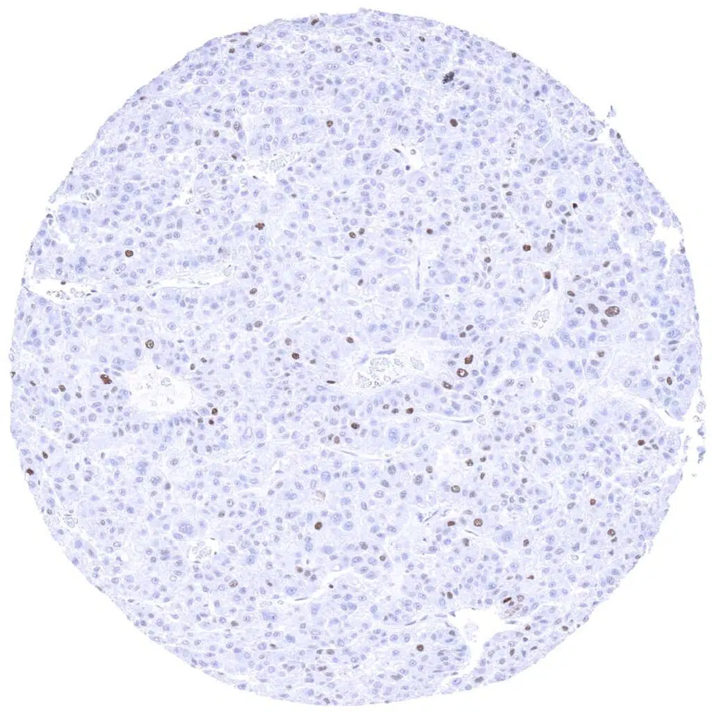
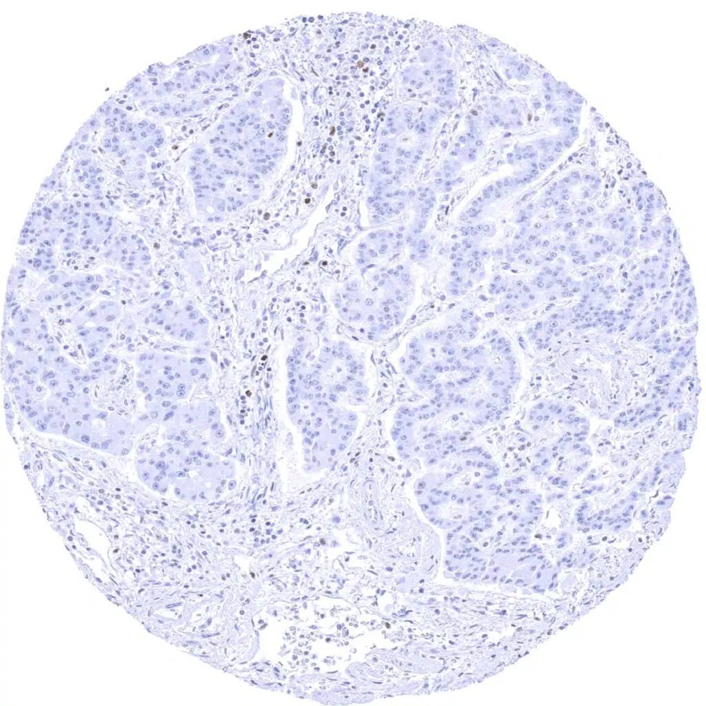
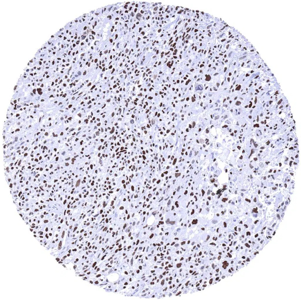
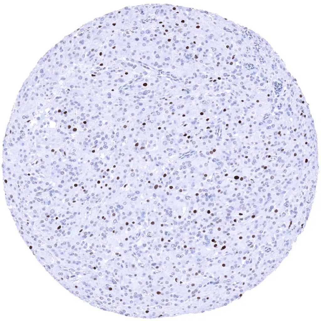
Liver- Hepatocellular carcinoma showing a mostly weak to moderate MCM3 positivity in only a small fraction of tumor cells.
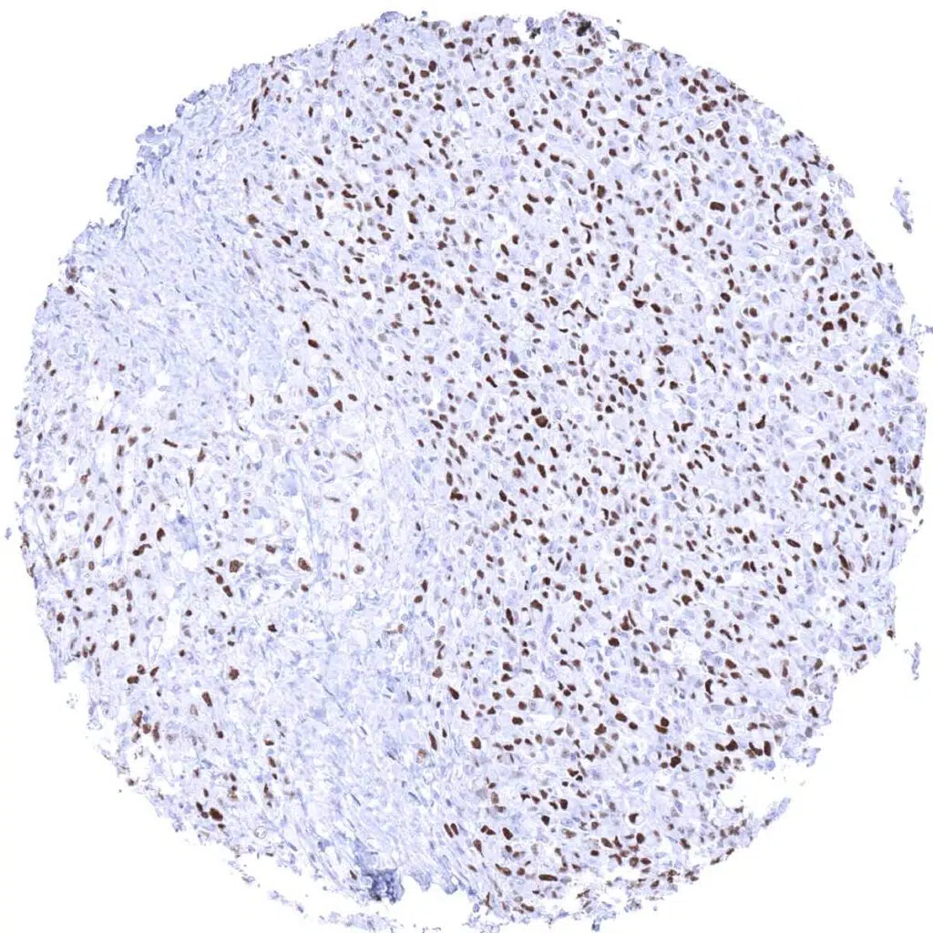
Lung- Adenocarcinoma with strong MCM3 immunostaining of most tumor cells.
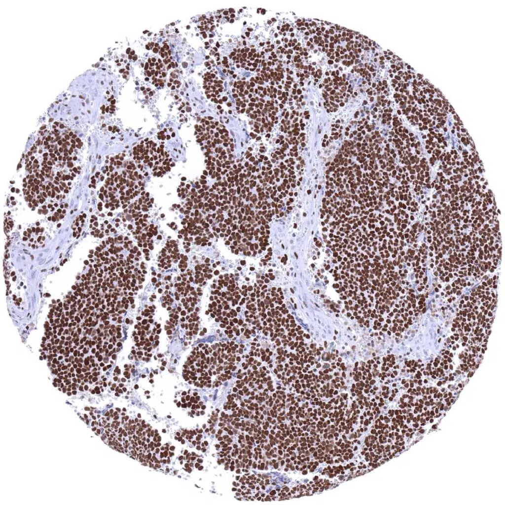
Lung- Small cell carcinoma with strong MCM3 positivity of all tumor cells.
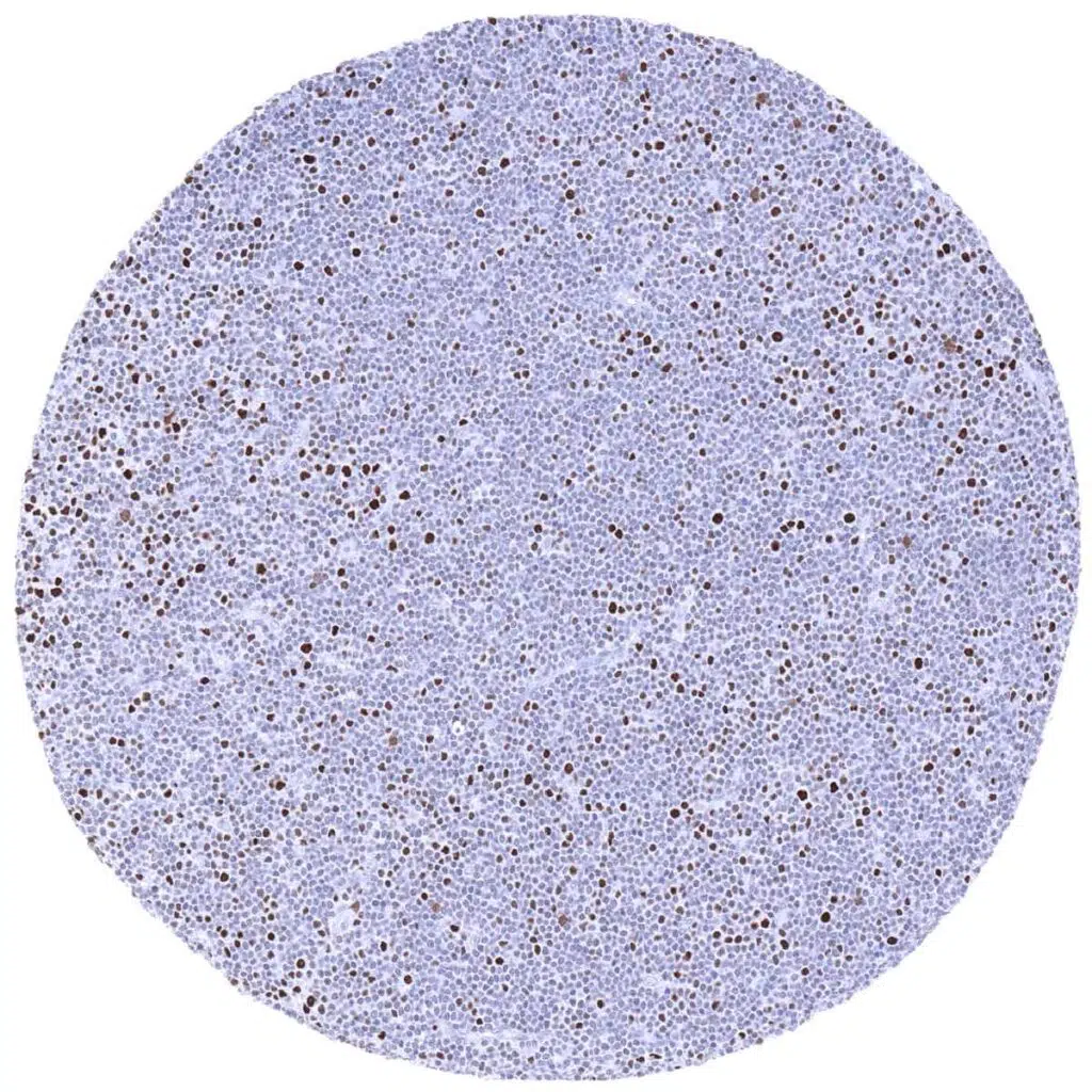
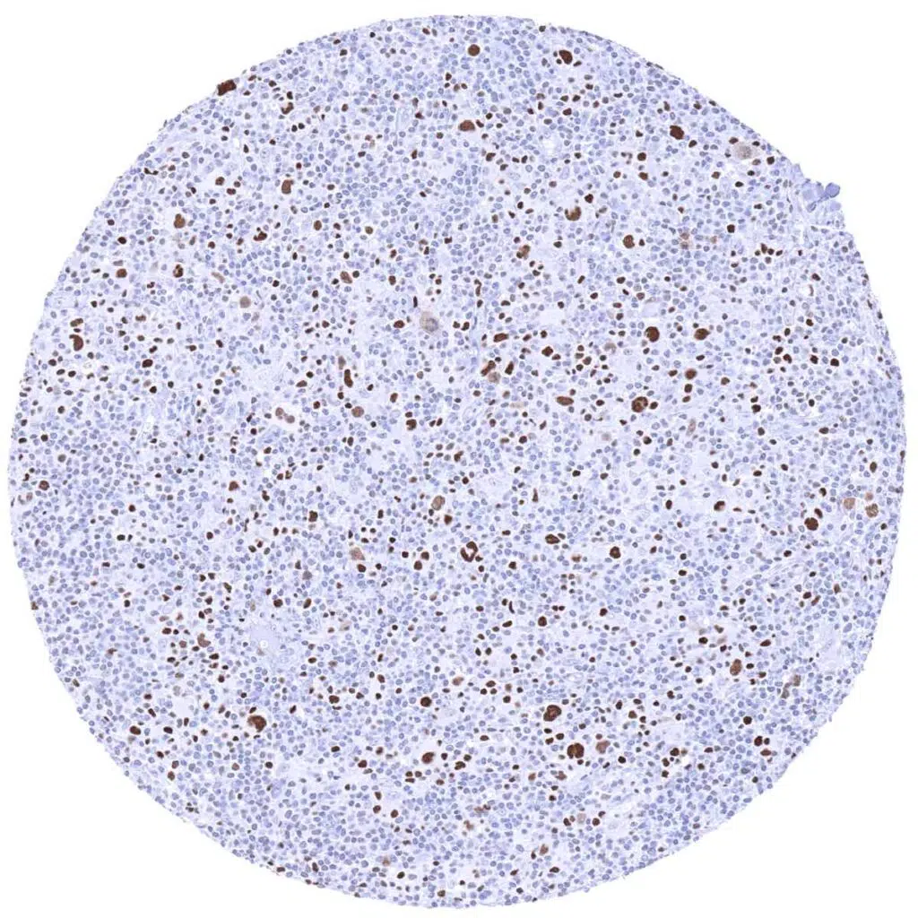
Lymph node- B-CLL showing MCM3 positivity in only a small fraction of tumor cells.
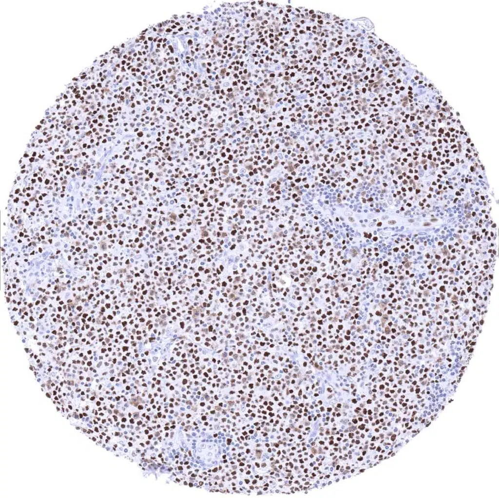
Lymph node- Diffuse large B-cell lymphoma with moderate MCM3 positivity of all tumor cells.
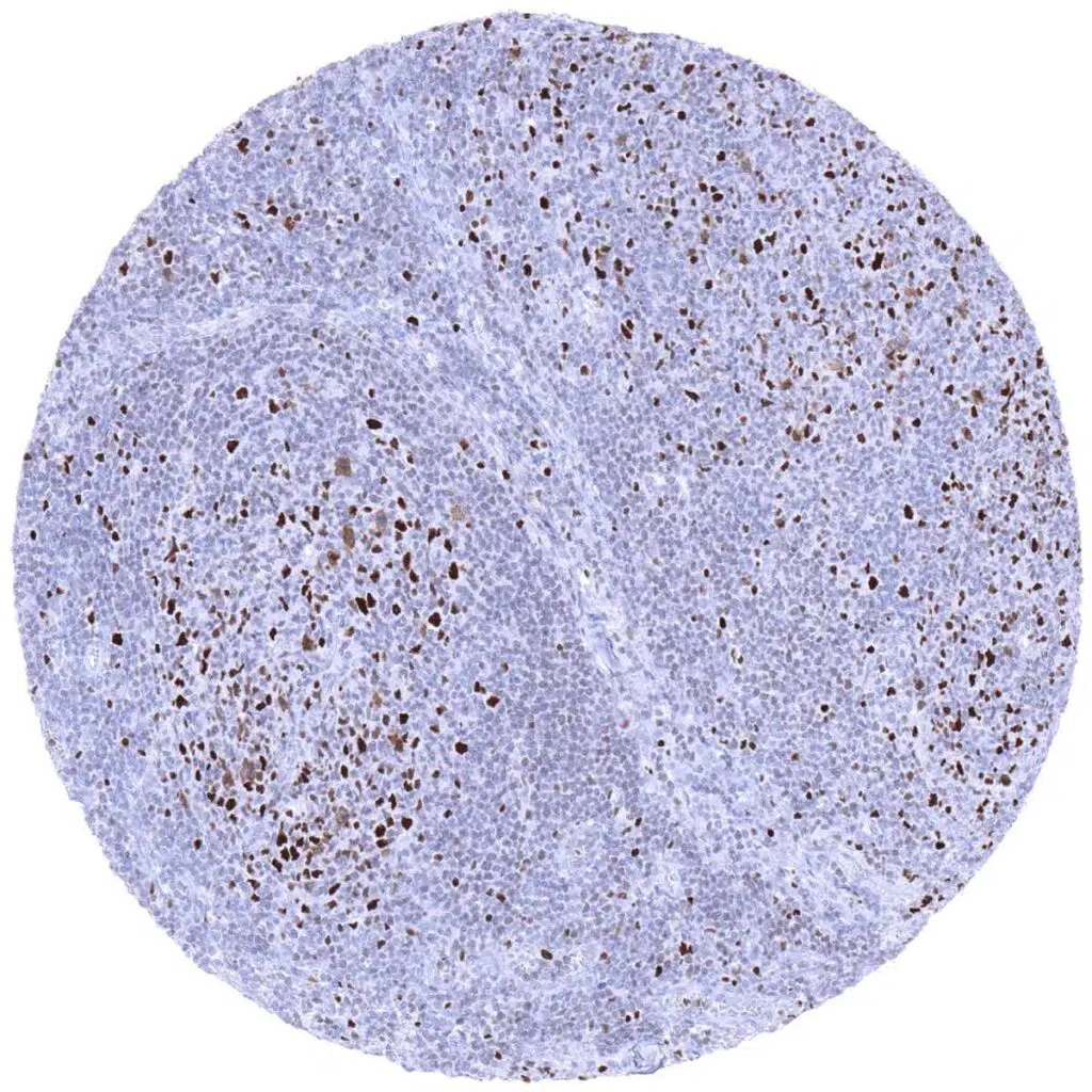
Lymph node- Follicular B-cell lymphoma with MCM3 positivity of few tumor cells, predominantly occurring within follicles.
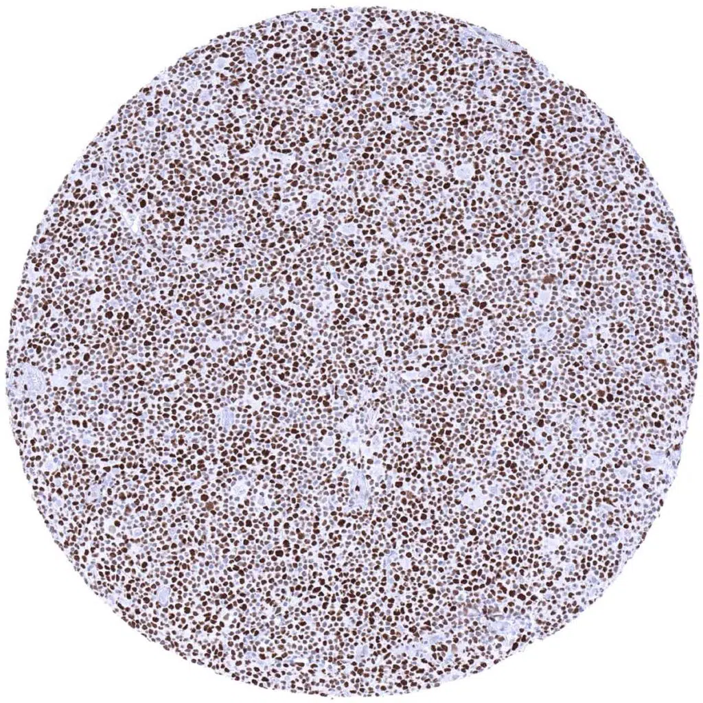
Lymph node- Hodgkin’s lymphoma with strong MCM3 immunostaining of most neoplastic cells.
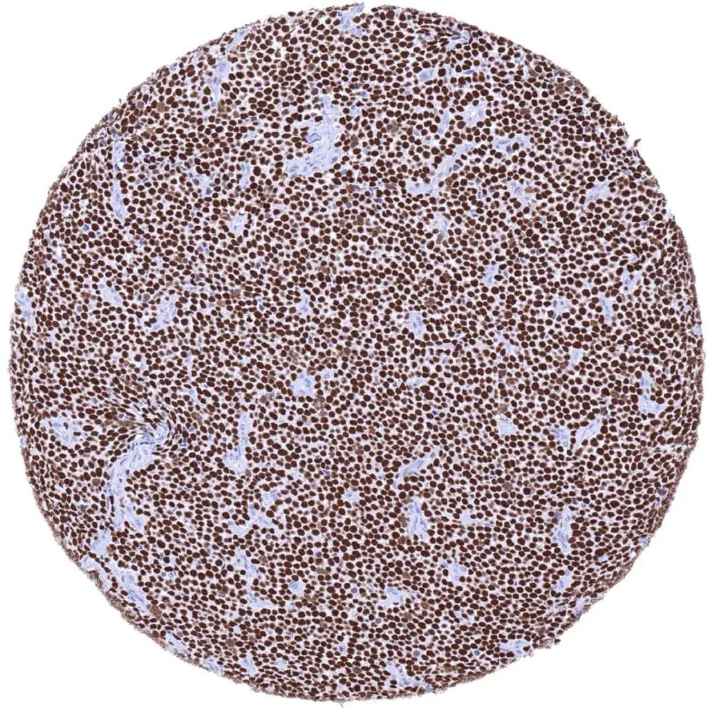
Lymph node- Mantle cell lymphoma with strong MCM3 immunostaining of all tumor cells.
Ovary- Serous high-grade carcinoma with focal accumulations of MCM3 positive tumor cells.
Pancreas- Ductal adenocarcinoma with strong MCM3 staining of almost all tumor cells while adjacent normal duct cells are mostly negative.
Pancreas- Neuroendocrine tumor containing only few MCM3 positive tumor cells.
Pharynx- Squamous cell carcinoma with strong MCM3 immunostaining of virtually all tumor cells.
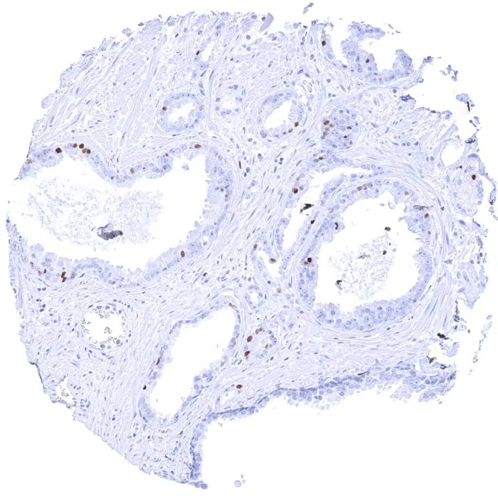
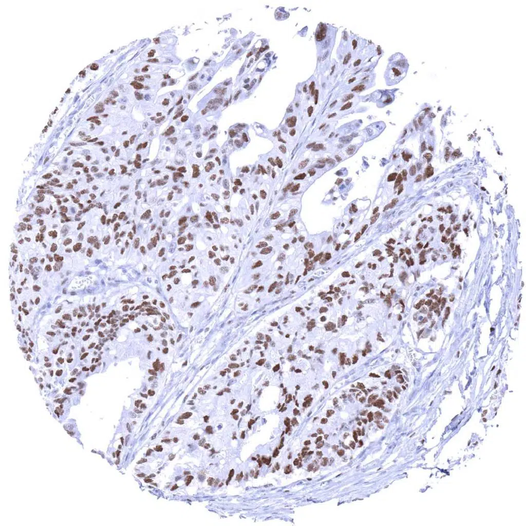
Prostate- Adenocarcinoma (Gleason 3+3=6) with strong MCM3 immunostaining of a limited number of tumor cells.
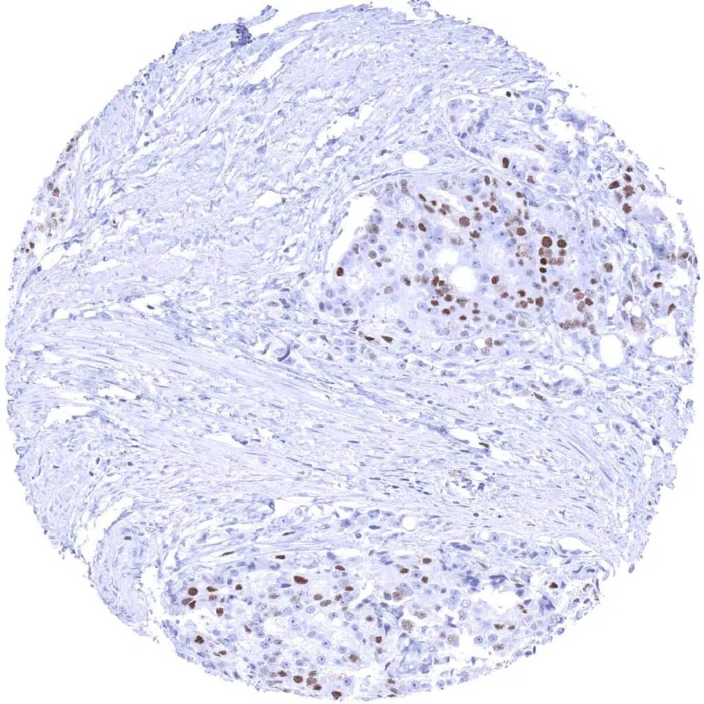
Prostate- Adenocarcinoma (Gleason 4+4=8) with MCM3 positivity of about 50_ of tumor cells.
Salivary gland- Warthin tumor displaying MCM3 positivity in few epithelial cells and few lymphocytes.
Skin- Basal cell carcinoma showing MCM3 positivity in a large fraction of tumor cells, predominantly in the basal layers.
Skin- Merkel cell carcinoma showing strong MCM3 staining in all tumor cells.
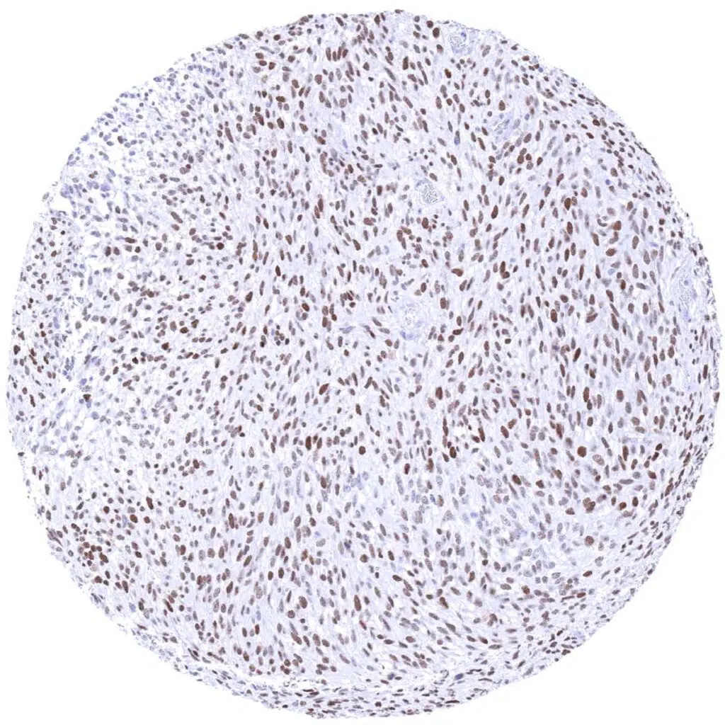
Soft tissue- Liposarcoma showing strong nuclear MCM3 positivity of all tumor cells.
Soft tissue- Liposarcoma with strong MCM3 immunostaining of almost all tumor cells.
Stomach- Gastric adenocarcinoma (diffuse type) with strong MCM3 immunostaining of a large fraction of tumor cells.
Stomach- Gastric adenocarcinoma (intestinal type) with strong MCM3 positivity of 95% of tumor cells.
Stomach- Gastrointestinal stromal tumor (GIST) showing moderate MCM3 staining in all tumor cells.
Testis- Leydig cell tumor with strong MCM3 immunostaining of tumor cells.
Testis- Seminoma with moderate MCM3 positivity in all tumor cells.
Thyroid- Medullary cancer with weak to moderate MCM3 positivity of a small fraction of tumor cells.
Thyroid- Papillary cancer with moderate MCM3 staining of few tumor cells.
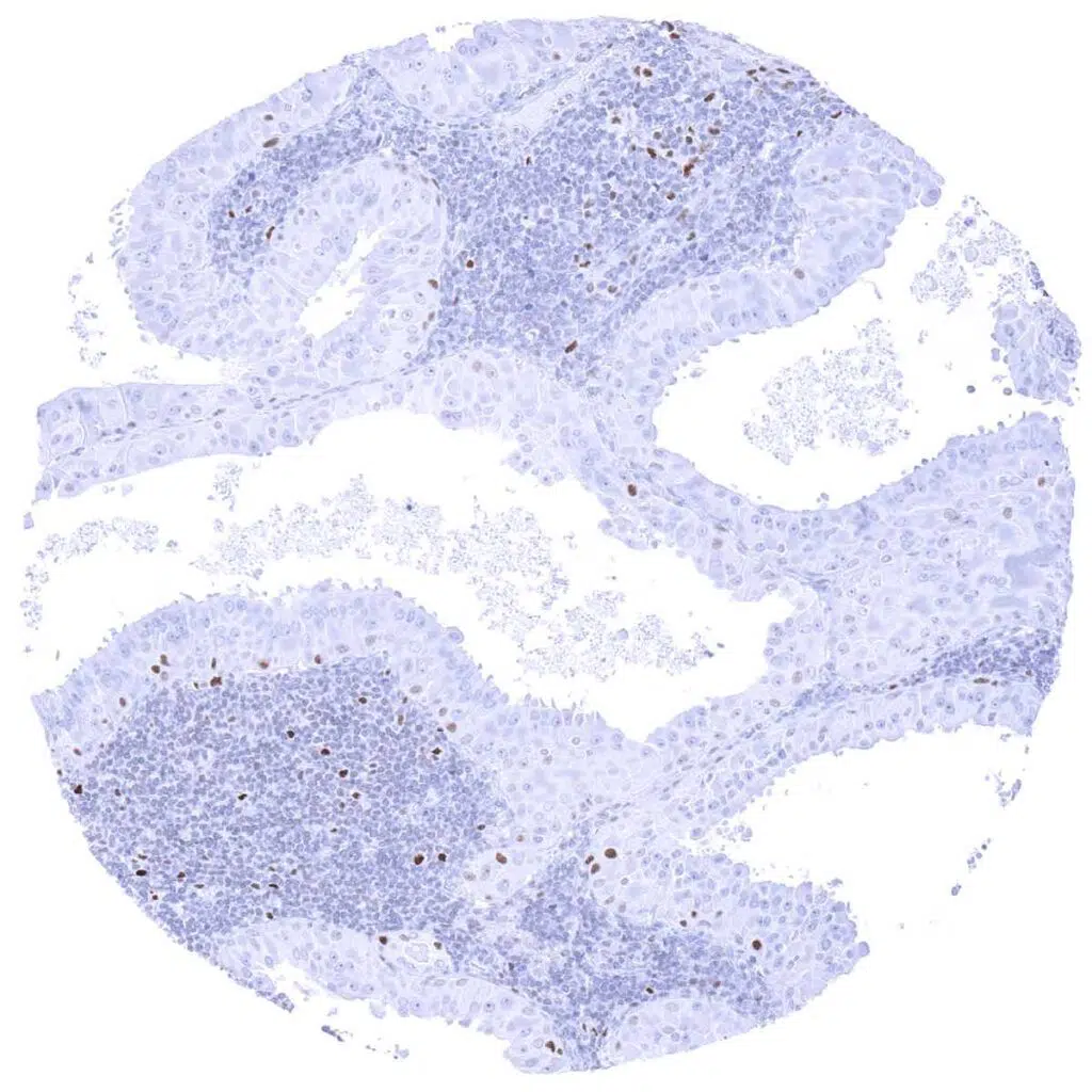
Urinary bladder- Muscle-invasive urothelial carcinoma showing strong MCM3 positivity in all tumor cells.
Uterus- Leiomyosarcoma with strong MCM3 immunostaining of all tumor cells.
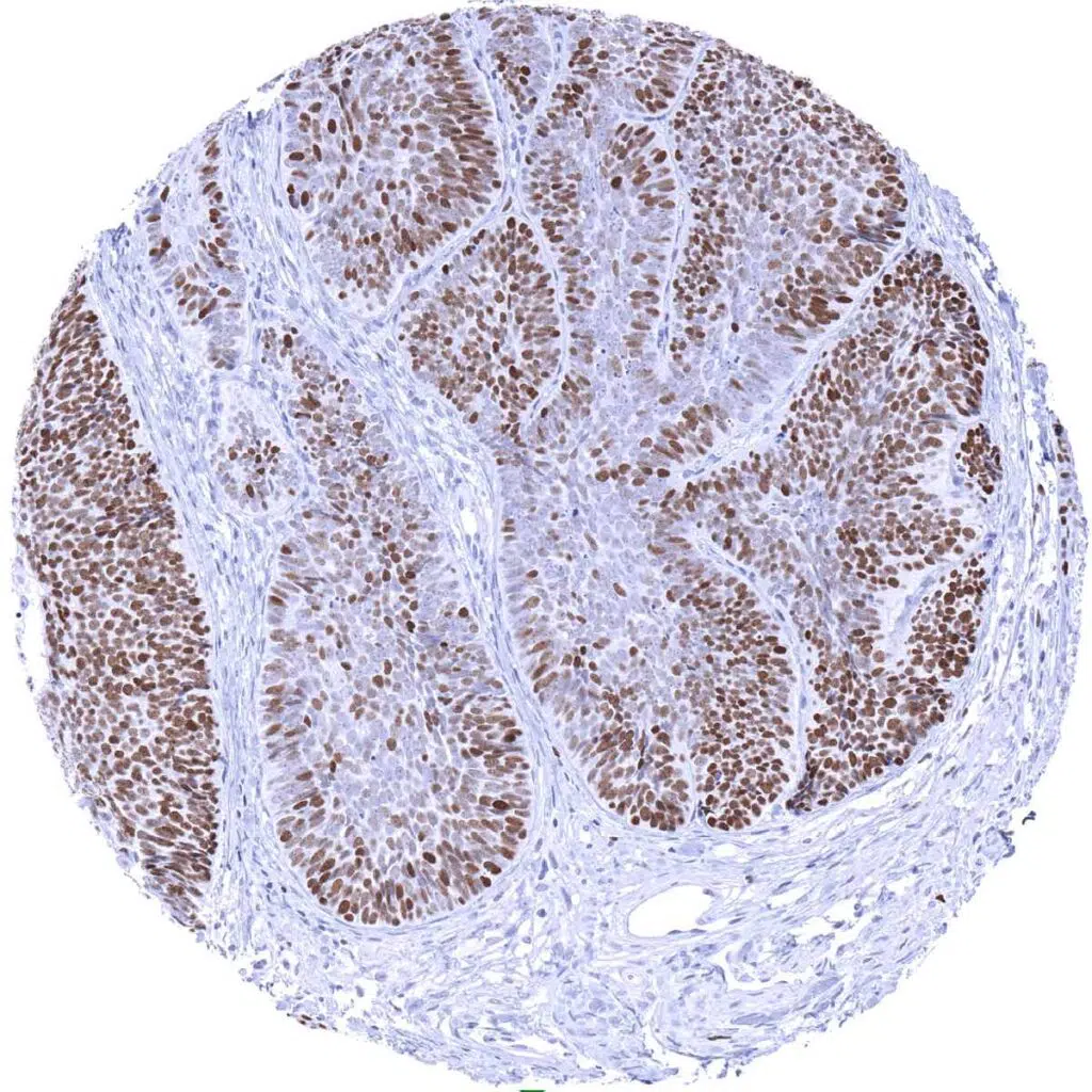
Uterus- Strong nuclear positivity in all tumor cells of a cervical adenocarcinoma.
| TOP |