Cancer Tissue Gallery- GTX639944

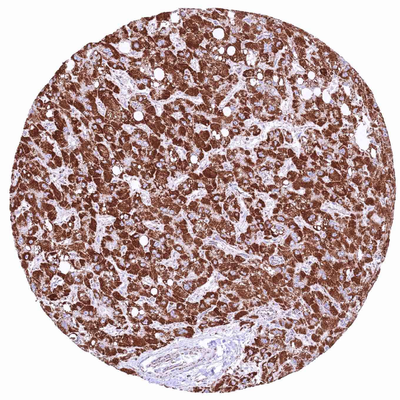
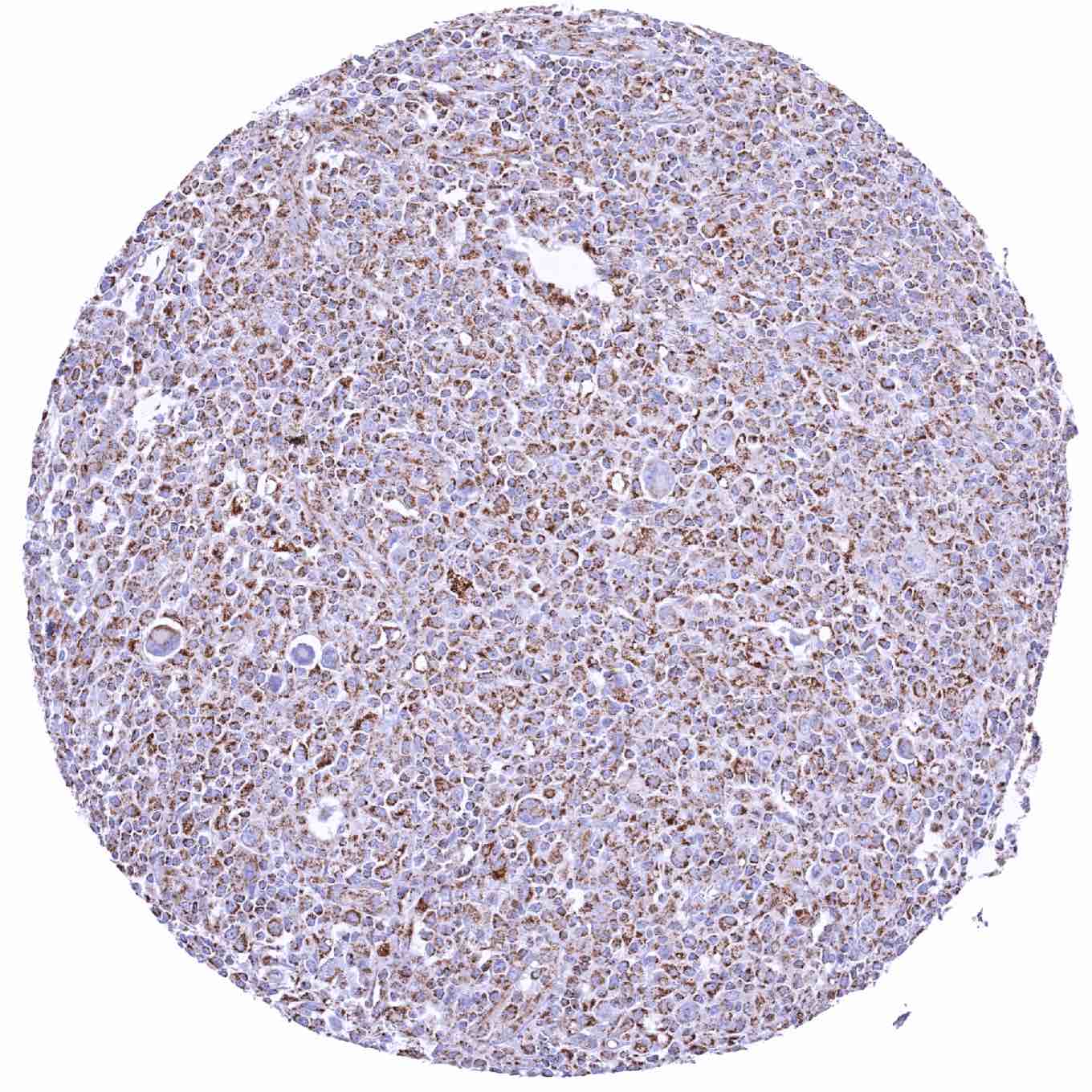
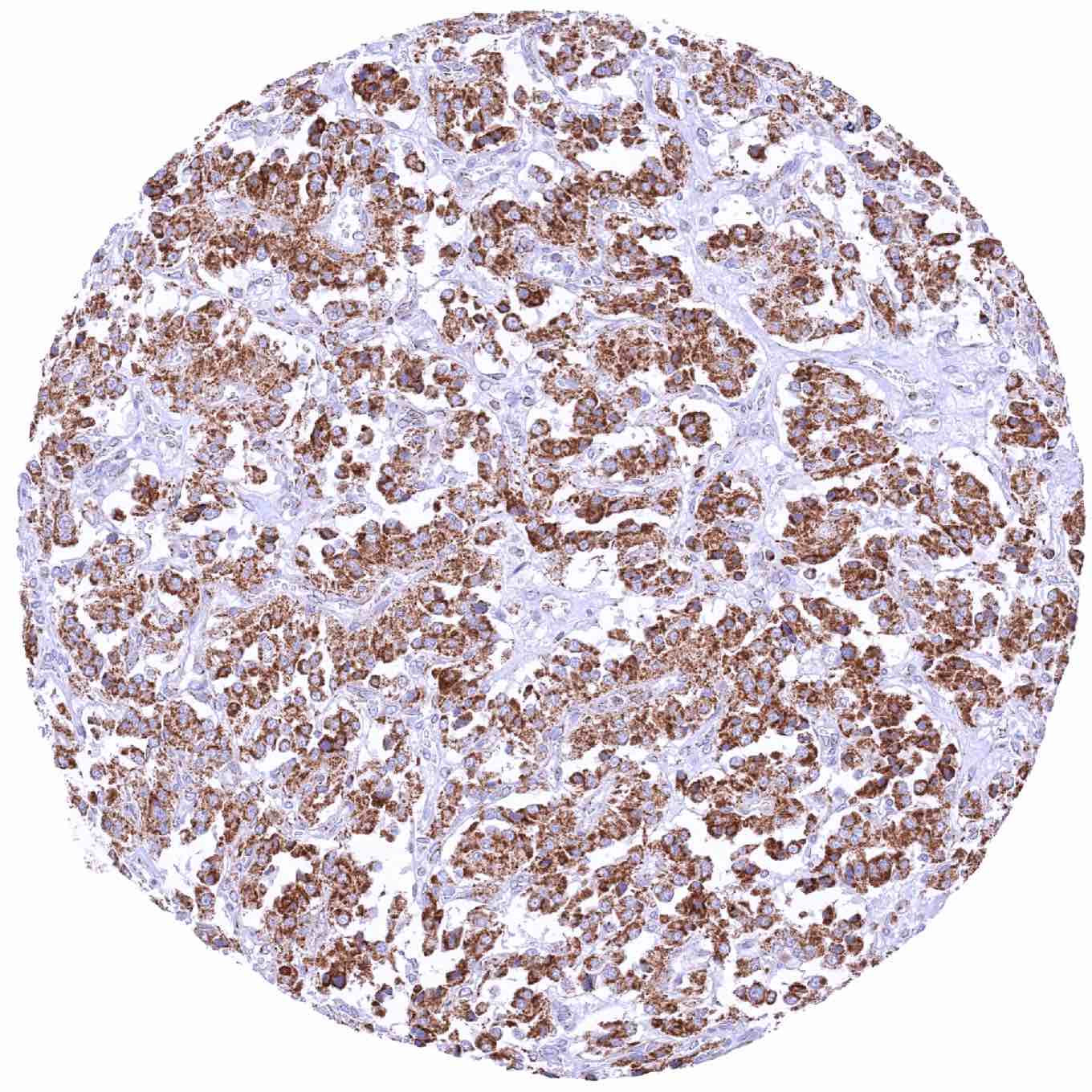
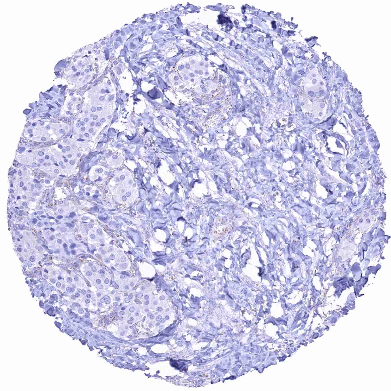
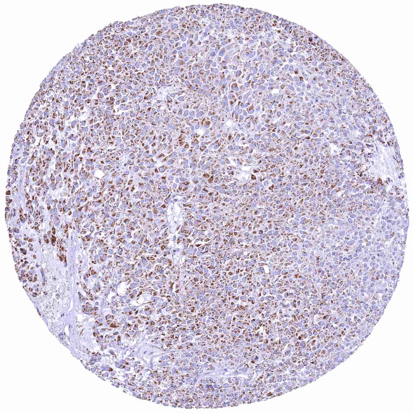
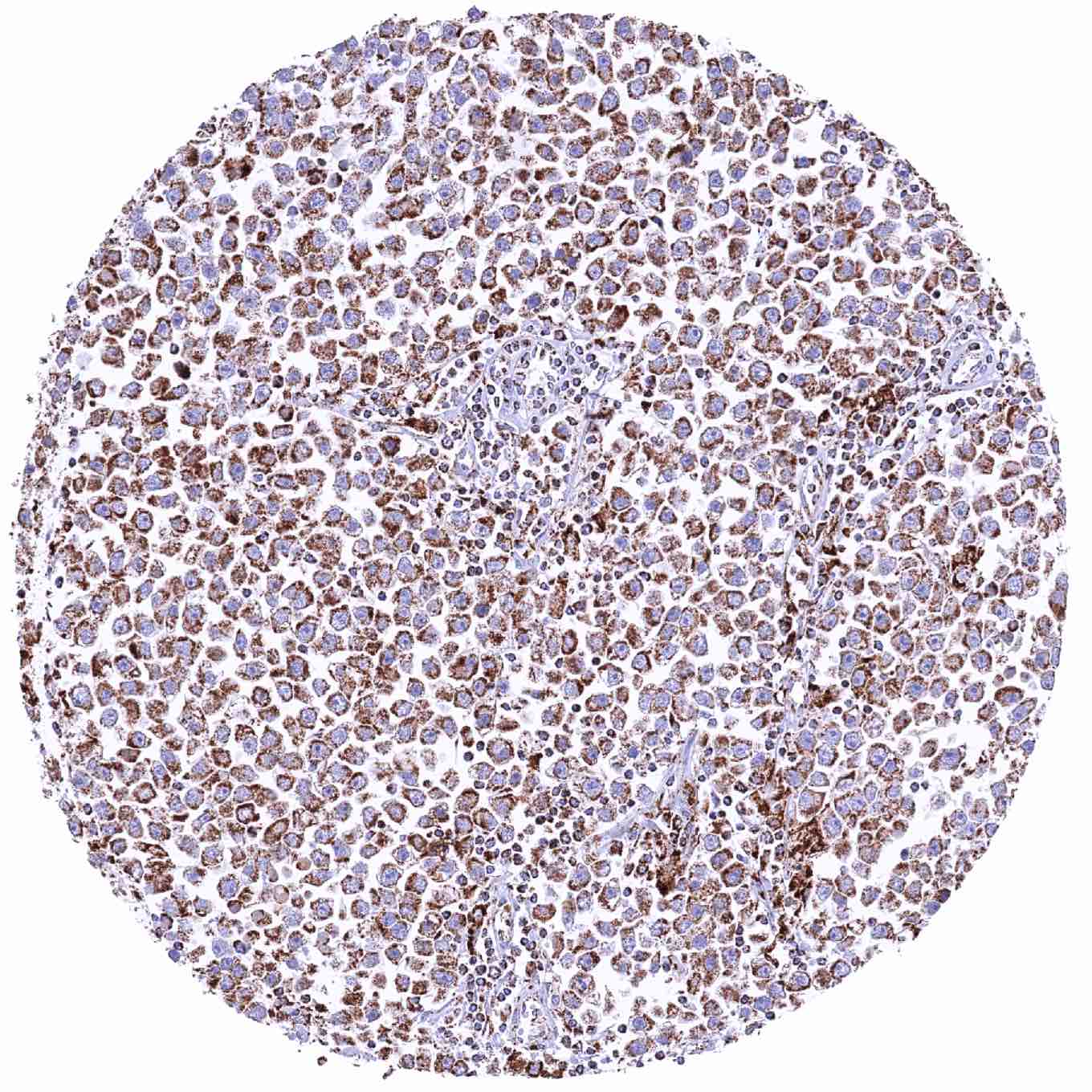
DHB antibody [HMV320] HistoMAXTM


Go to Normal Tissue Gallery
  
 Go back to product page
  

Go to HistoMAX page


Adrenal gland – Adrenocortical adenoma with moderate SDHB immunostaining of tumor cells

Adrenal gland – Adrenocortical carcinoma with intense SDHB staining of all tumor cells

Adrenal gland – Pheochromocytoma with moderate SDHB immunostaining of tumor cells

Breast – Invasive breast cancer of no special type (NST) with strong SDHB immunostaining of tumor cells


Colon – Colorectal adenocarcinoma with strong SDHB staining of tumor cells

Esophagus – Adenocarcinoma with strong SDHB staining of tumor cells

Kidney – Absence of granular SDHB staining in tumor cells of an SDH deficient renal cell carcinoma

Kidney – Chromophobe renal cell carcinoma with strong SDHB labeling of tumor cells


Kidney – Clear cell renal cell carcinoma exhibiting a strong SDHB immunostaining of tumor cells

Kidney – Clear cell renal cell carcinoma showing moderate SDHB immunostaining of tumor cells

Kidney – Papillary renal cell carcinoma with moderate to strong SDHB staining of tumor cells

Kidney – SDHB mutated renal cell carcinoma lacking granular SDHB staining of tumor cells


Larynx – Squamous cell carcinoma with intense SDHB positivity of tumor cells

Liver – Cholangiocellular carcinoma with strong SDHB immunostaining of tumor cells

Liver – Hepatocellular carcinoma with intense SDHB staining of tumor cells

Lymph node – Diffuse large B-cell lymphoma with strong SDHB staining of tumor cells


Lymph node – Hodgkin's lymphoma with strong SDHB positivity of tumor associated inflammatory cells. Tumor cells are also positive

Pancreas – Acinar cell carcinoma showing SDHB staining of variable intensity in tumor cells

Pancreas – Ductal adenocarcinoma with intense SDHB tumor cell positivity

Pancreas – Neuroendocrine tumor showing moderate to strong SDHB positivity of tumor cells


Parathyroidal gland – Adenoma exhibiting strong SDHB positivity of tumor cells

Prostate – Adenocarcinoma (Gleason 3+3=6) with moderate intensity SDHB staining of tumor cells

Prostate – Adenocarcinoma (Gleason 4+4=8) showing marked SDHB positivity of tumor cells

Salivary gland – Warthin tumor with intensive SDHB immunostaining of epithelial tumor cells


Soft tissue – Paraganglioma showing intense SDHB positivity of all tumor cells

Soft tissue – Paraganglioma with complete loss of granular SDHB staining of tumor cells. Intense STHB staining of inflammatory and stroma cells

Soft tissue – Paraganglioma with diffuse SDHB positivity of tumor cells

Soft tissue – Paraganglioma with strong SDHB immunostaining of all tumor cells


Soft tissue – Paraganglioma with strong SDHB positivity of tumor cells

Soft tissue – Paraganglioma with weak to moderate SDHB staining of tumor cells

Soft tissue – SDH deficient paraganglioma lacking granular SDHB staining in all tumor cells. Stroma cells are SDHB positive

Soft tissue – SDH deficient Paraganglioma lacking granular SDHB staining of tumor cells while stromal cells are positive


Stomach – Gastric adenocarcinoma (diffuse type) with strong SDHB positivity of tumor cells

Stomach – Gastrointestinal stromal tumor (GIST) with strong SDHB immunostaining of tumor cells

Stomach – Gastrointestinal stromal tumor (GIST) with strong SDHB staining of tumor cells

Stomach – Gastrointestinal stromal tumor (GIST) with strong SDHB staining of tumor cells


Testis – Leydig cell tumor with intense SDHB staining of tumor cells

Testis – Seminoma with intensepositivity SDHB immunostaining of tumor cells

Thyroid – Medullary cancer with moderate SDHB positivity of tumor cells

Thyroid – Papillary cancer with moderate SDHB immunostaining of tumor cells


Urinary bladder – Muscle-invasive urothelial carcinoma showing weak to moderate SDHB immunostaining of tumor cells

Uterus – Leiomyosarcoma with moderate SDHB staining of tumor cells


| TOP |