Country / Location Selection
Search suggestions
Synaptophysin antibody [MSVA-462R] HistoMAXTM
Go to HistoMAX page
Adrenal gland – Adrenocortical adenoma with a weak to moderate , mostly membranous Synaptophysin immunostaining in about 50% of tumor cells.
Adrenal gland – Adrenocortical adenoma with a weak Synaptophysin immunostaining in few tumor cells.
Adrenal gland – Moderate to strong Synaptophysin positivity in an adrenocortical adenoma.
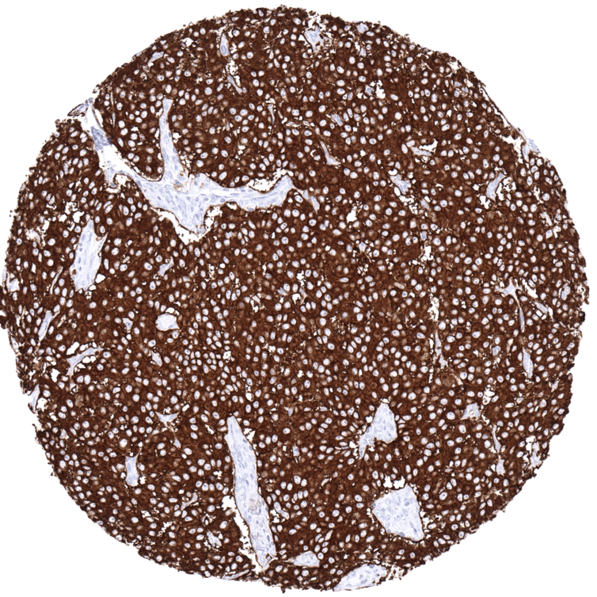
Adrenal gland – Pheochromocytoma with strong, diffuse Synaptophysin positivity in all tumor cells.
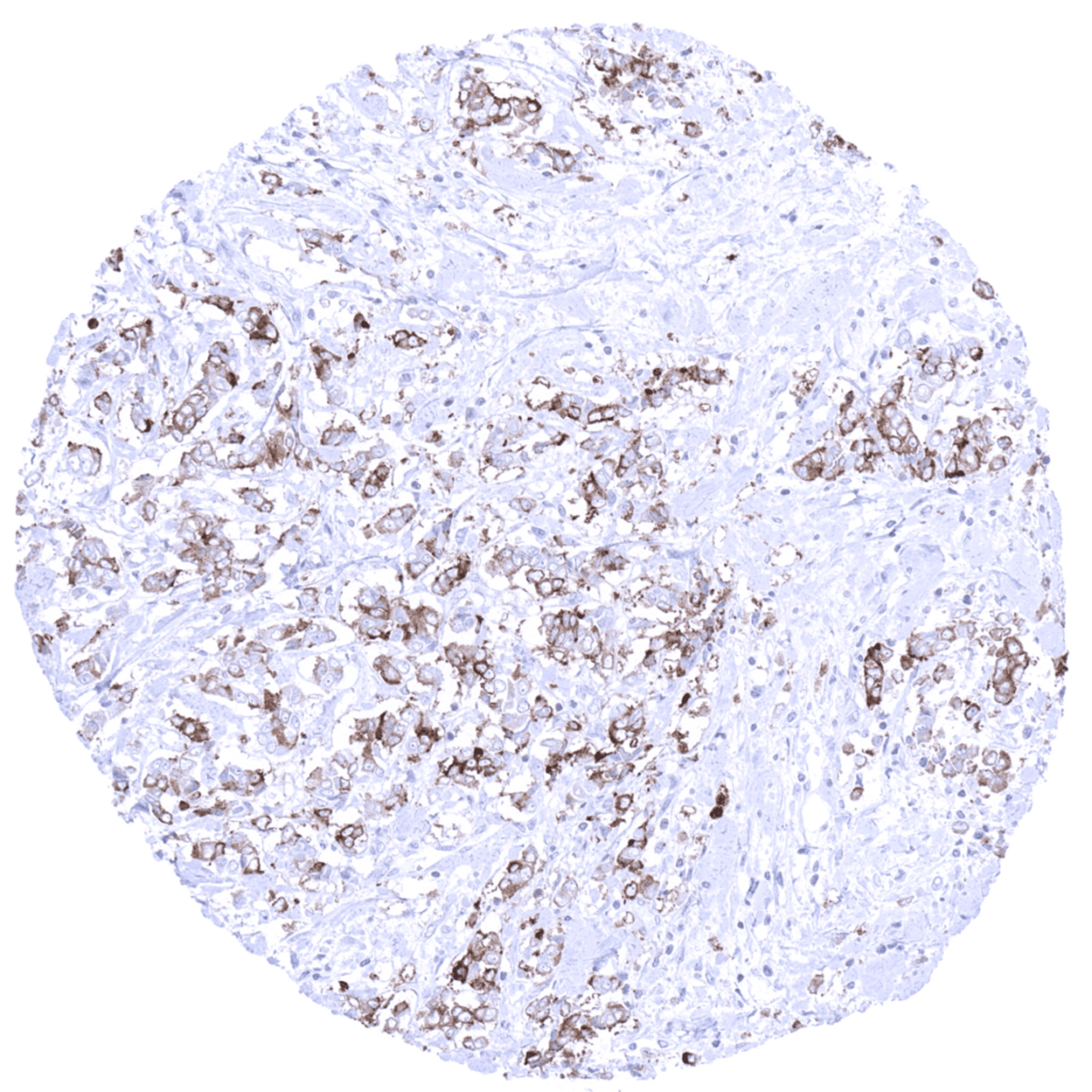
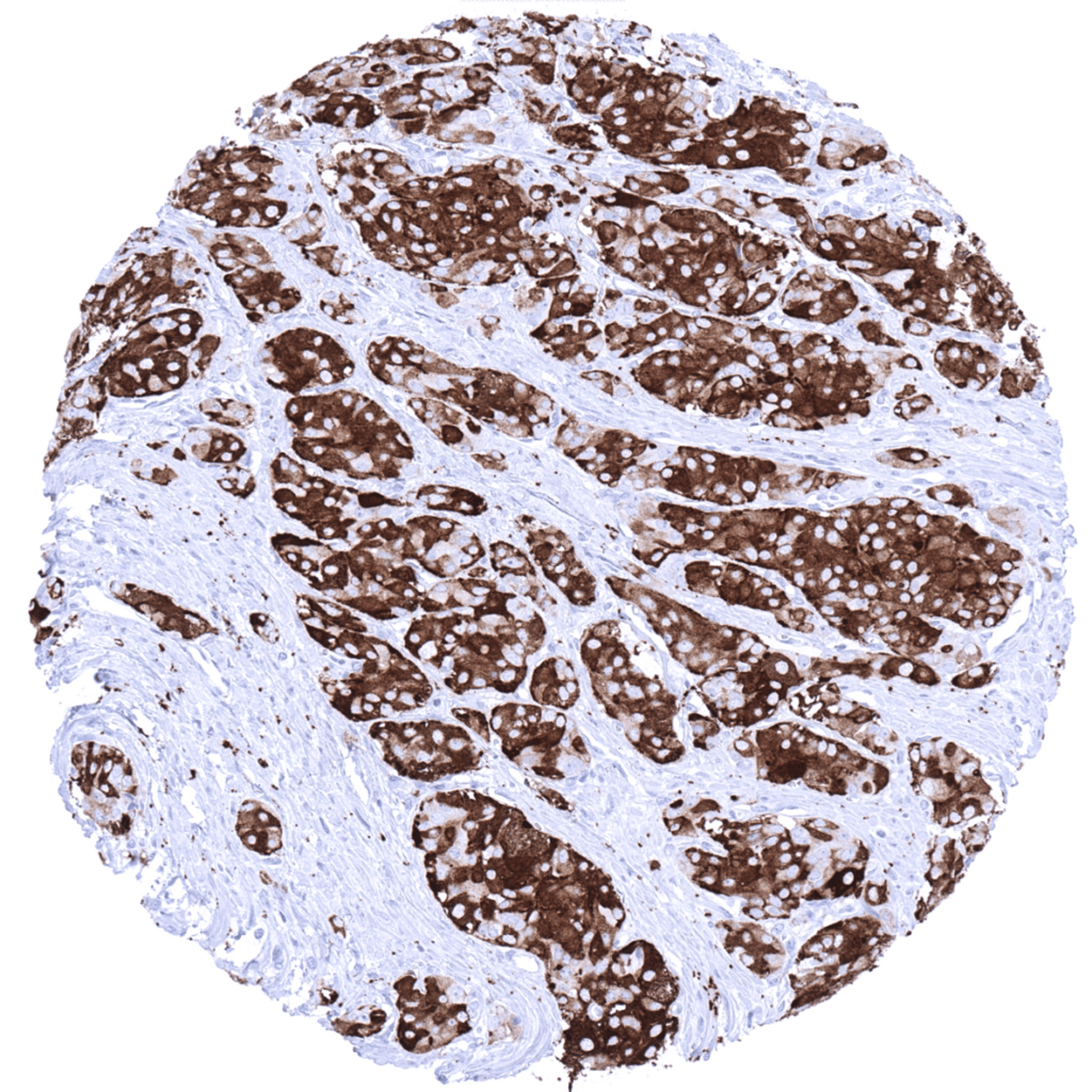
Breast – Invasive breast cancer of no special type (NST) exhibiting a weak to moderate Synaptophysin immunostaining of all tumor cells.
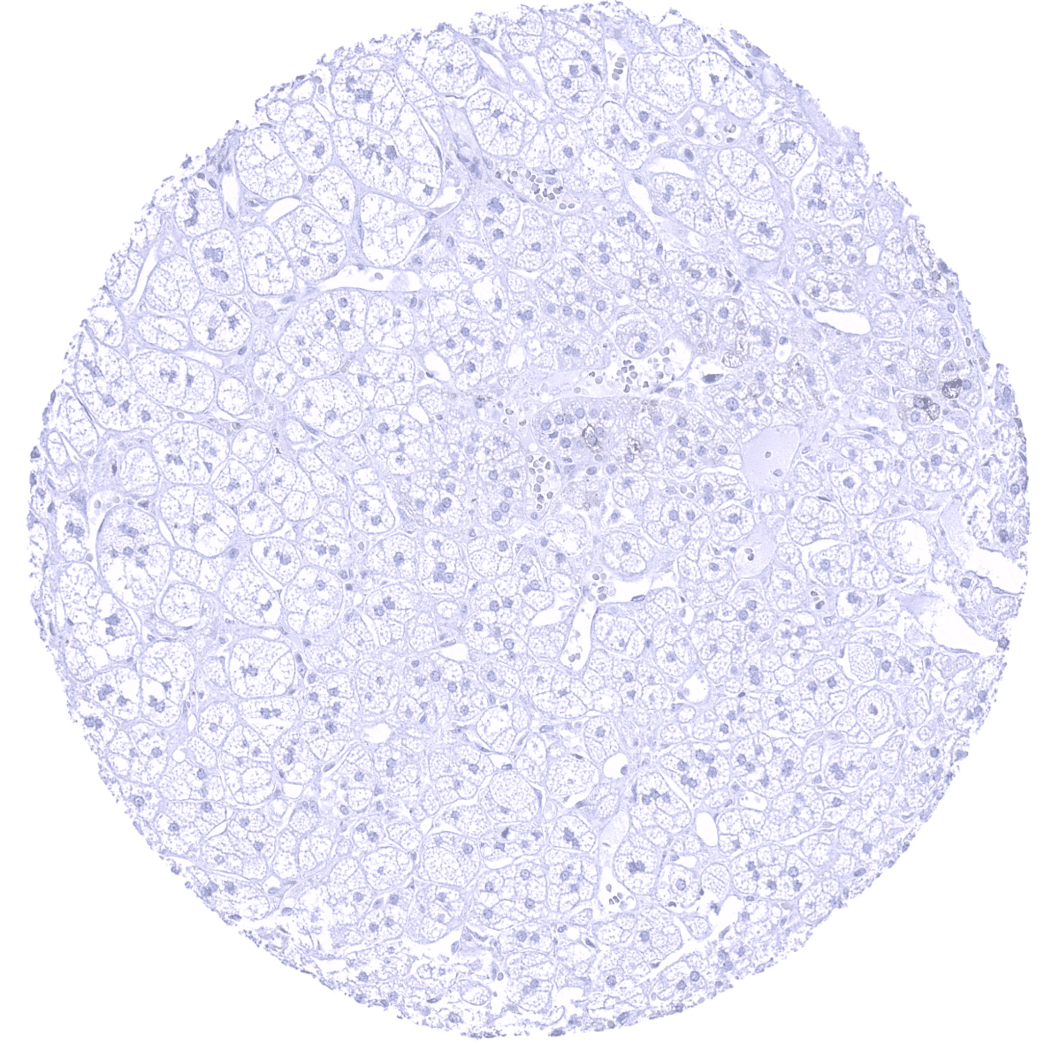
Breast – Synaptophysin negative invasive breast cancer of no special type (NST).
Lung – Small cell carcinoma showing a moderate Synaptophysin immunostaining of all tumor cells.
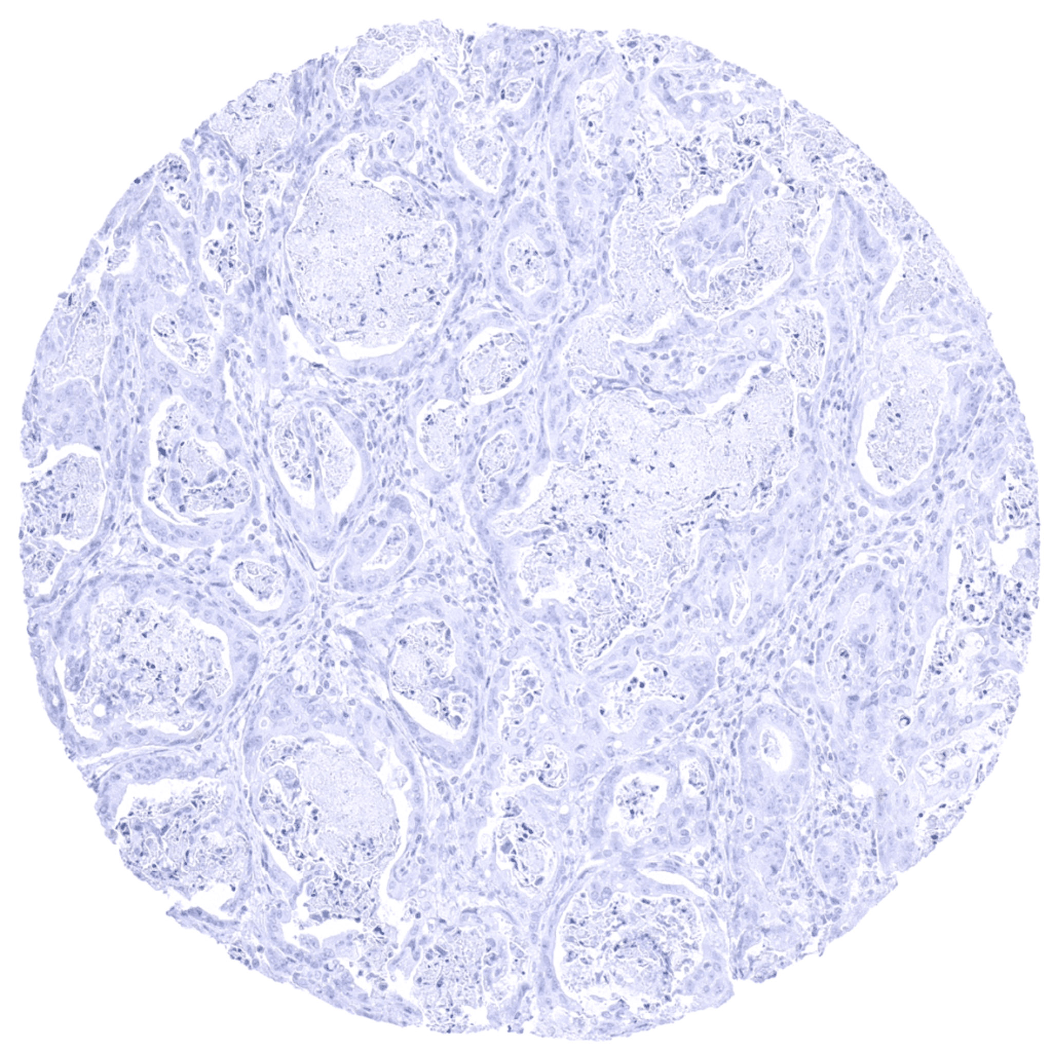
Lung – Synaptophysin negative adenocarcinoma.
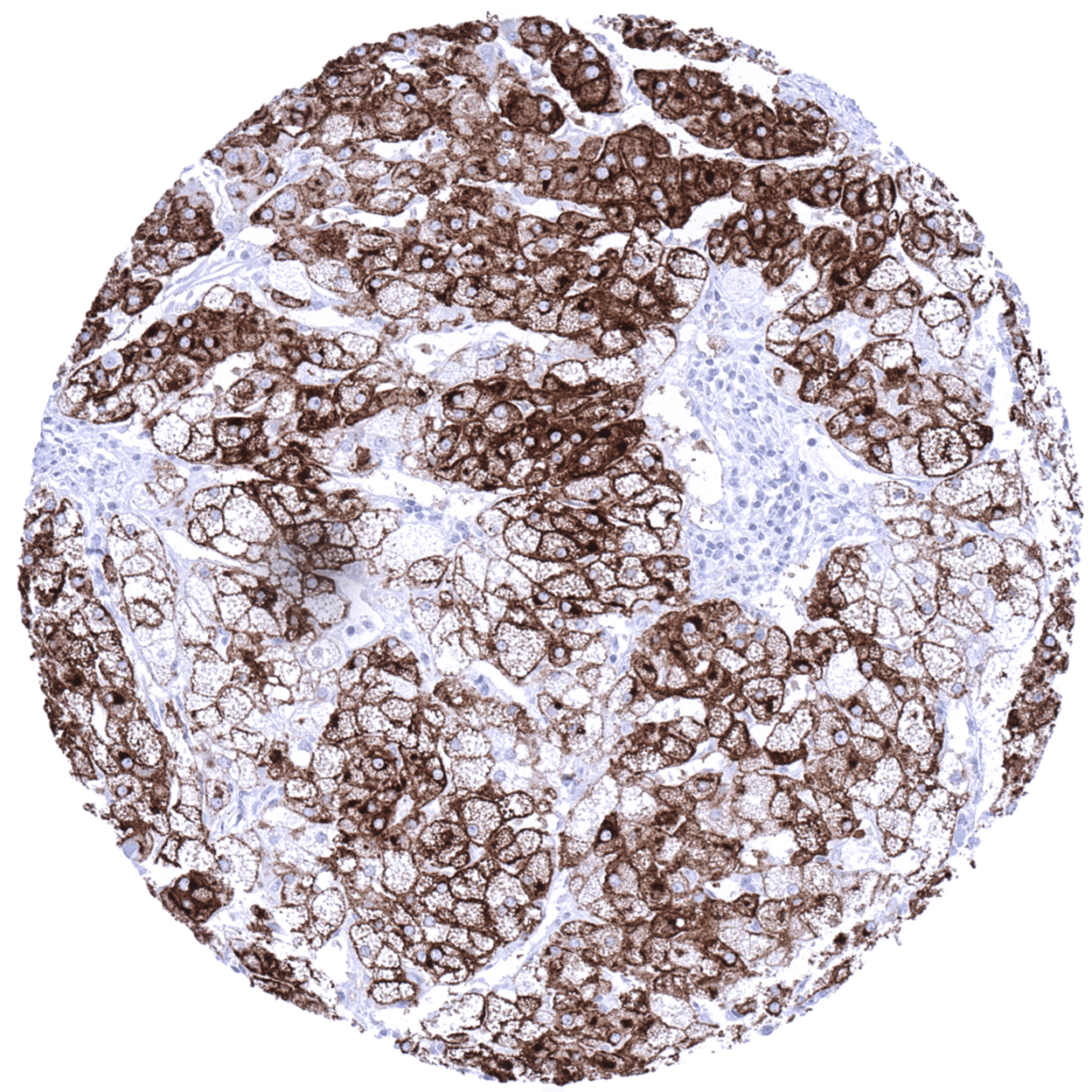
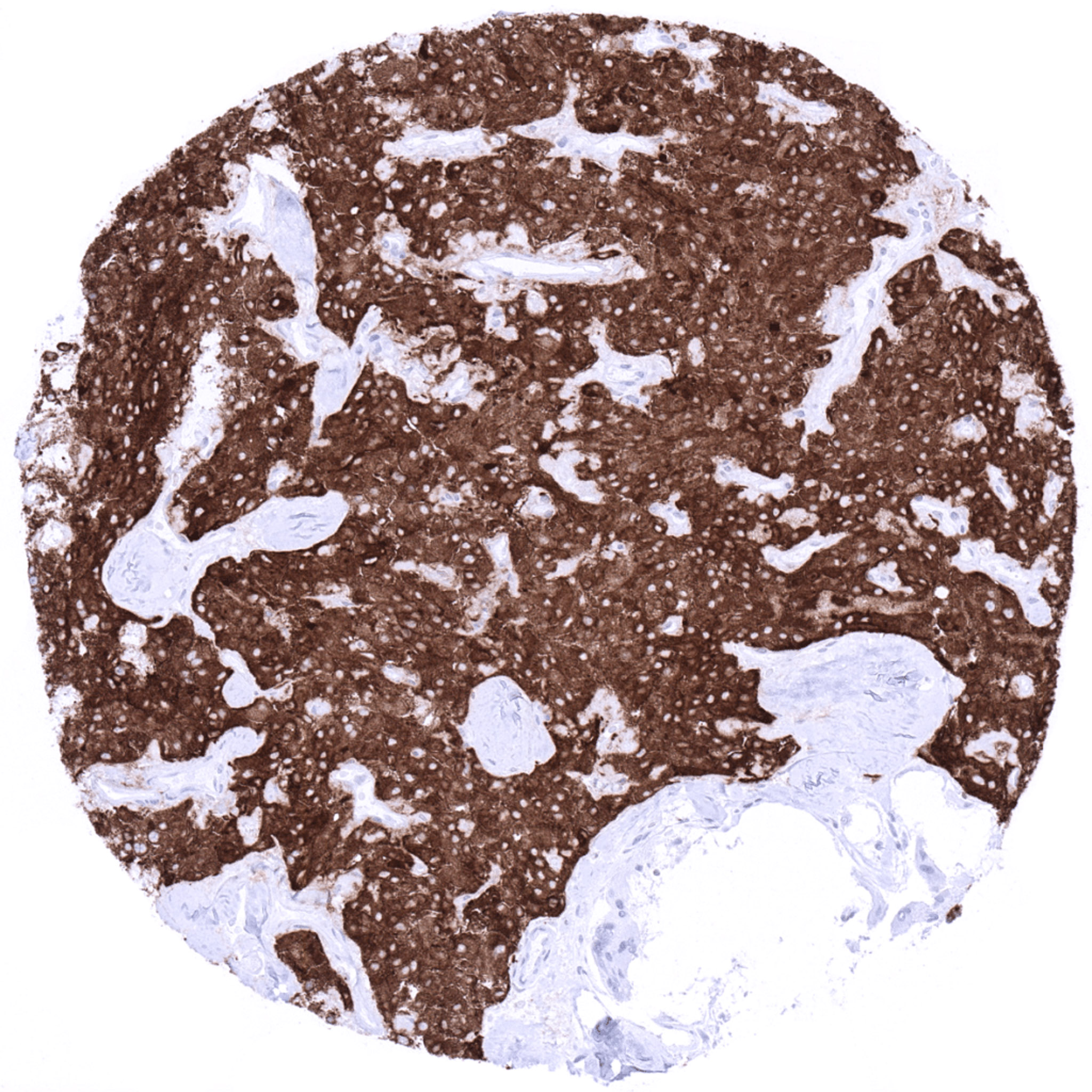
Pancreas – Neuroendocrine tumor exhibiting strong cytoplasmic Synaptophysin immunostaining of tumor cells.
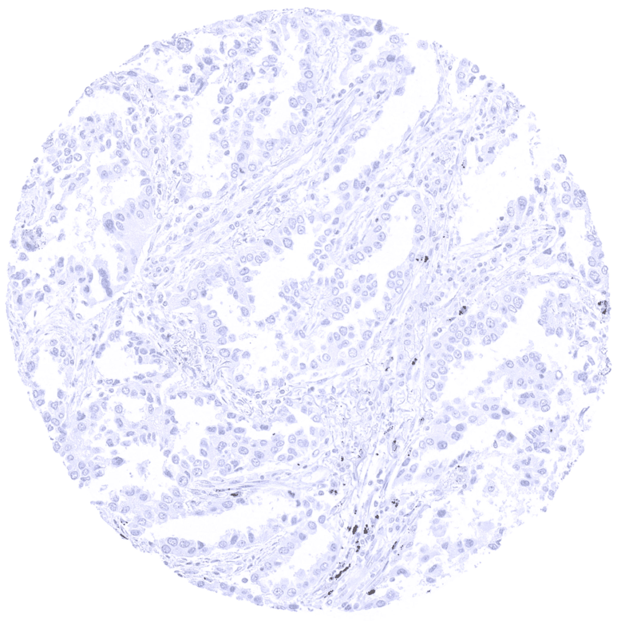
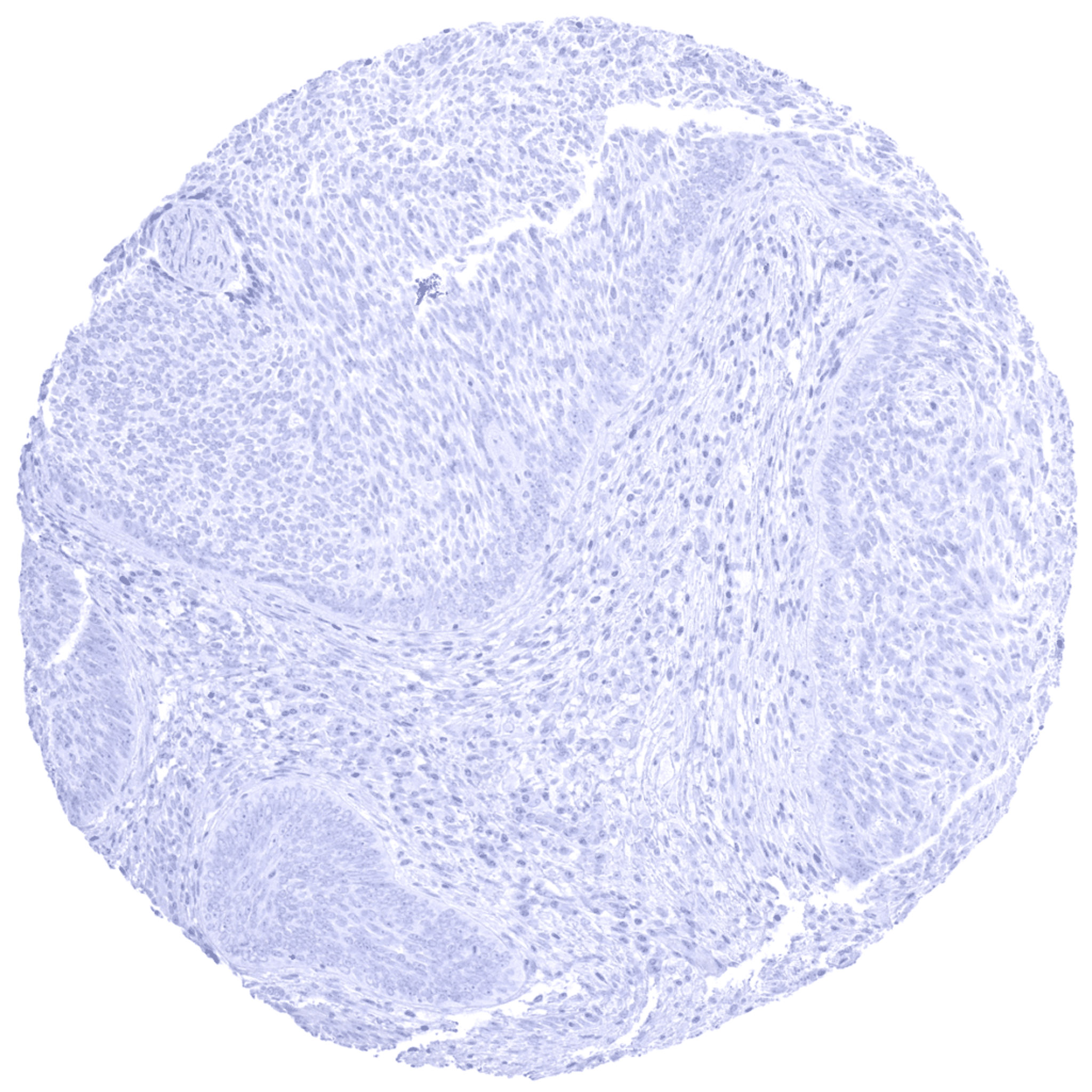
Prostate – Synaptophysin negative adenocarcinoma (Gleason 3+3=6).
Prostate – Synaptophysin positive adenocarcinoma (Gleason 5+5=10).
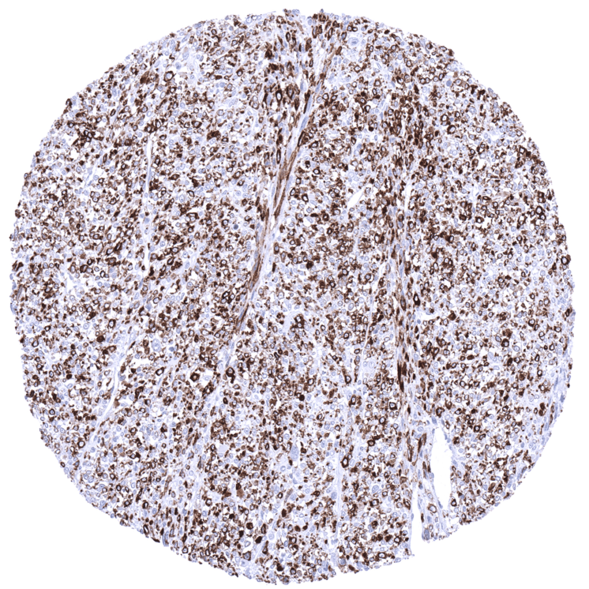
Retroperitoneum – Diffuse, moderate to strong Synaptophysin immunostaining of all cells of a leiomyosarcoma.
Skin – Merkel cell carcinoma displaying an intense cytoplasmic Synaptophysin immunostaining of all tumor cells.
Skin – Synaptophysin negative basal cell carcinoma.
Stomach – Synaptophysin negative adenocarcinoma (intestinal type).
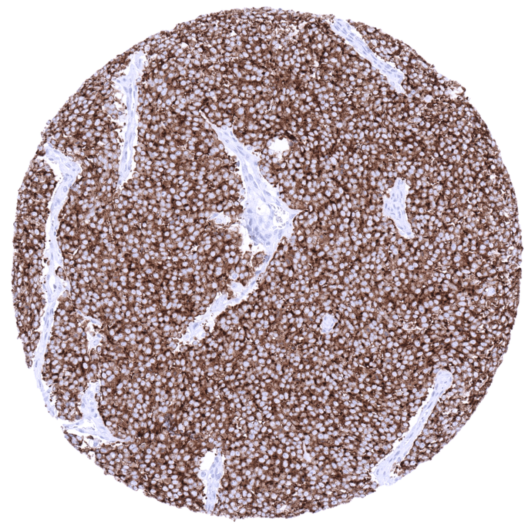
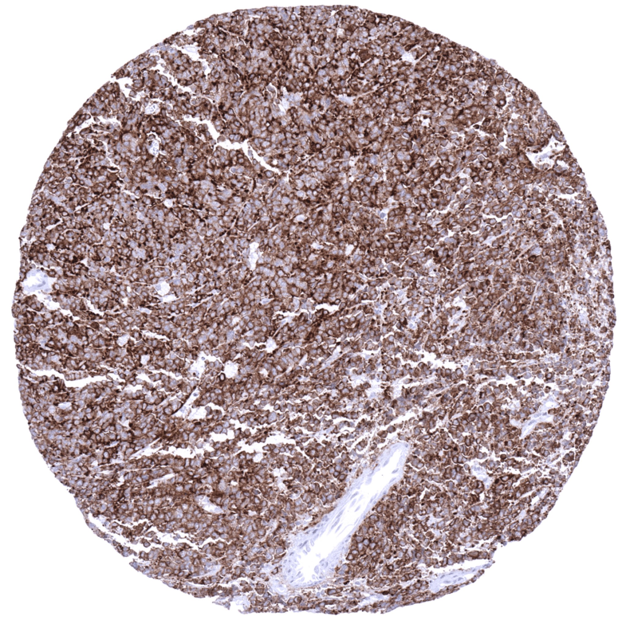
Thyroid – Medullary carcinoma with strong cytoplasmic synaptophysin immunostaining of all tumor cells.
Urinary bladder – Small cell carcinoma with diffuse, intense cytoplasmic Synaptophysin positivity.
| TOP |