Normal Tissue Gallery- GTX639933

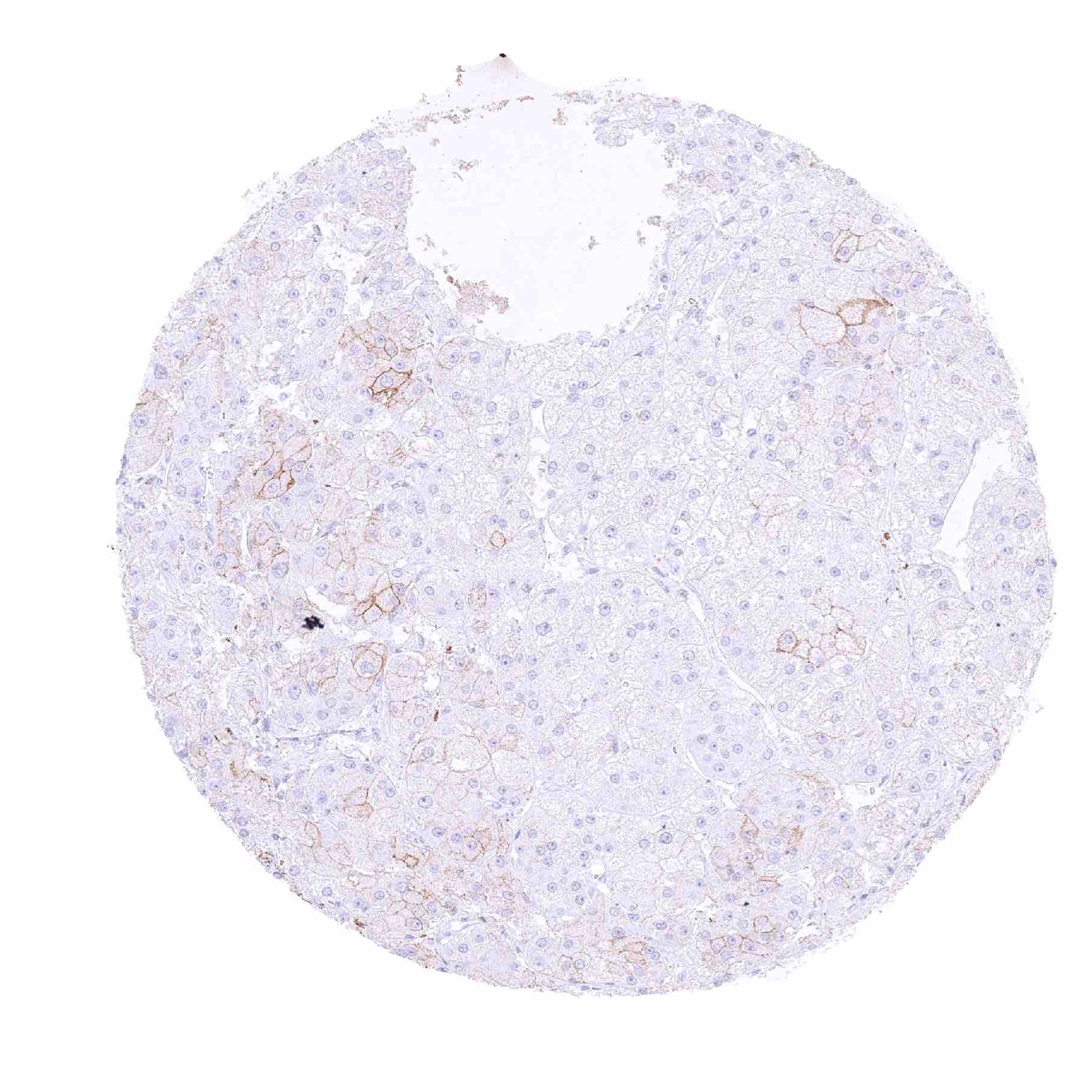
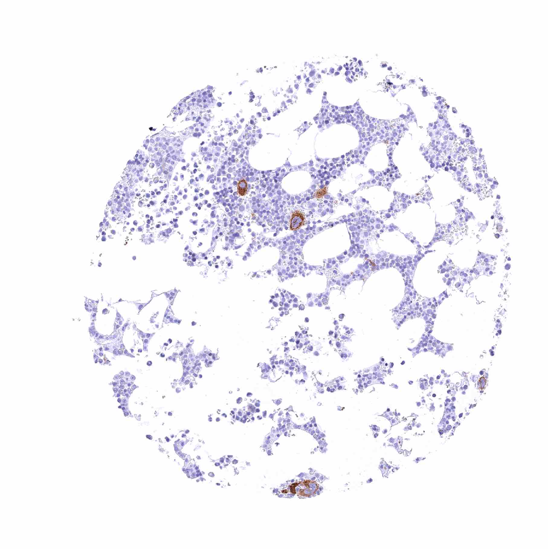
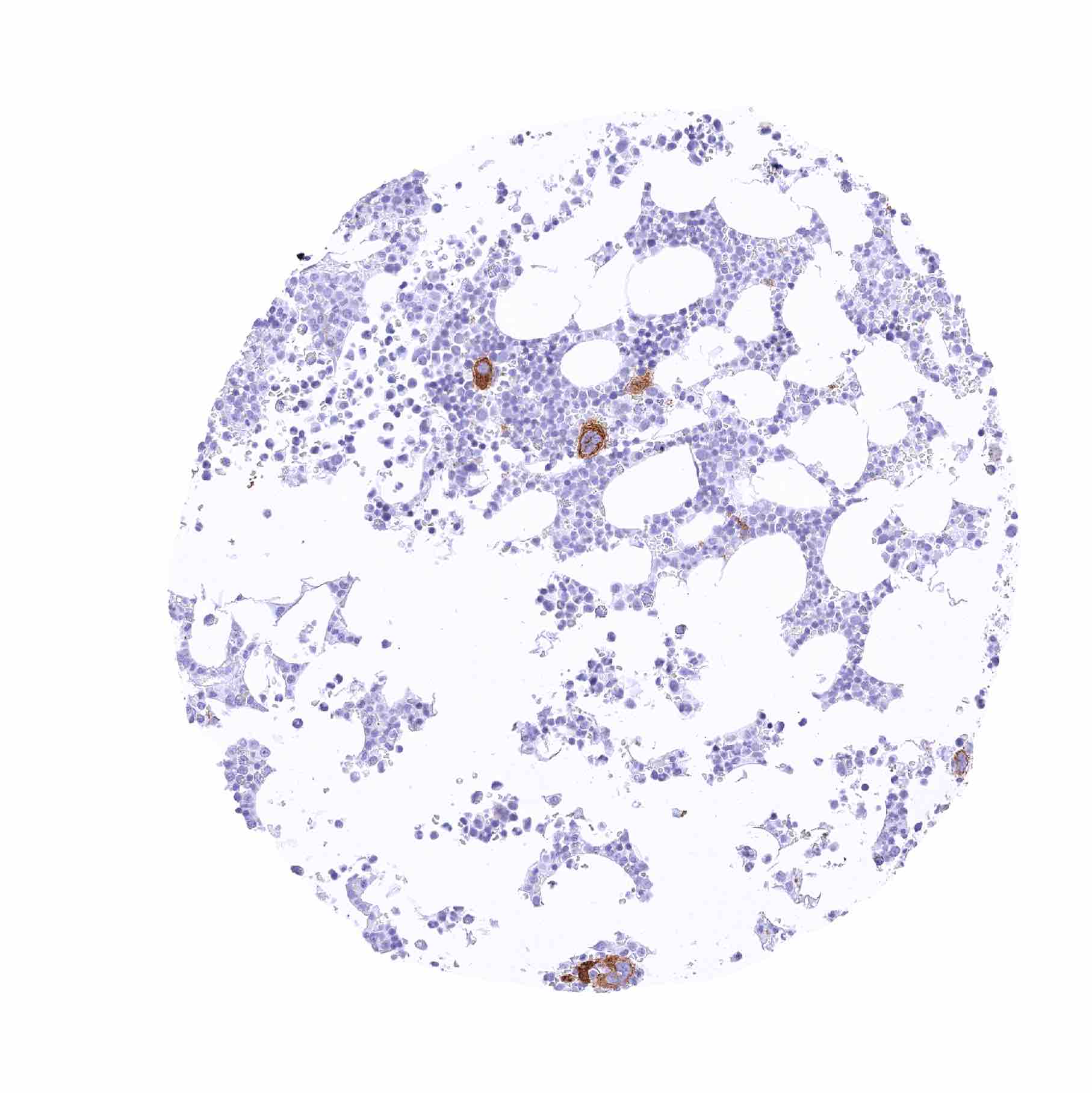
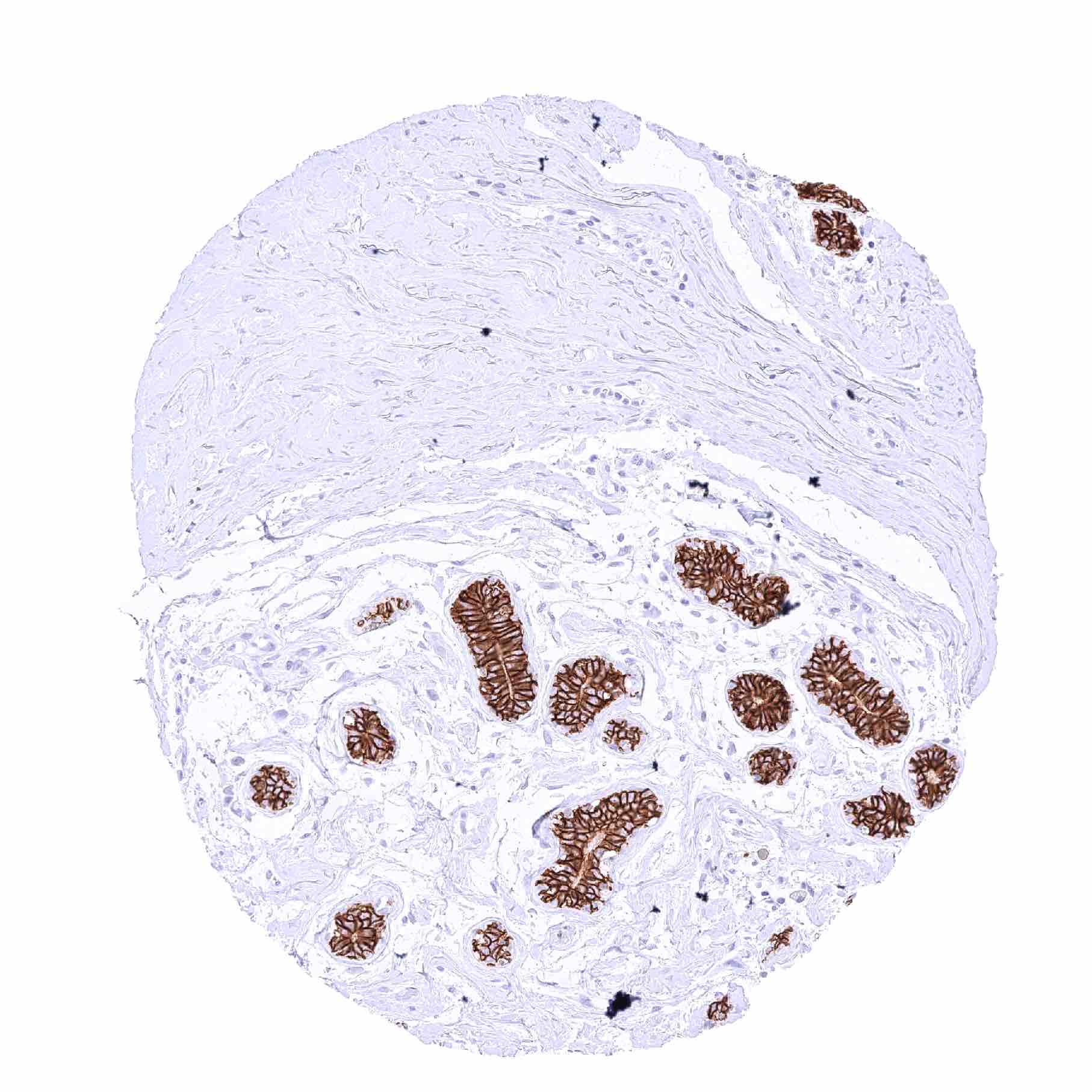
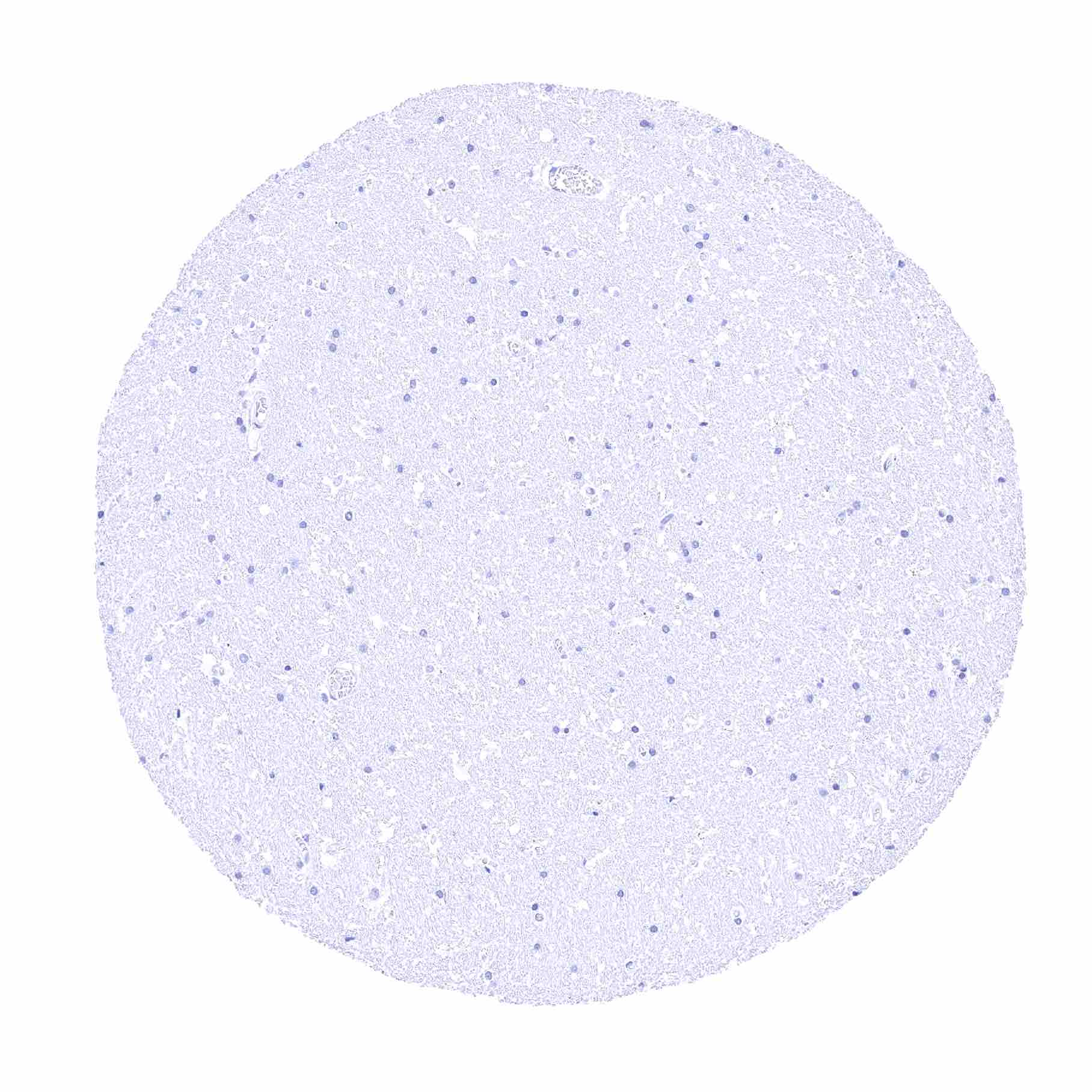
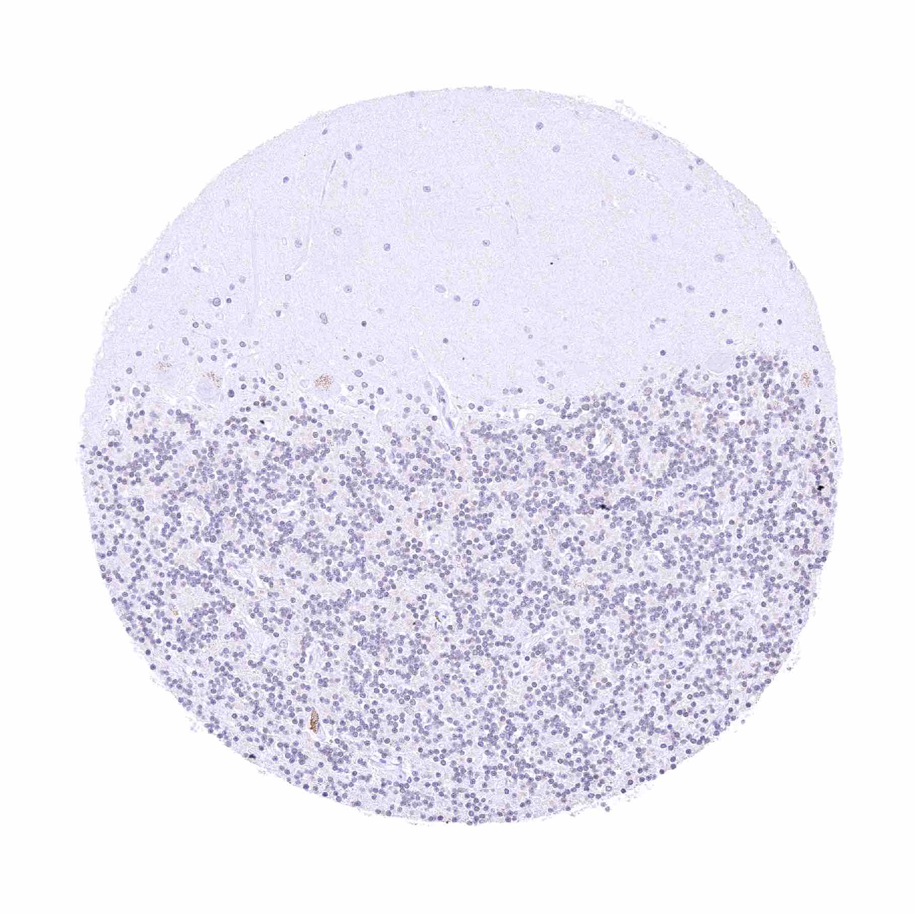
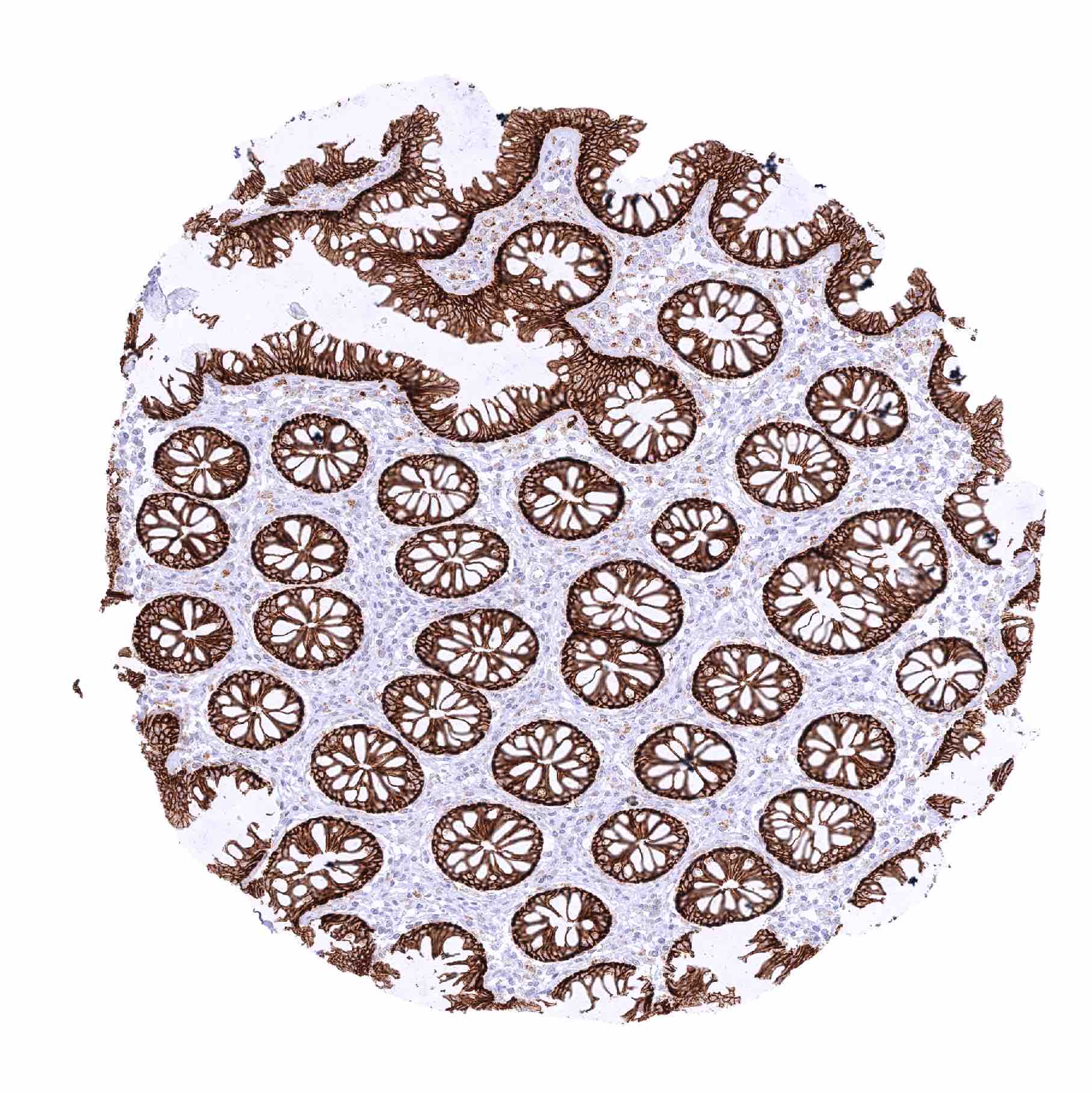
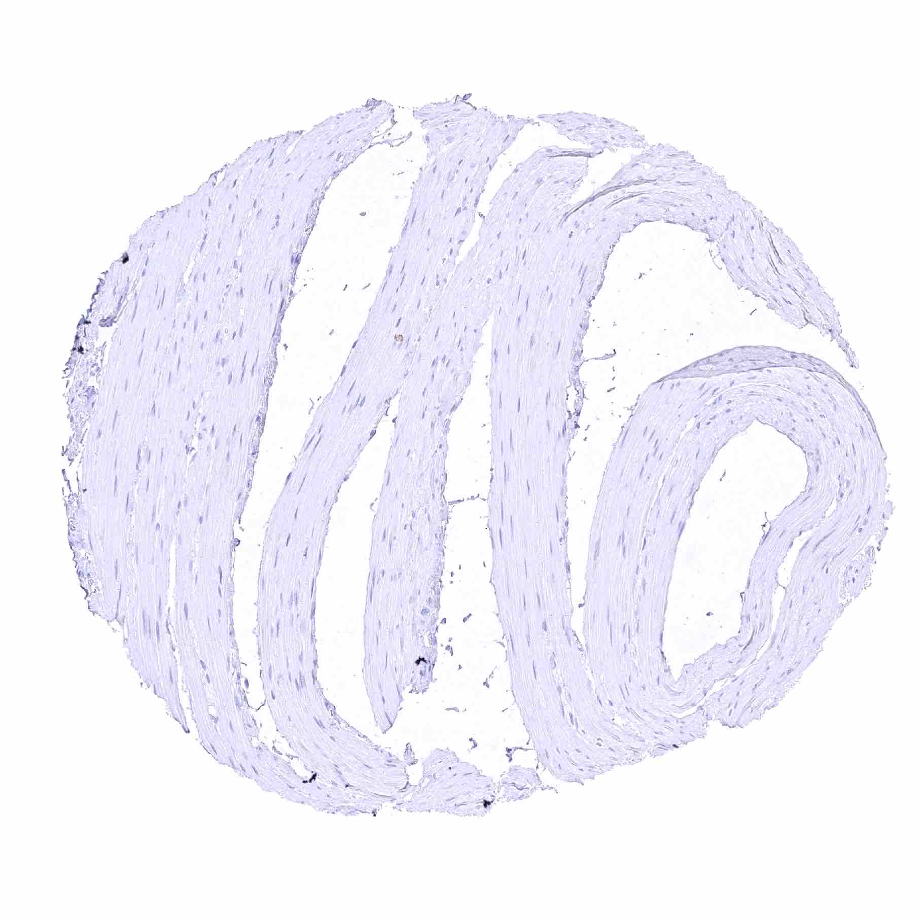
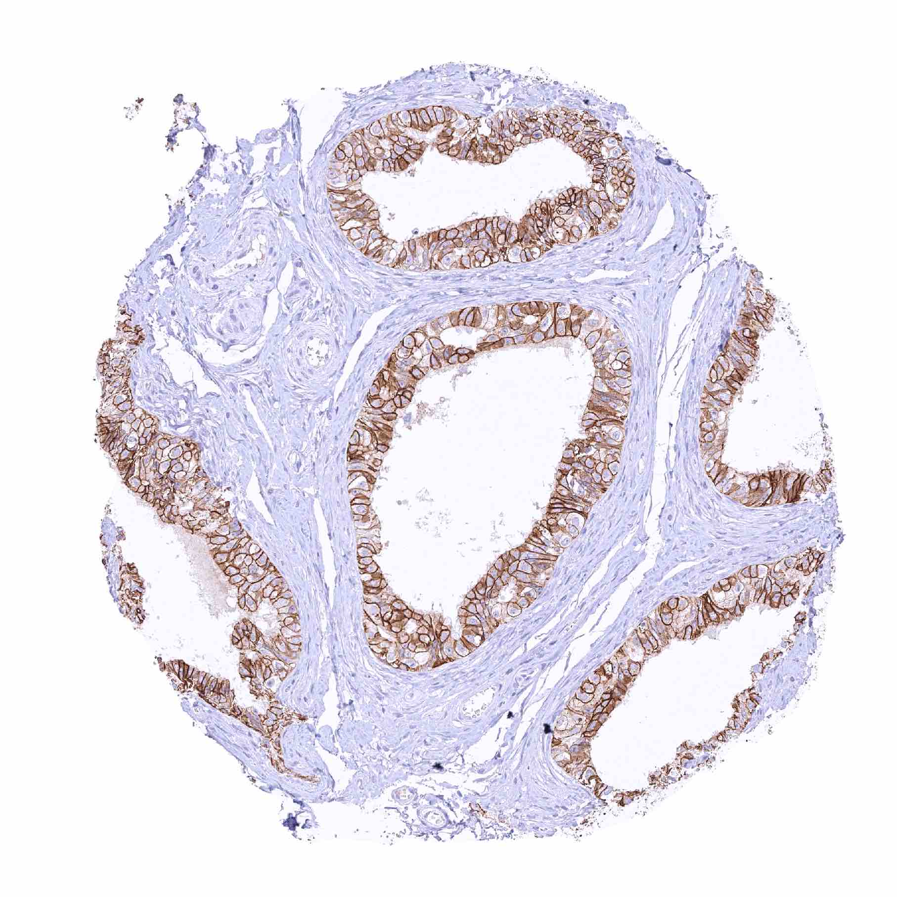
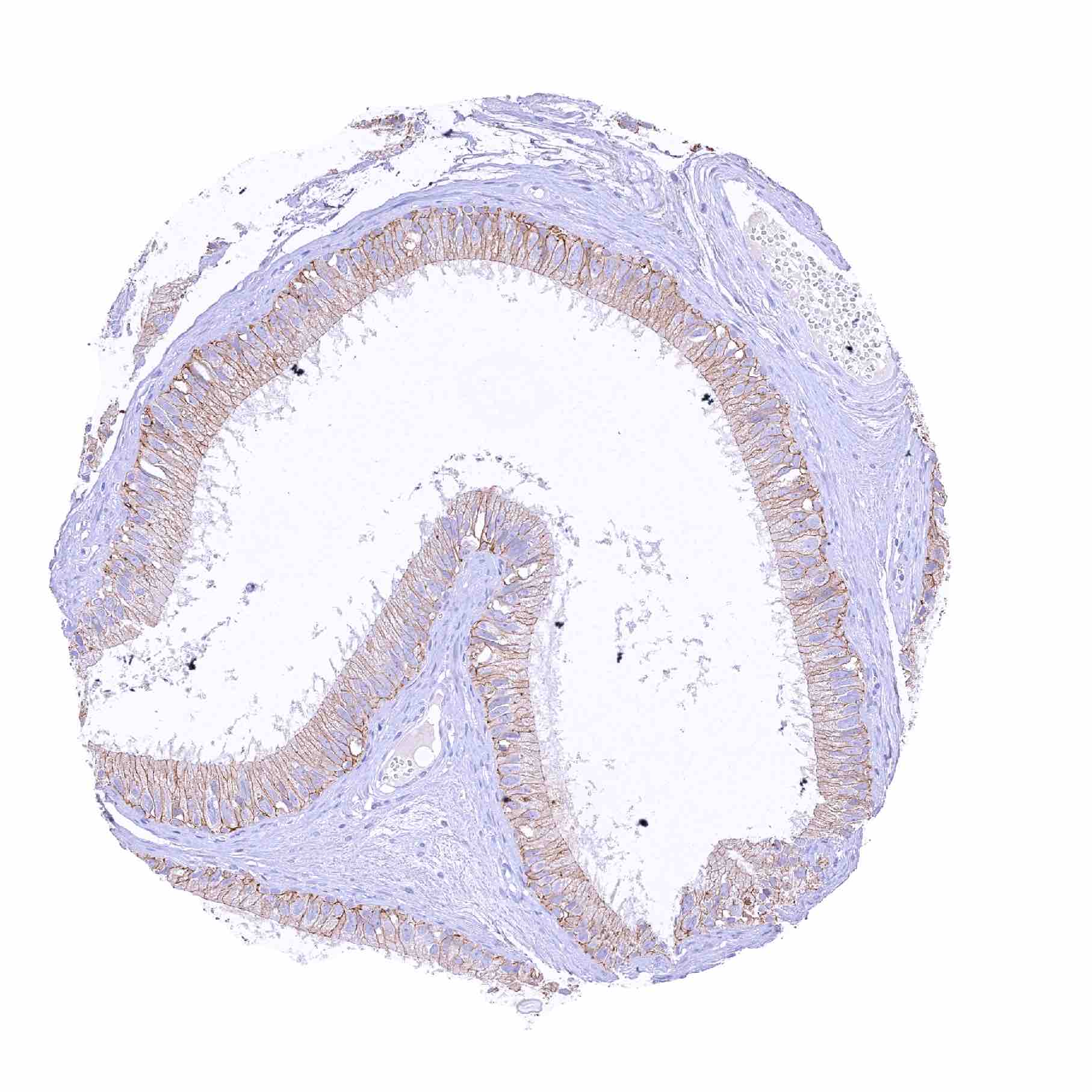
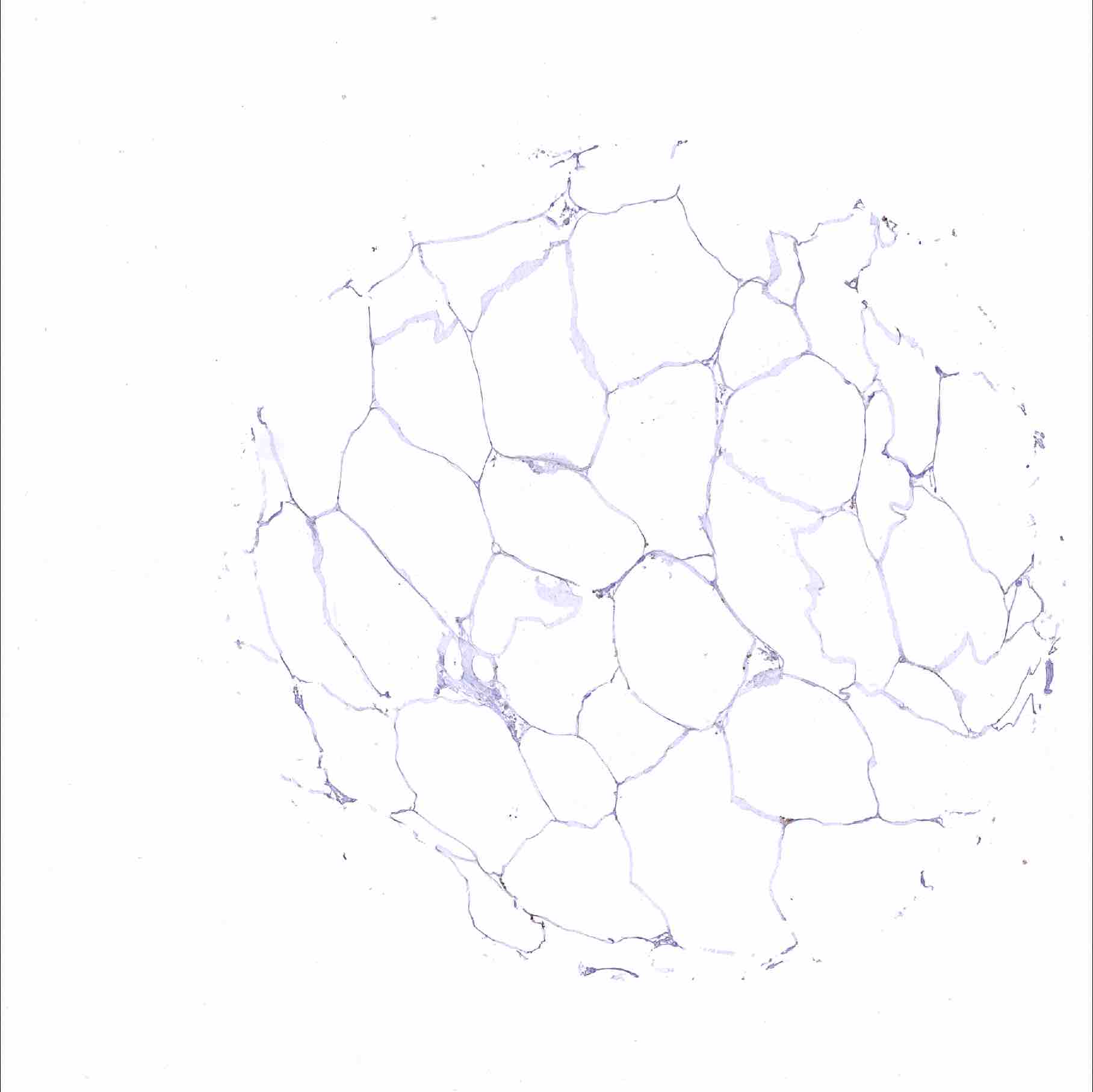
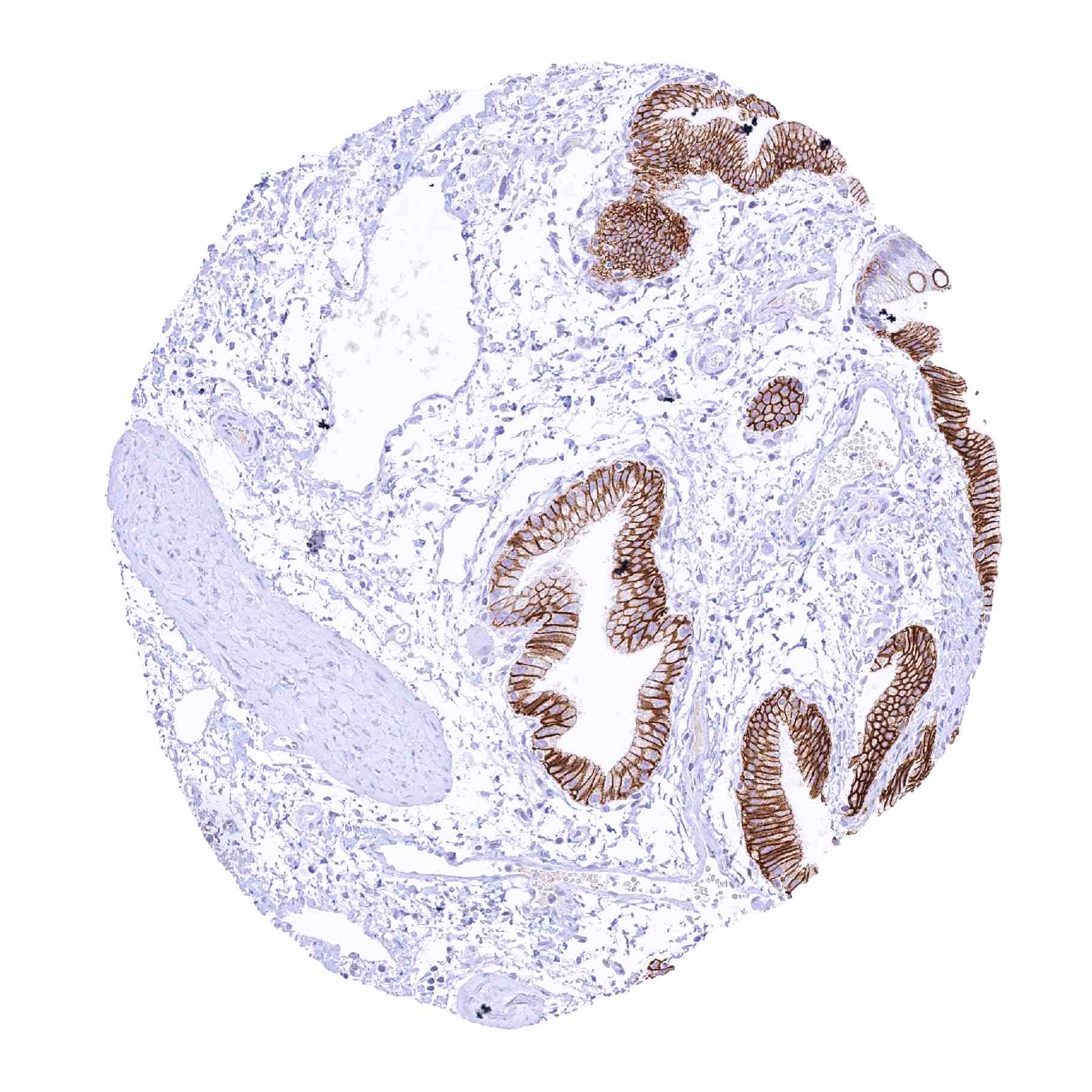
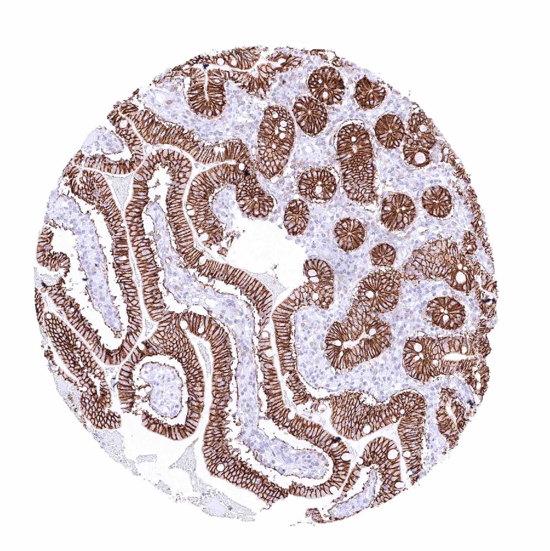
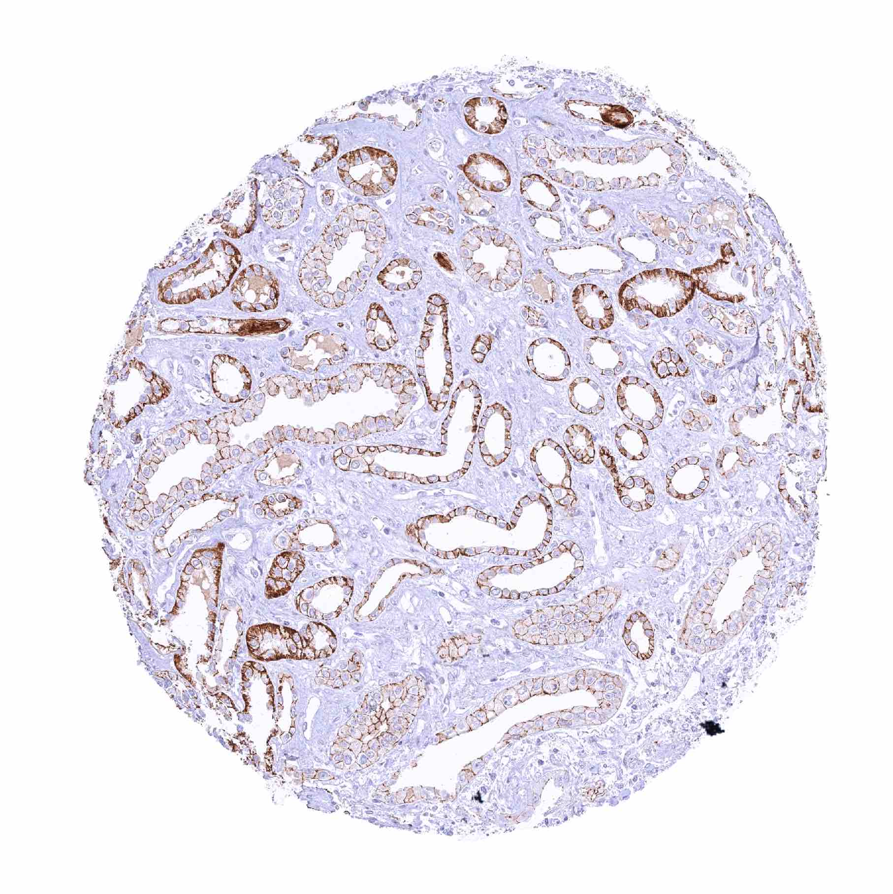
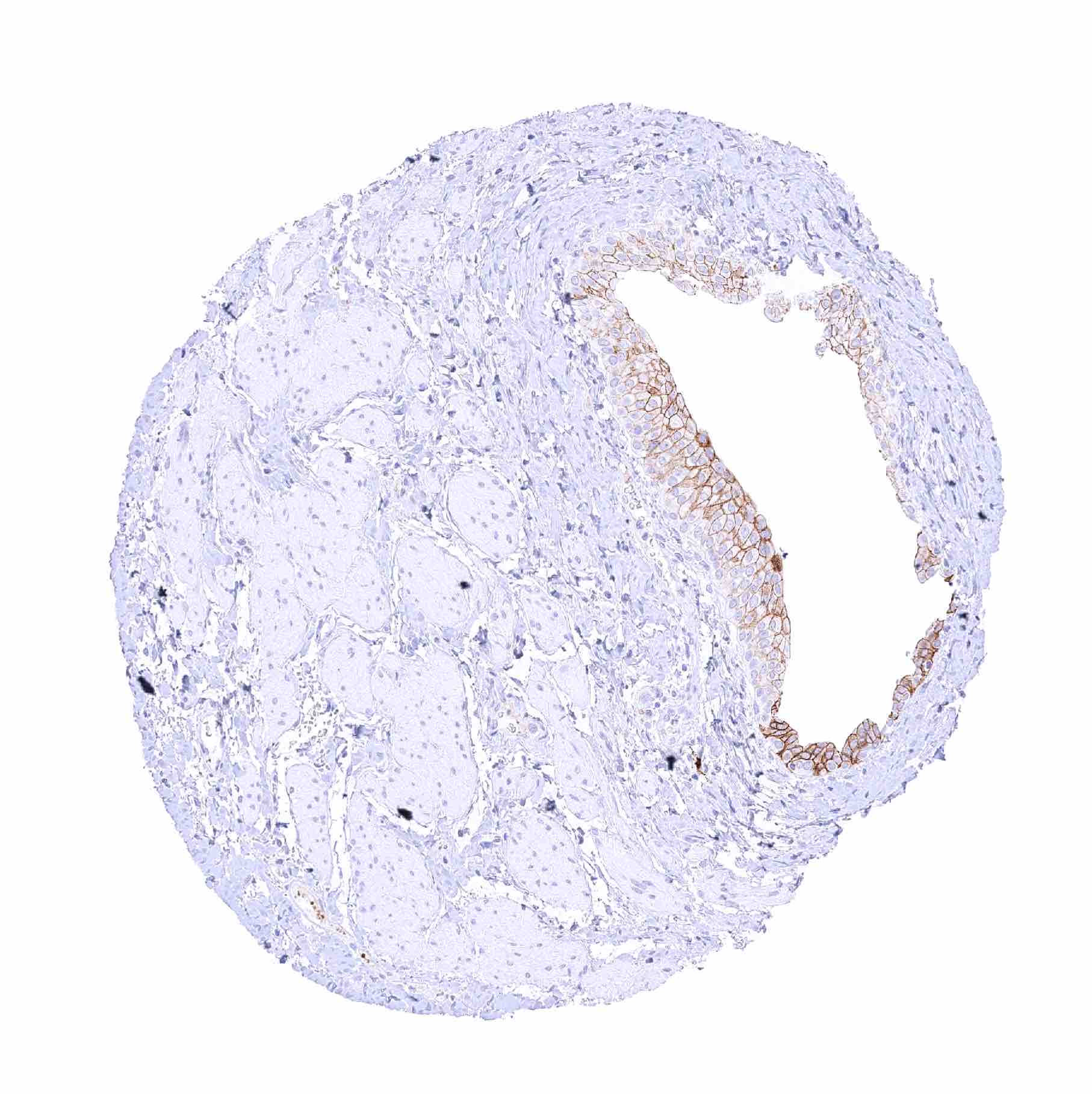
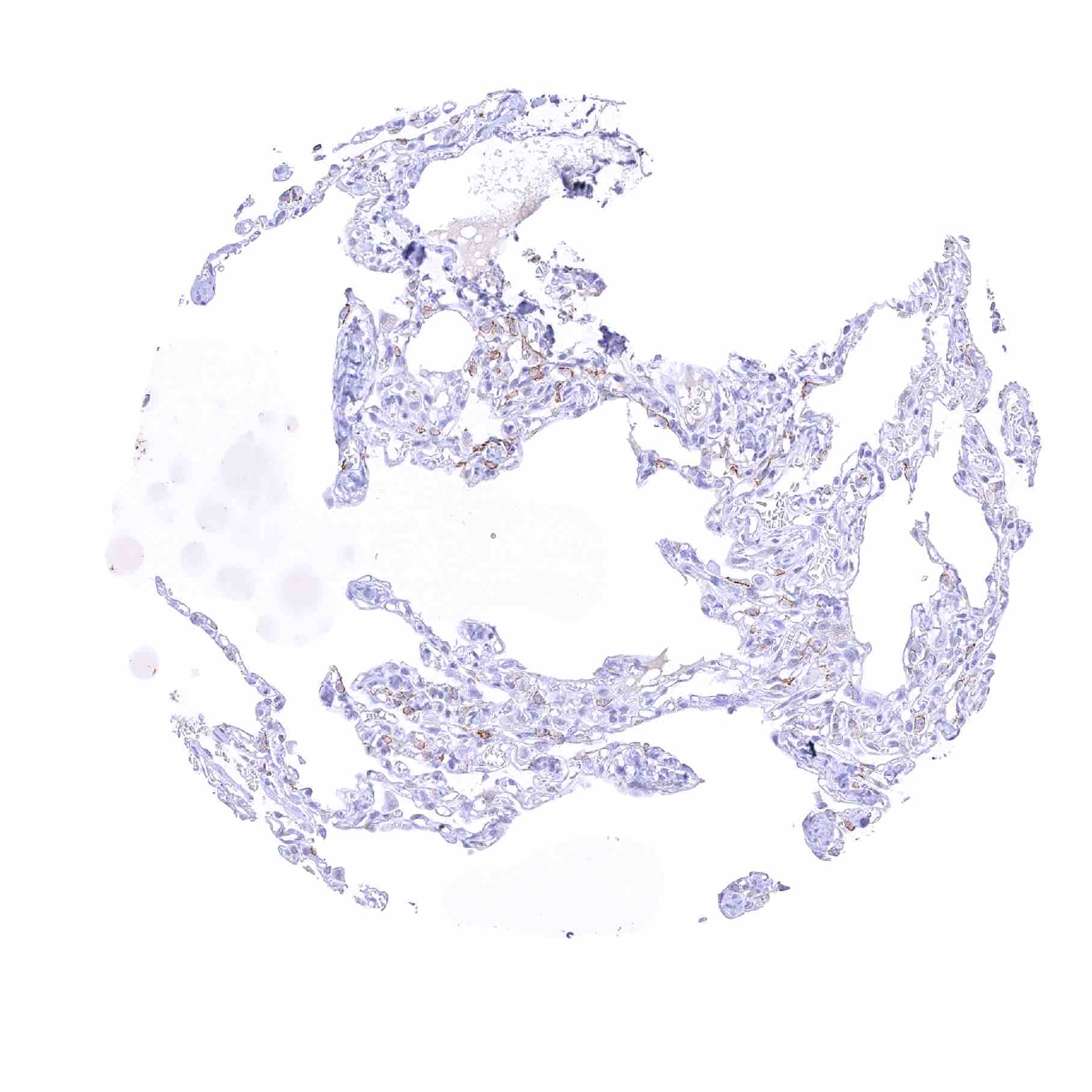
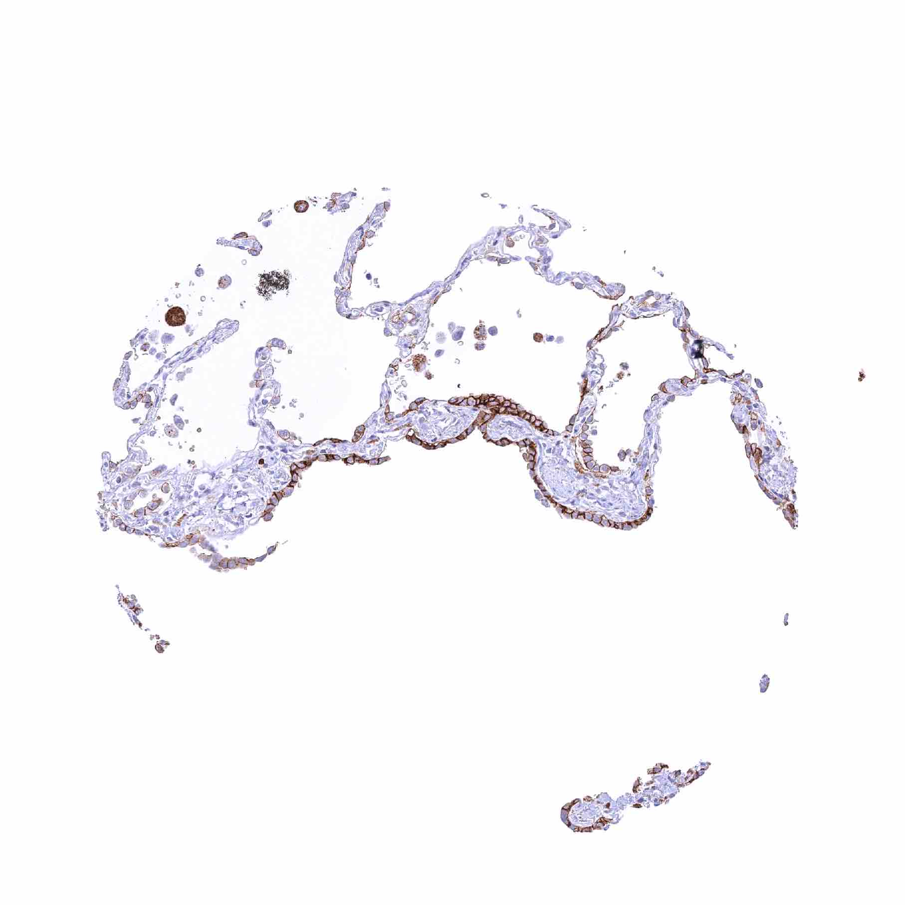
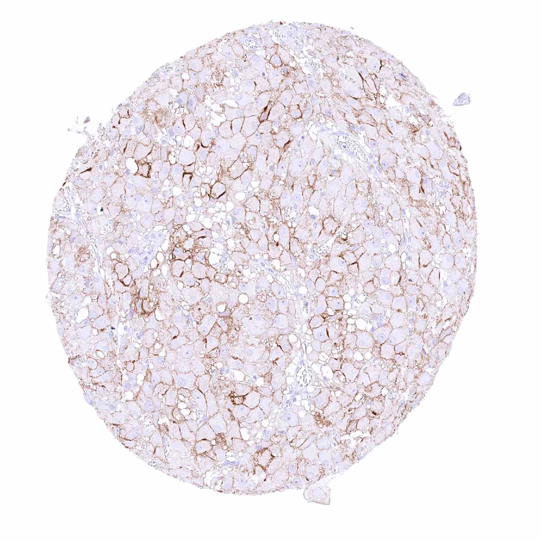
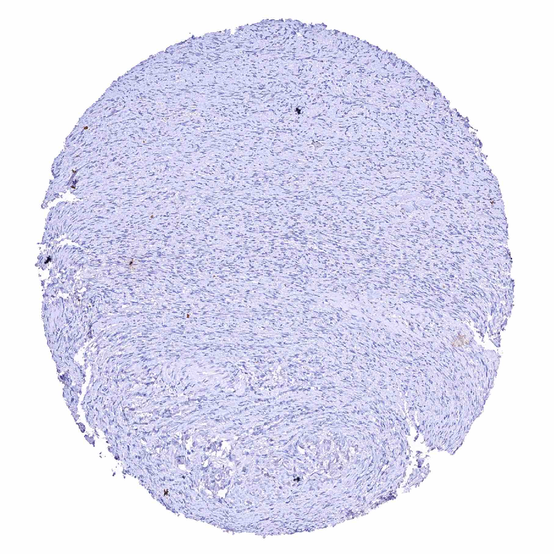
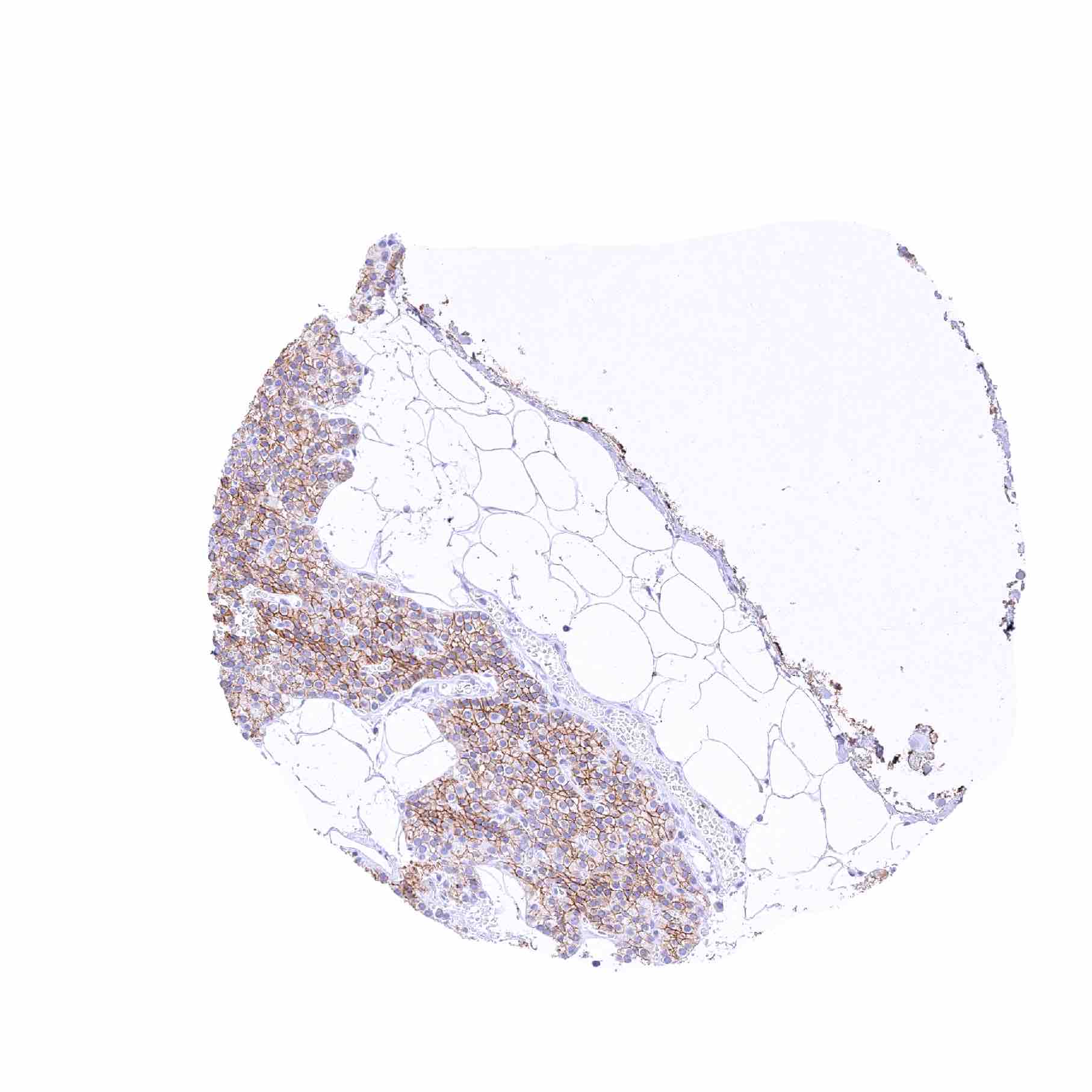
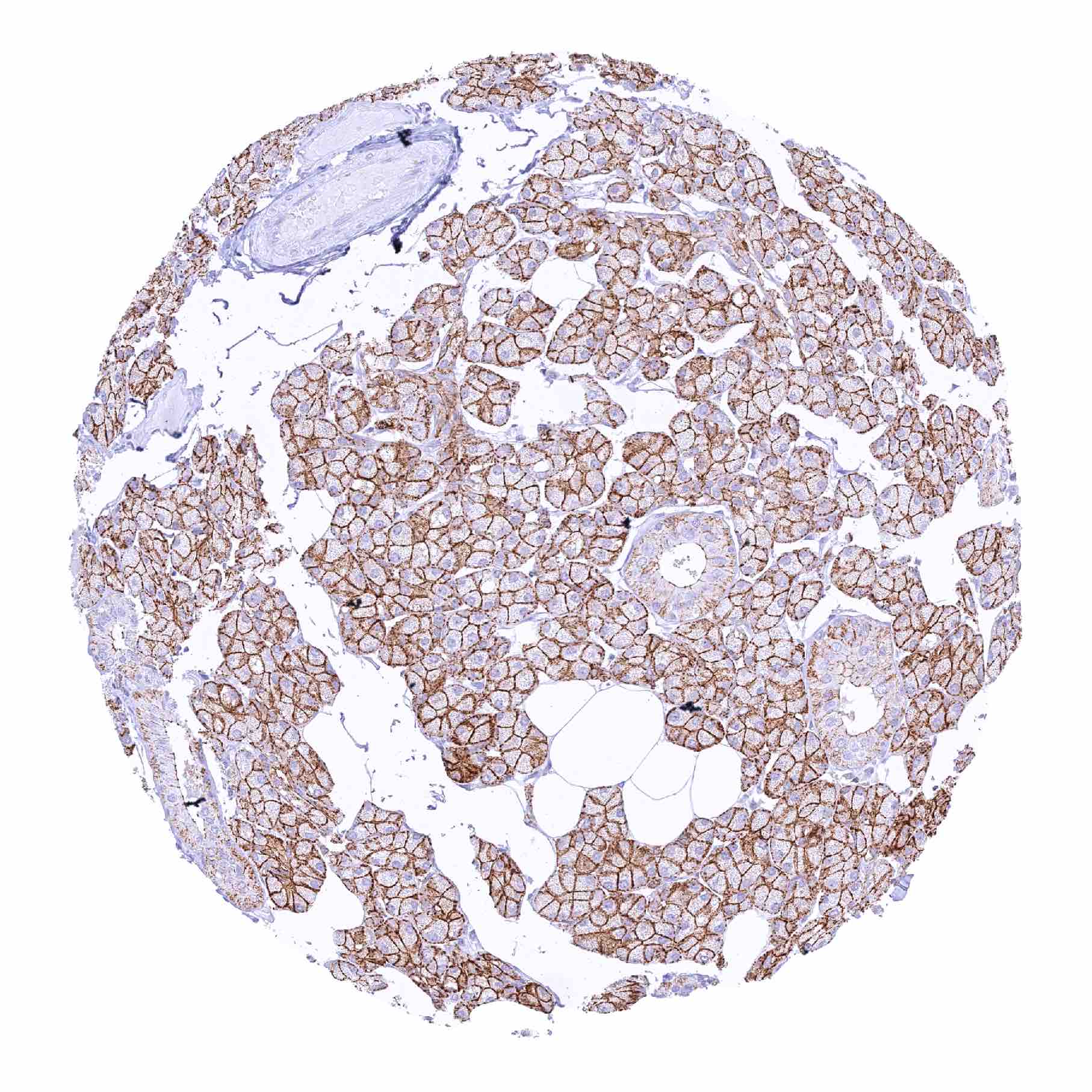
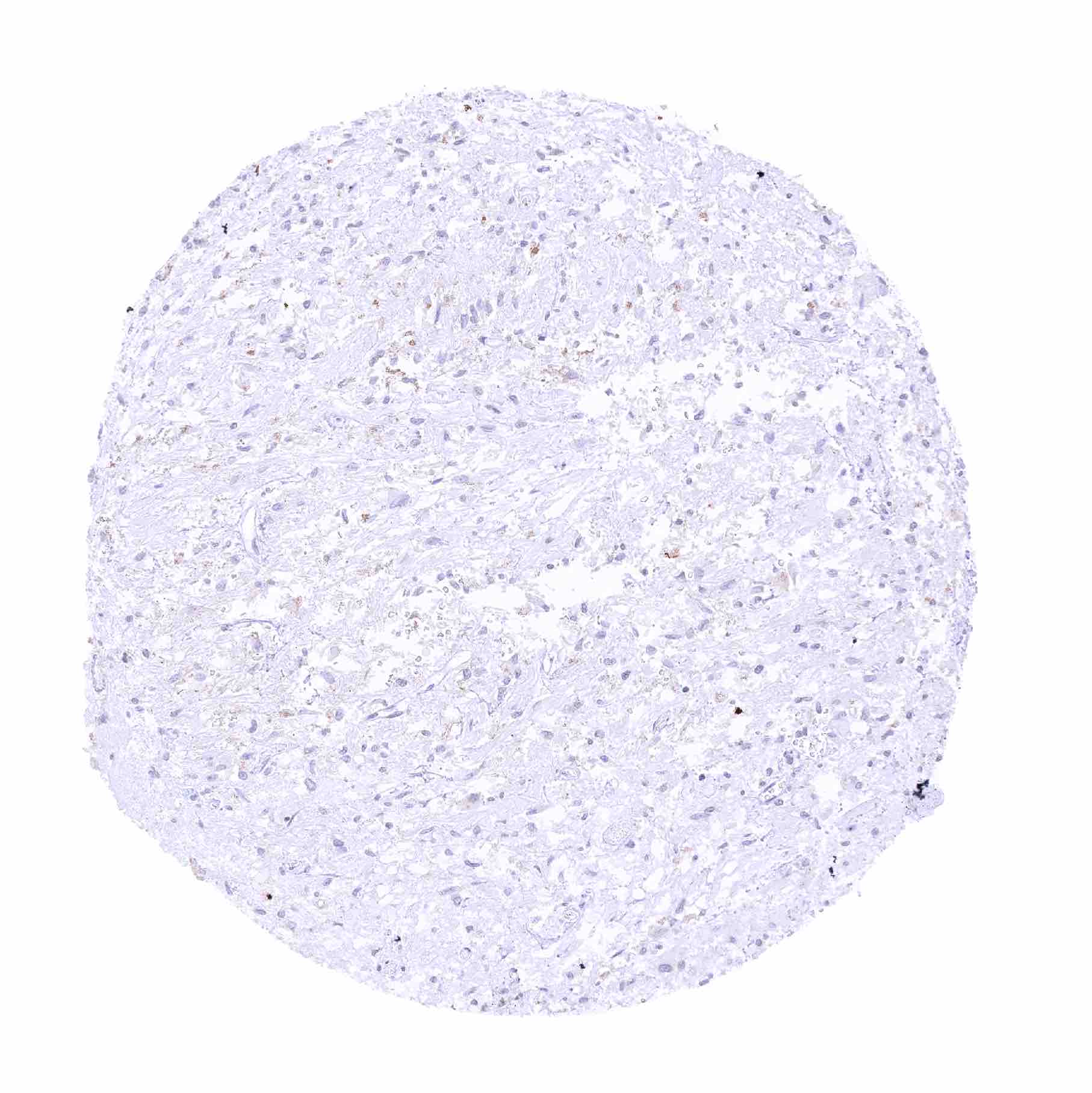
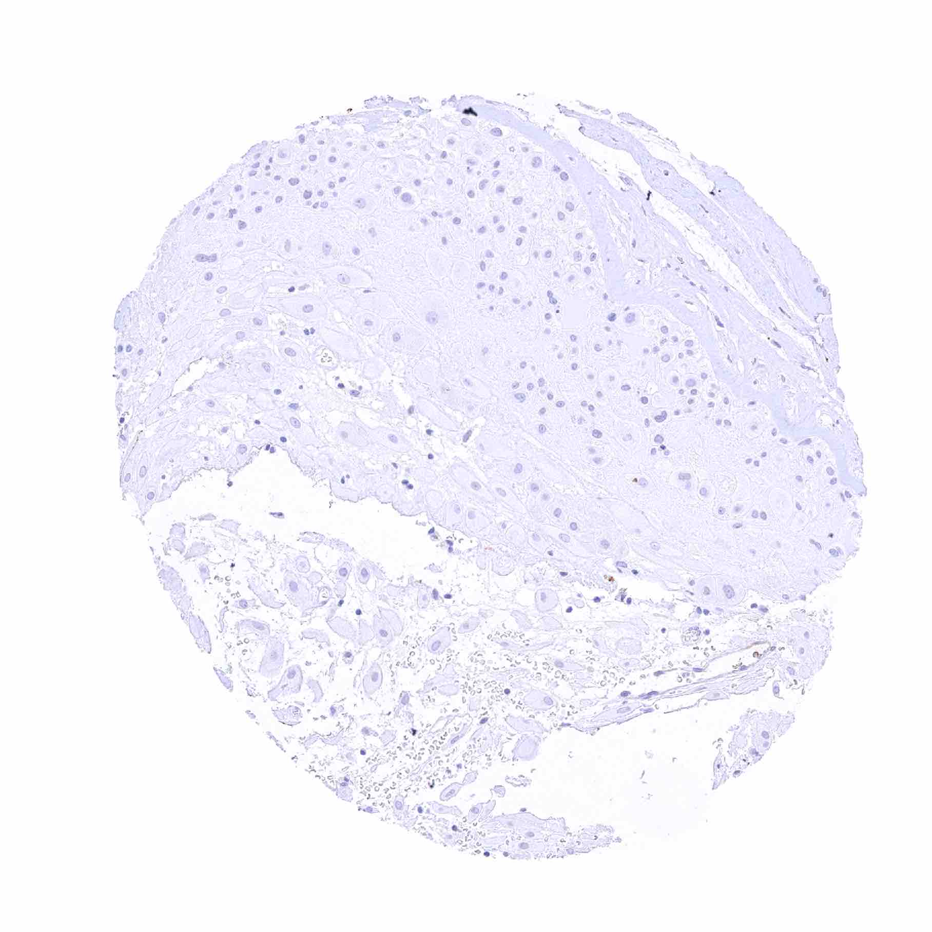
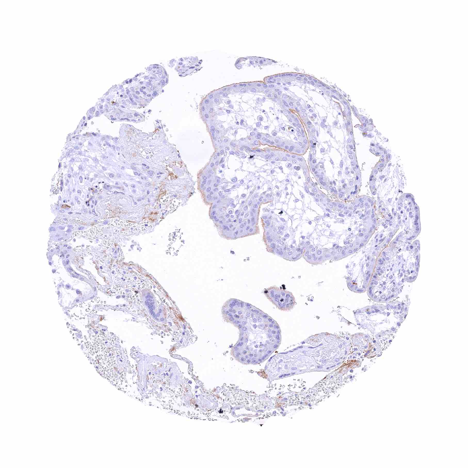
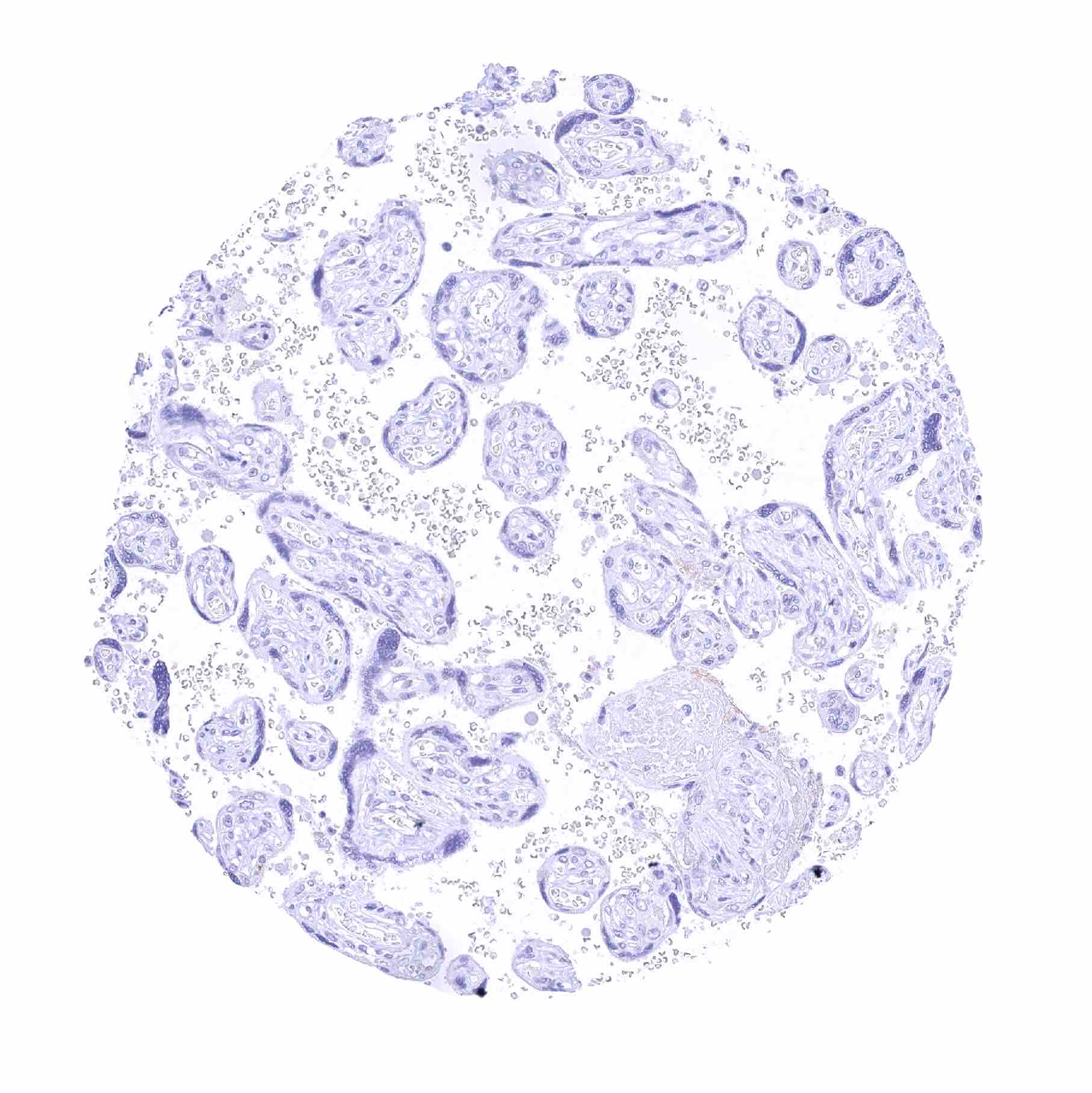
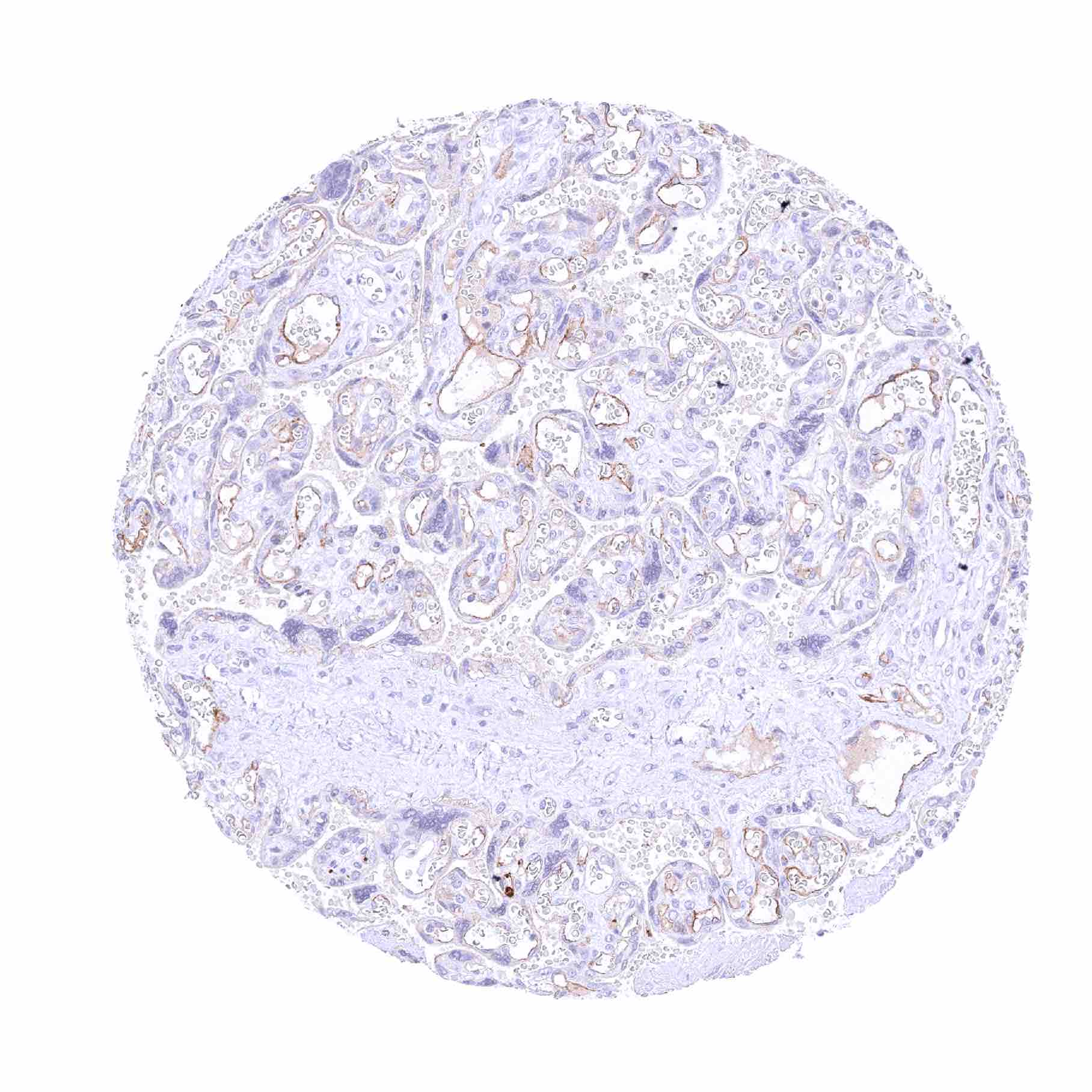
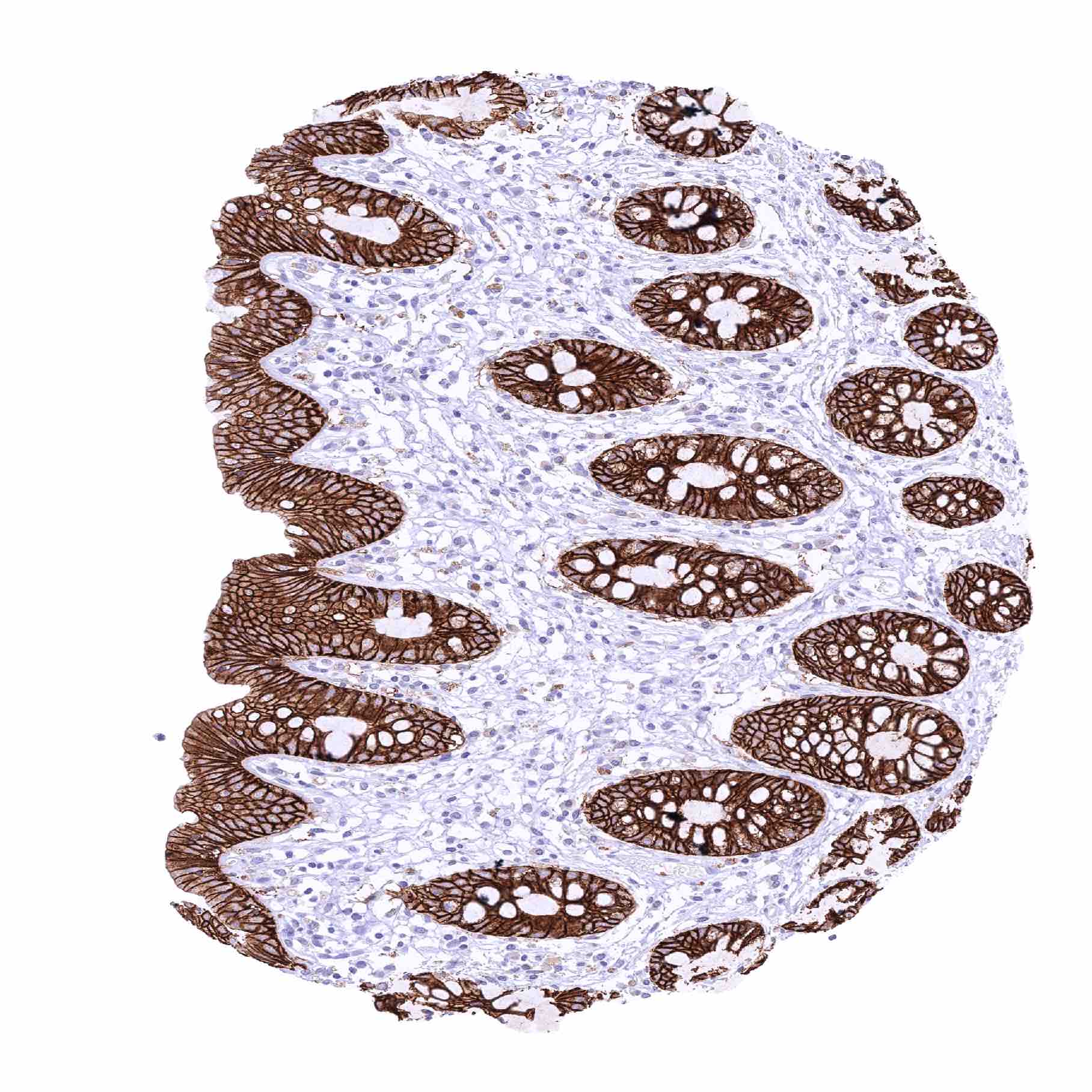
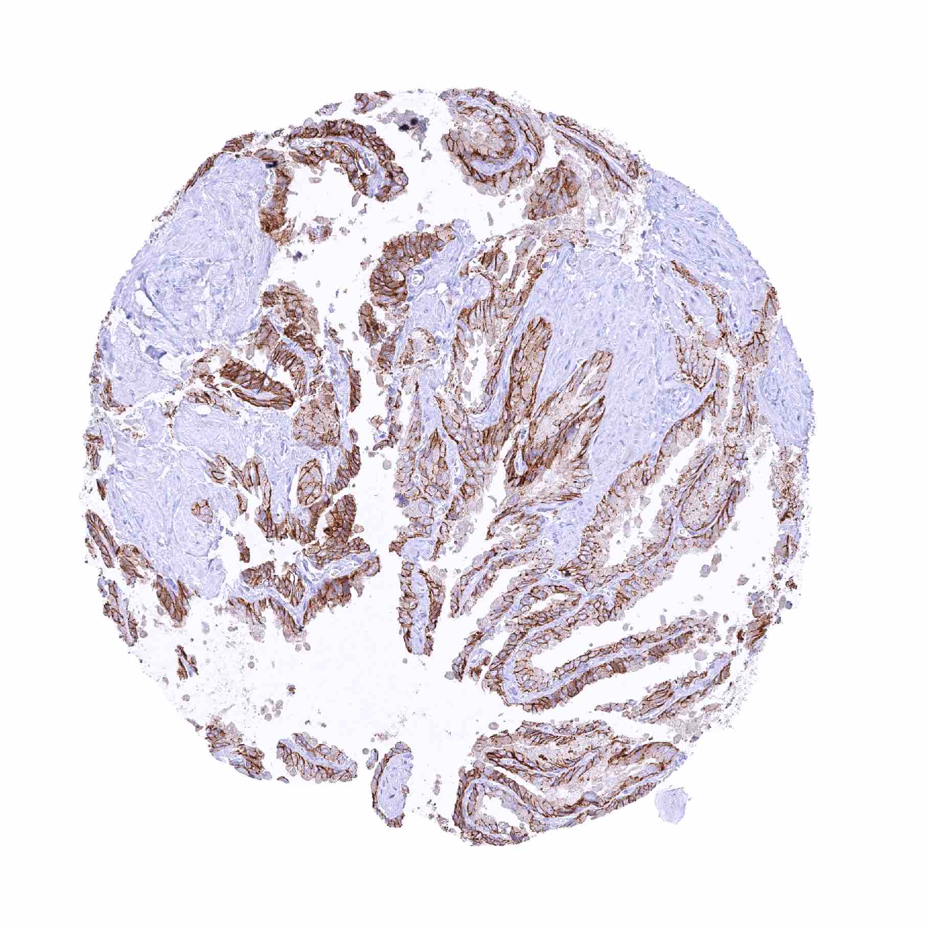
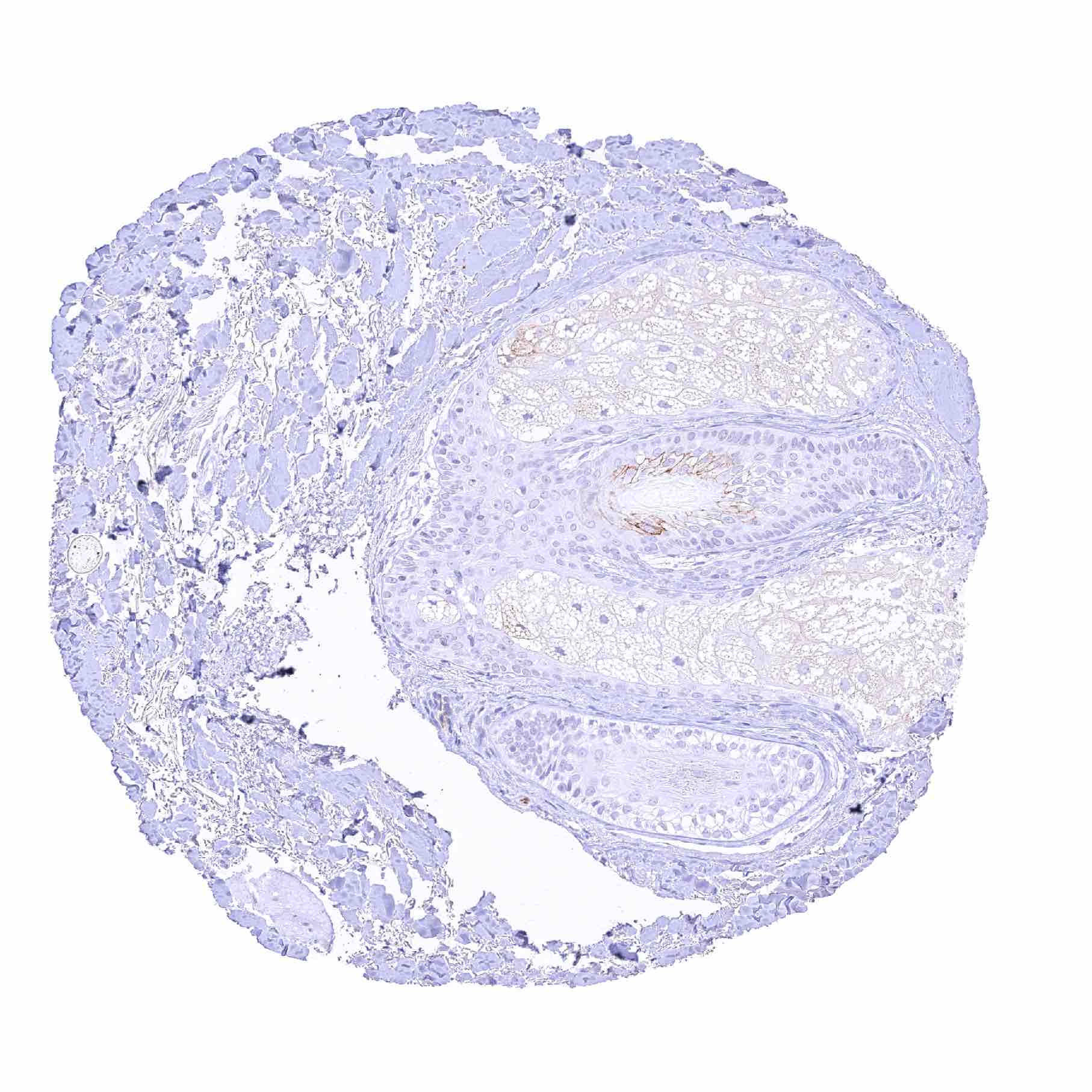
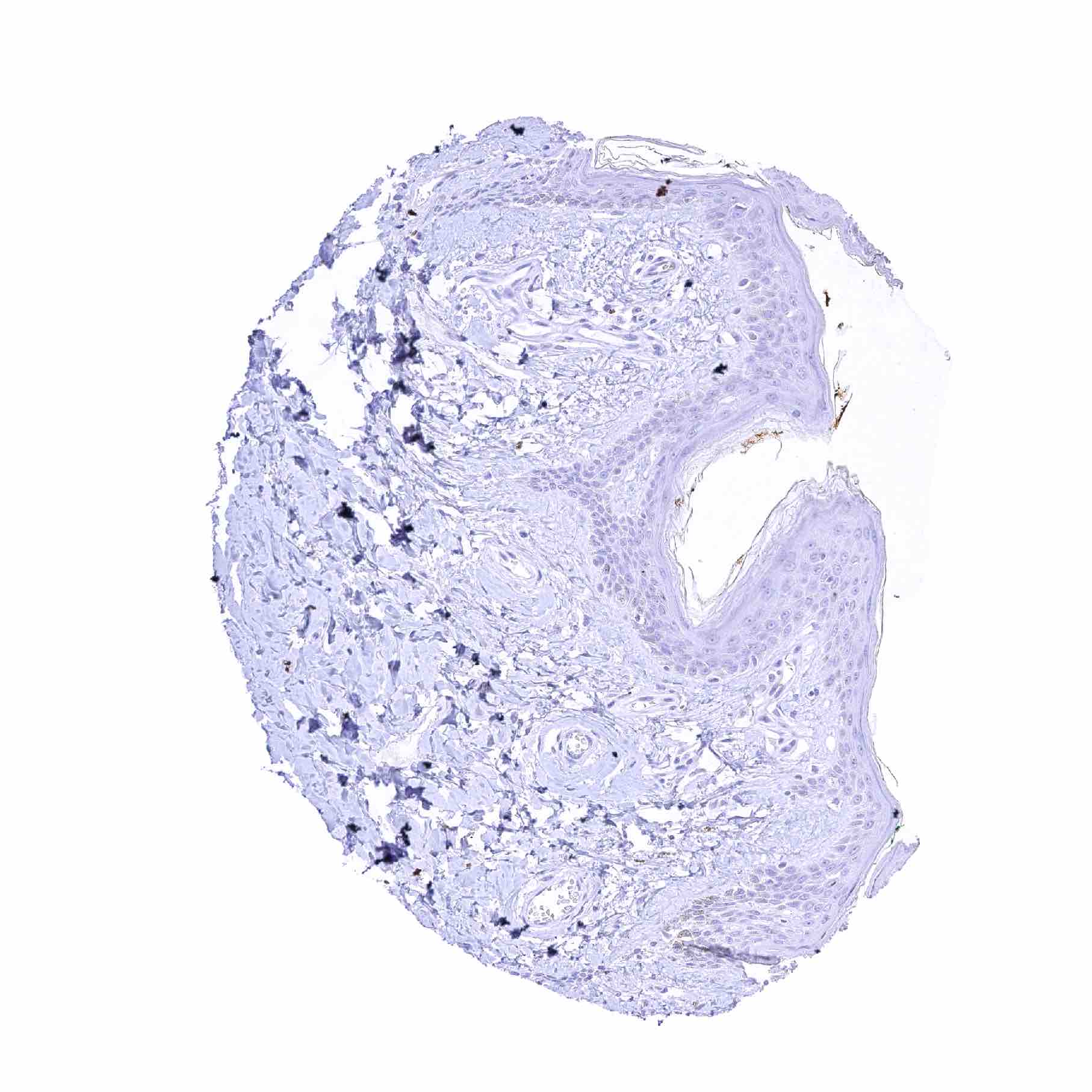
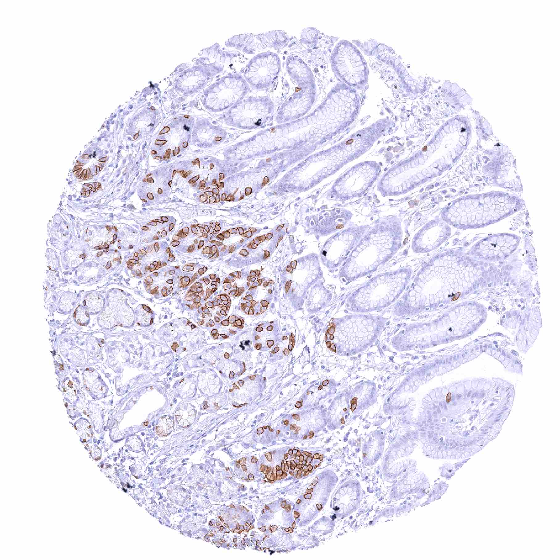
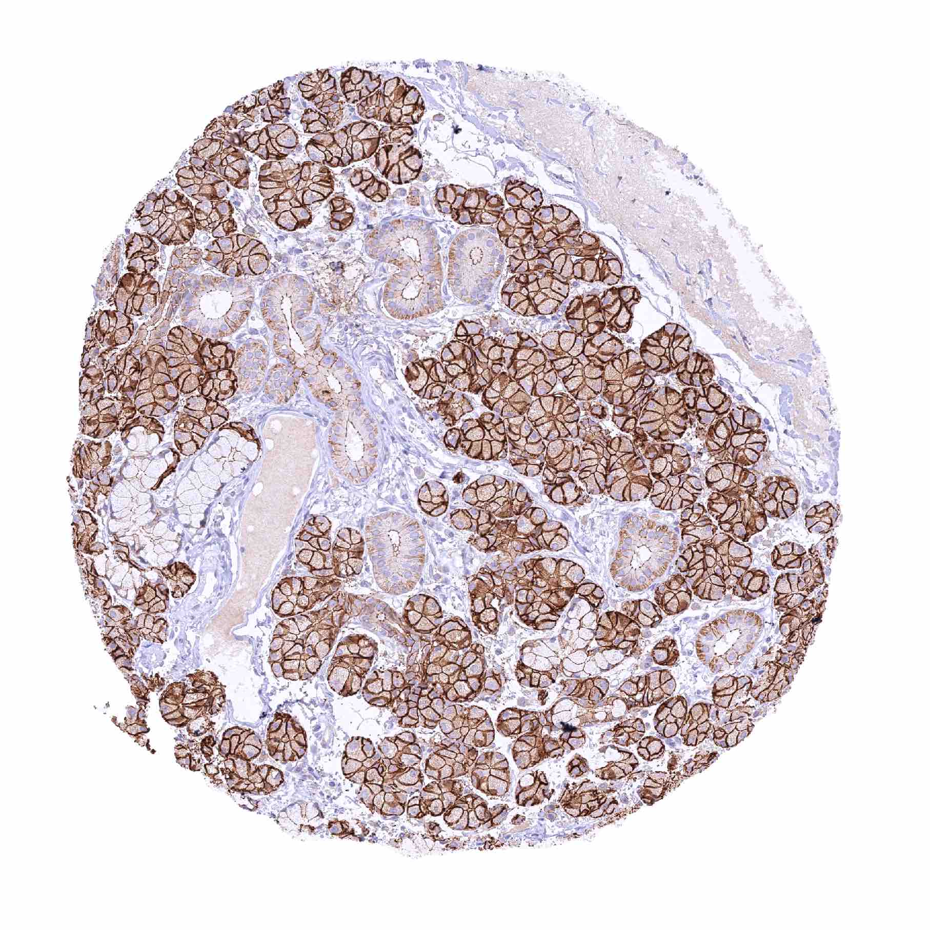
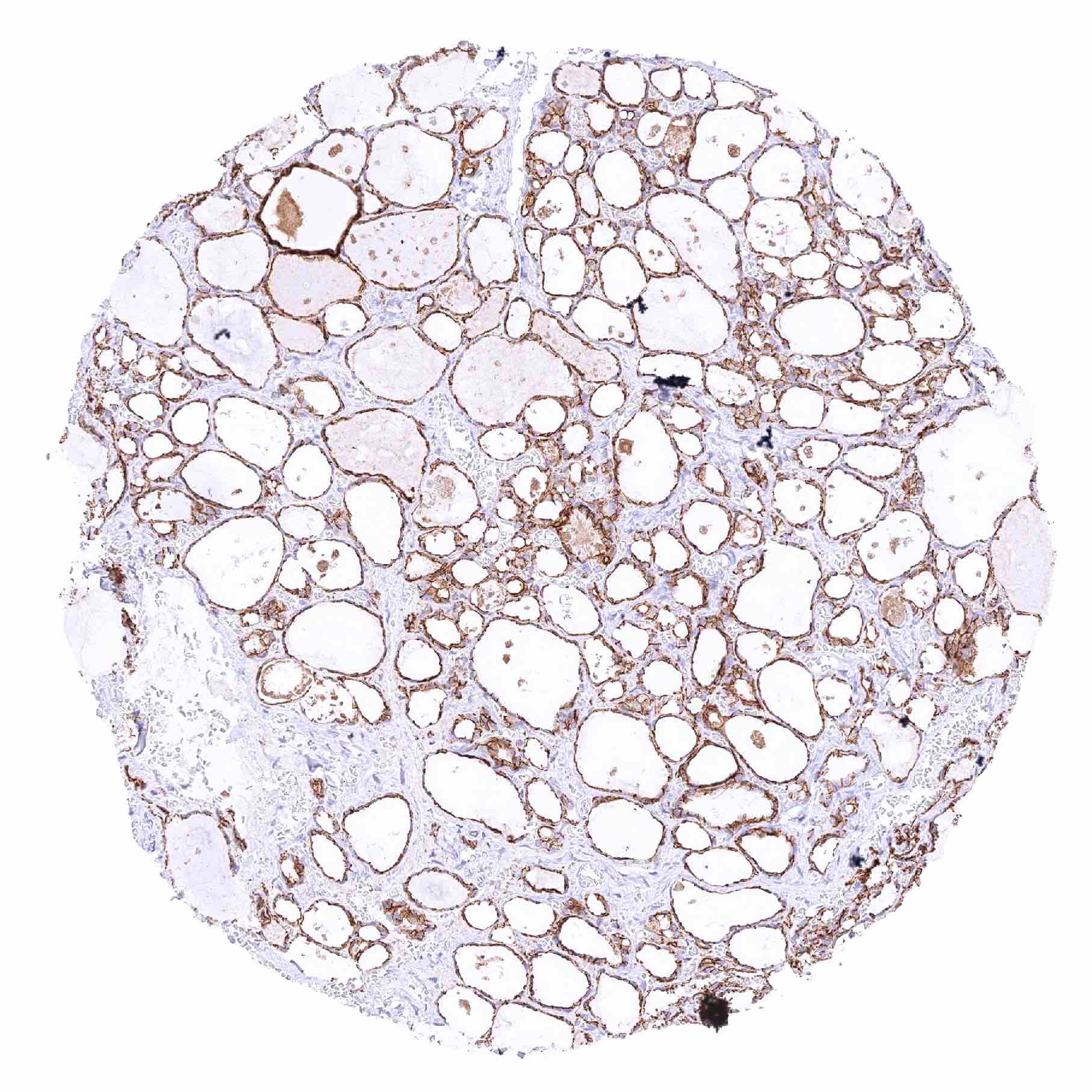
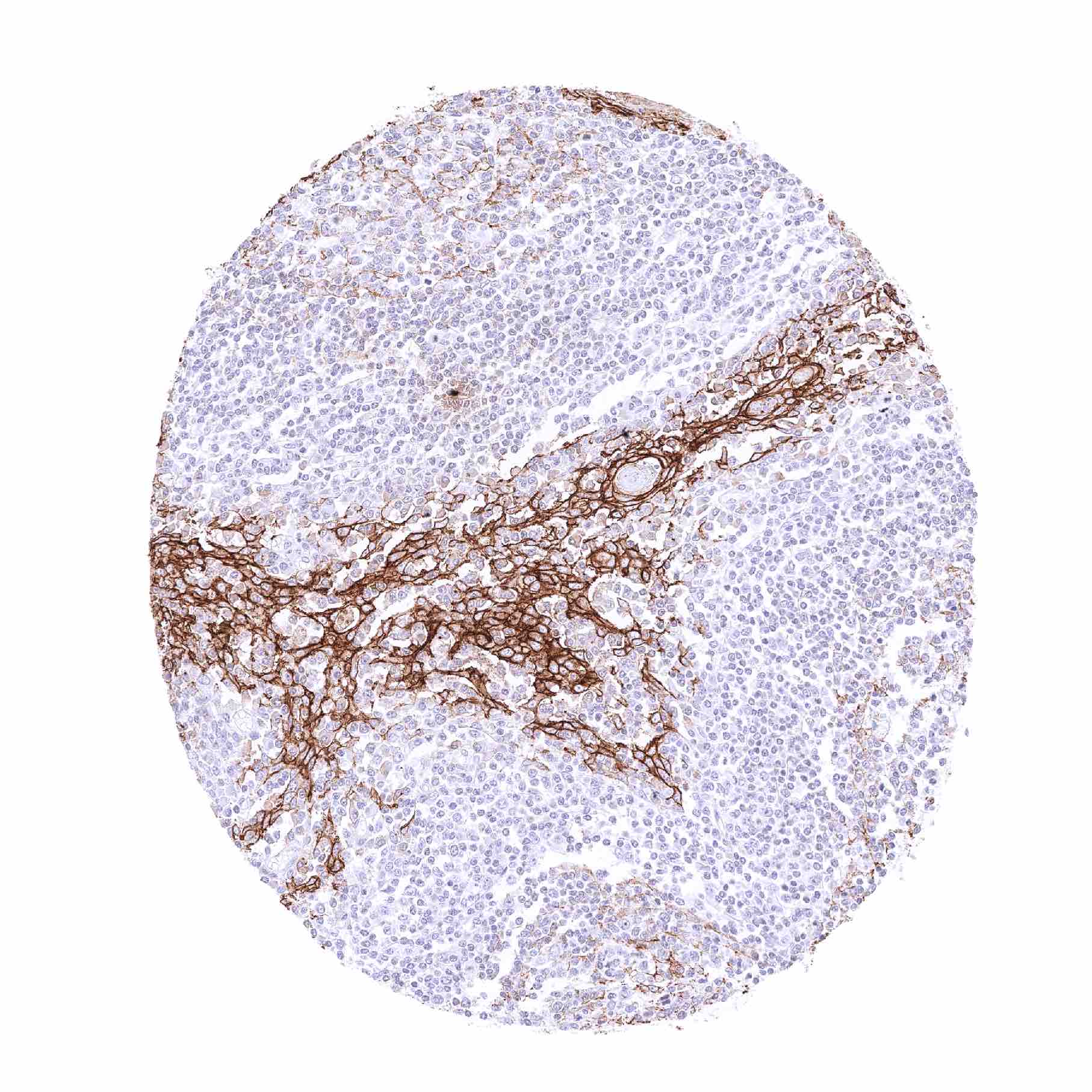
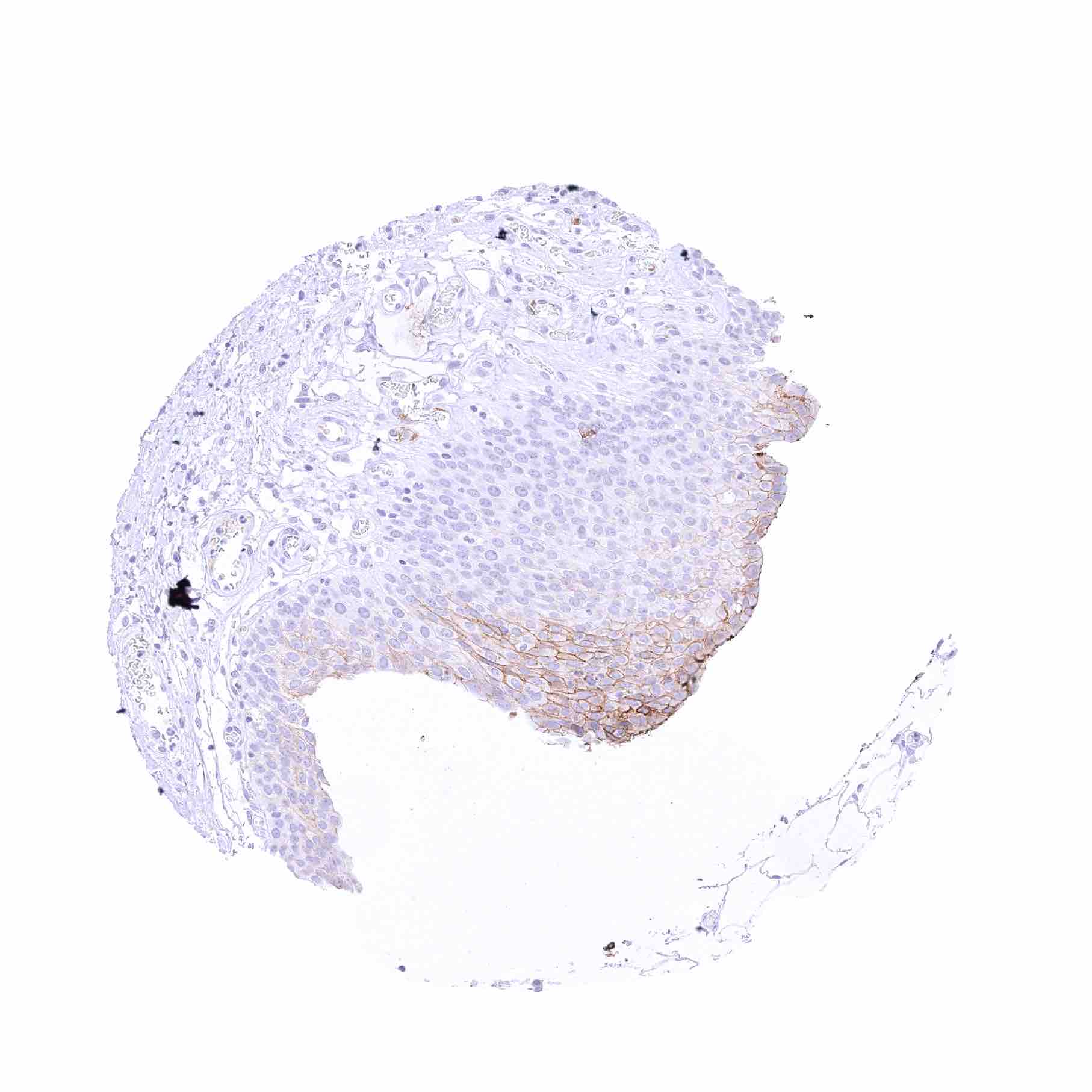
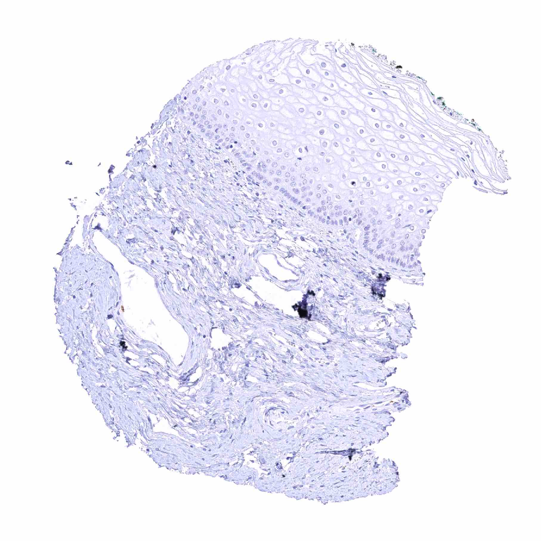
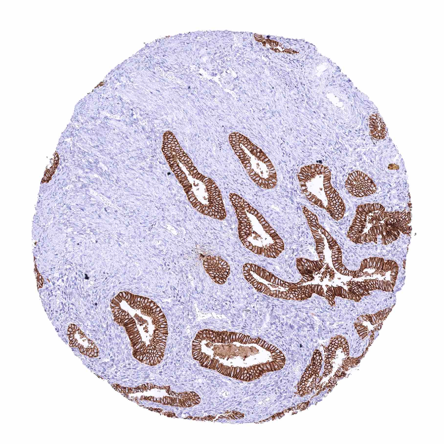
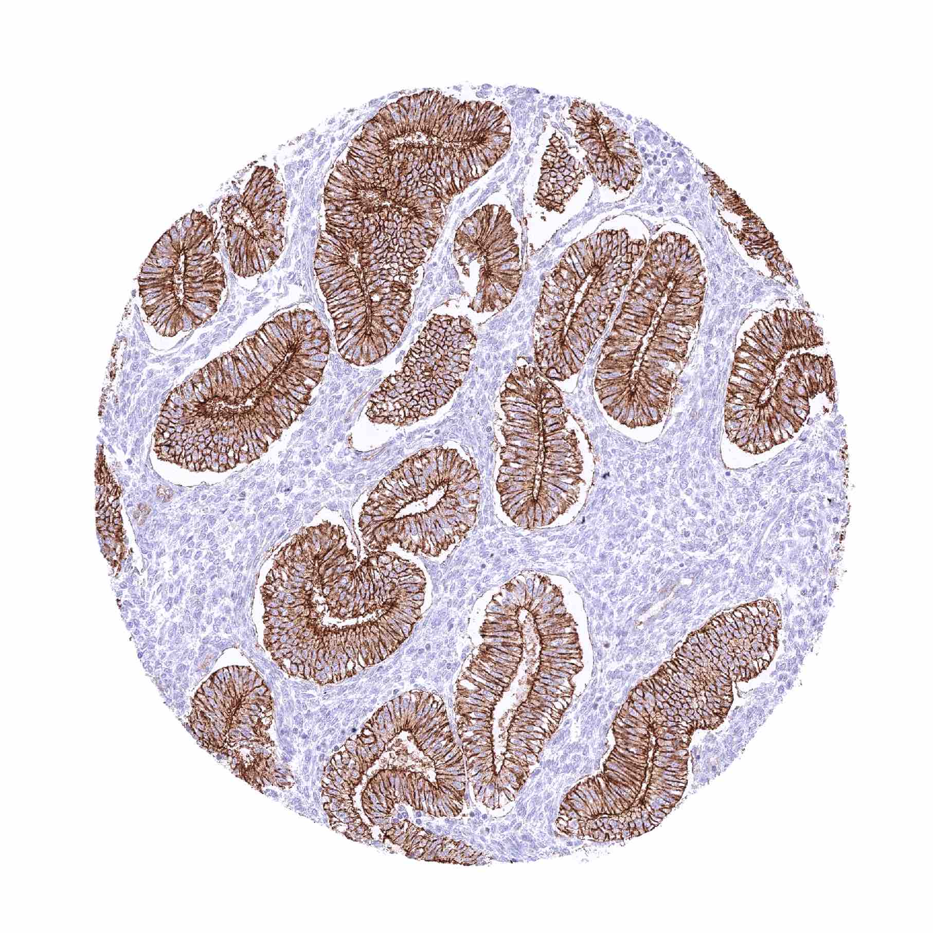
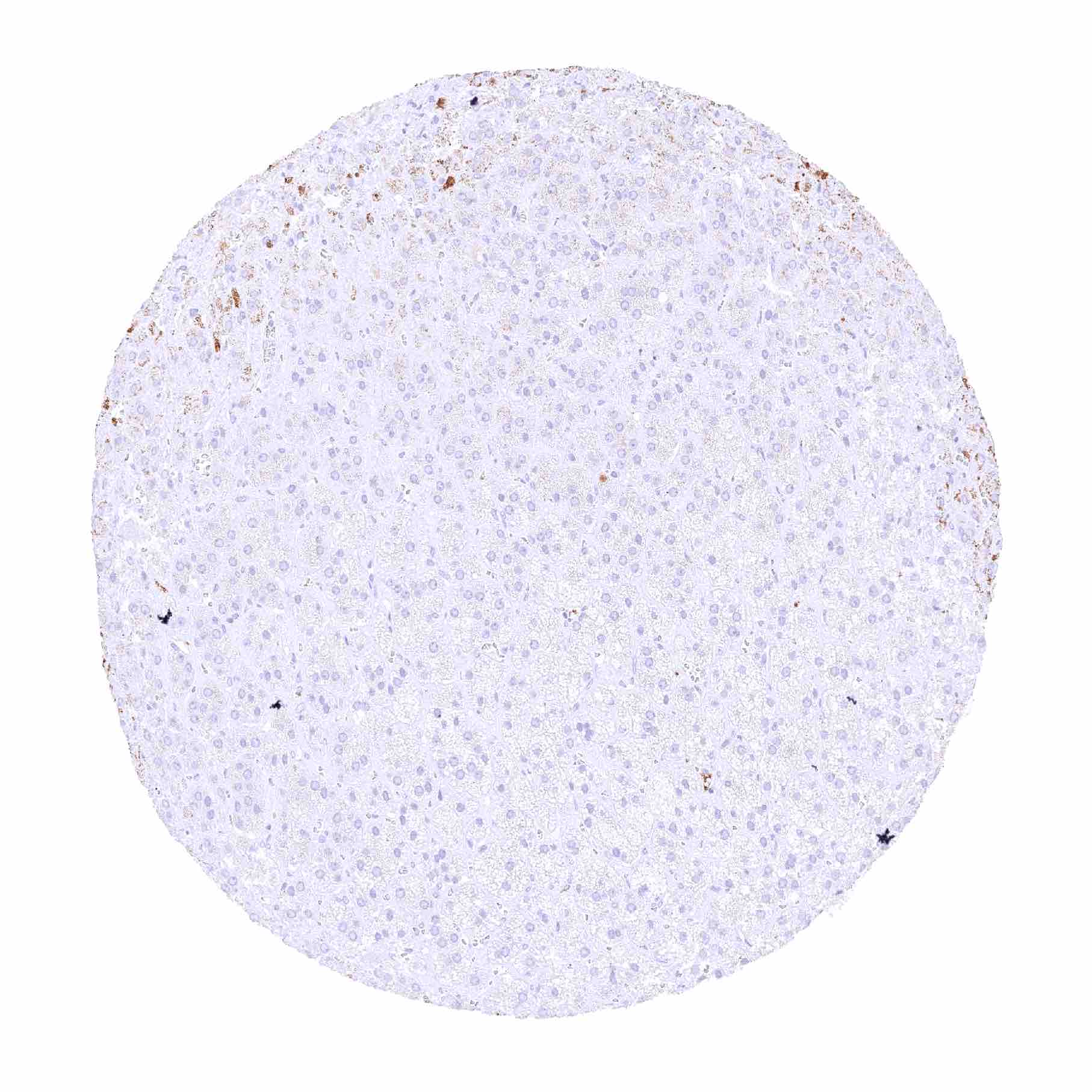
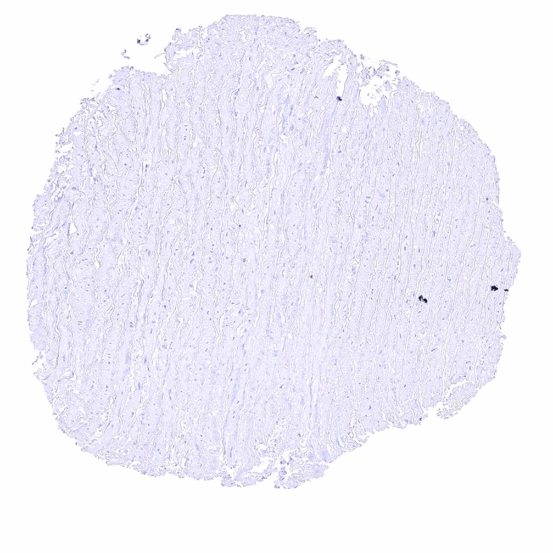
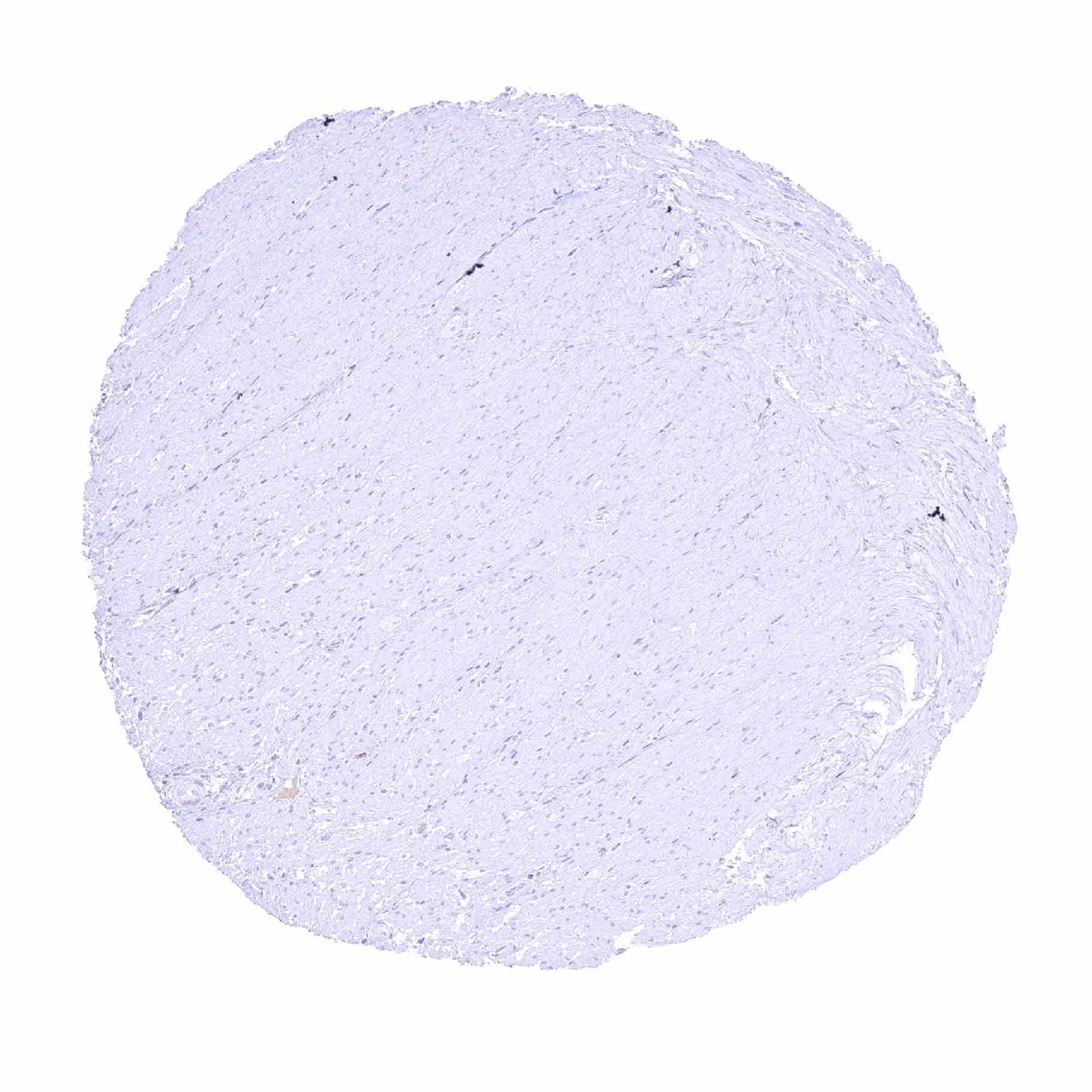
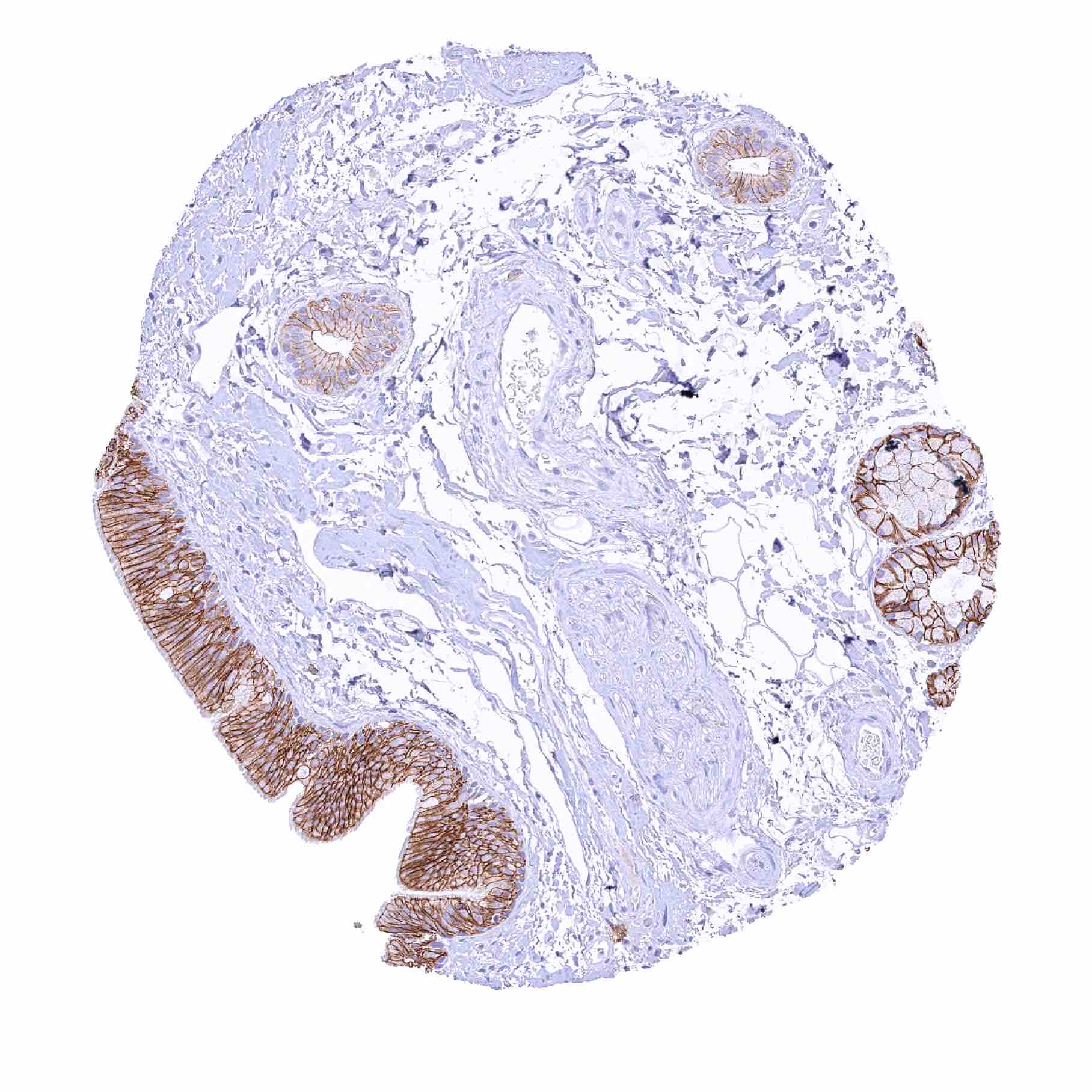
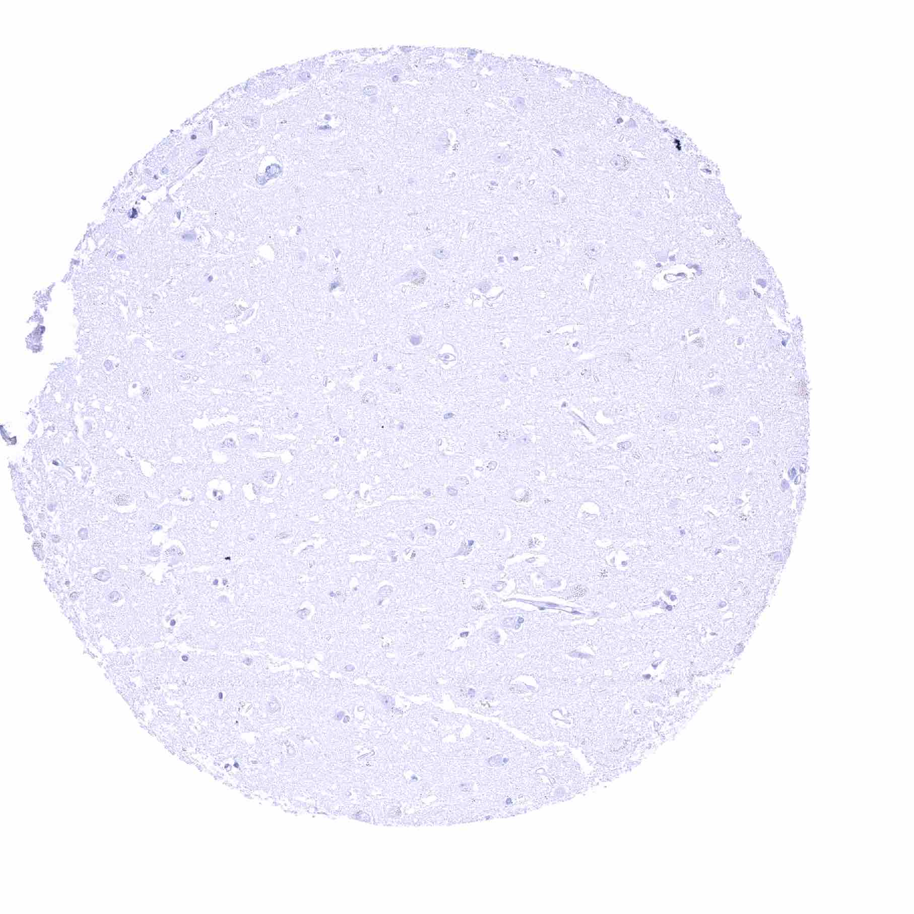
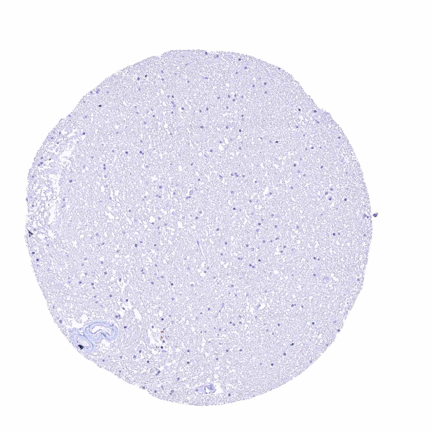
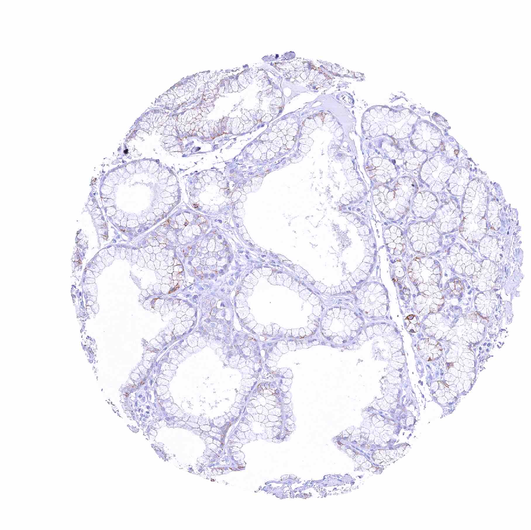
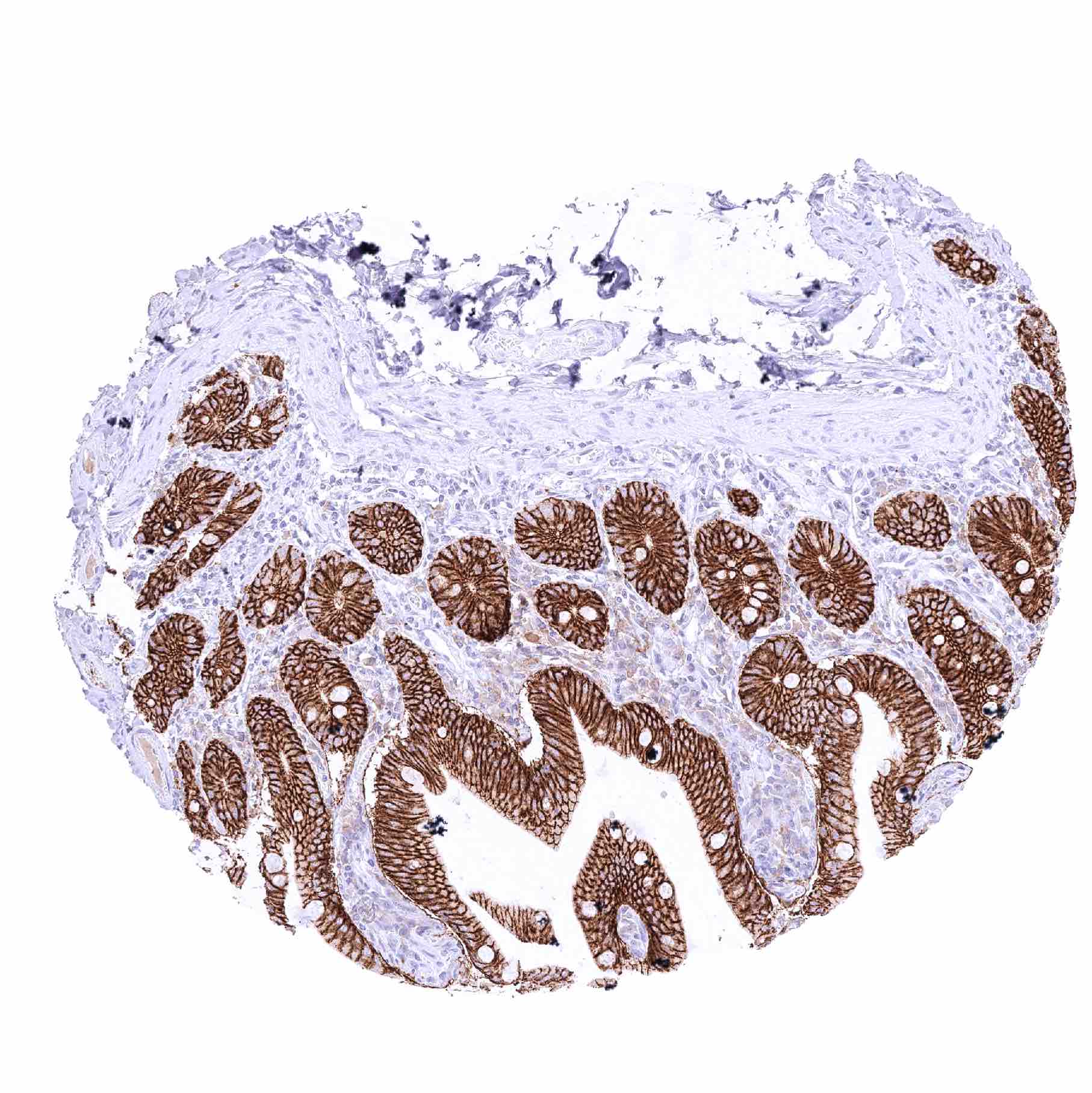
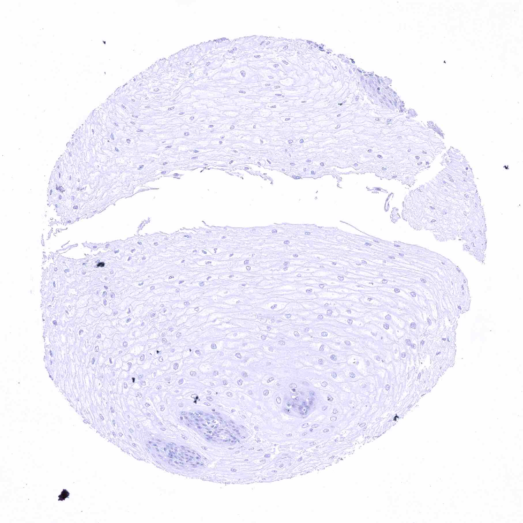
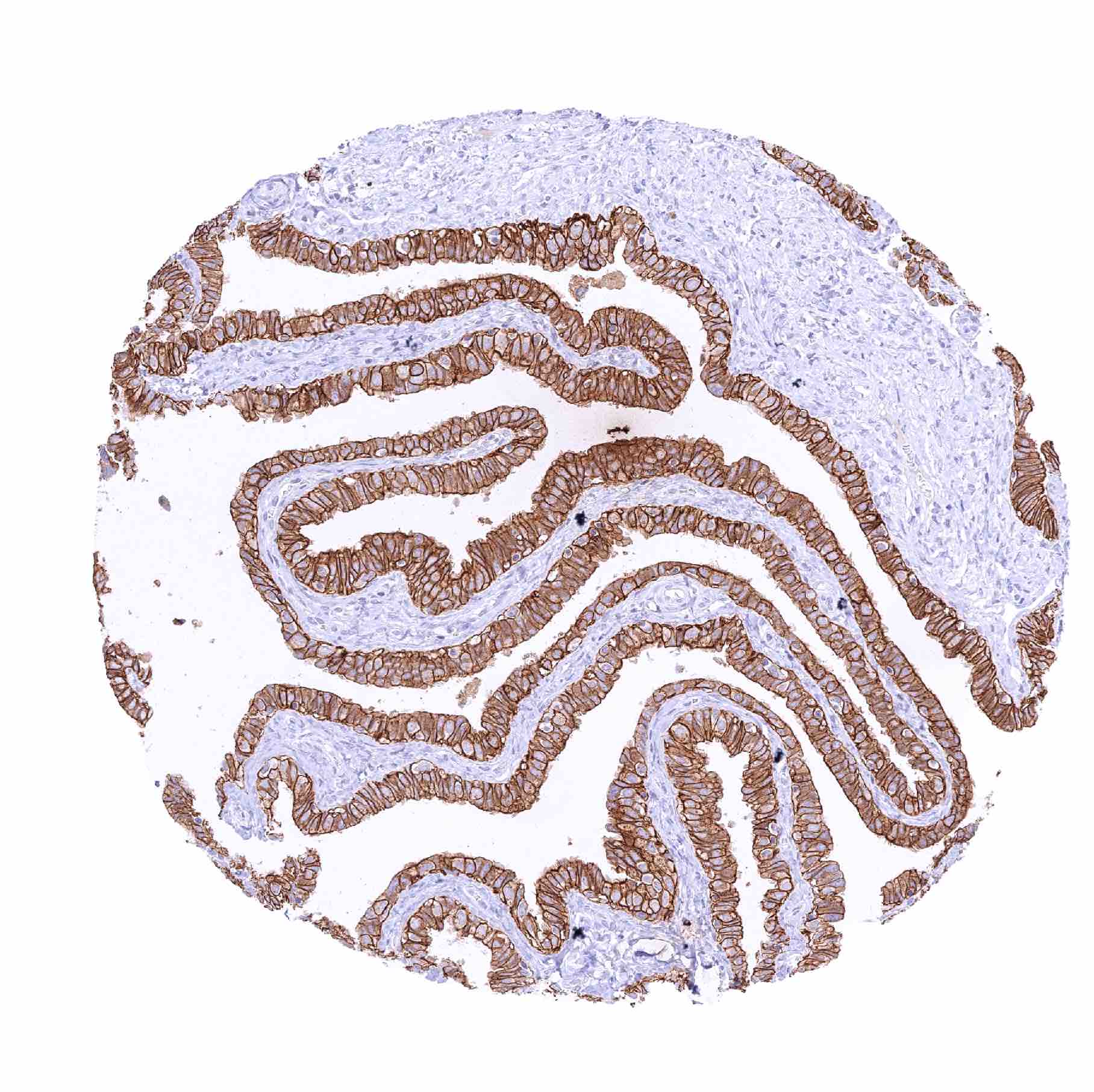
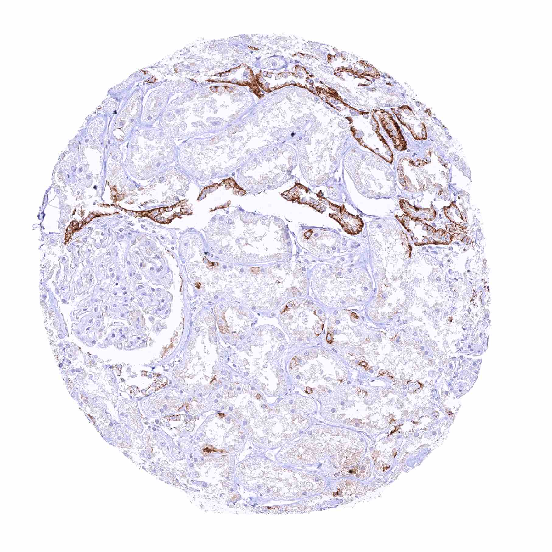
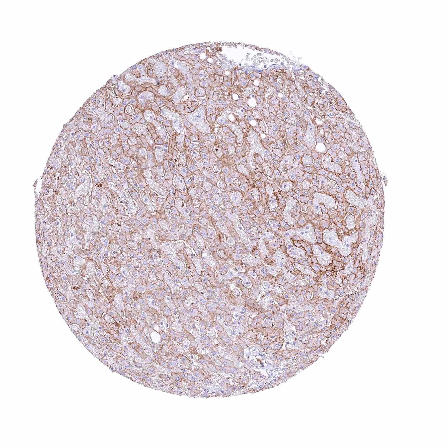
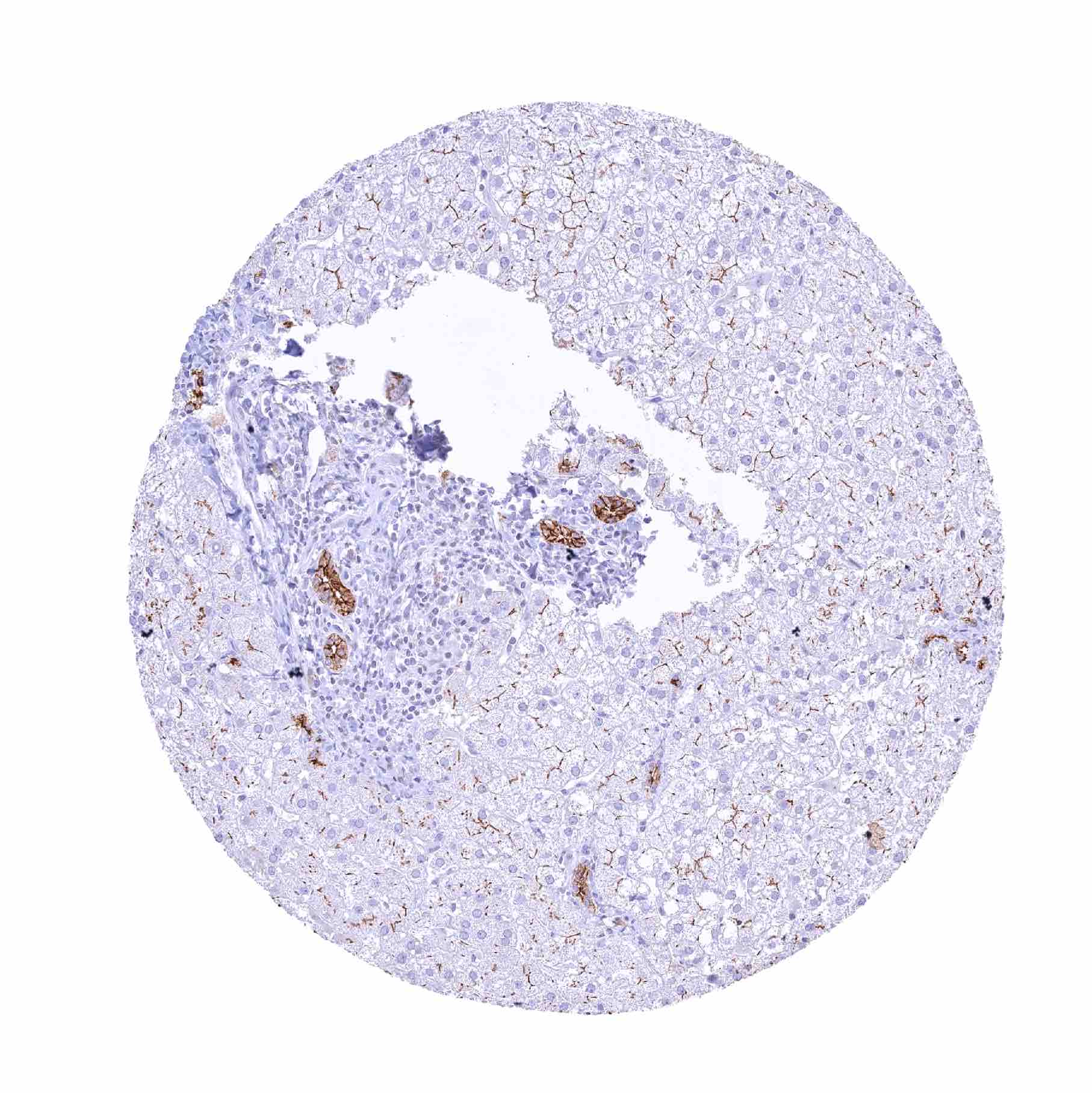
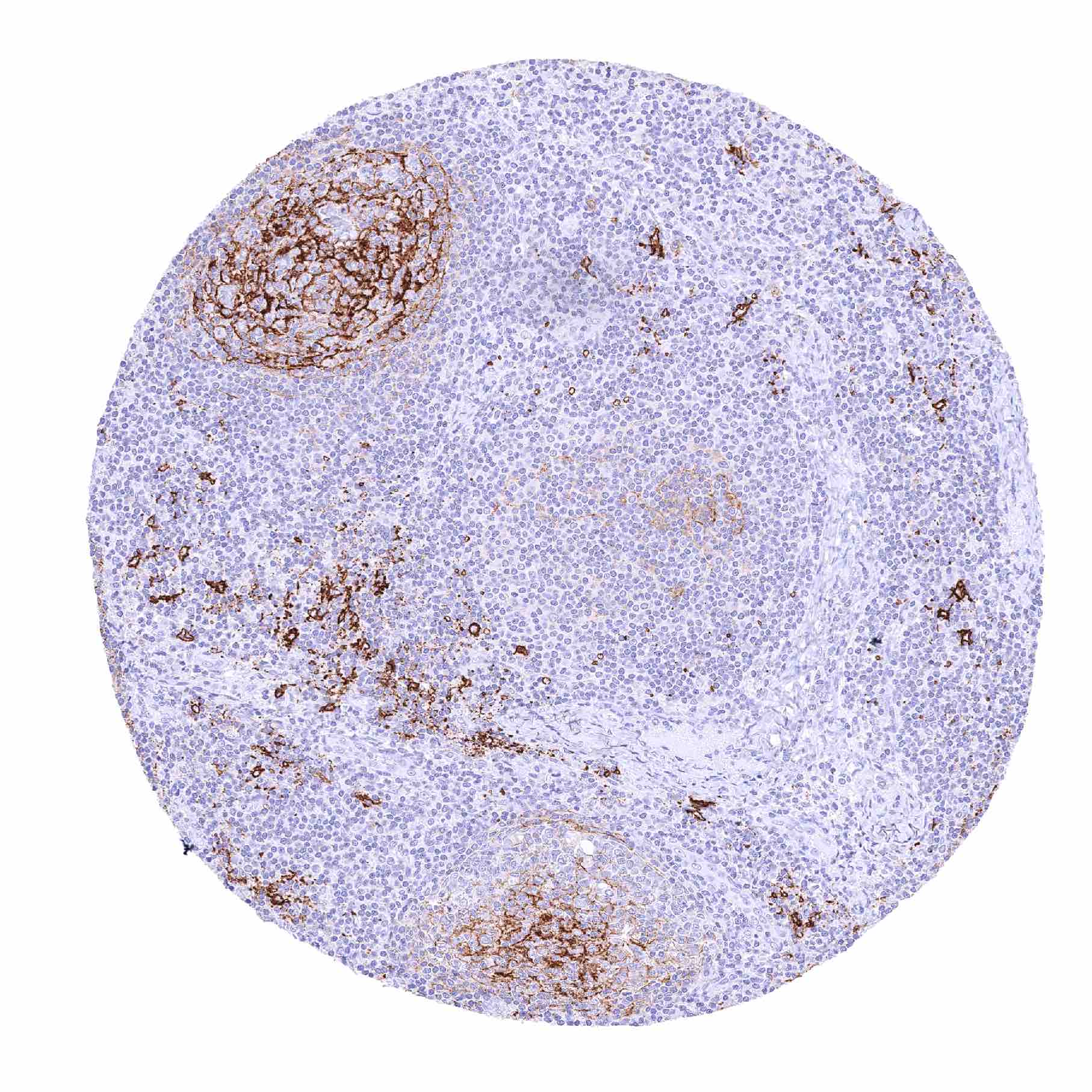
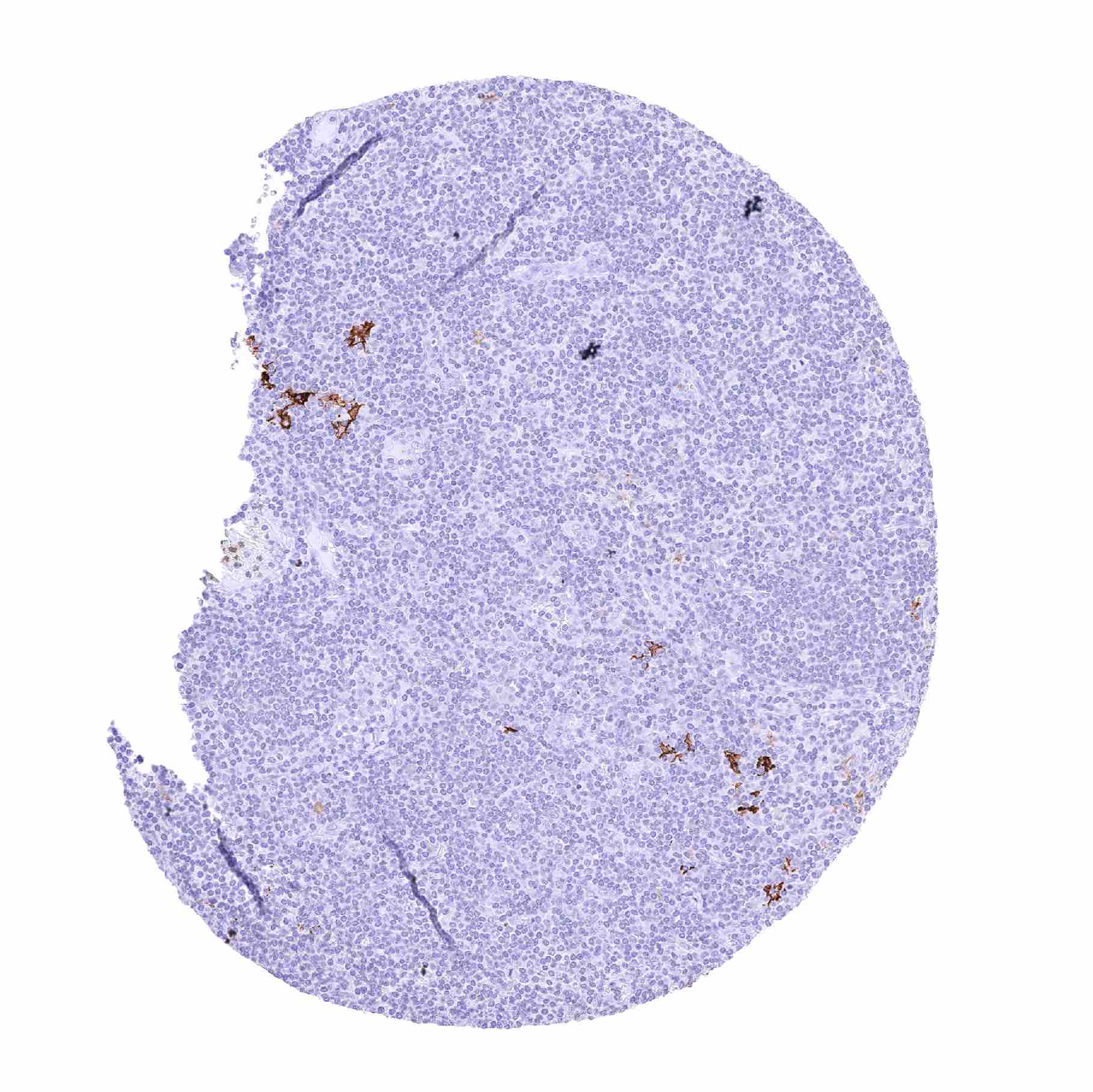
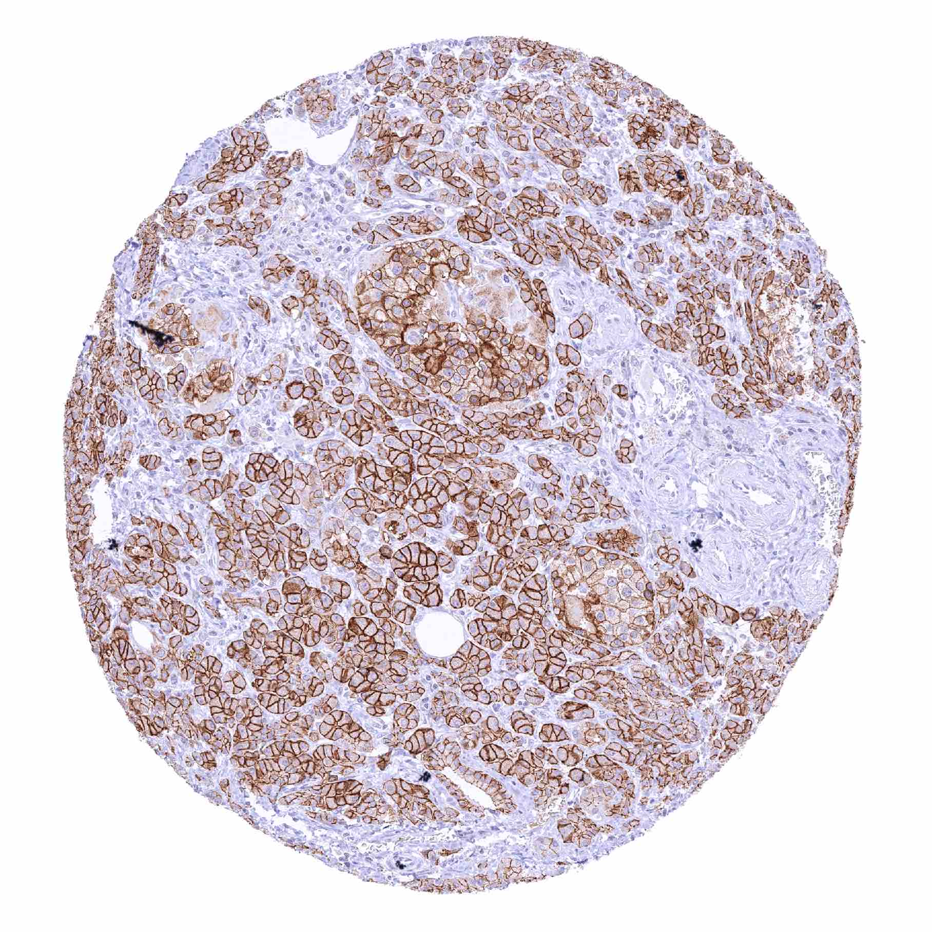
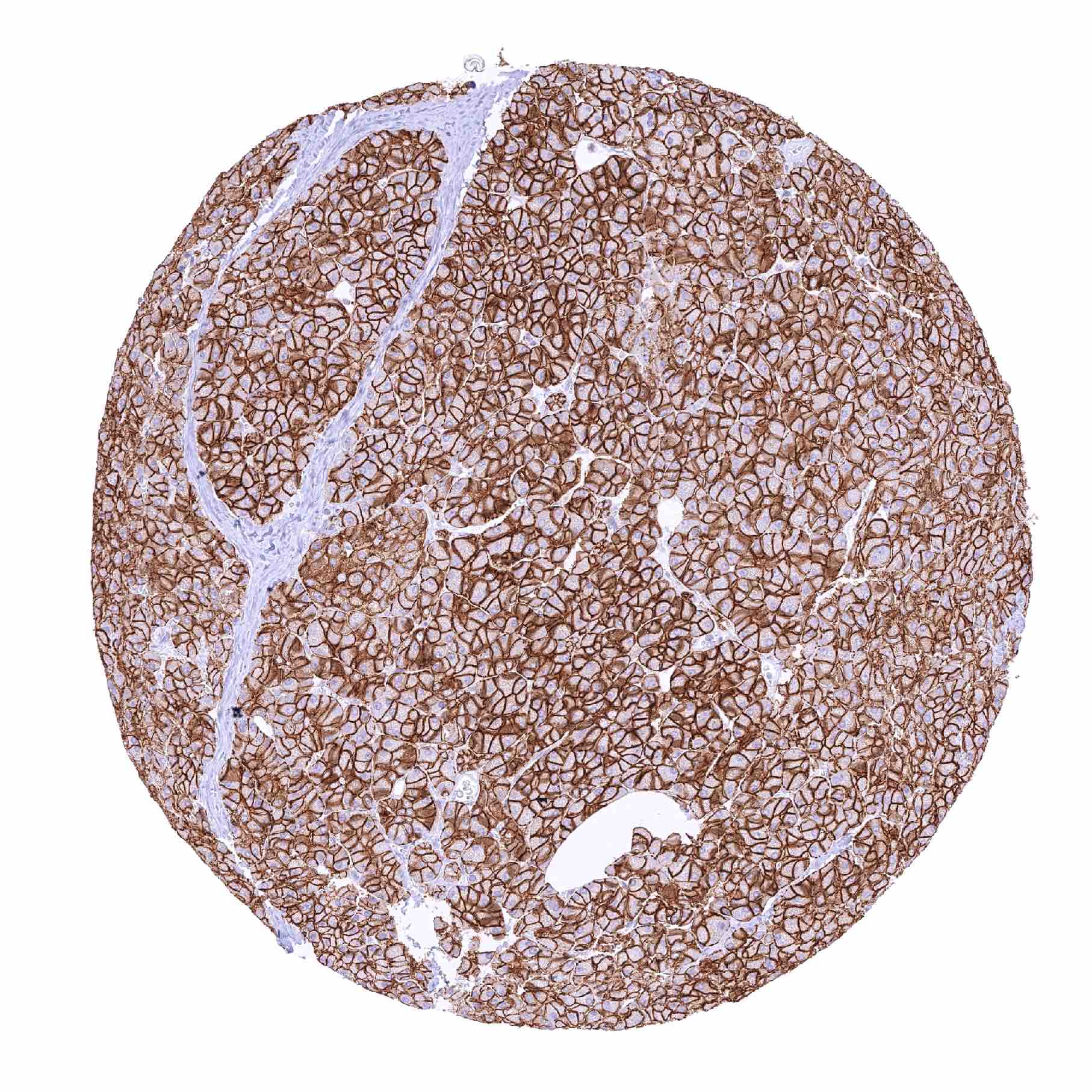
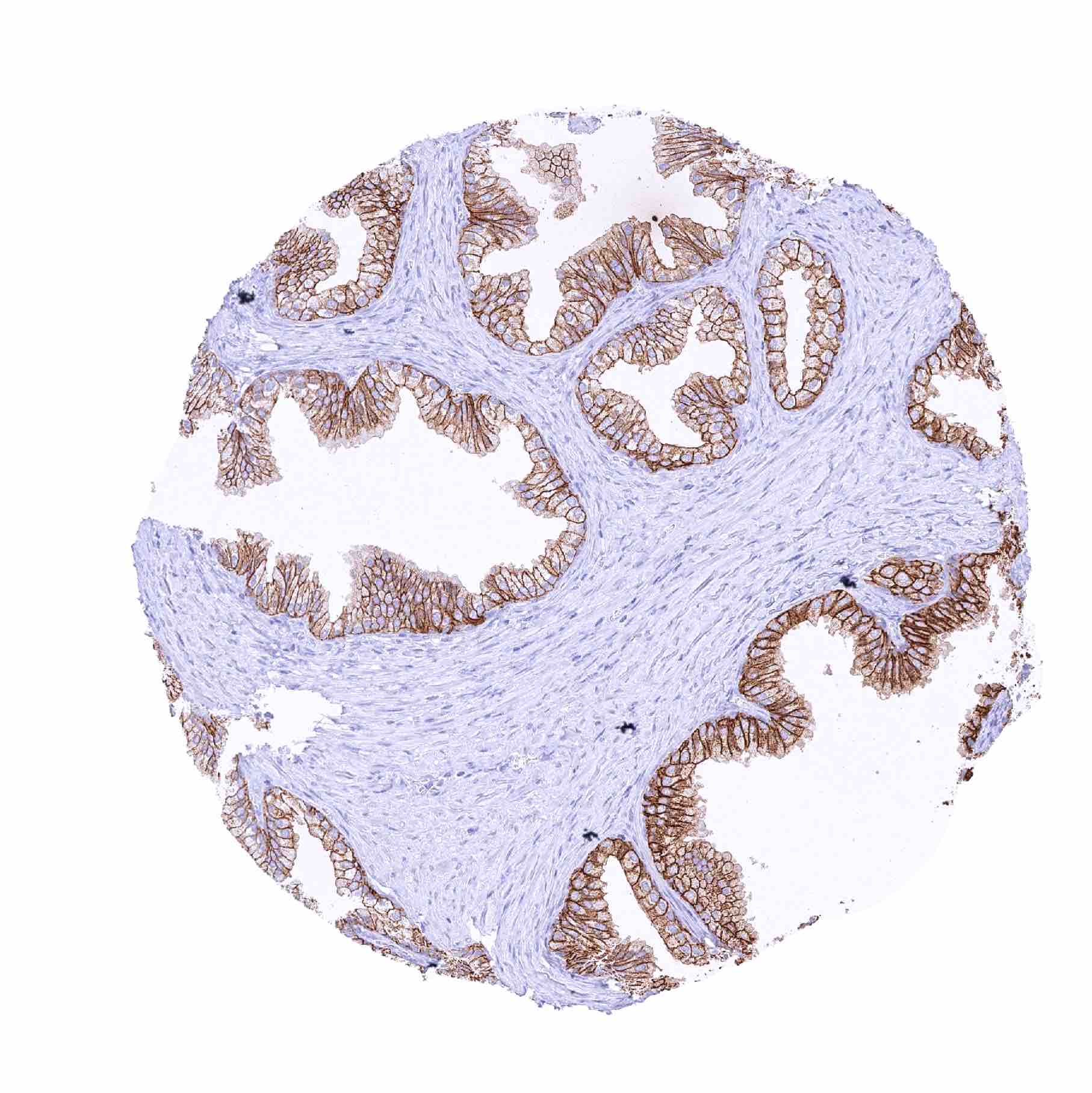
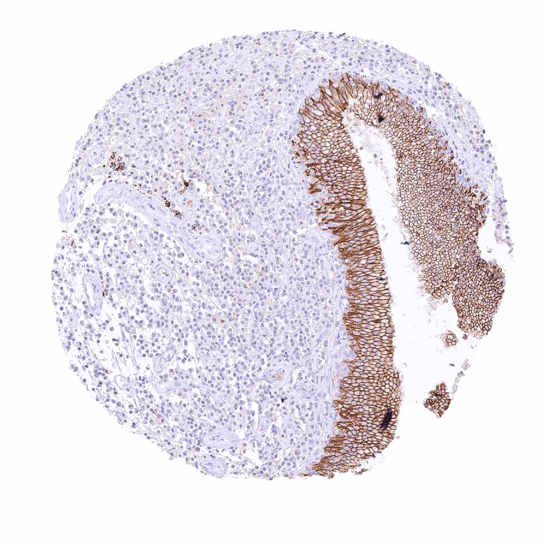
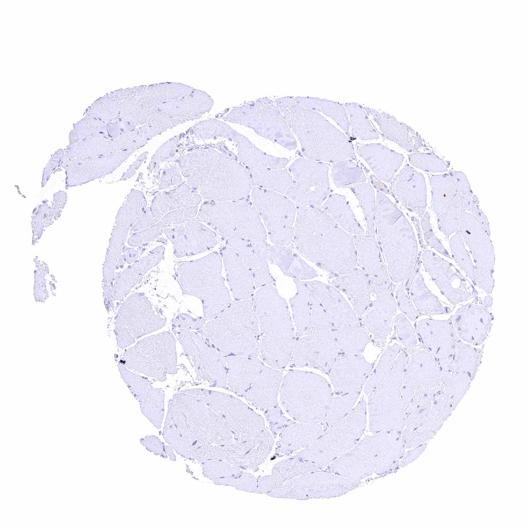
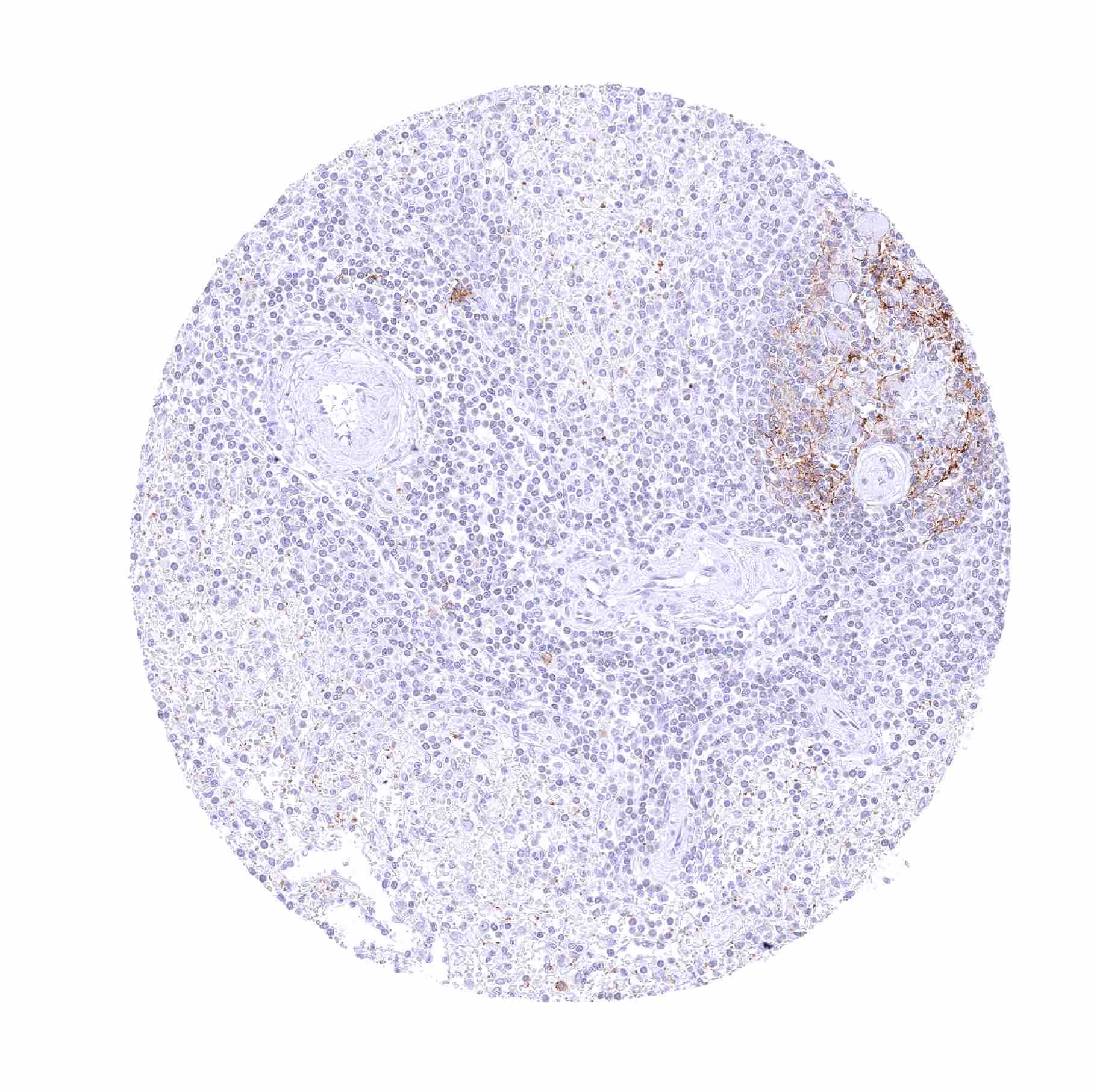
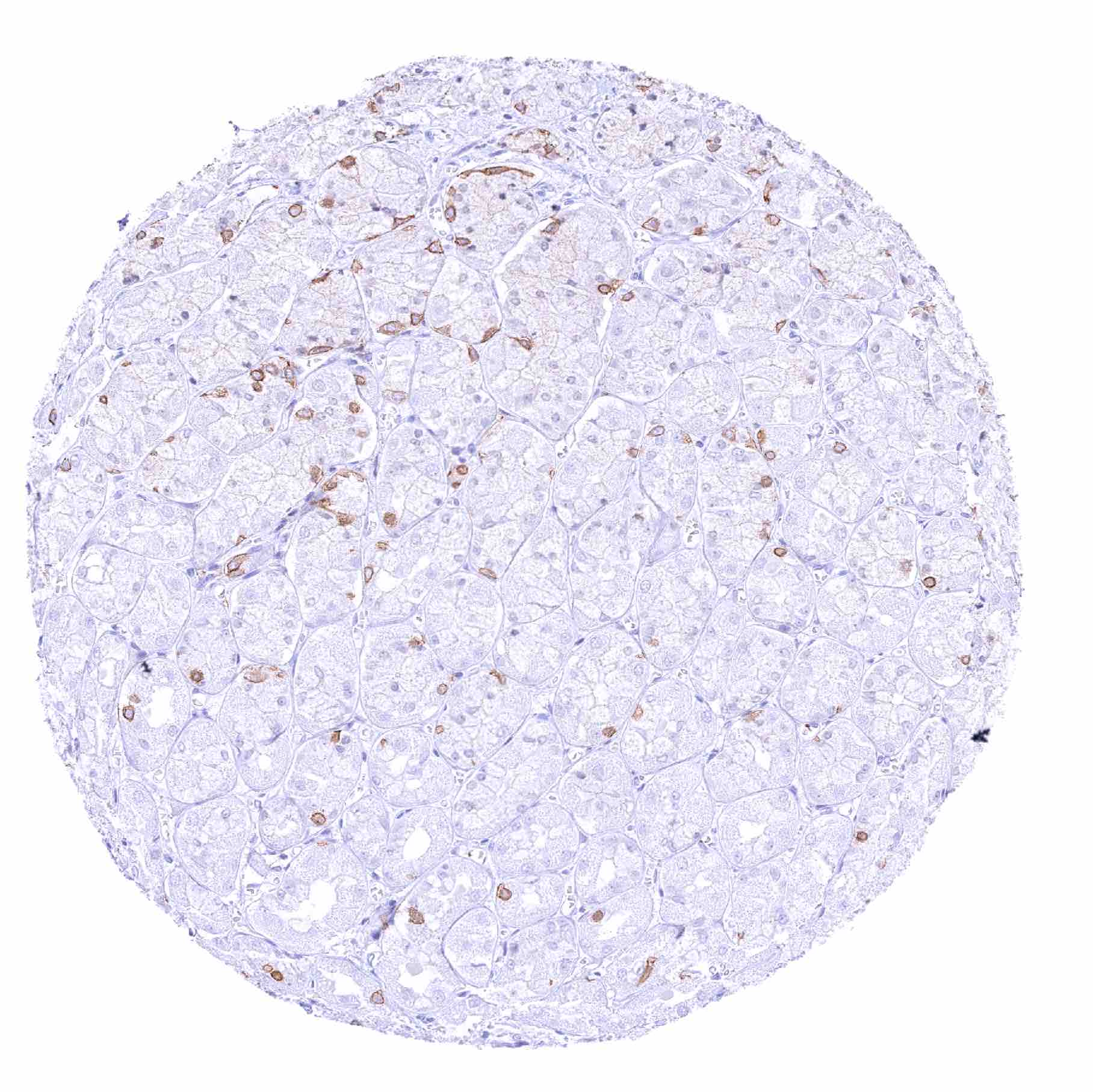
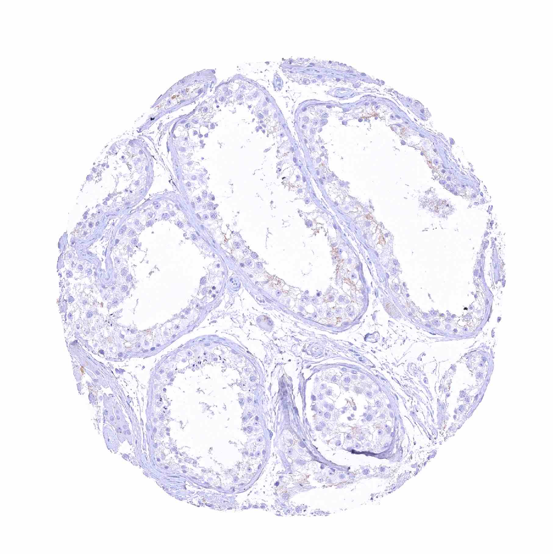
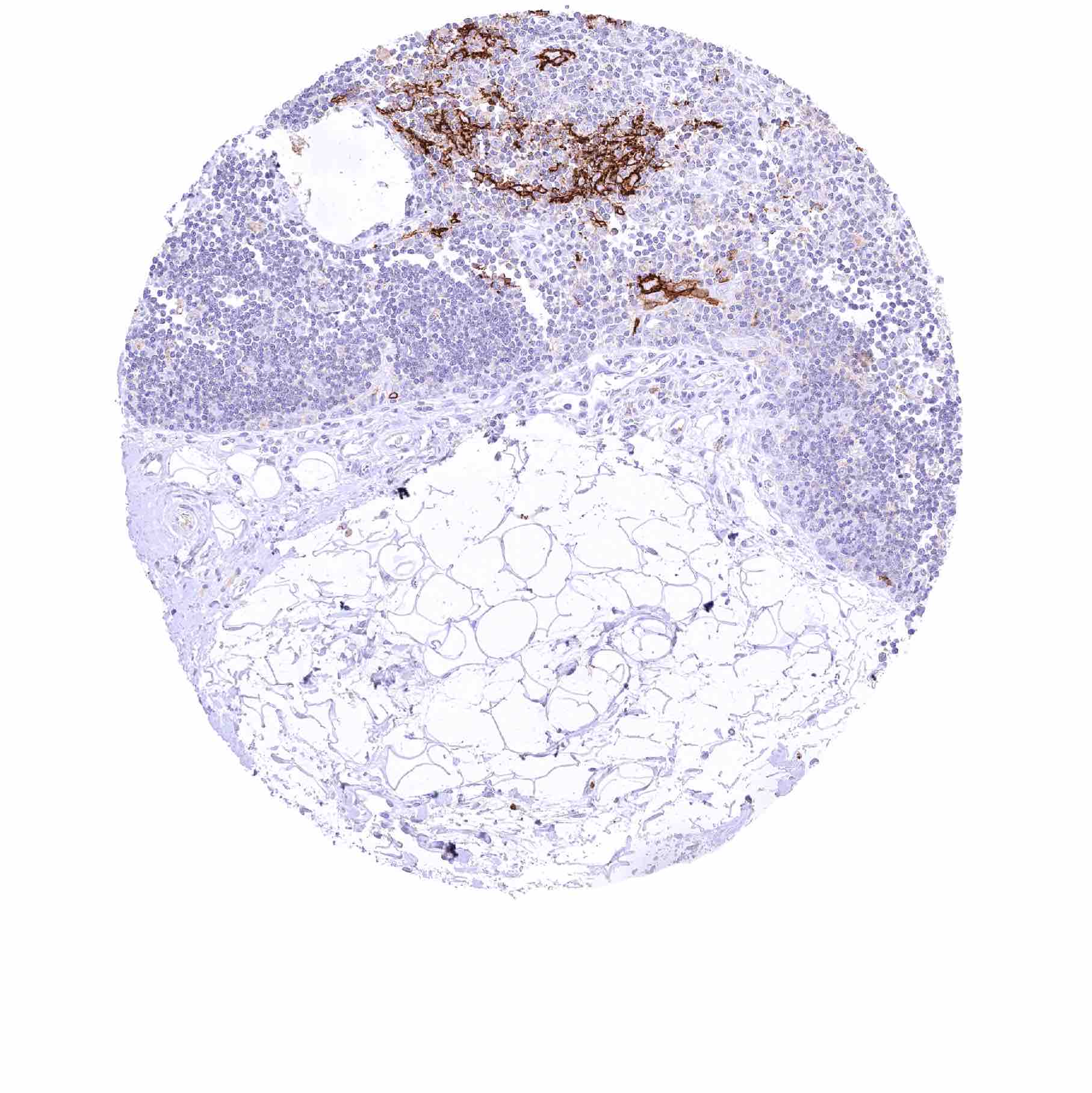
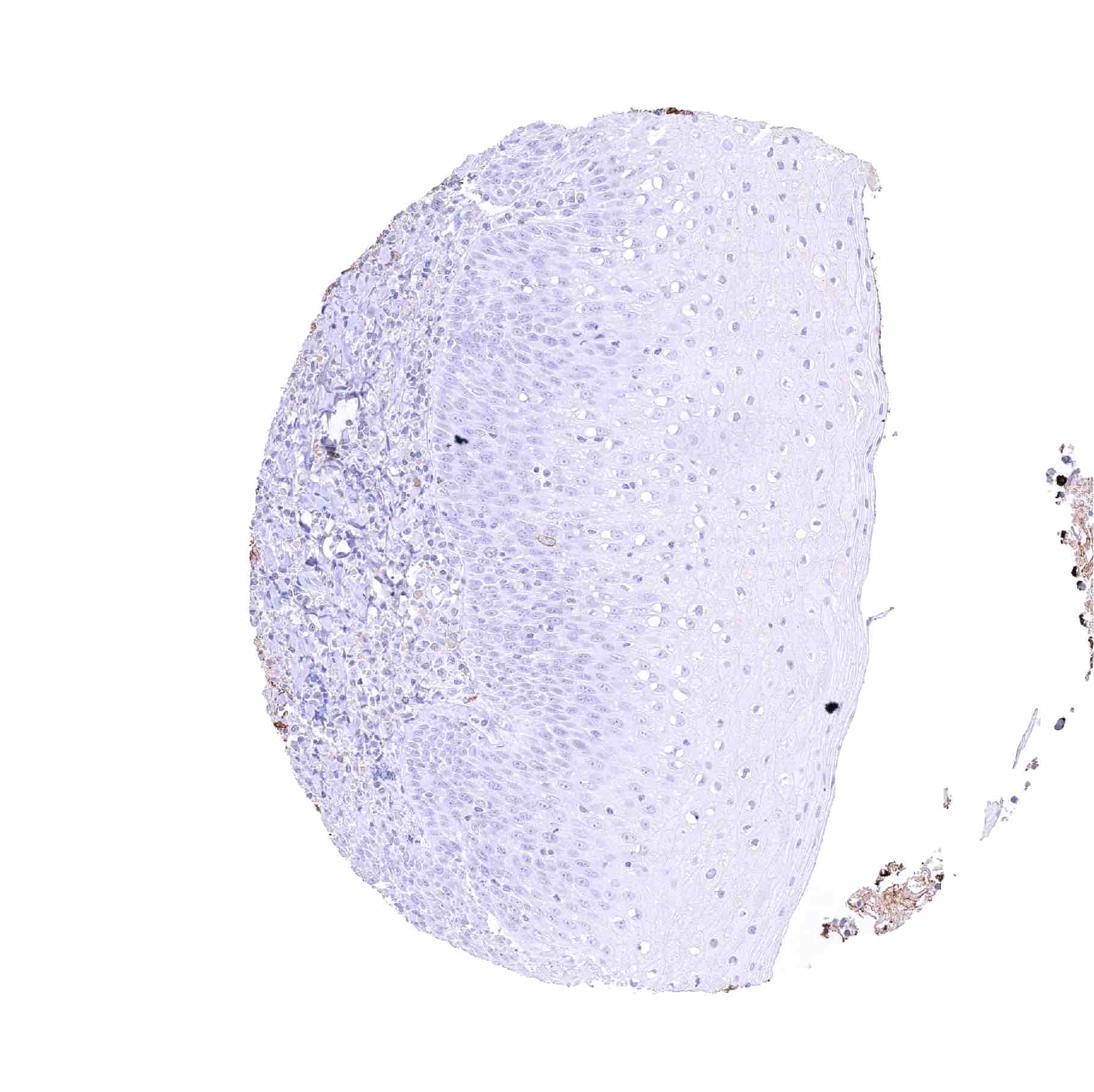
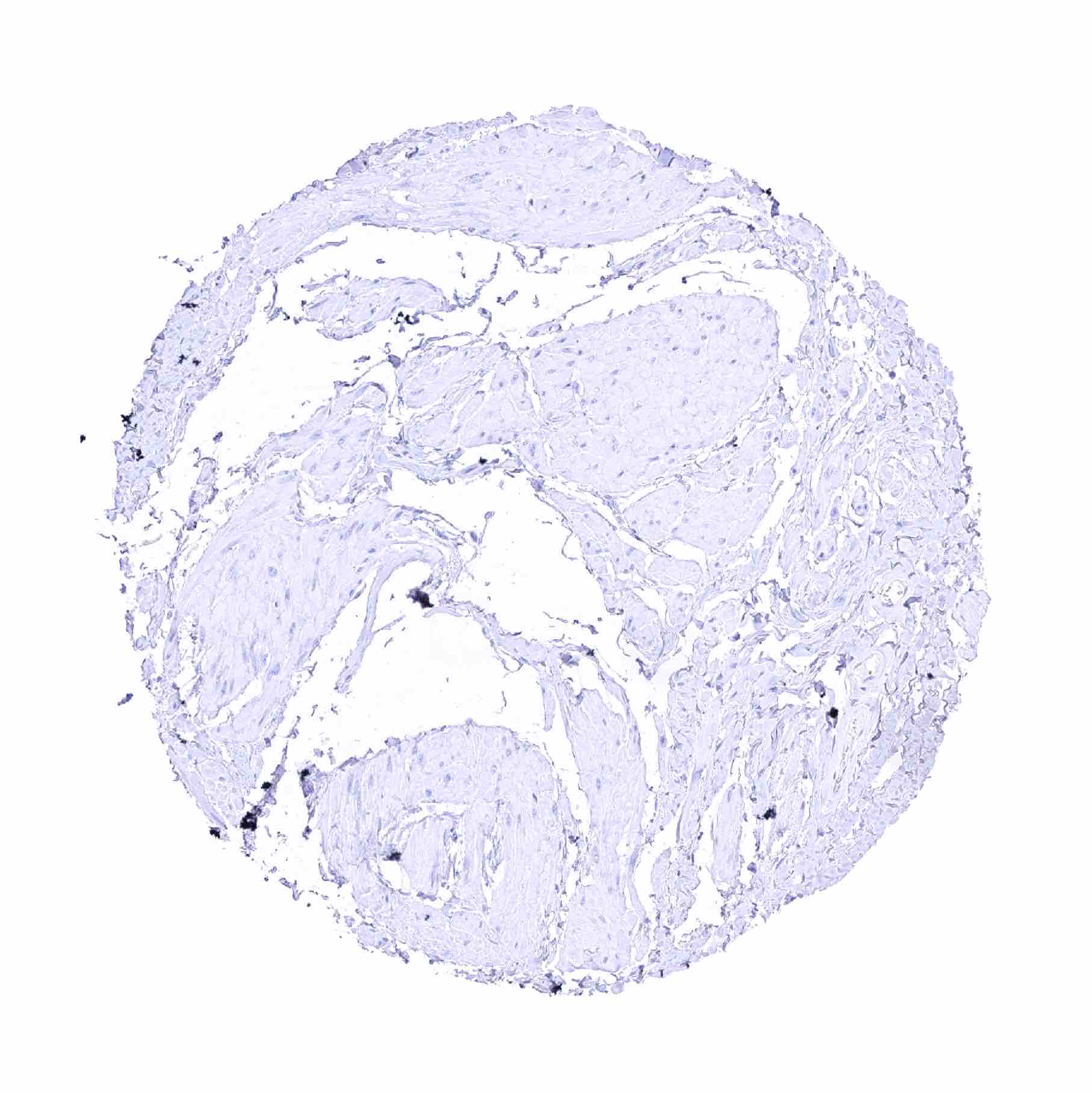
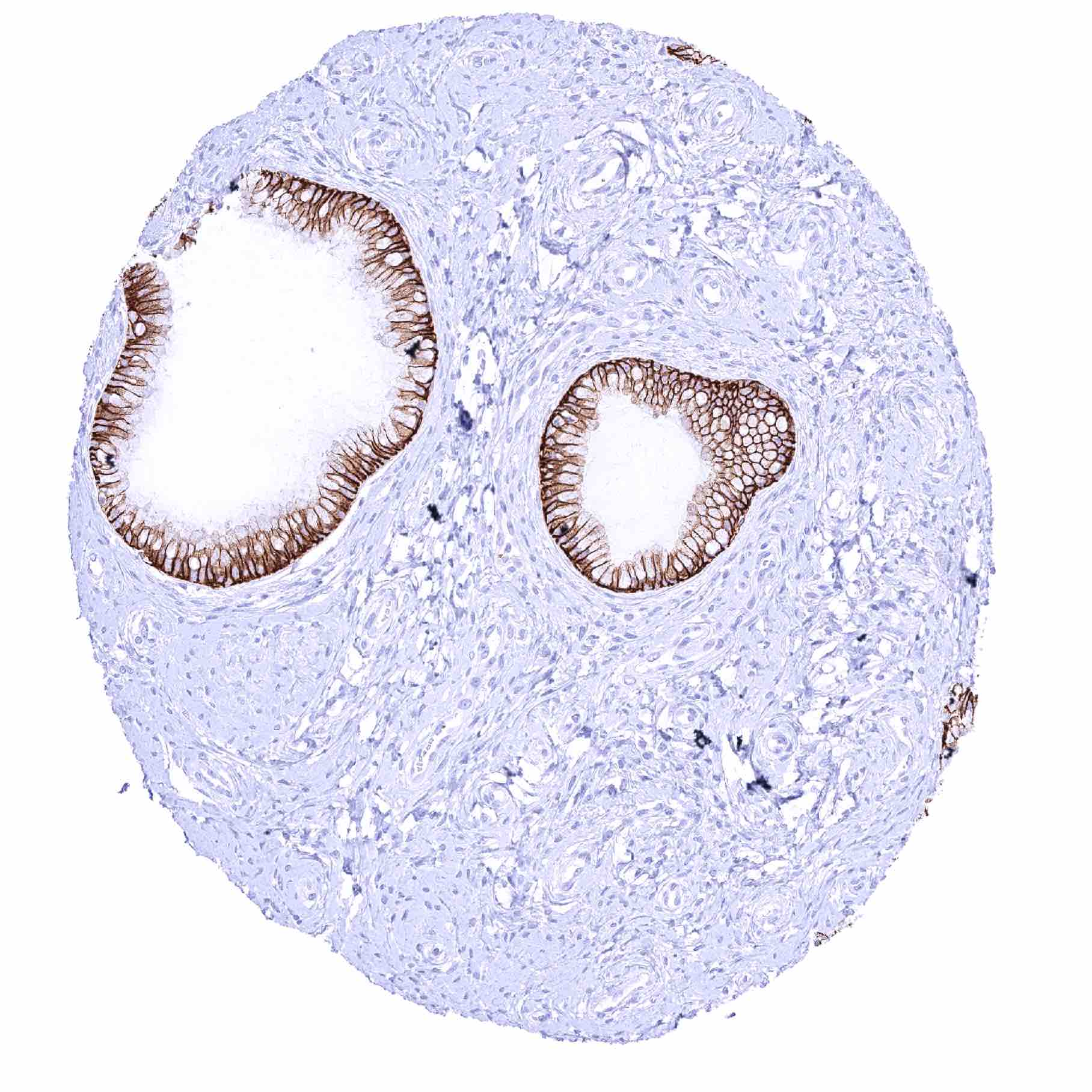
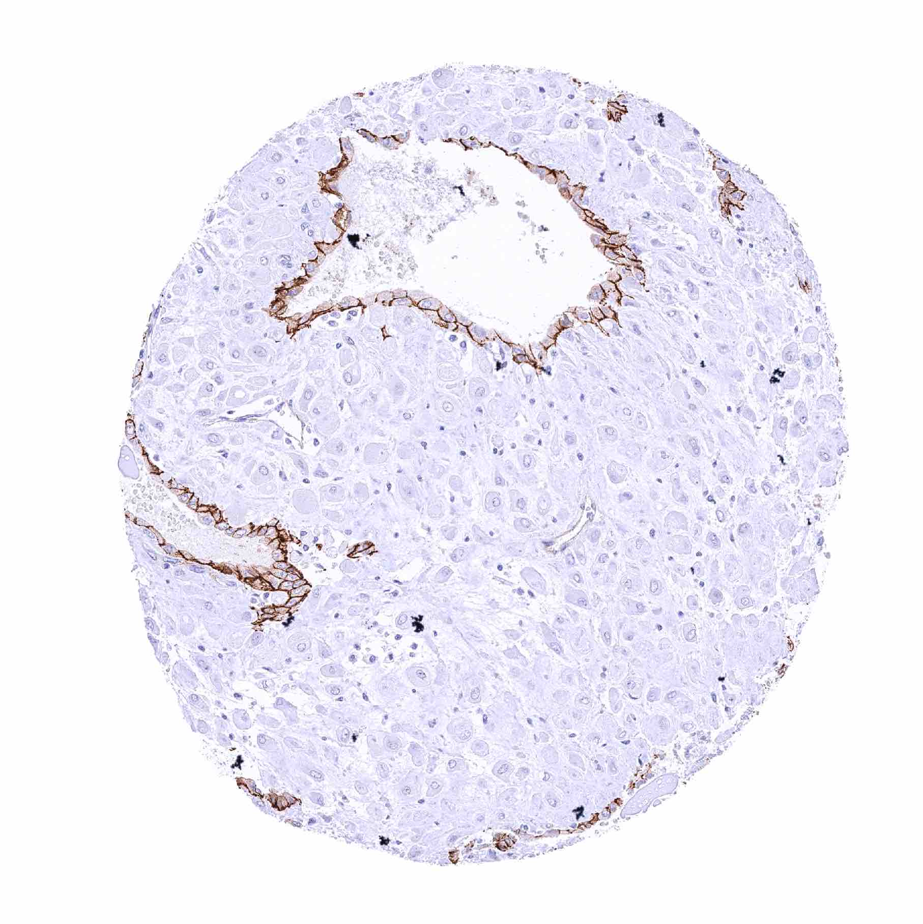
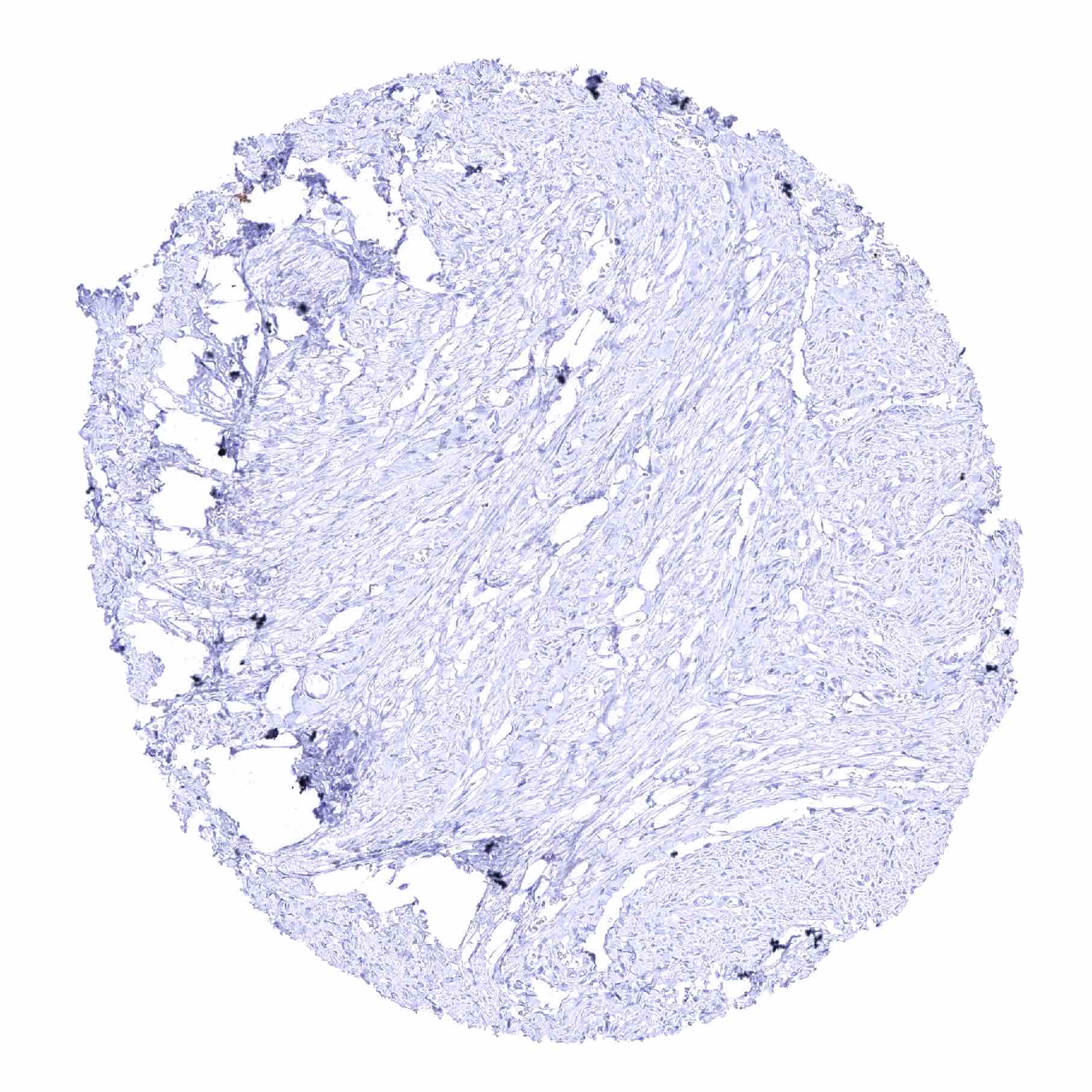
Claudin 3 antibody [HMV309] HistoMAXTM


Go to Cancer Tissue Gallery
  
 Go back to product page
  

Go to HistoMAX page


Adrenal gland – Faint membranous CLDN3 staining of few epithelial cells

Adrenal gland – No CLDN3 staining in this sample

Aorta, media

Appendix, muscular wall


Bone marrow – Distinct membranous CLDN3 staining of megacaryocytes.

Bone marrow – Distinct membranous CLDN3 staining of megacaryocytes

Breast – Strong membranous CLDN3 staining of luminal cells

Bronchus, mucosa – Strong membranous CLDN3 staining of respiratory epithelial cells


Cerebellum (white matter)

Cerebellum, cortex (molecular layer, Purkinje cell layer, granule cell layer)

Cerebrum, grey matter

Cerebrum, white matter


Colon descendens, mucosa – Strong membranous CLDN3 staining of all epithelial cells

Colon descendens, muscular wall

Duodenum, Brunner gland

Duodenum, mucosa – Strong membranous CLDN3 staining of all epithelial cells


Epididymis (Cauda) – Strong membranous CLDN3 staining of most epithelial cells

Epididymis (Corpus) – Faint membranous CLDN3 staining of chief cells

Esophagus, squamous epithelium

Fallopian tube, mucosa – Strong membranous CLDN3 staining of epithelial cells


Fat

Gallbladder, epithelium – Moderate to strong membranous CLDN3 staining of epithelial cells

Ileum, mucosa – Strong membranous CLDN3 staining of all epithelial cells

Kidney, cortex – A strong membranous CLDN3 staining preferentially occurs in collecting ducts but can also be seen in individual tubular cells


Kidney, medulla – A membranous CLDN3 staining of variable intensity occurs in collecting ducts

Kidney, pelvis, urothelium – Weak to strong membranous CLDN3 staining of a subset of urothelial cells, predominantly in the upper half of the urothelium

Liver – Moderate membranous CLDN3 staining of hepatocytes

Liver – Strong membranous CLDN3 staining of bile ducts. Weak to moderate membranous CLDN3 staining of hepatocytes, predominantly at the bile secreting pole


Lung – Faint membranous CLDN3 staining of some pneumocytes

Lung – Strong membranous CLDN3 staining of bronchiolized alveolar cell. CLDN3 staining is only faint in regular pneumocytes

Lymph node – Strong luminal membranous CLDN3 staining of at least a subset of high endothelial venules. Moderate to strong CLDN3 staining of a subset of cells in germinal centres

Lymph node – Strong luminal membranous CLDN3 staining of at least a subset of high endothelial venules


Ovary, corpus luteum – Weak to moderate, membranous CLDN3 staining of at least a large subset of corpus luteum cells

Ovary, stroma

Pancreas – Strong membranous CLDN3 staining of acinar cells. Staining is weaker in cells of islets of Langerhans

Pancreas – Strong membranous CLDN3 staining of acinar cells


Parathyroid gland – Weak to moderate membranous CLDN3 staining of epithelial cells

Parotid gland – Strong membranous CLDN3 staining of glandular cells. CLDN3 staining is only very faint in excretory duct cells

Pituitary gland, anterior lobe – Moderate membranous CLDN3 staining of a subset of epithelial cells

Pituitary gland, posterior lobe


Placenta (amnion and chorion)

Placenta, early – Faint to moderate, membranous CLDN3 staining of the surface membrane of the syncytiotrophoblast

Placenta, mature – No CLDN3 staining in this sample

Placenta, mature – Various endothelial cells show a distinct CLDN3 staining


Prostate – Strong membranous CLDN3 staining of basal and acinar epithelial cells

Rectum, mucosa – Strong membranous CLDN3 staining of all epithelial cells

Seminal vesicle – Moderate to strong membranous CLDN3 staining of epithelial cells

Sinus paranasales – Strong membranous CLDN3 staining of respiratory epithelial cells


Skeletal muscle

Skin, hairfollicel and sebaceous glands

Skin

Spleen – Moderate to strong CLDN3 staining of a subset of cells in the white pulp


Stomach, antrum – Distinct membranous CLDN3 positivity of few epithelial cells in the neck and in glandular pits

Stomach, antrum – Strong membranous CLDN3 staining of a small subset of epithelial cells in the neck and in glandular pits

Submandibular gland – Strong membranous CLDN3 staining of glandular cells. CLDN3 staining is only very faint in excretory duct cells

Testis


Thymus – Strong membranous CLDN3 staining of cells of corpuscles of Hassall's

Thyroid gland – Moderate to strong membranous CLDN3 staining of follicular epithelial cells

Tonsil – Strong membranous CLDN3 staining of squamous epithelial cells of the crypts

Tonsil, surface epithelium


Urinary bladder, muscular wall

Urinary bladder, urothelium – Weak to strong membranous CLDN3 staining of a subset of urothelial cells, predominantly in the upper half of the urothelium

Uterus, ectocervix

Uterus, endocervix – Strong, predominantly basolateral membranous CLDN3 staining of epithelial cells


Uterus, endometrium (pregnancy) – Strong membranous CLDN3 staining of epithelial cells

Uterus, endometrium (proliferation) – Strong membranous CLDN3 staining of epithelial cells

Uterus, endometrium (secretion) – Strong membranous CLDN3 staining of epithelial cells

Uterus, myometrium


| TOP |