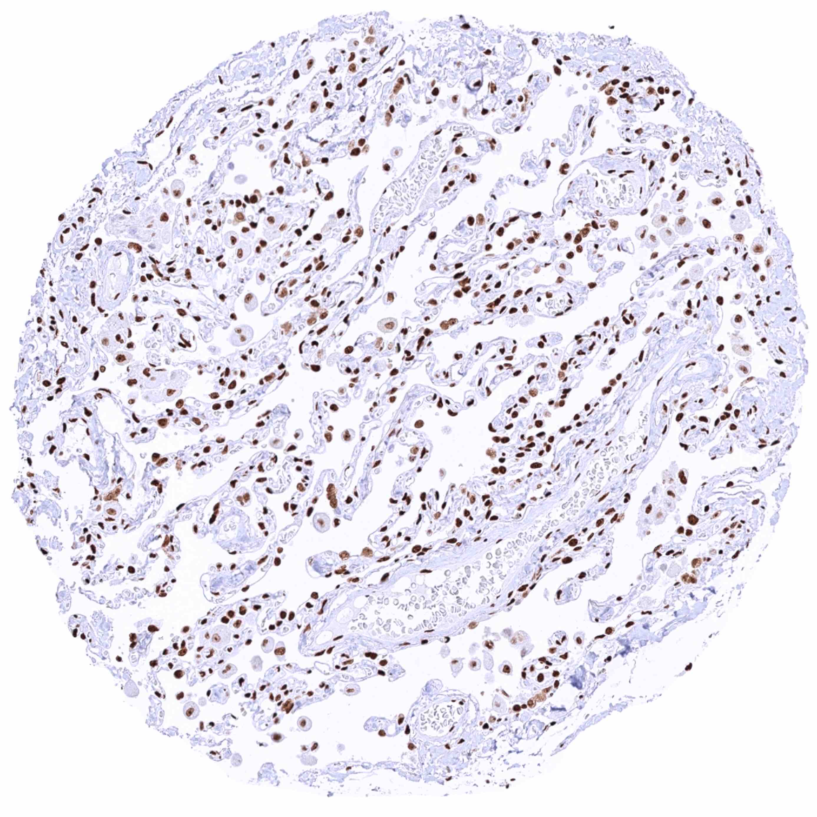
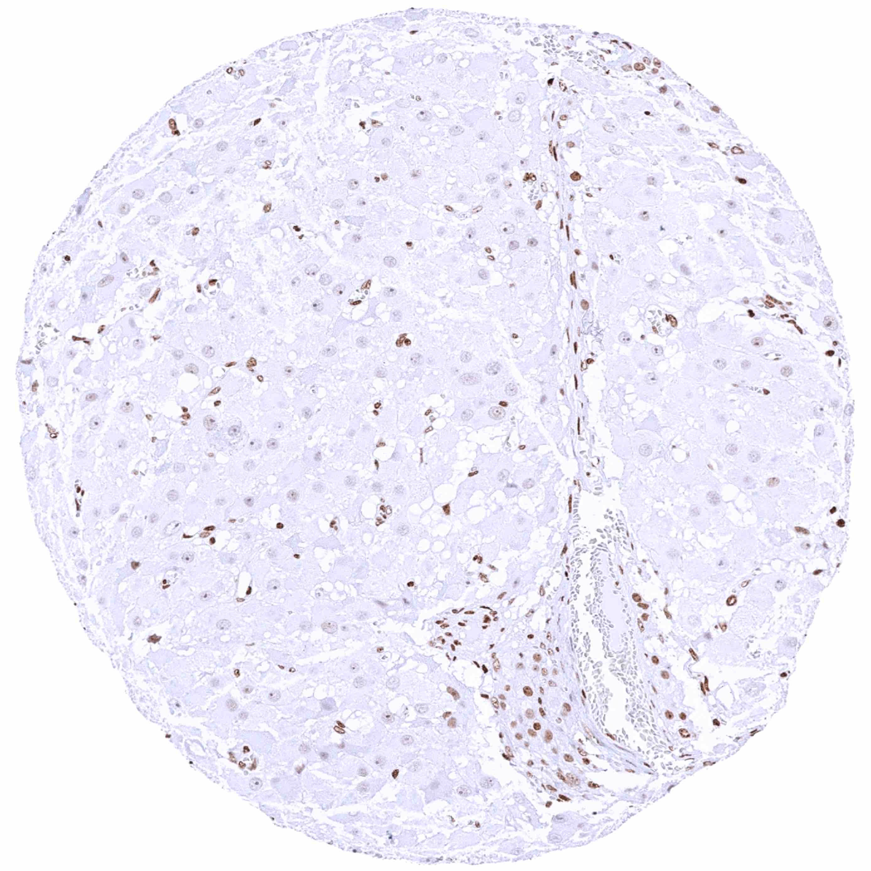
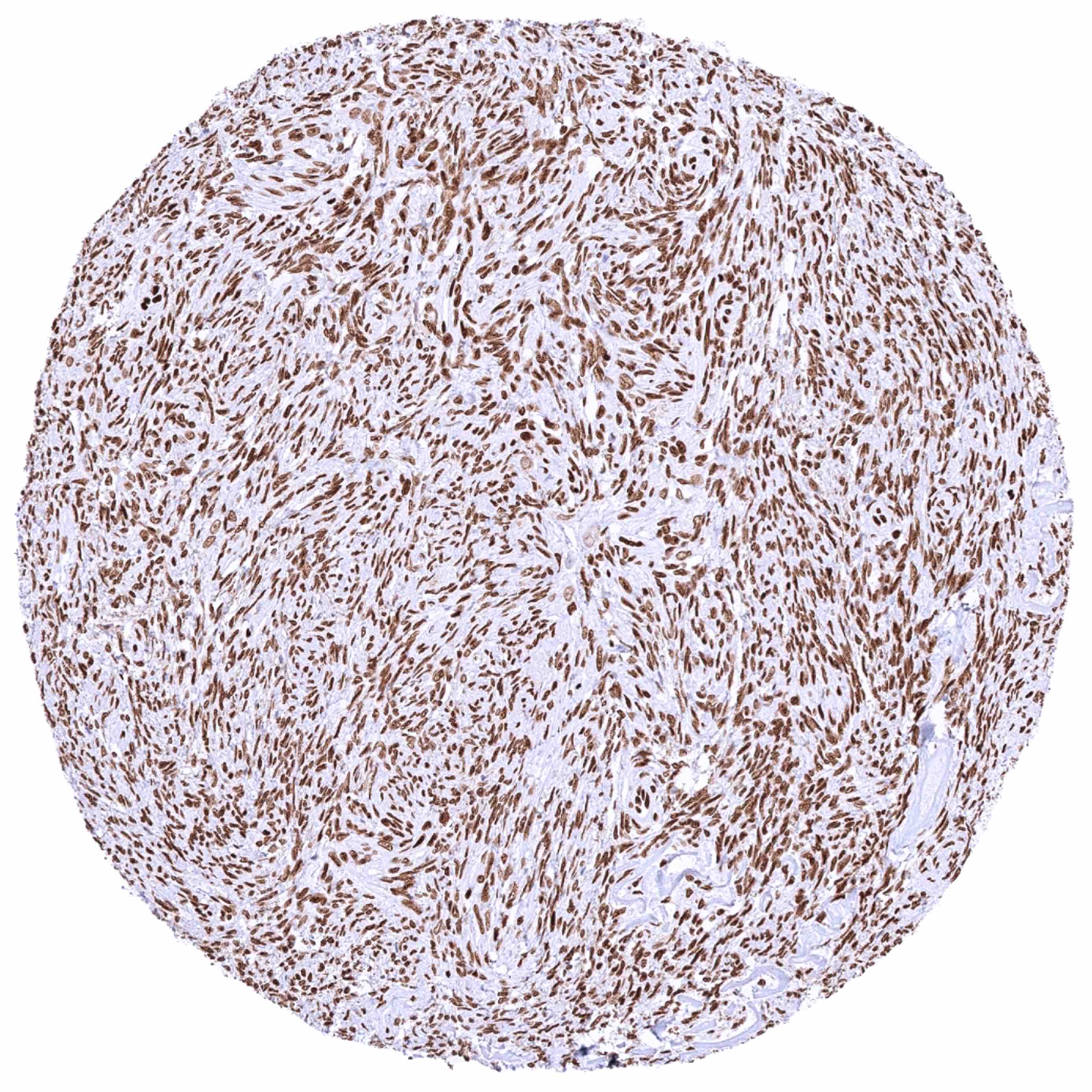
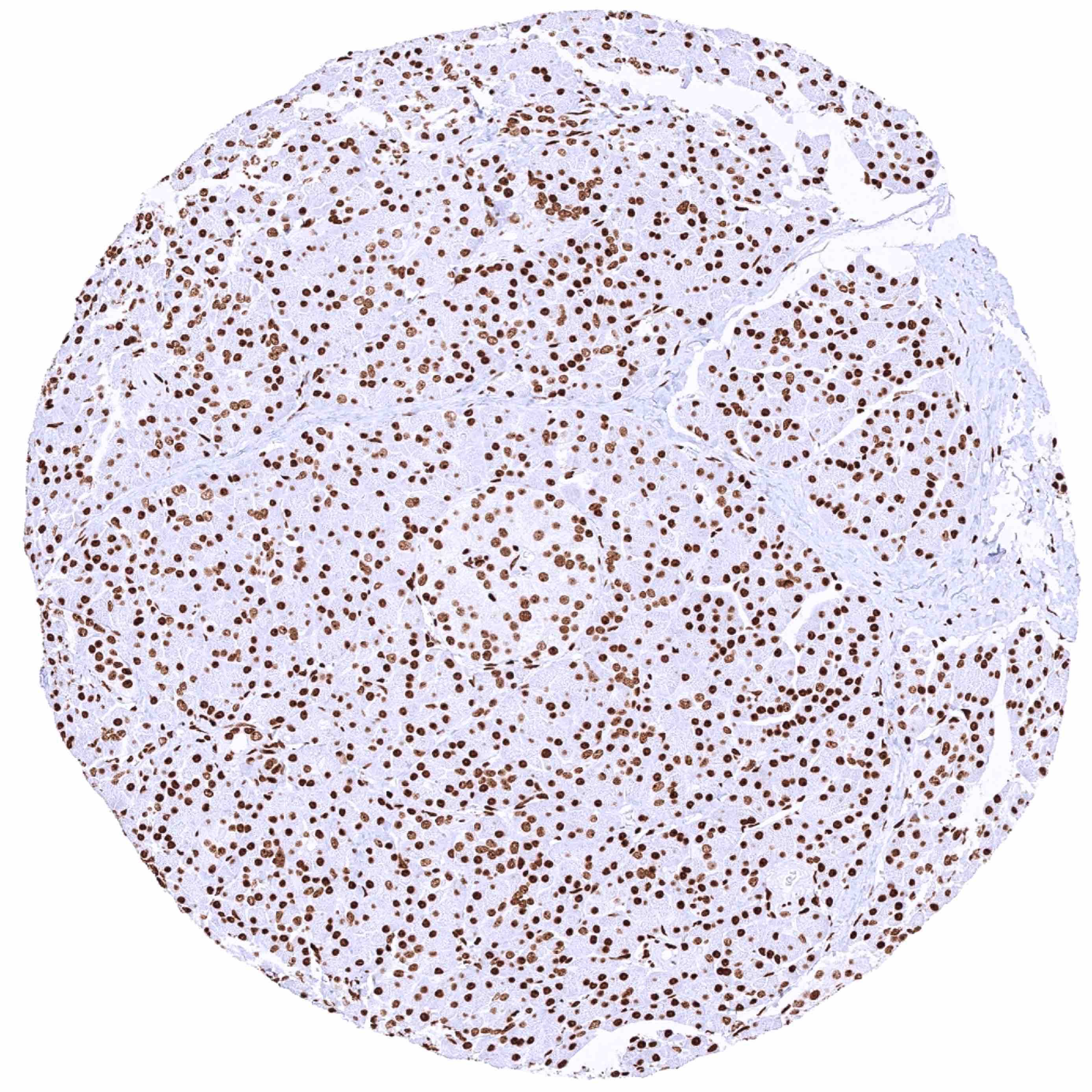
Normal Tissue Gallery- GTX639941

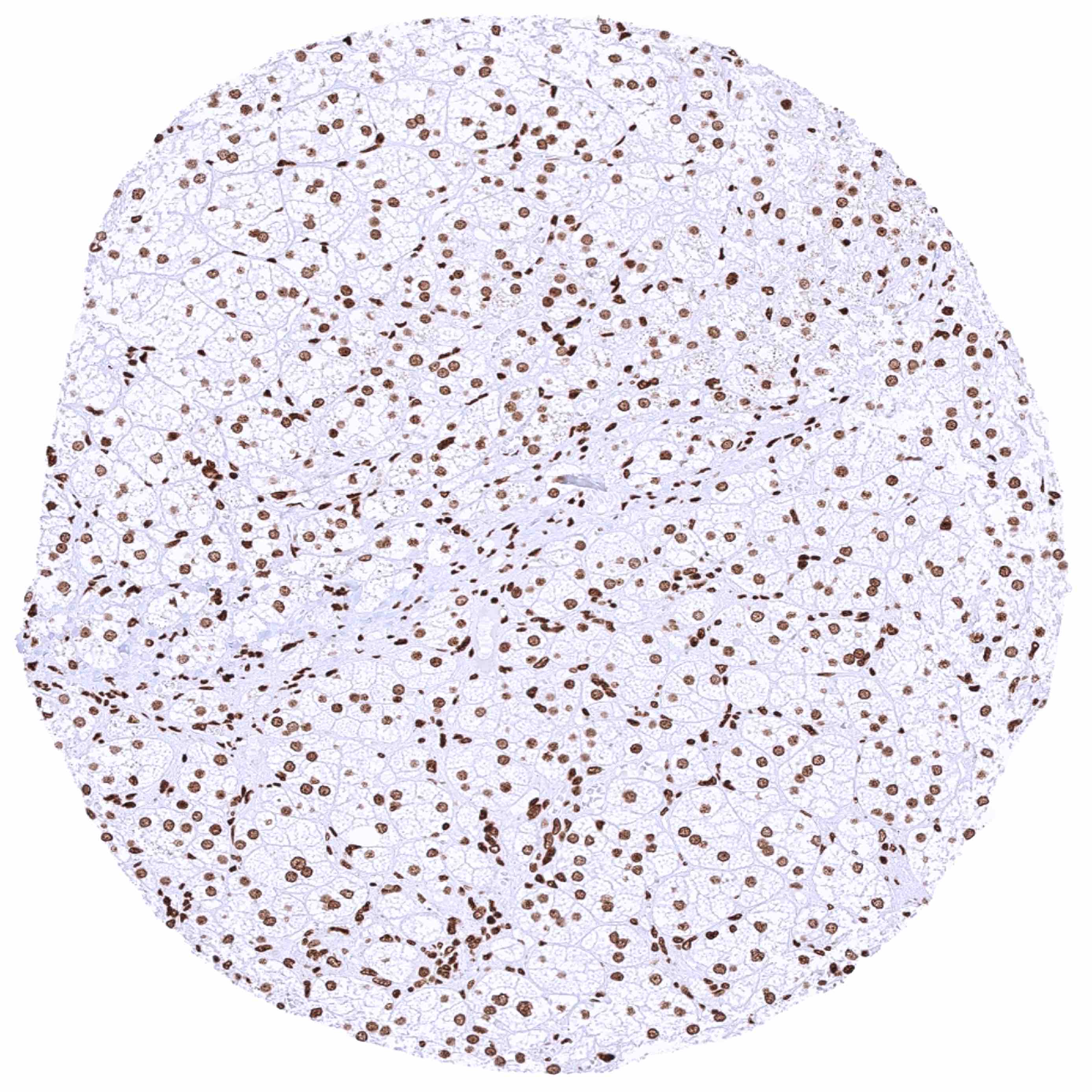
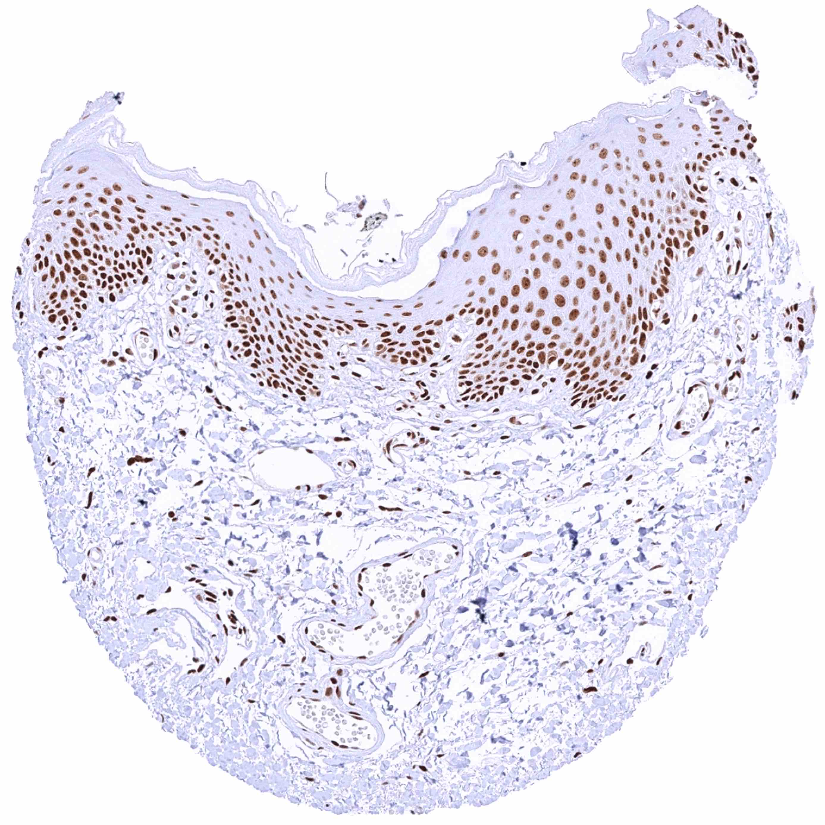
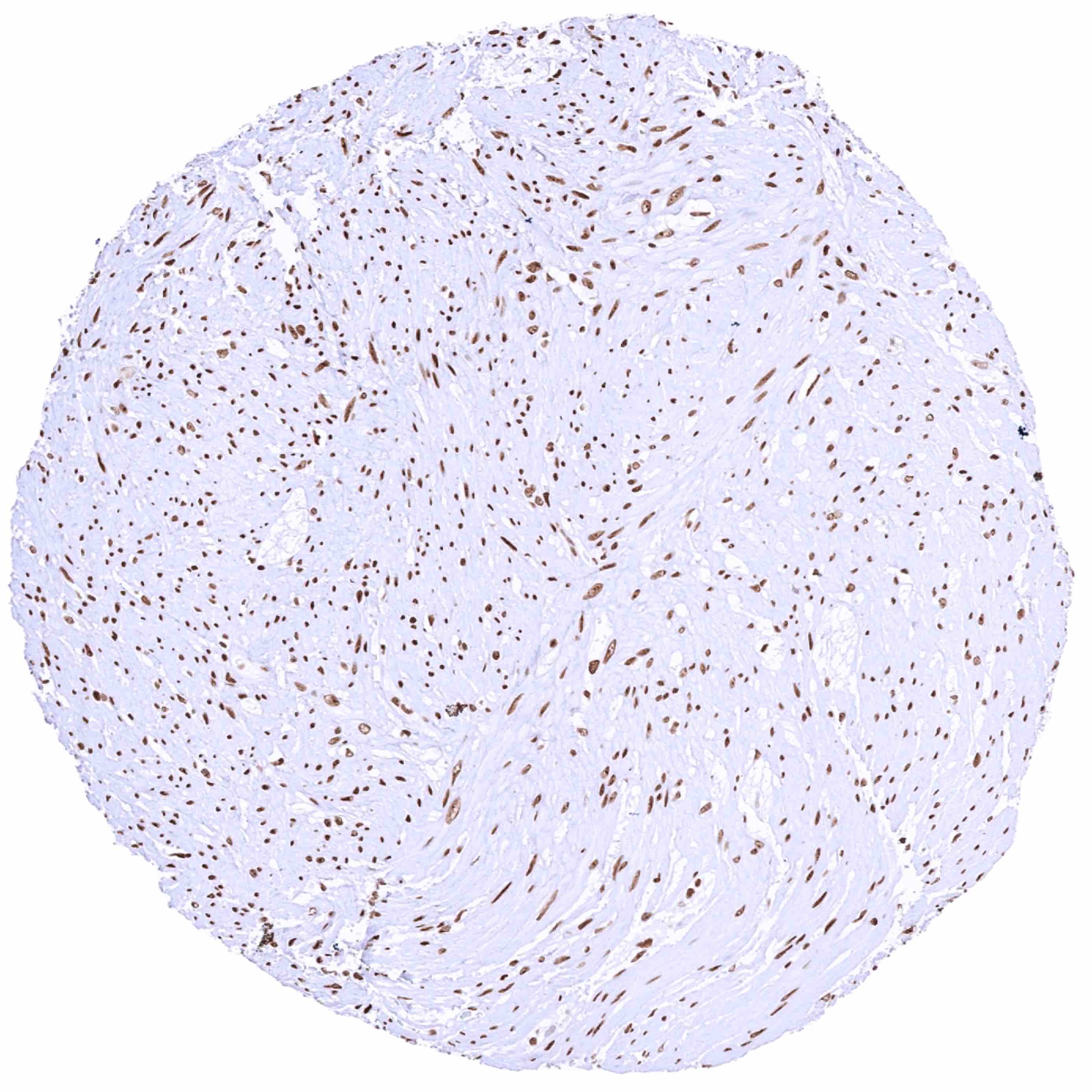
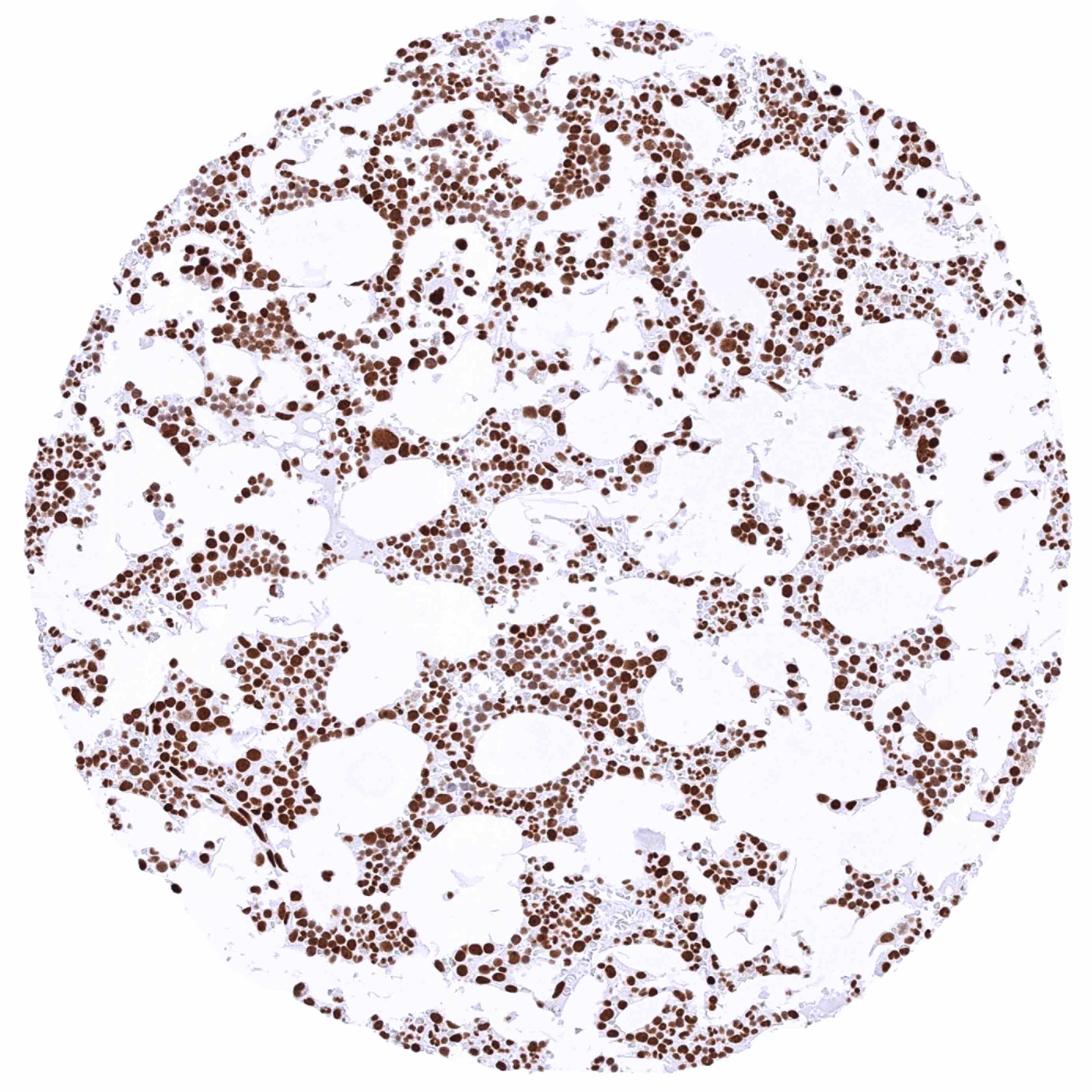
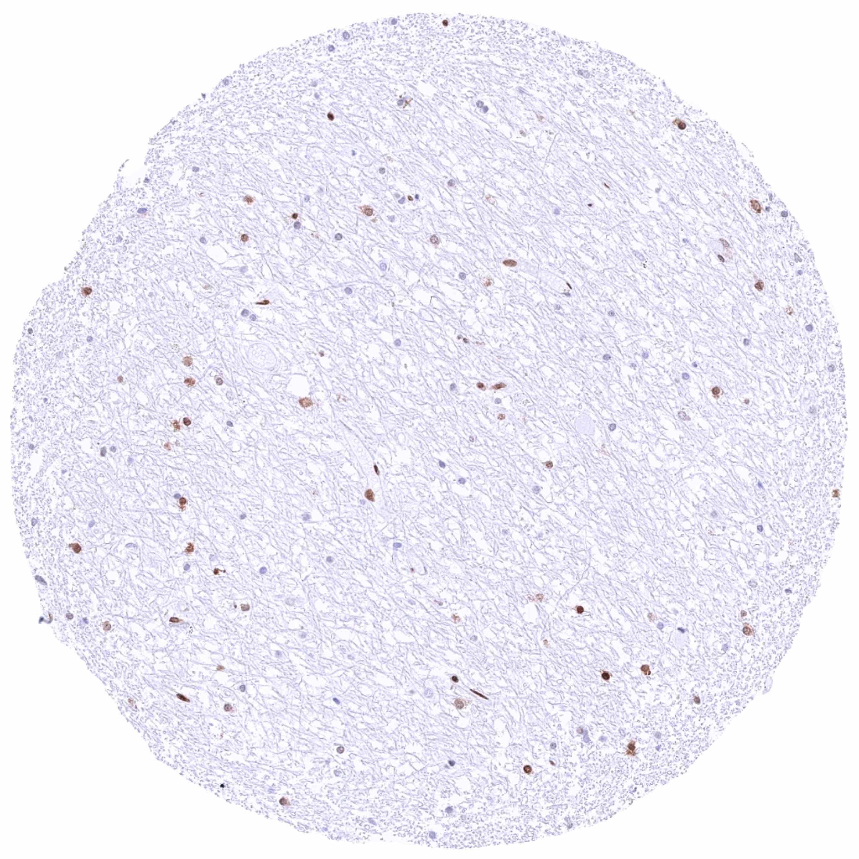
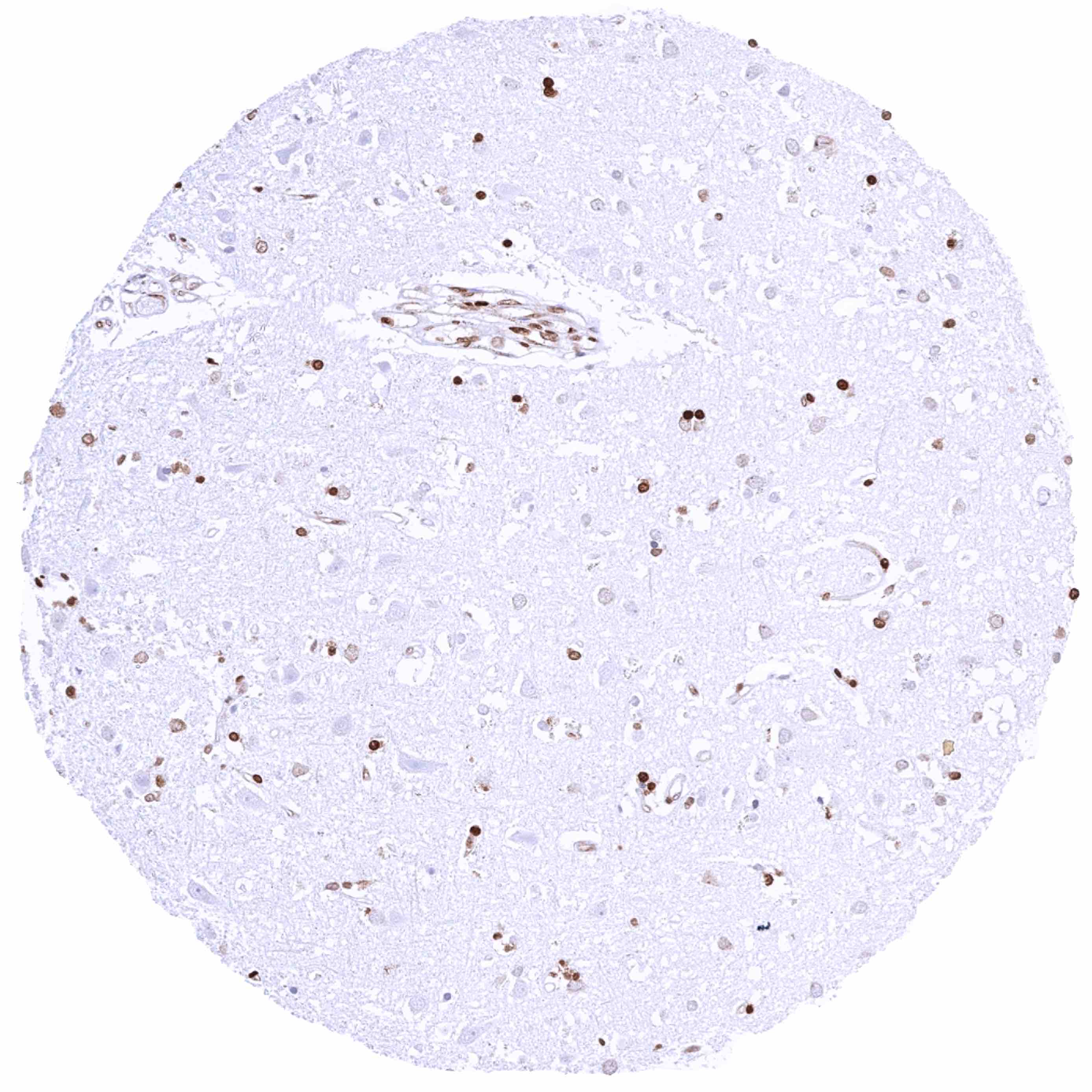
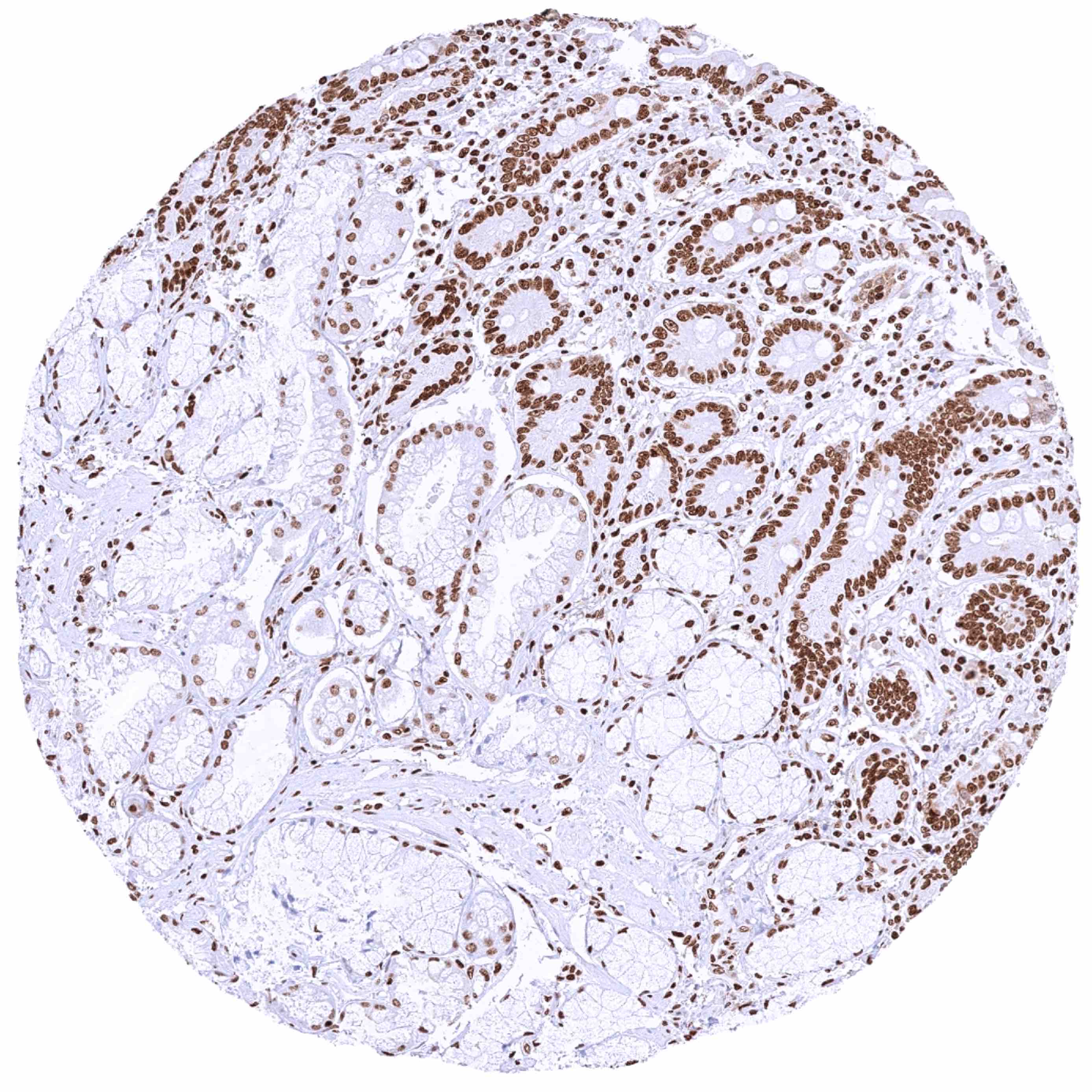
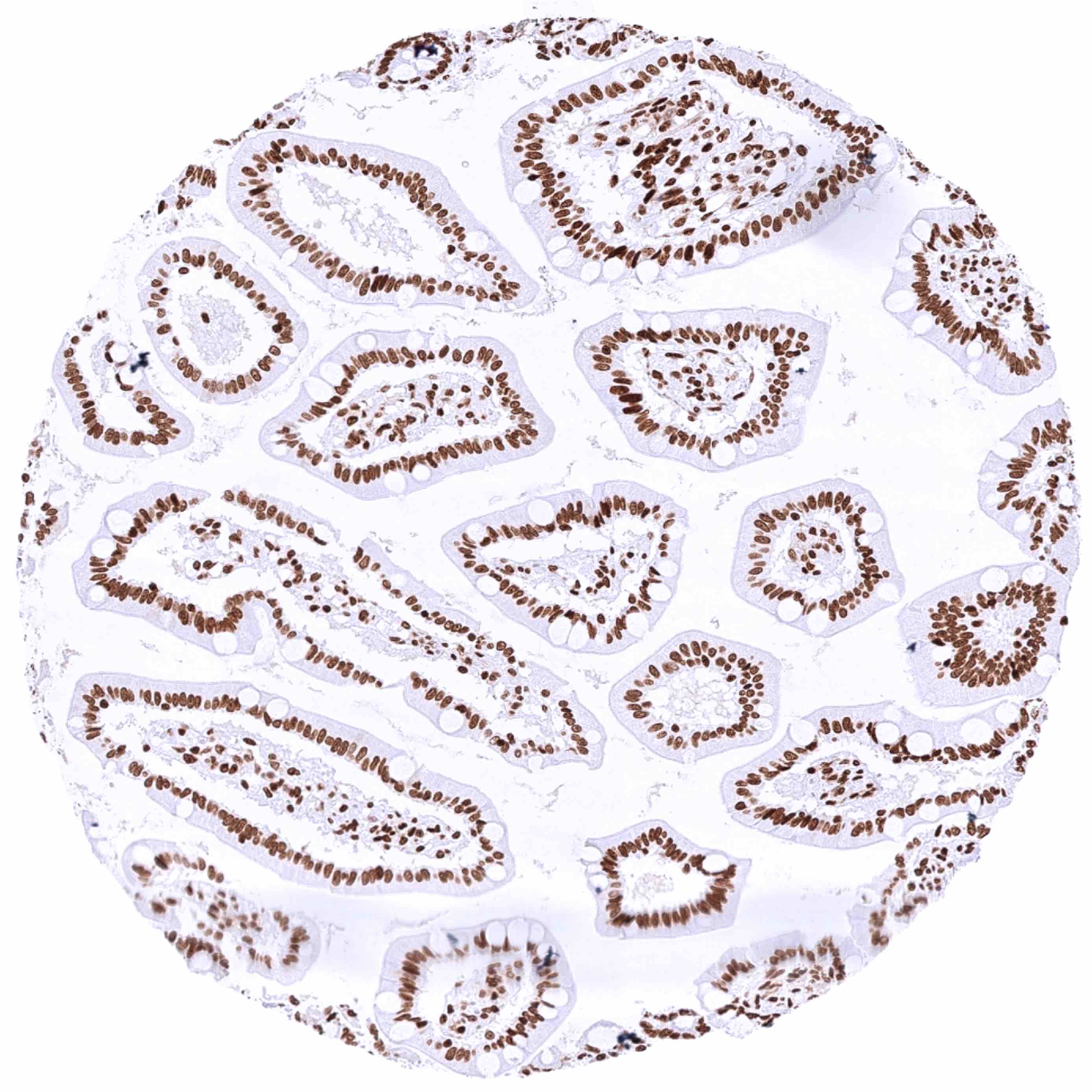
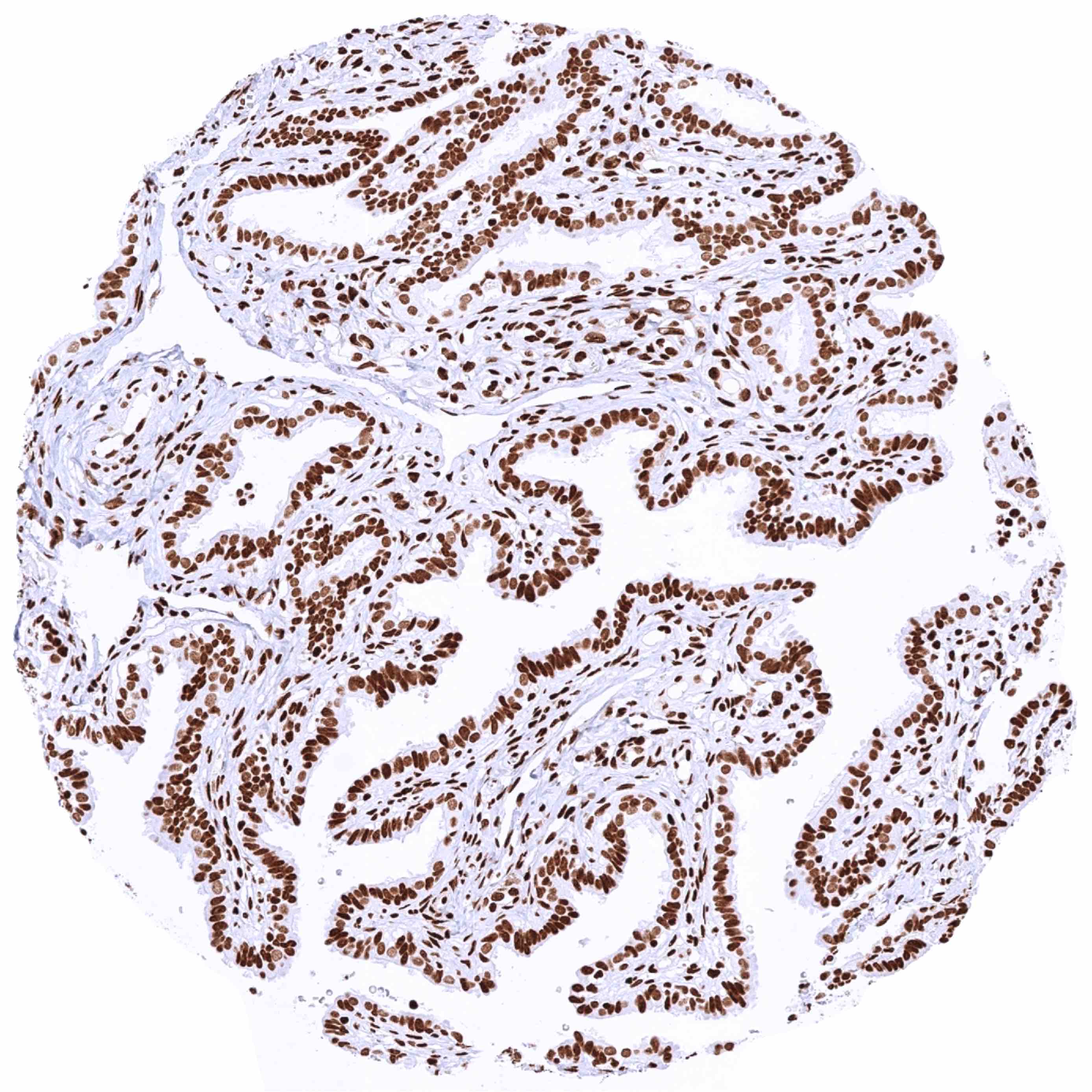
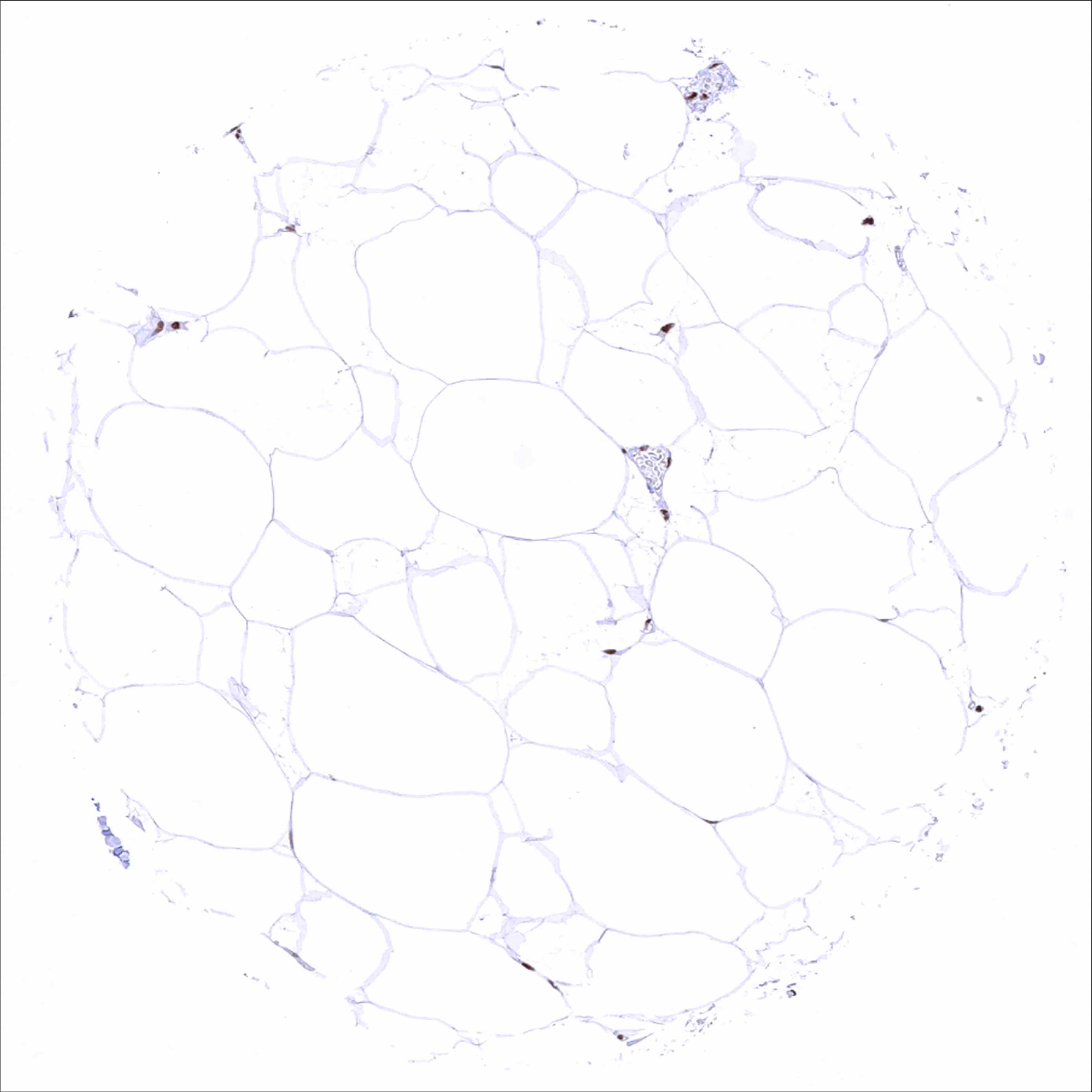
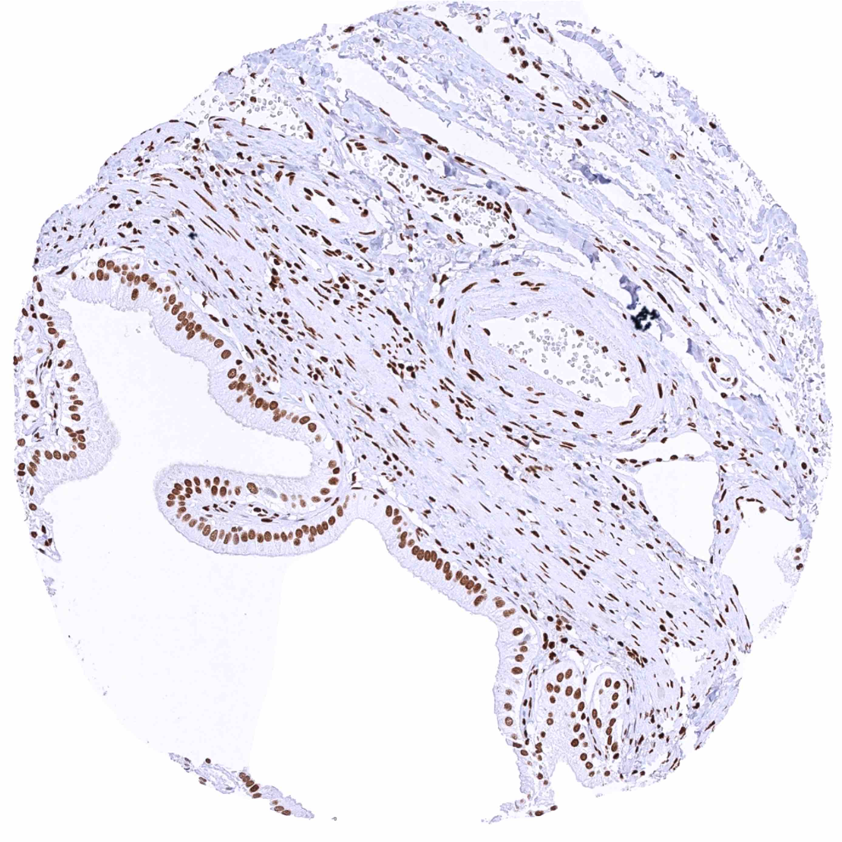
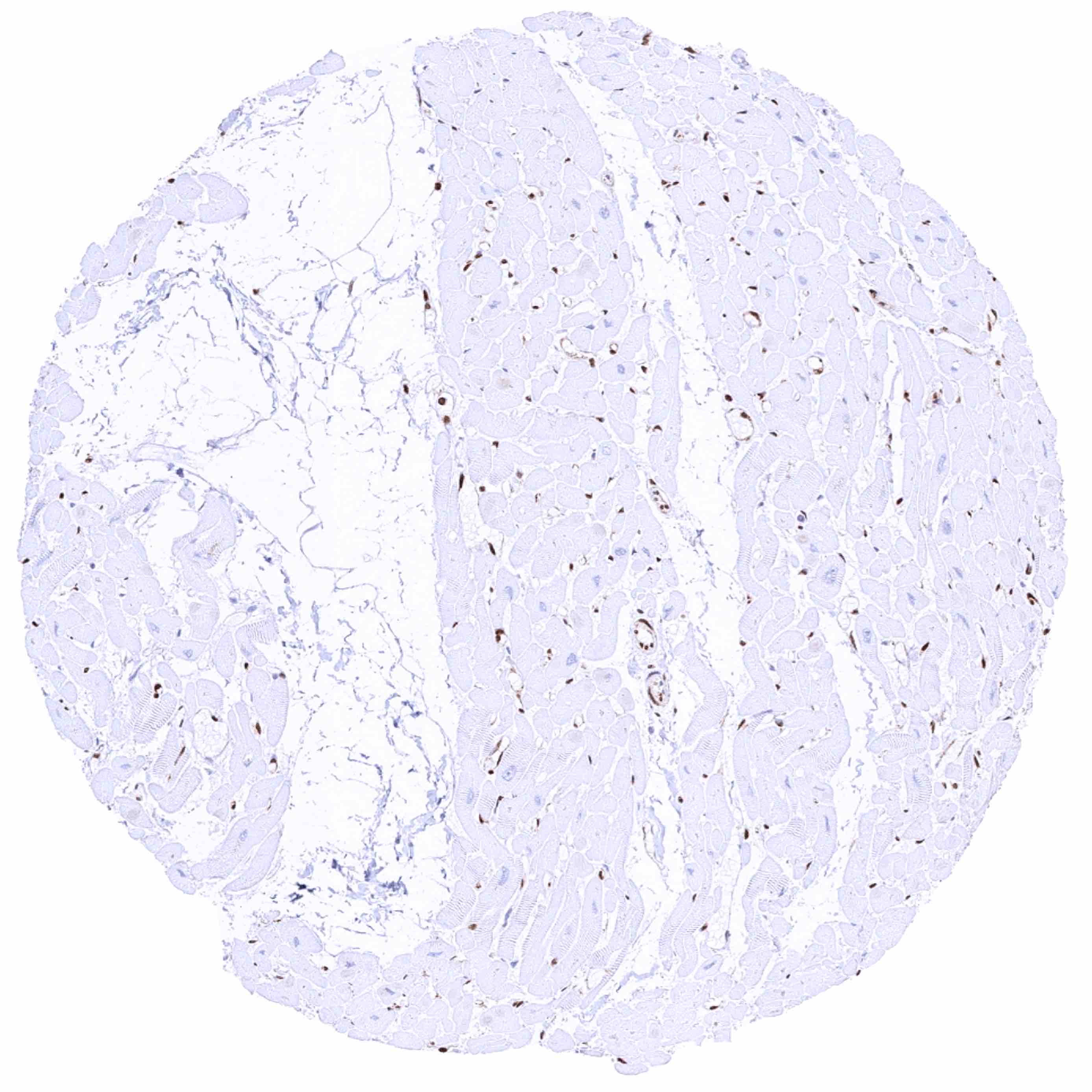
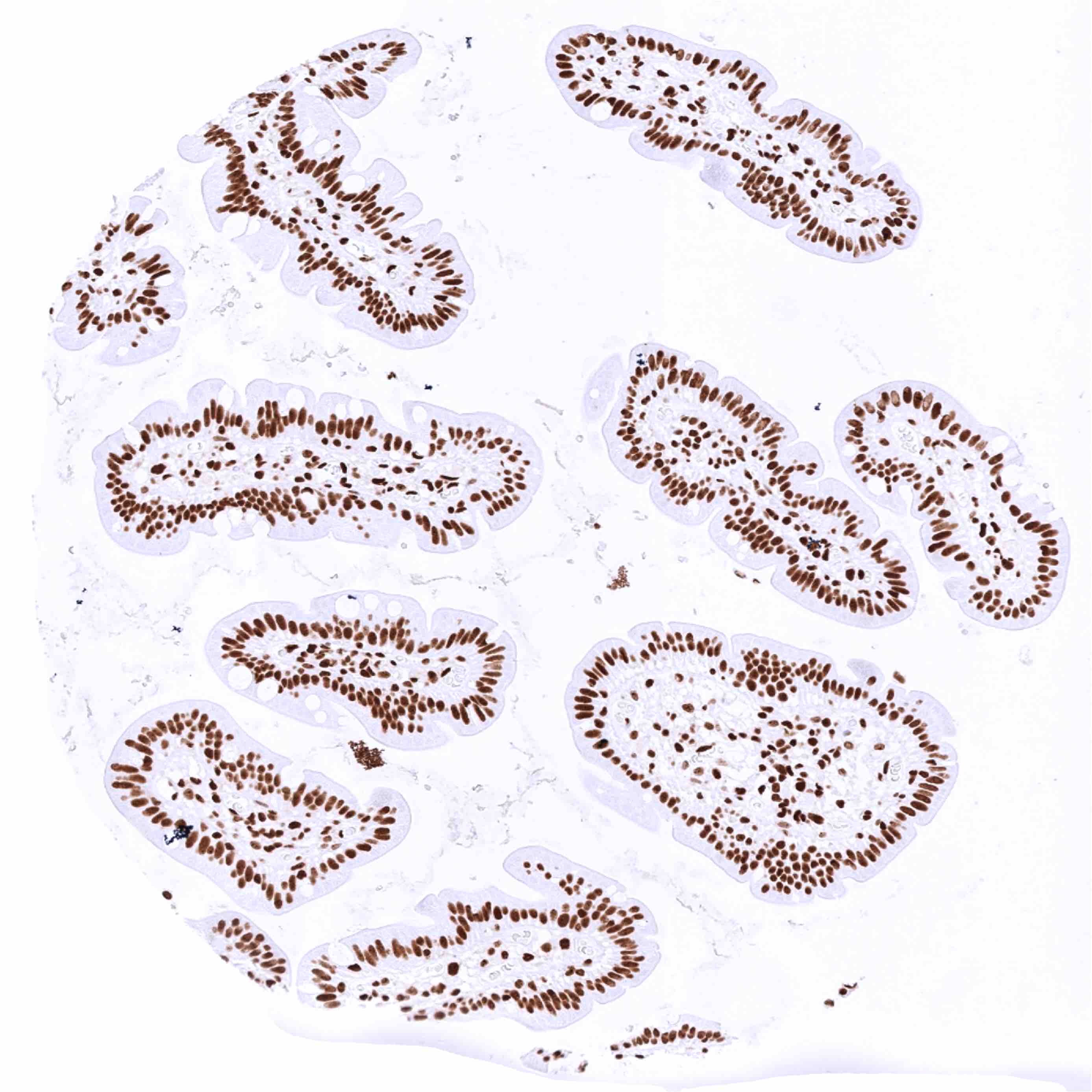
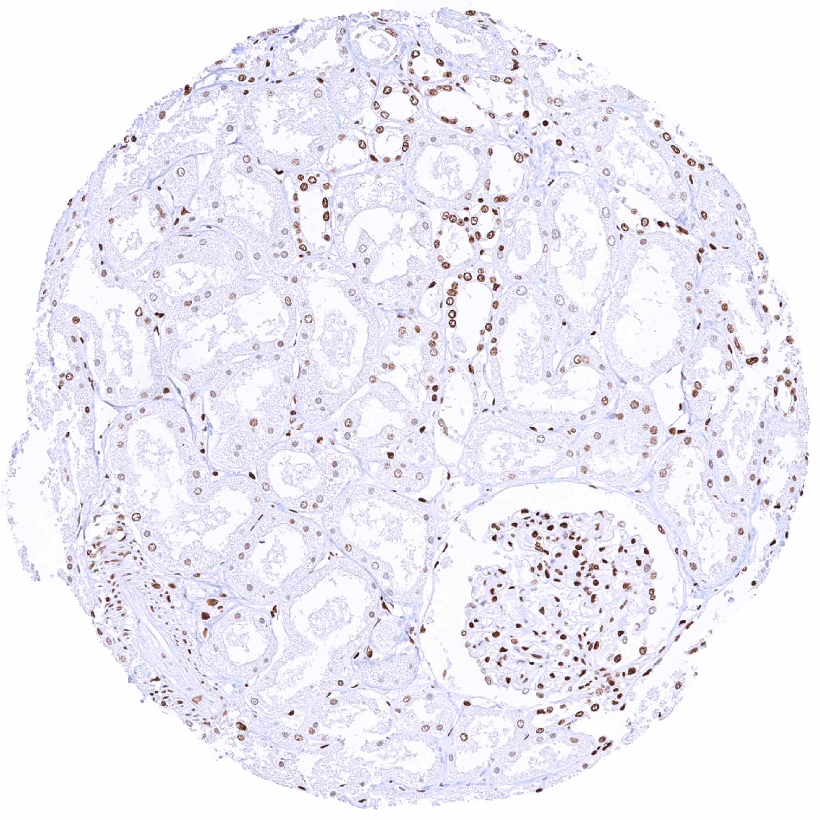
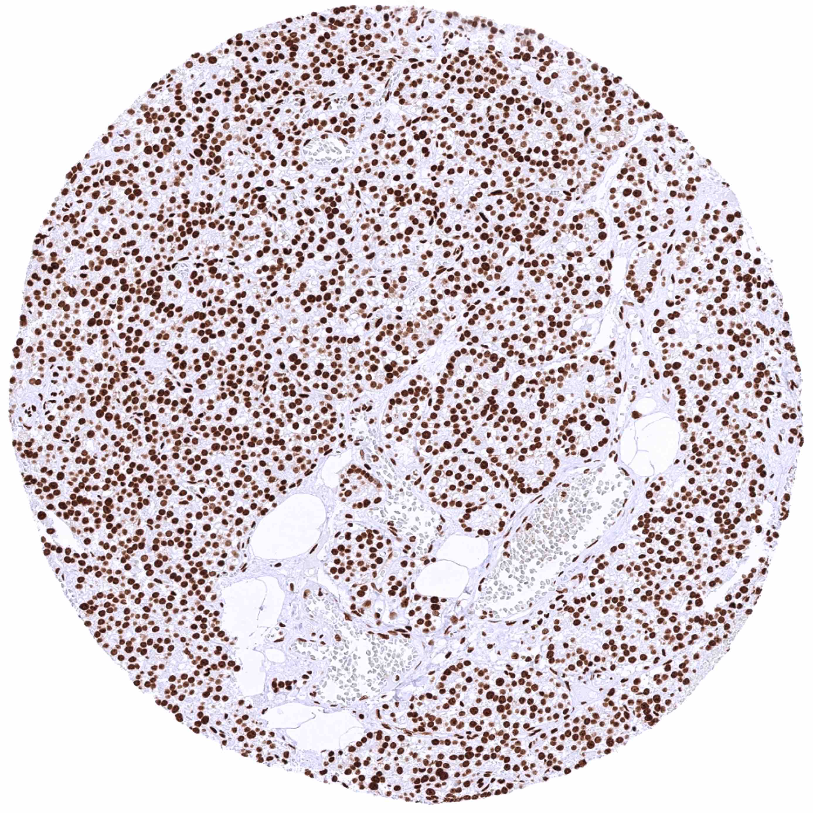
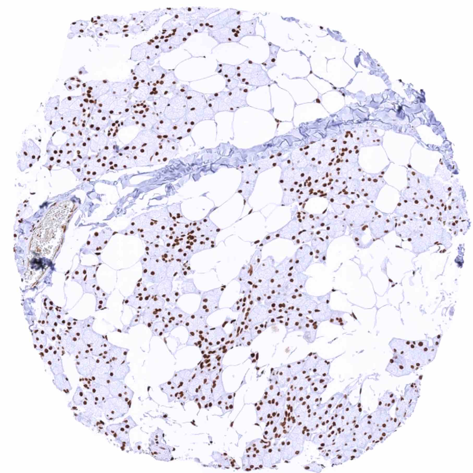
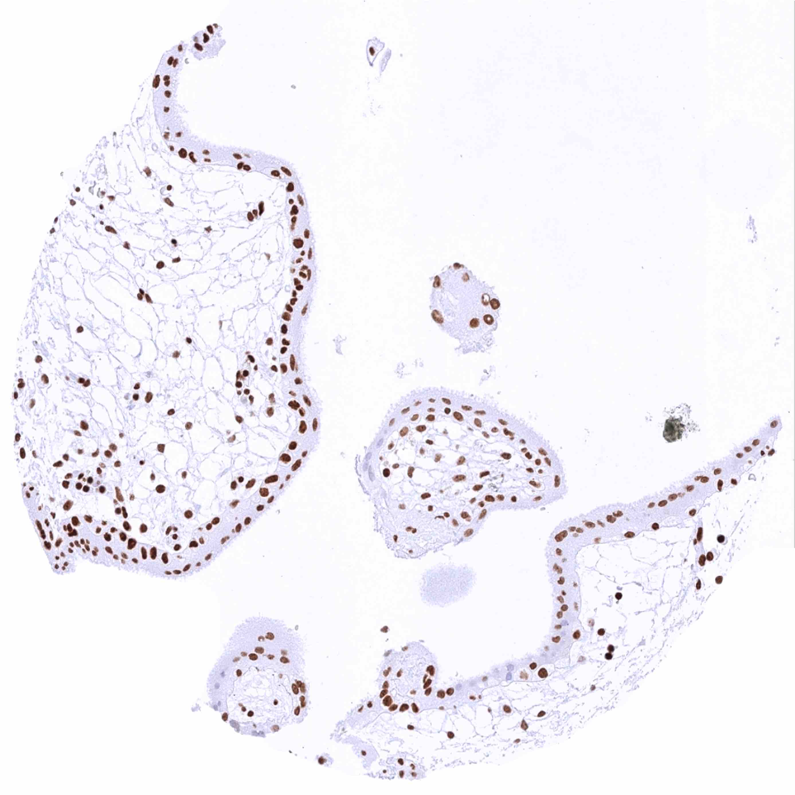
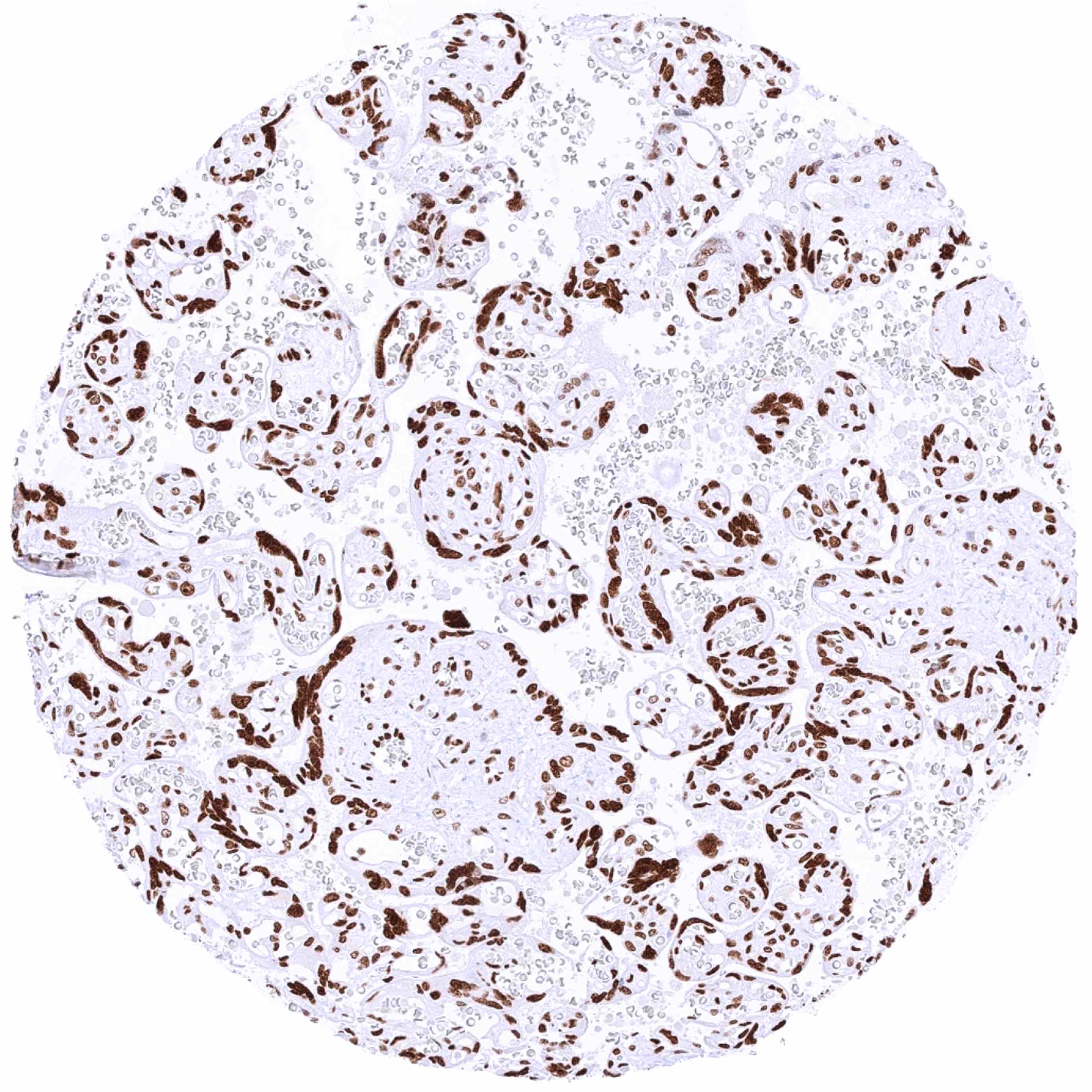
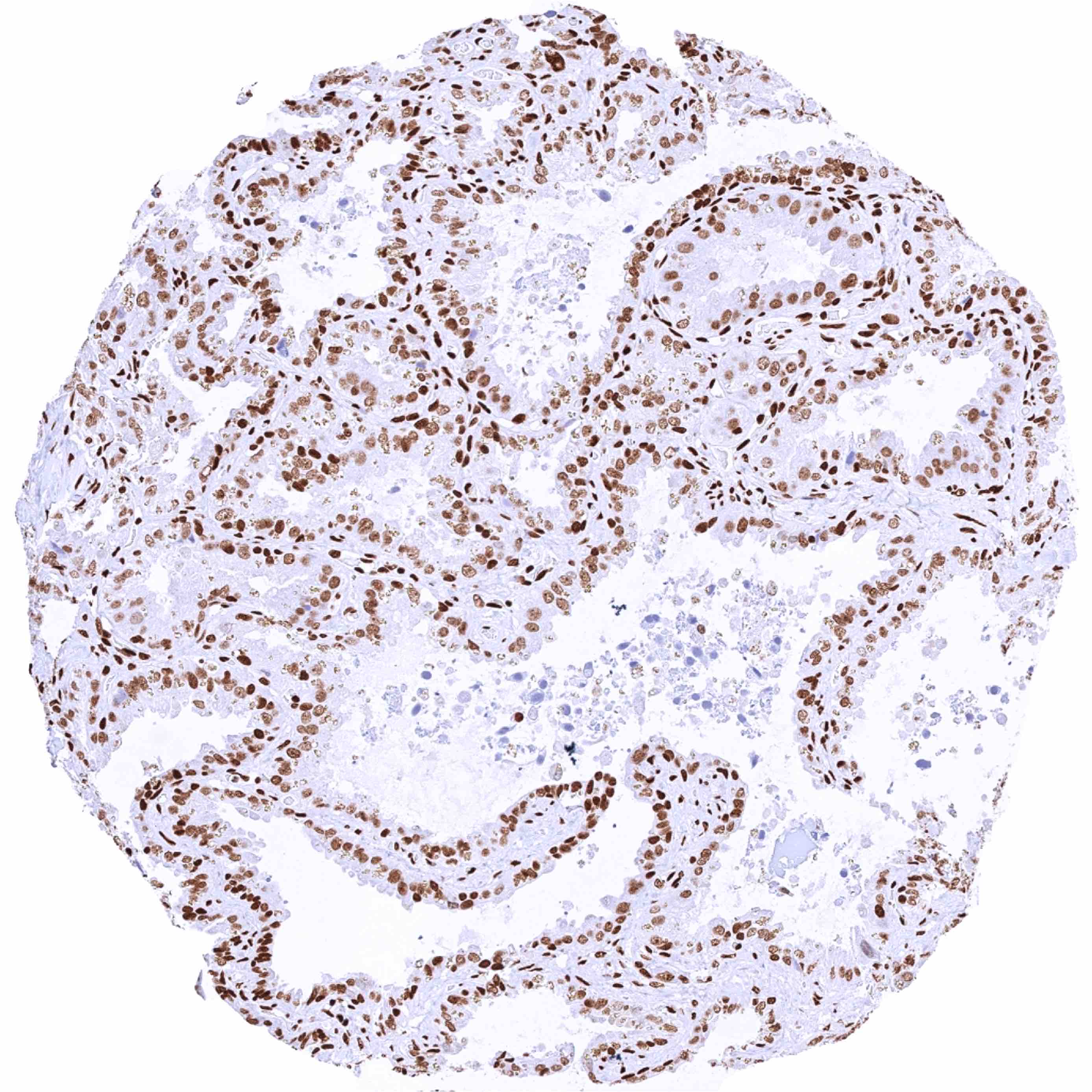
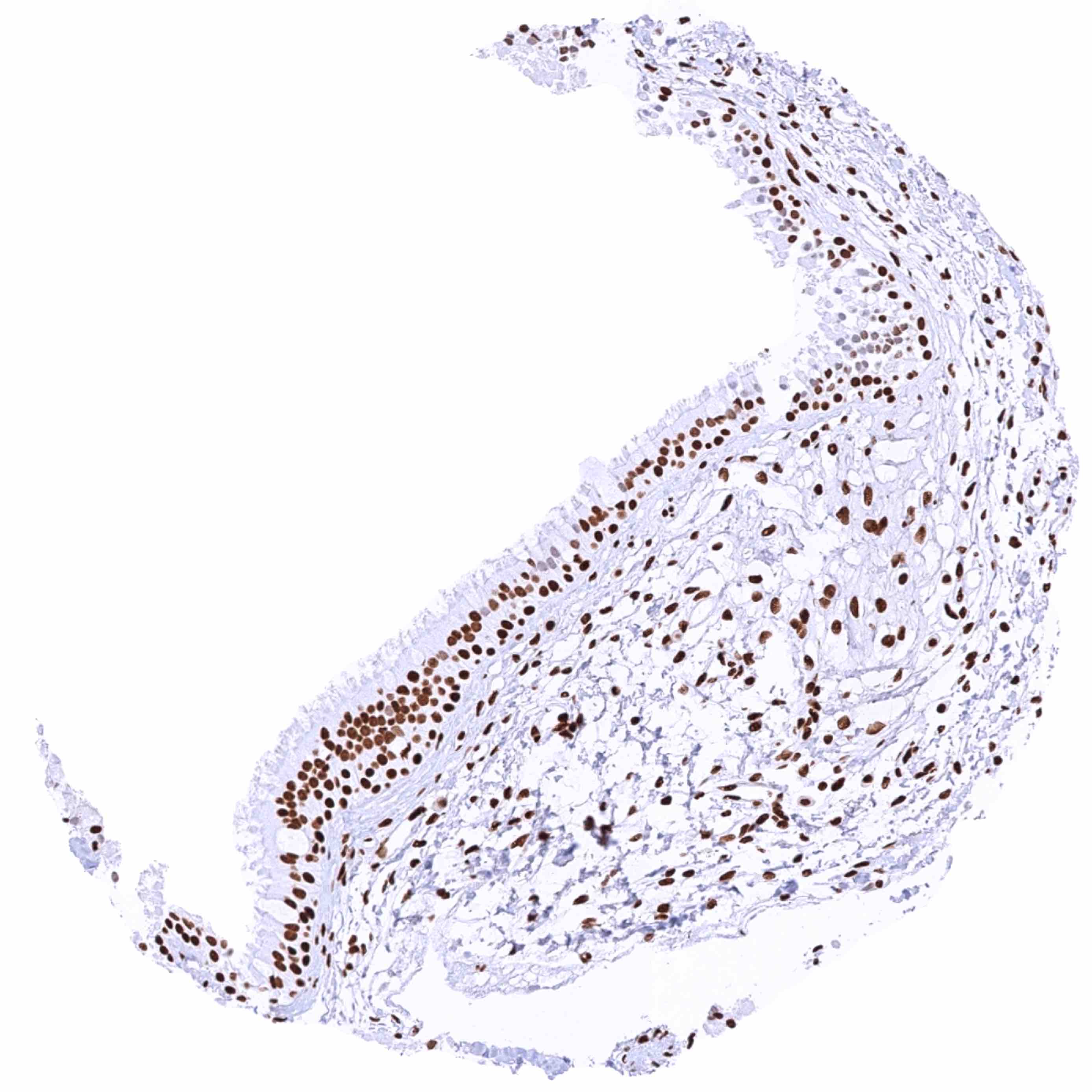
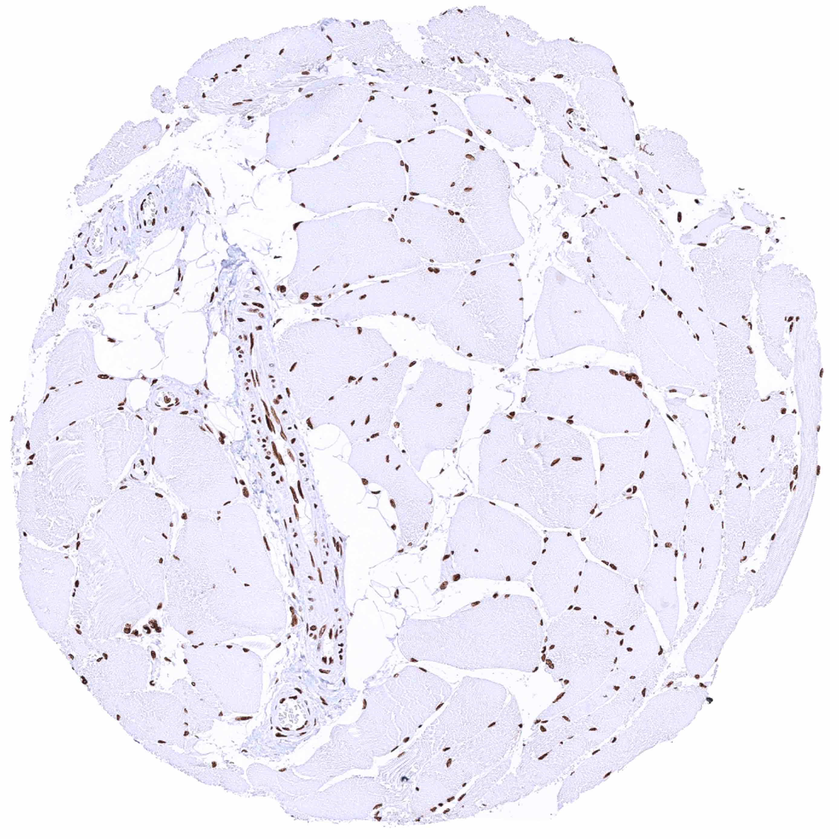
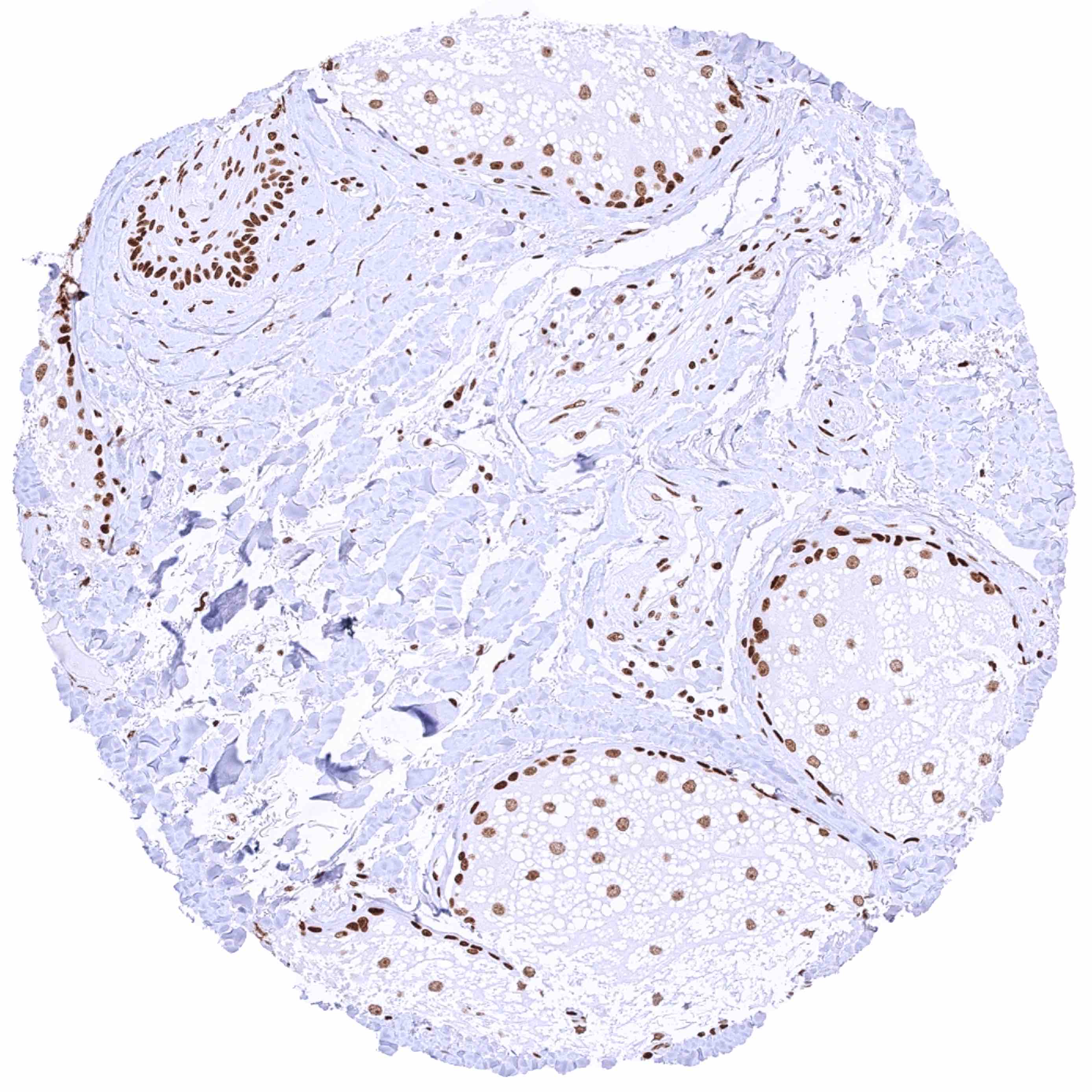
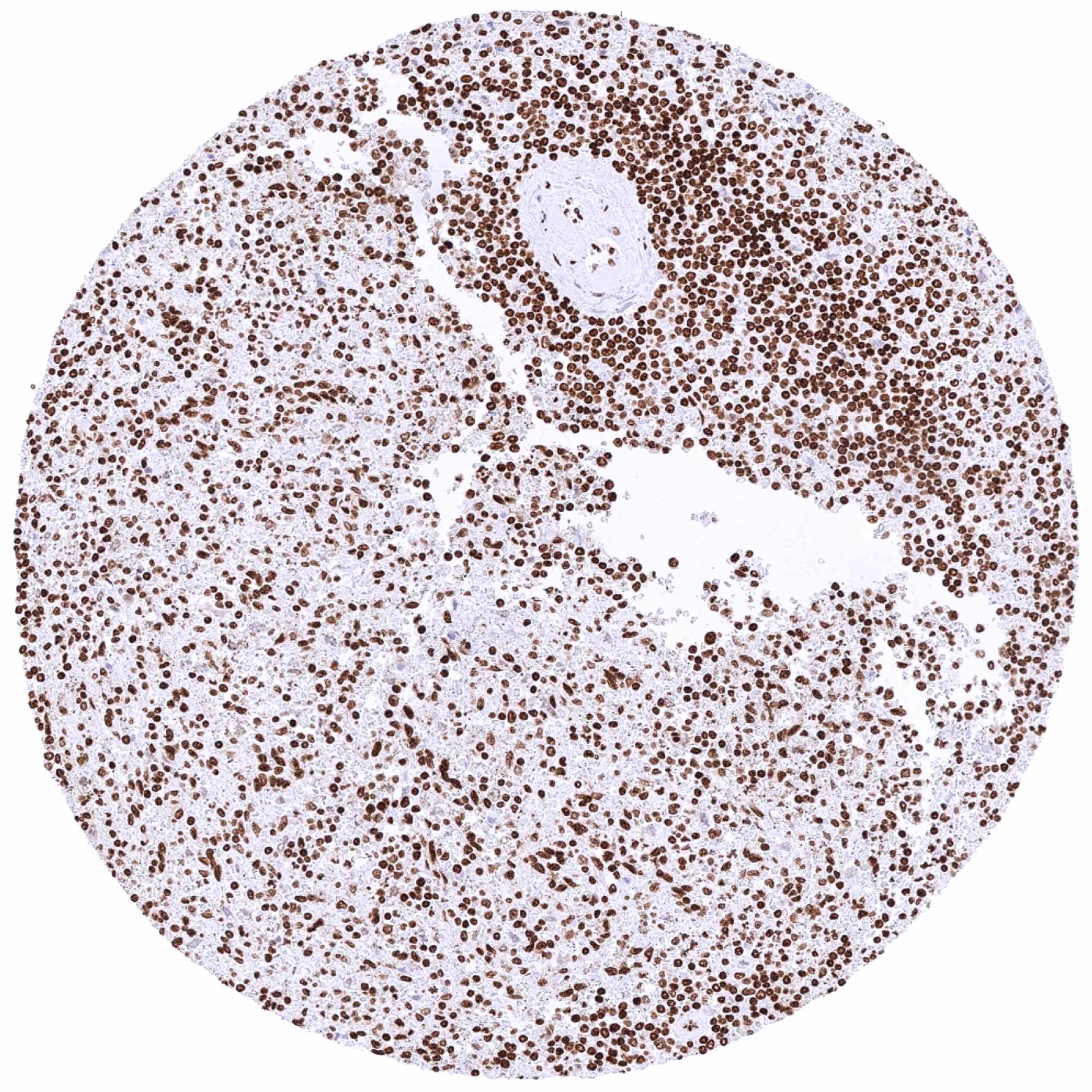
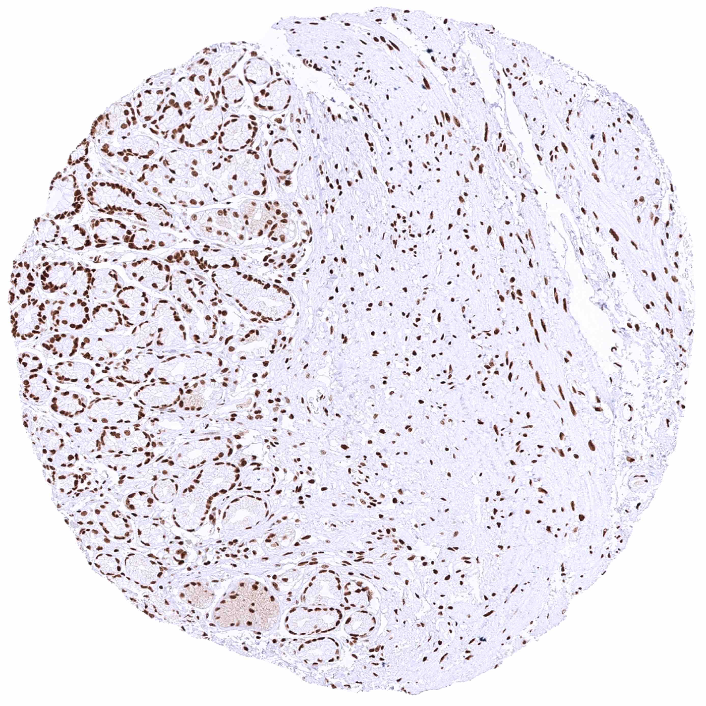
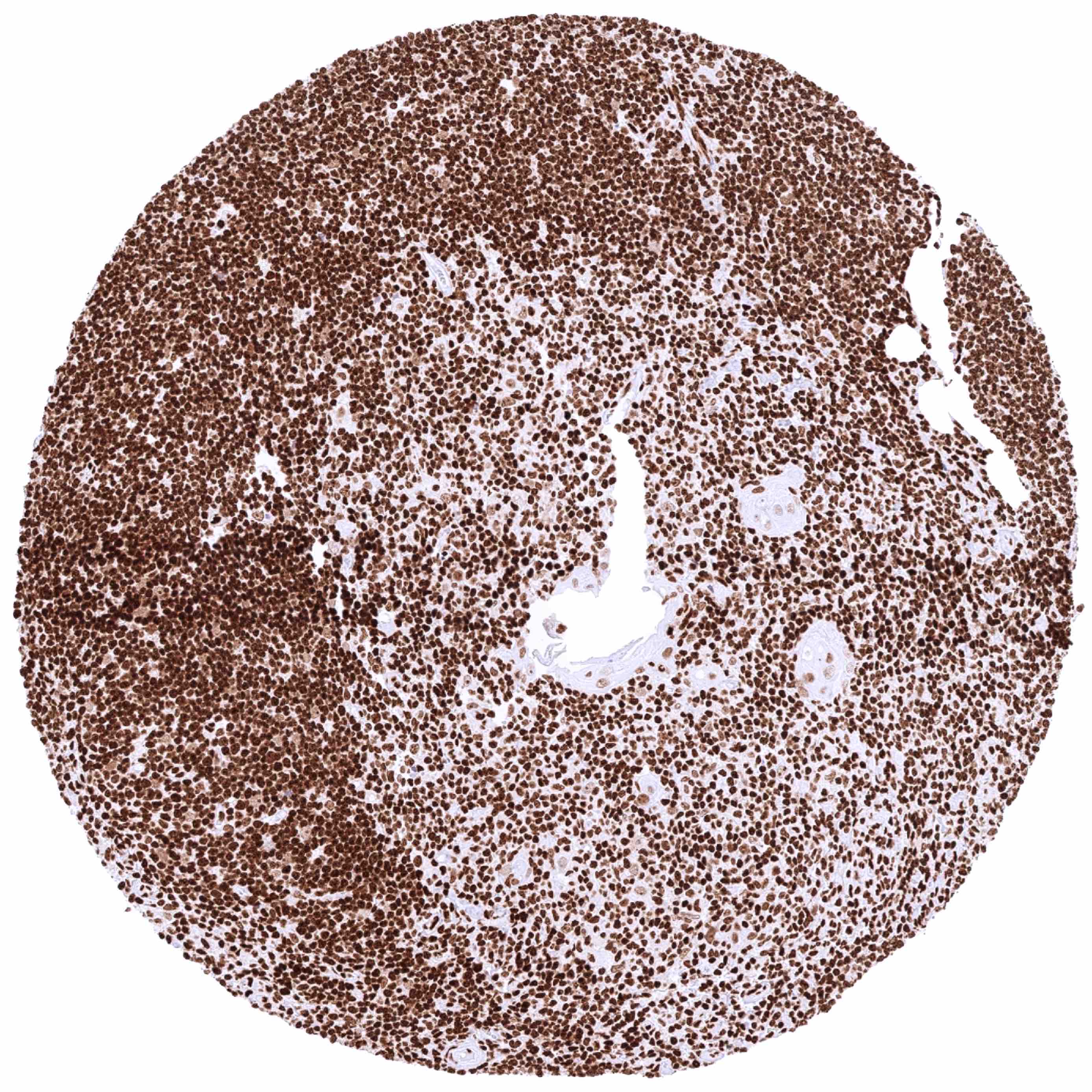
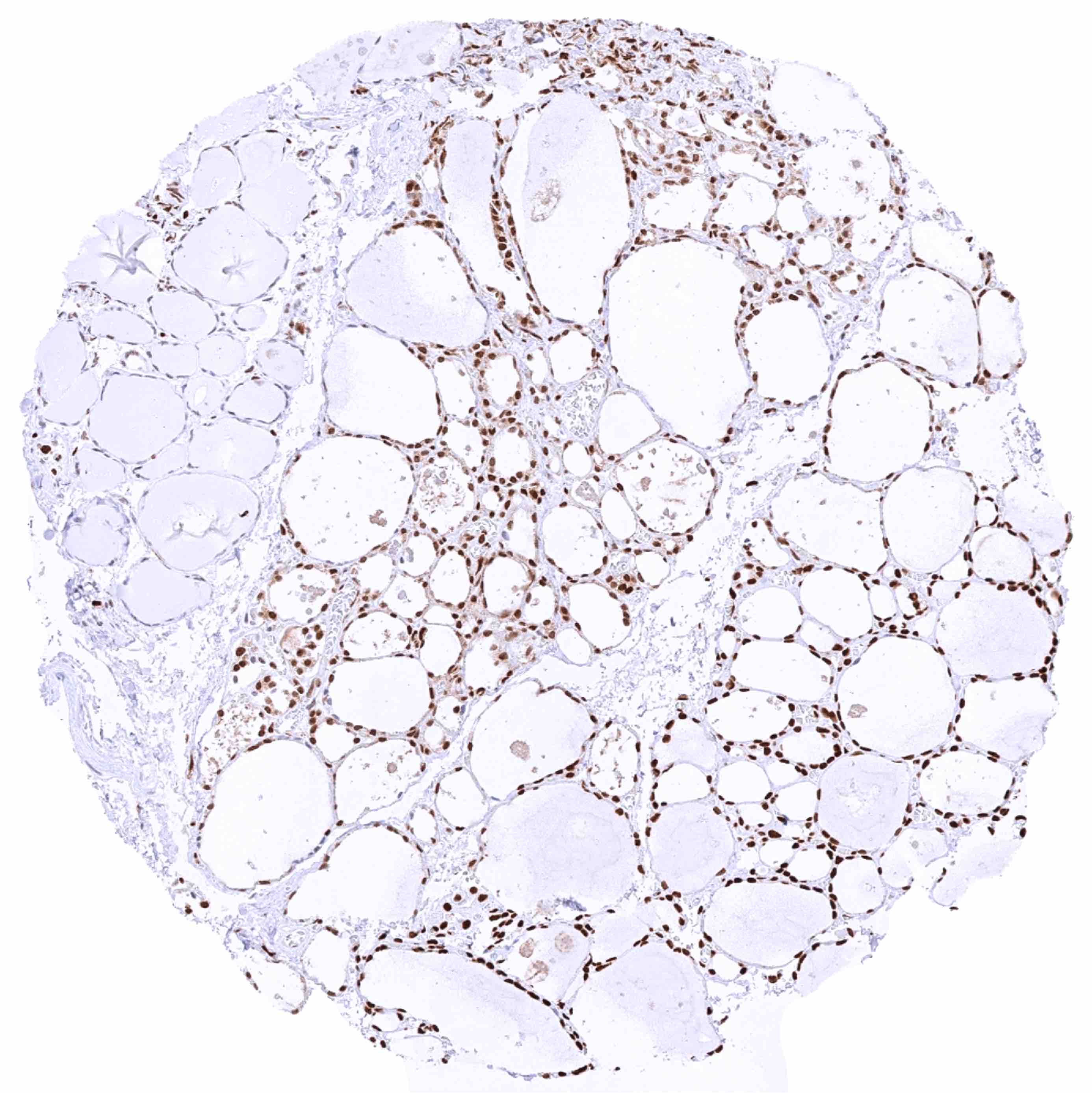
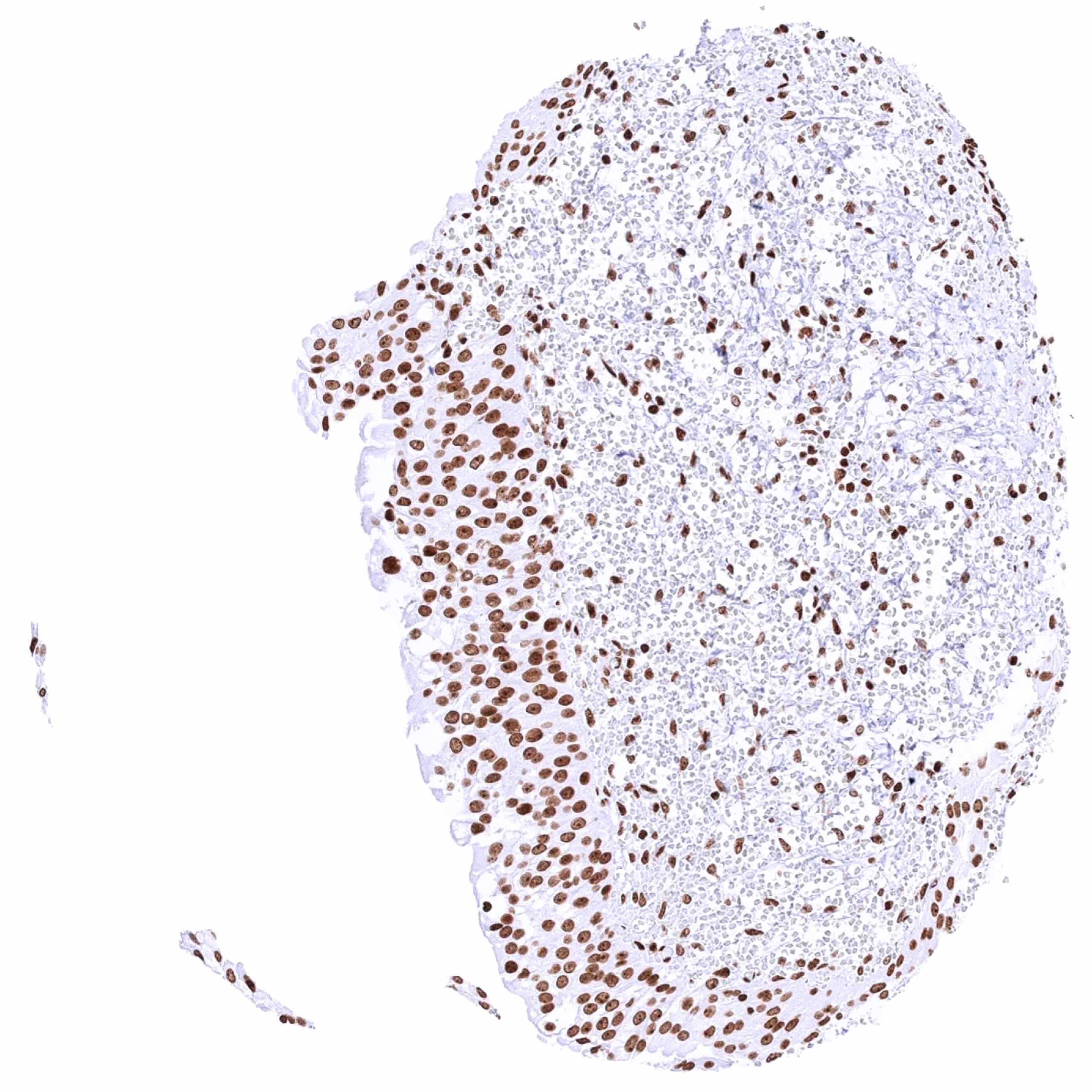
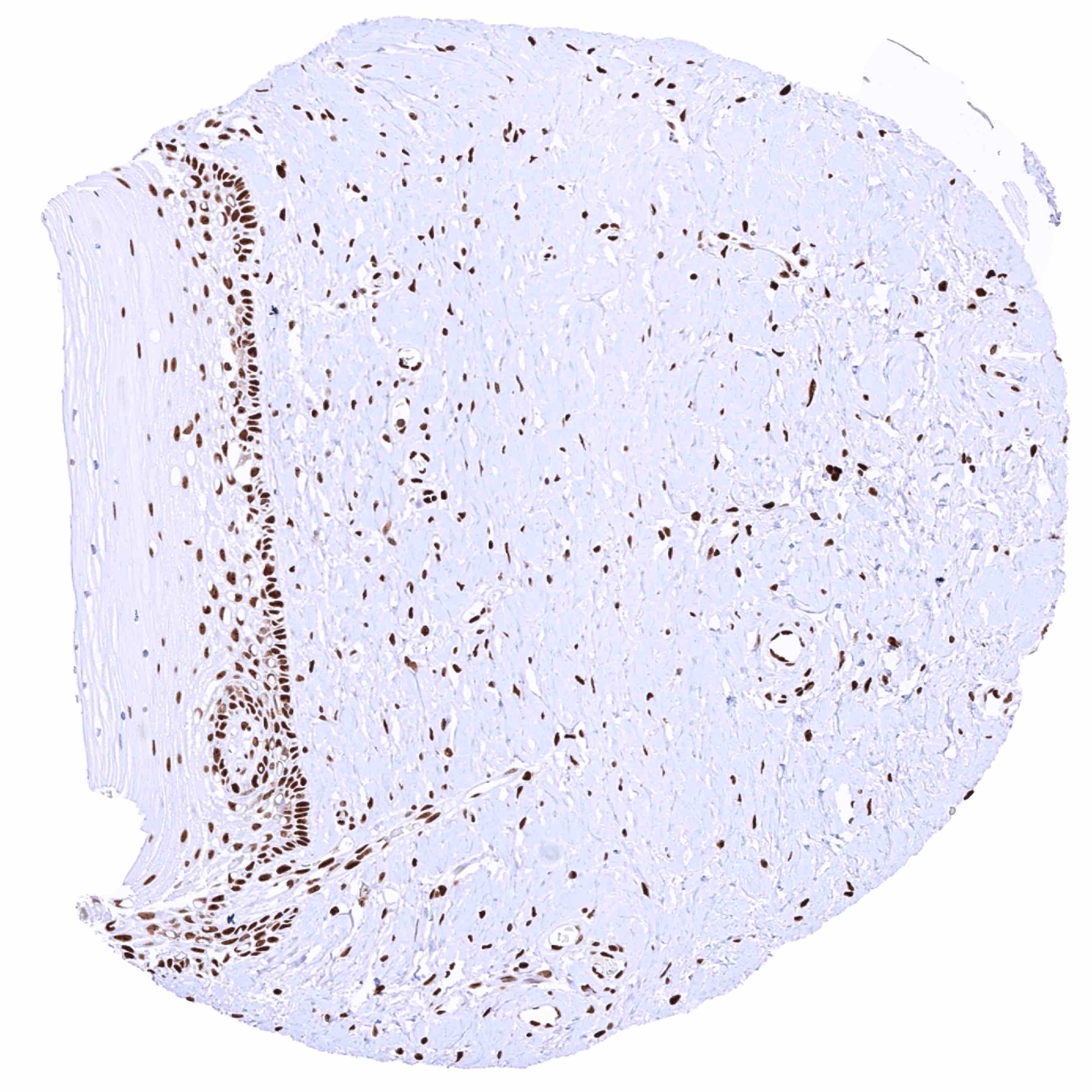
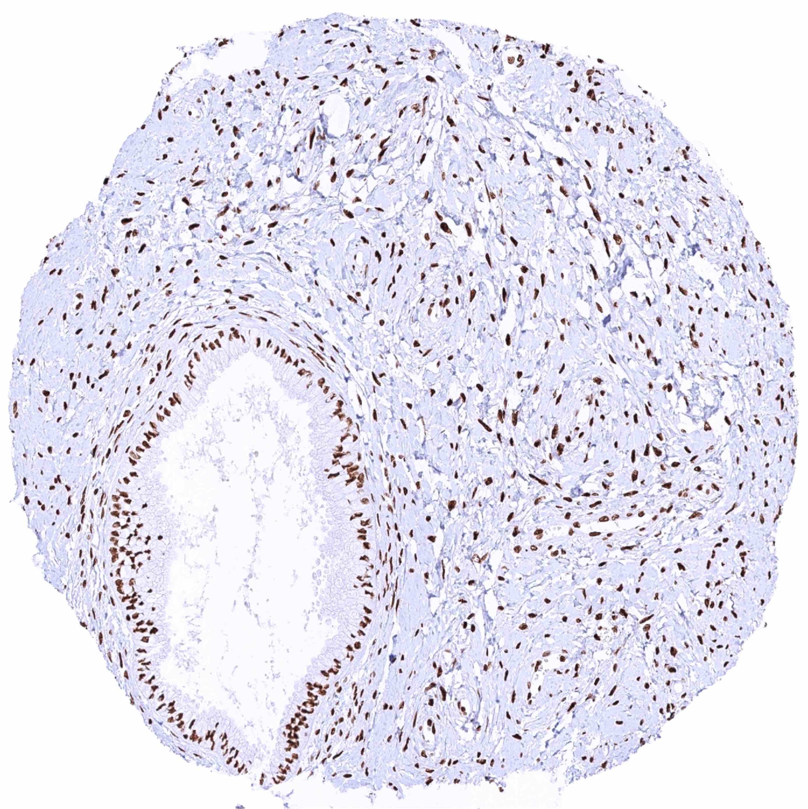
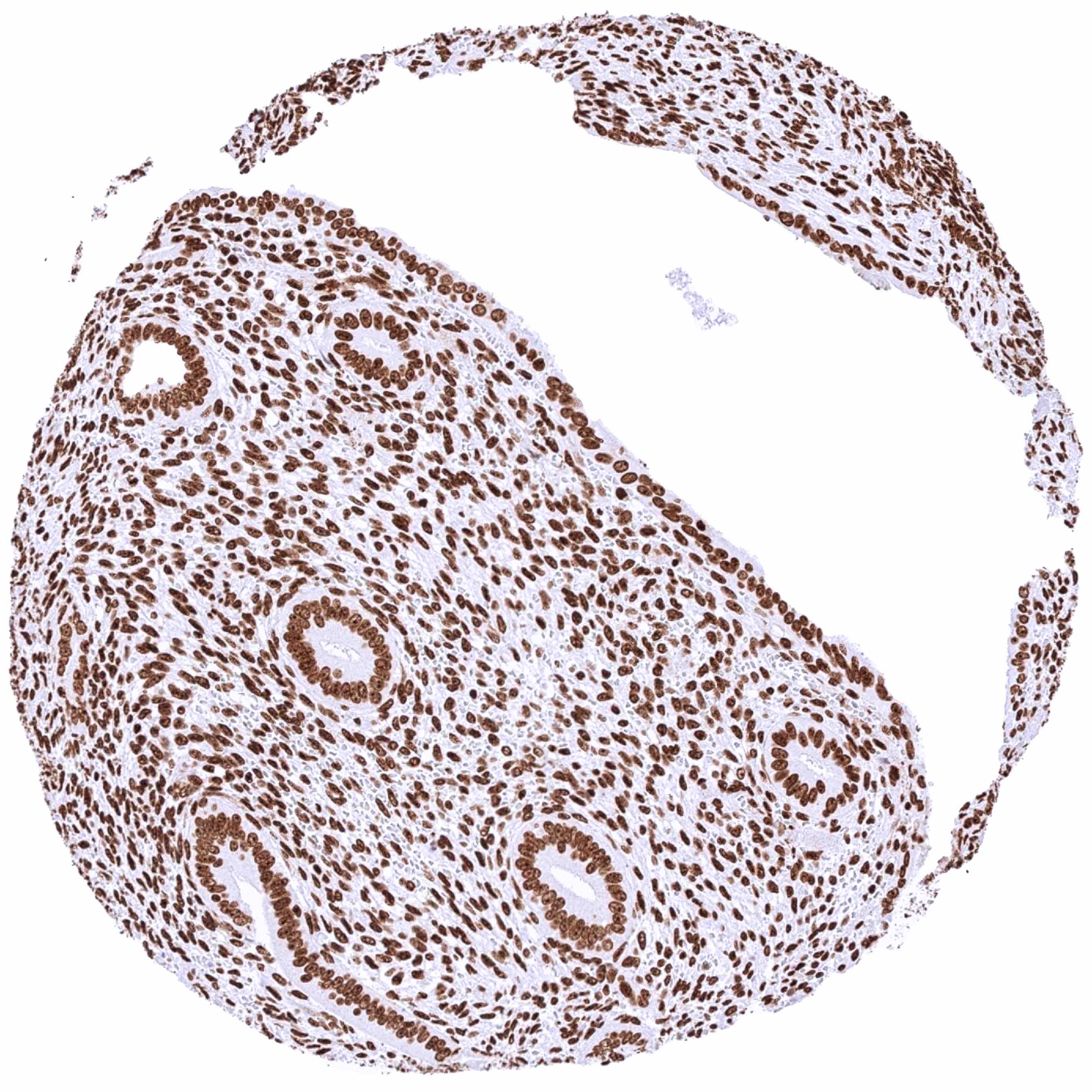
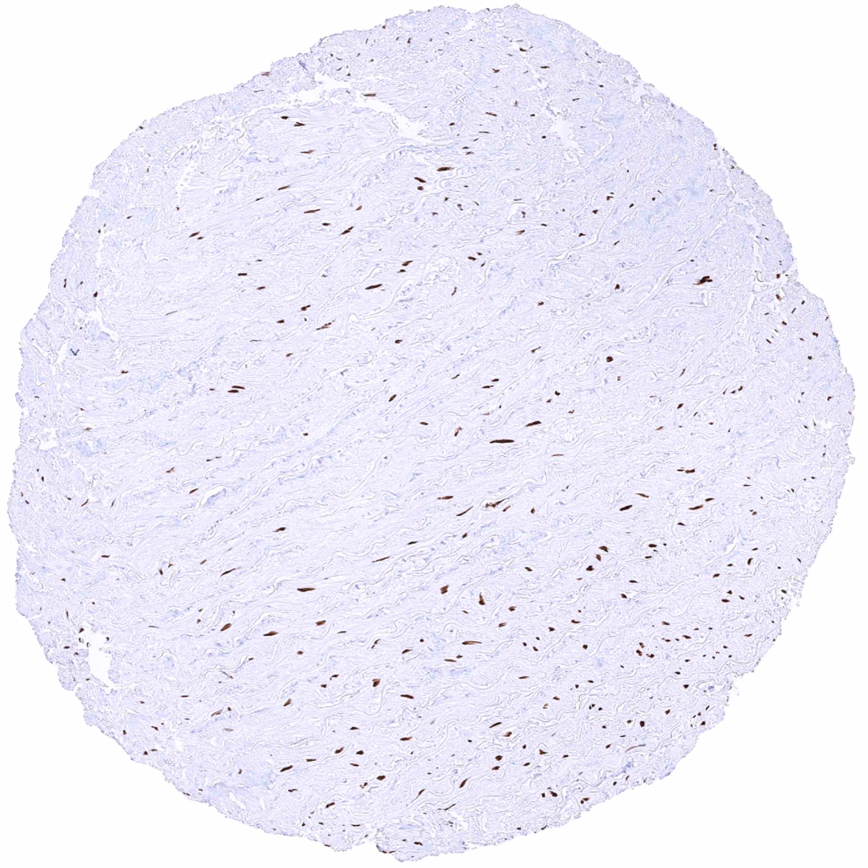
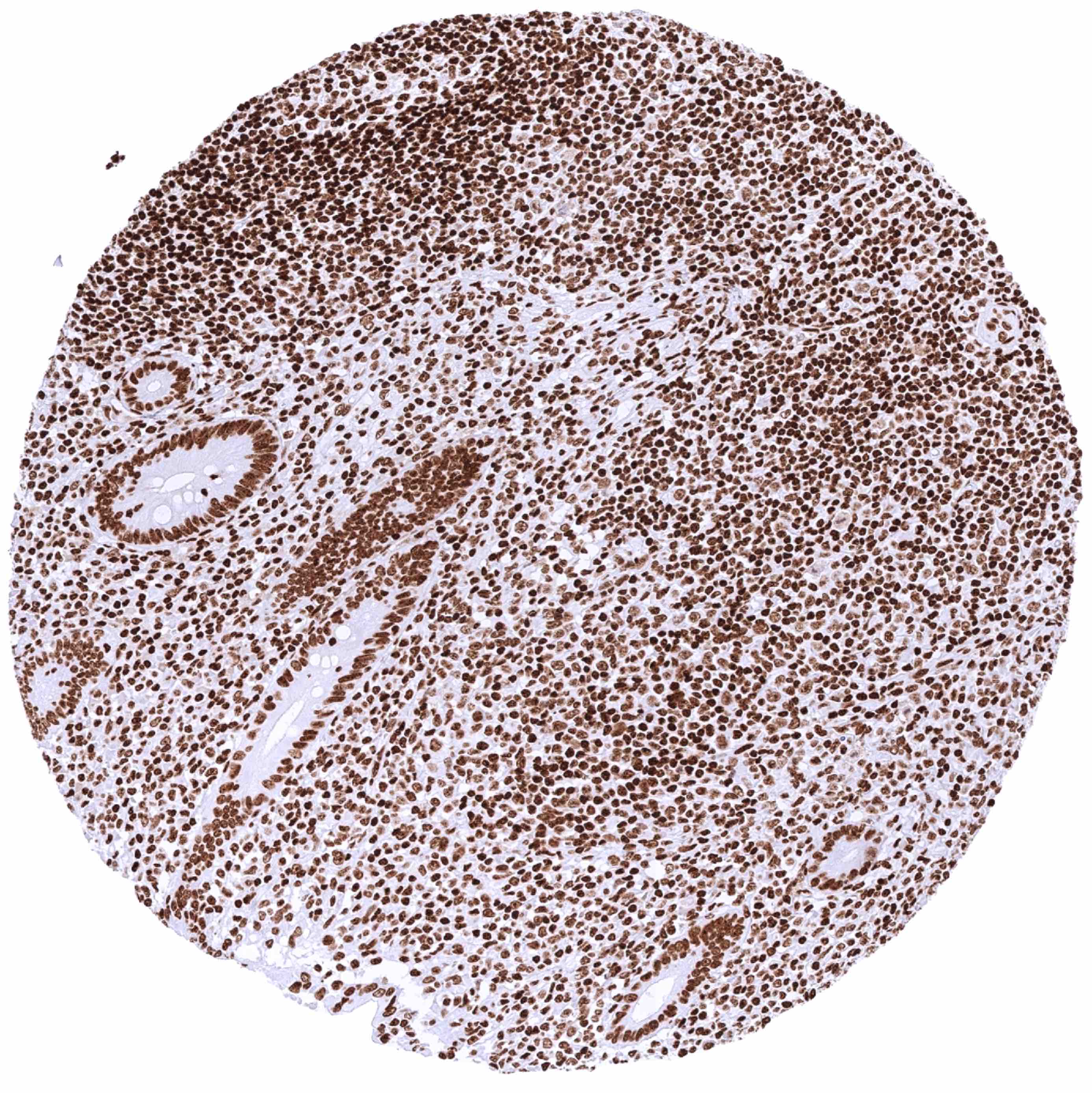
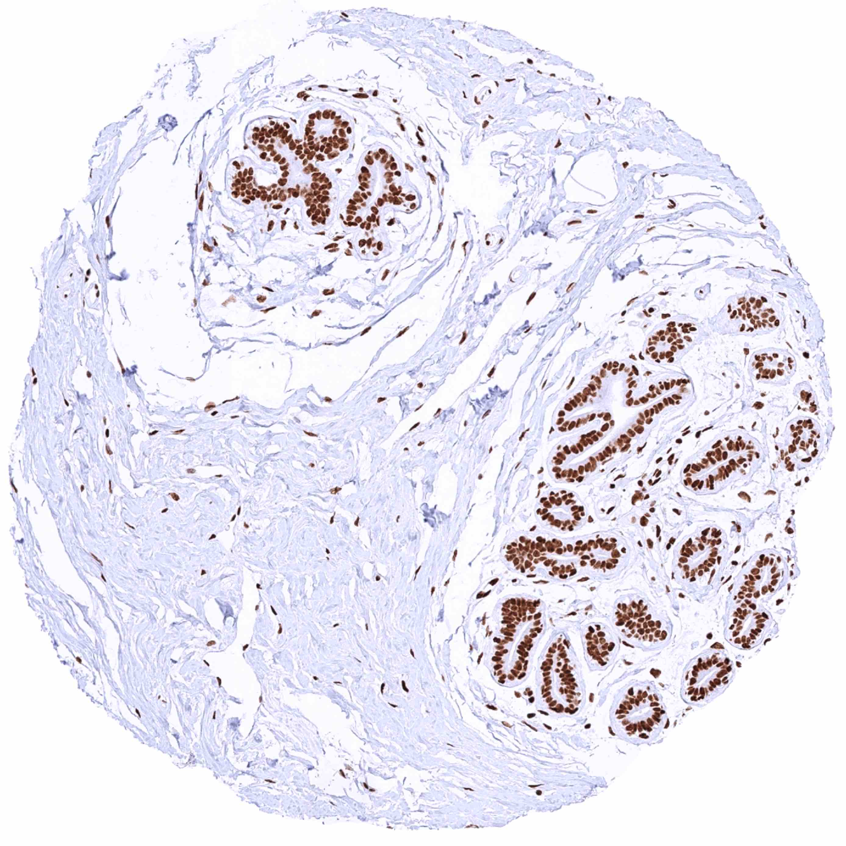
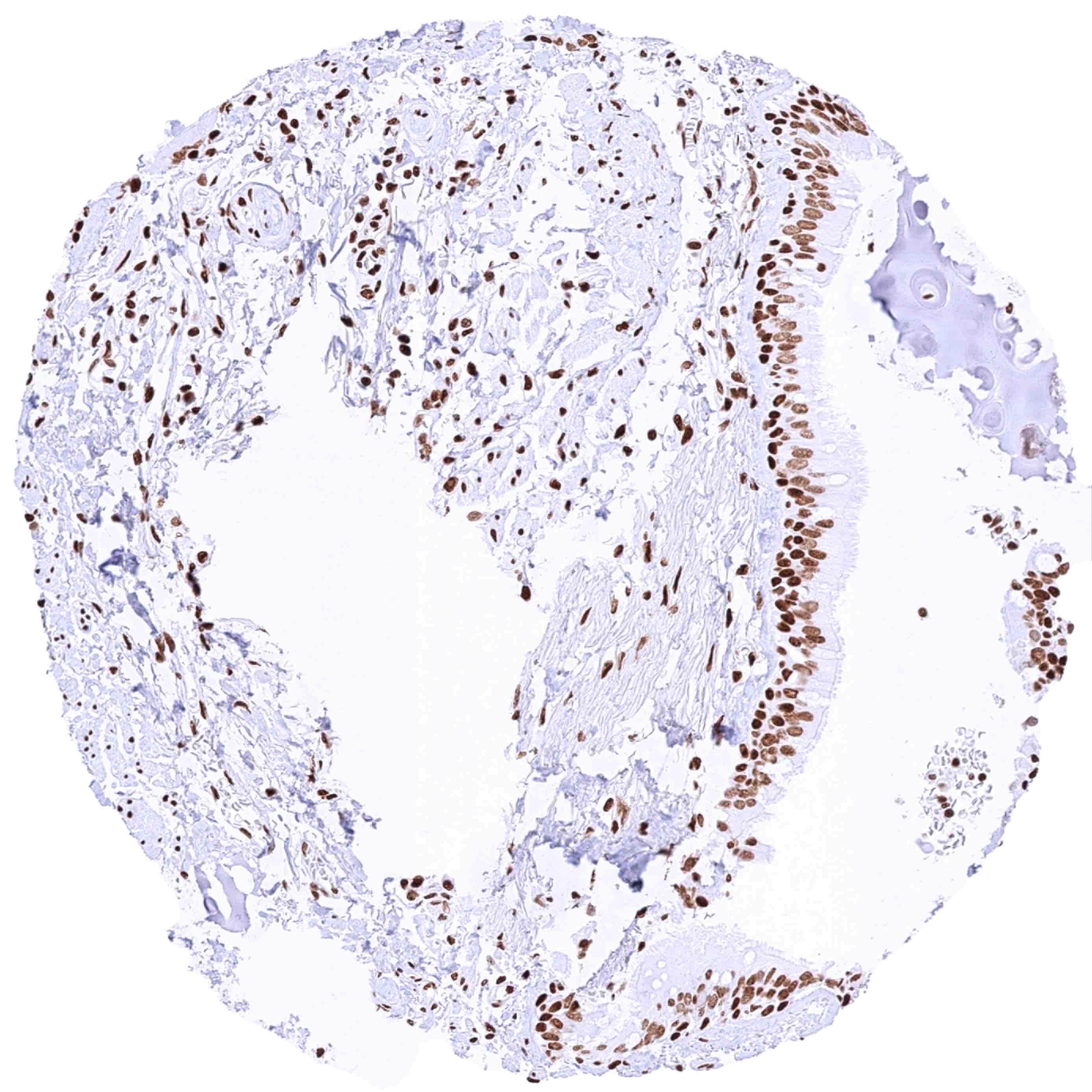
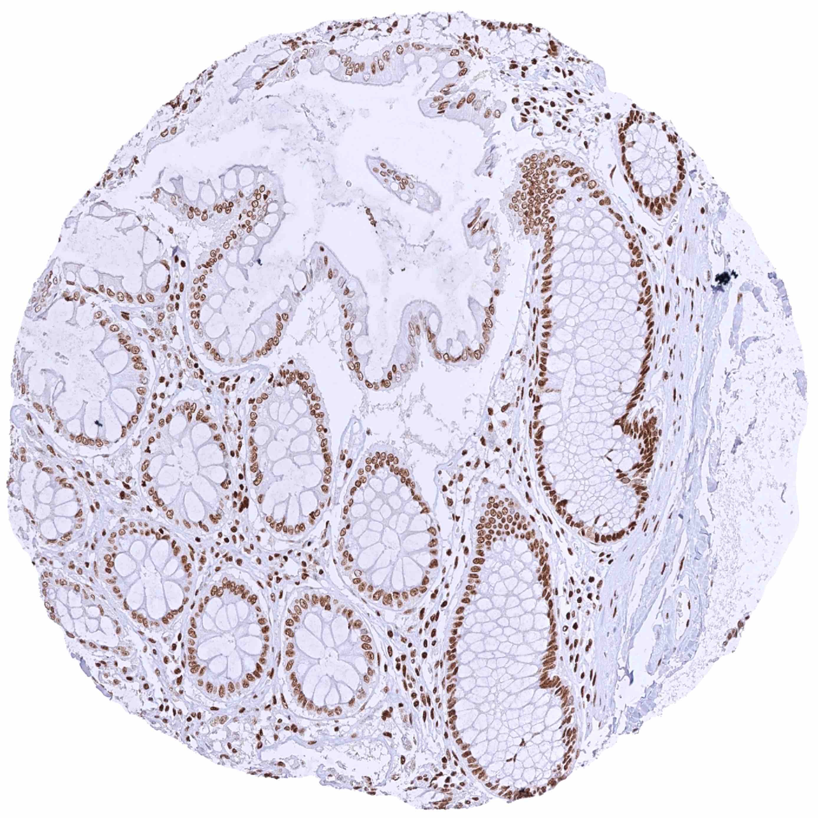
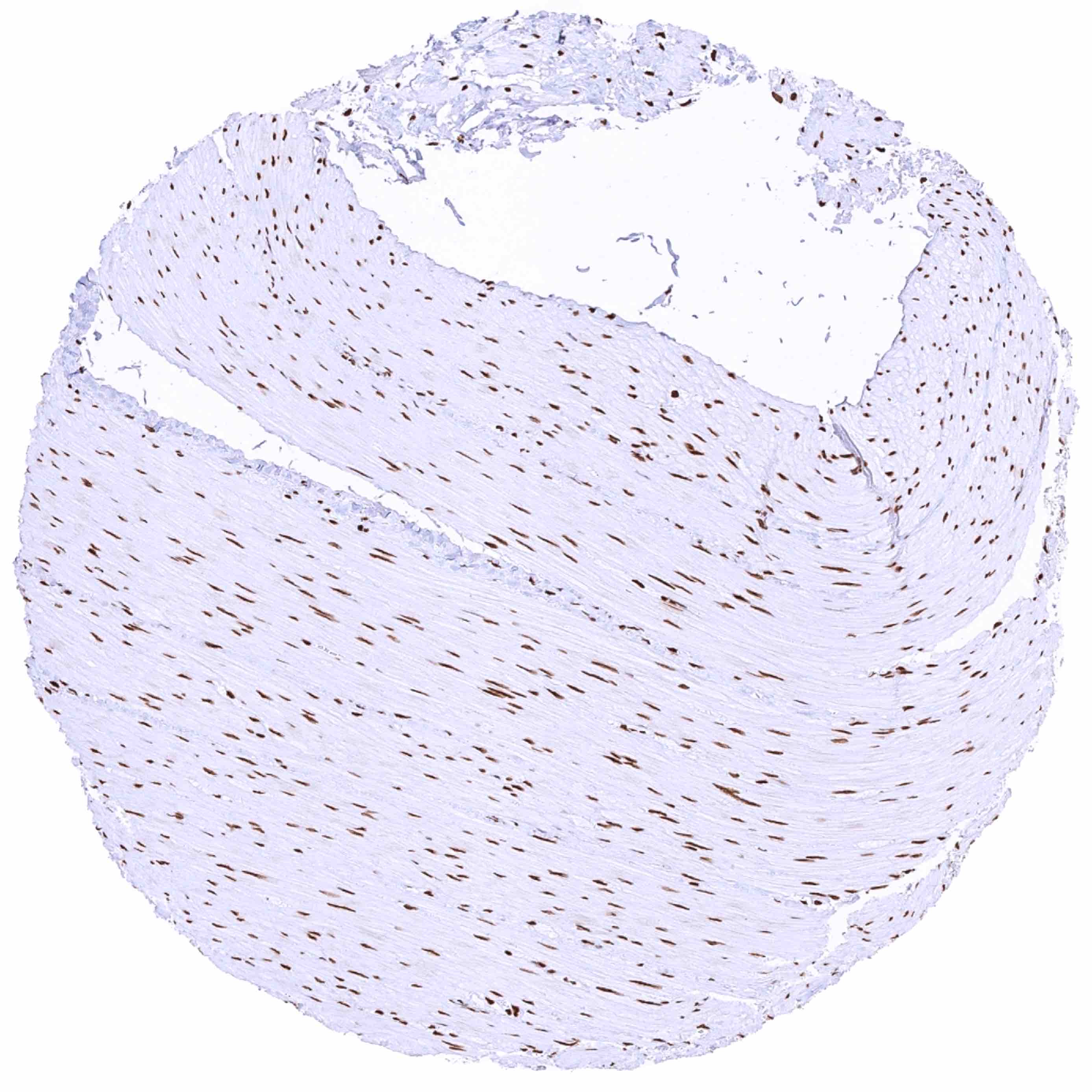
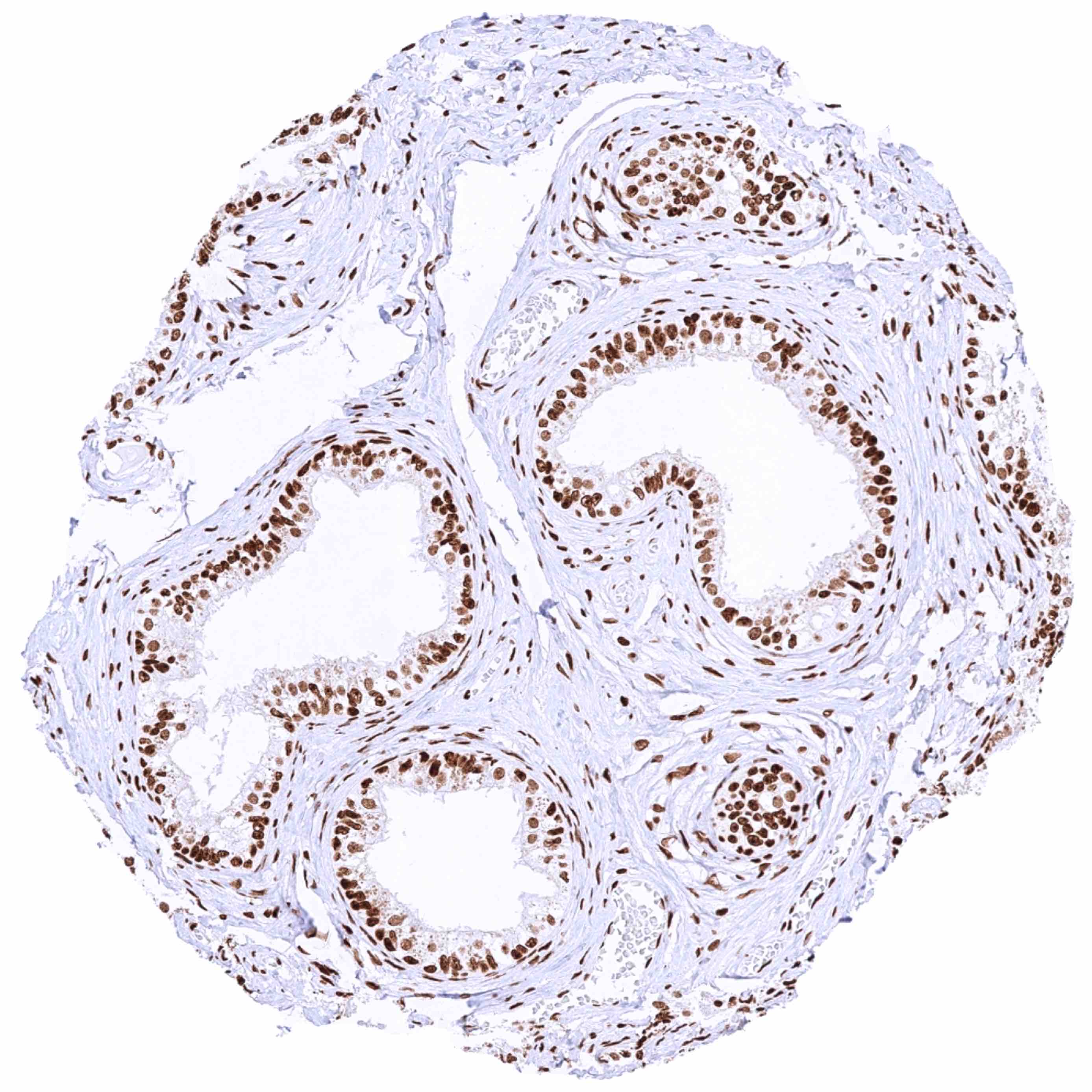
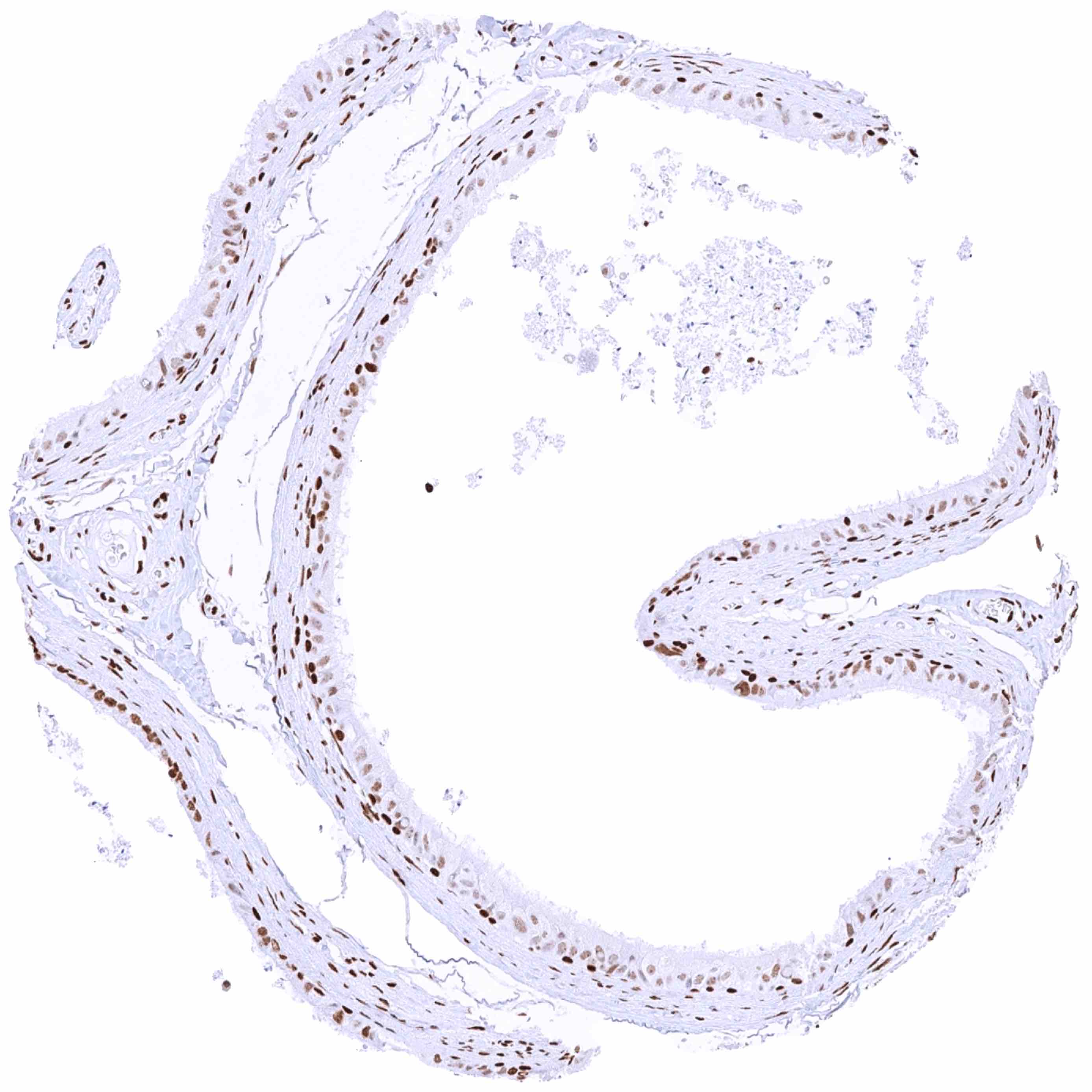
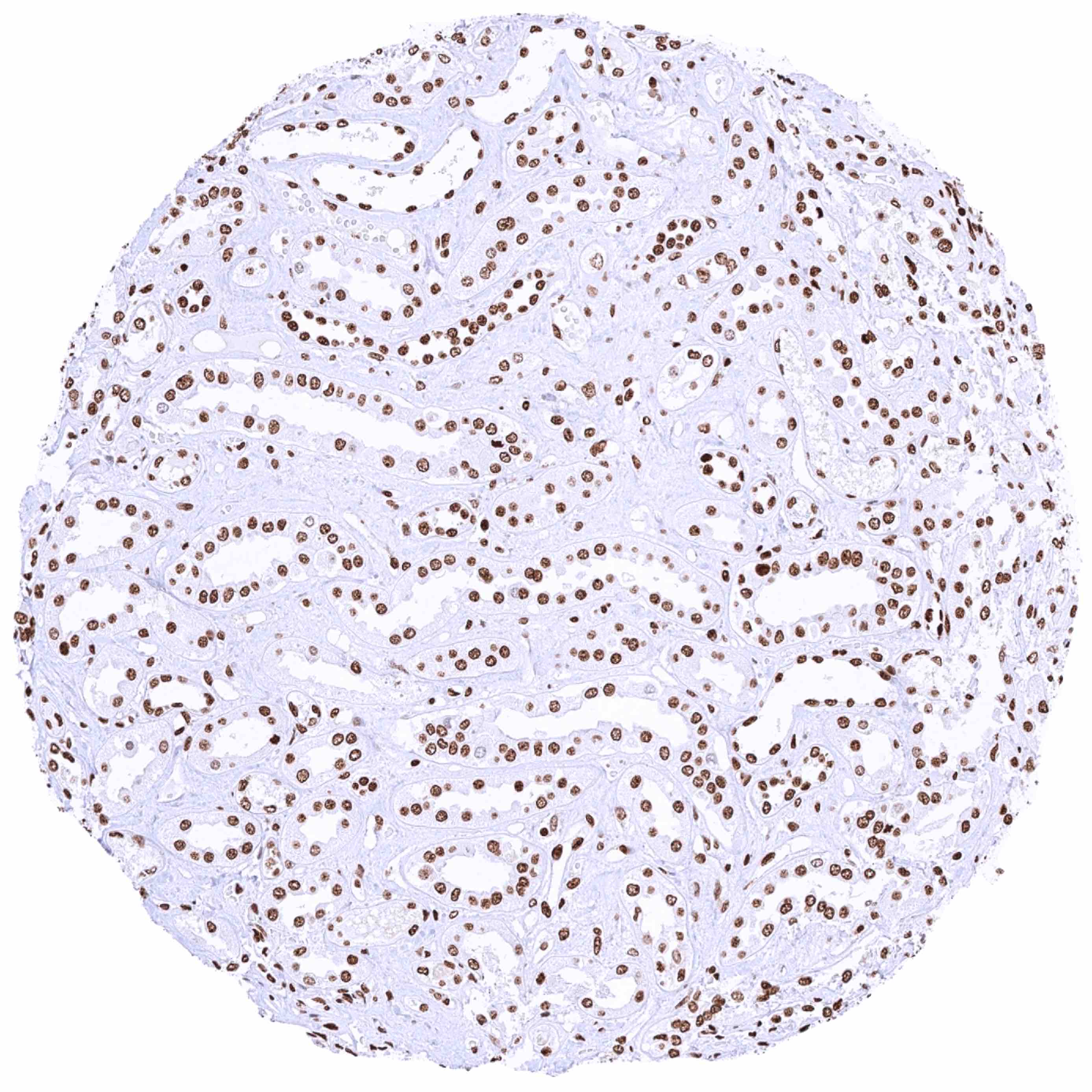
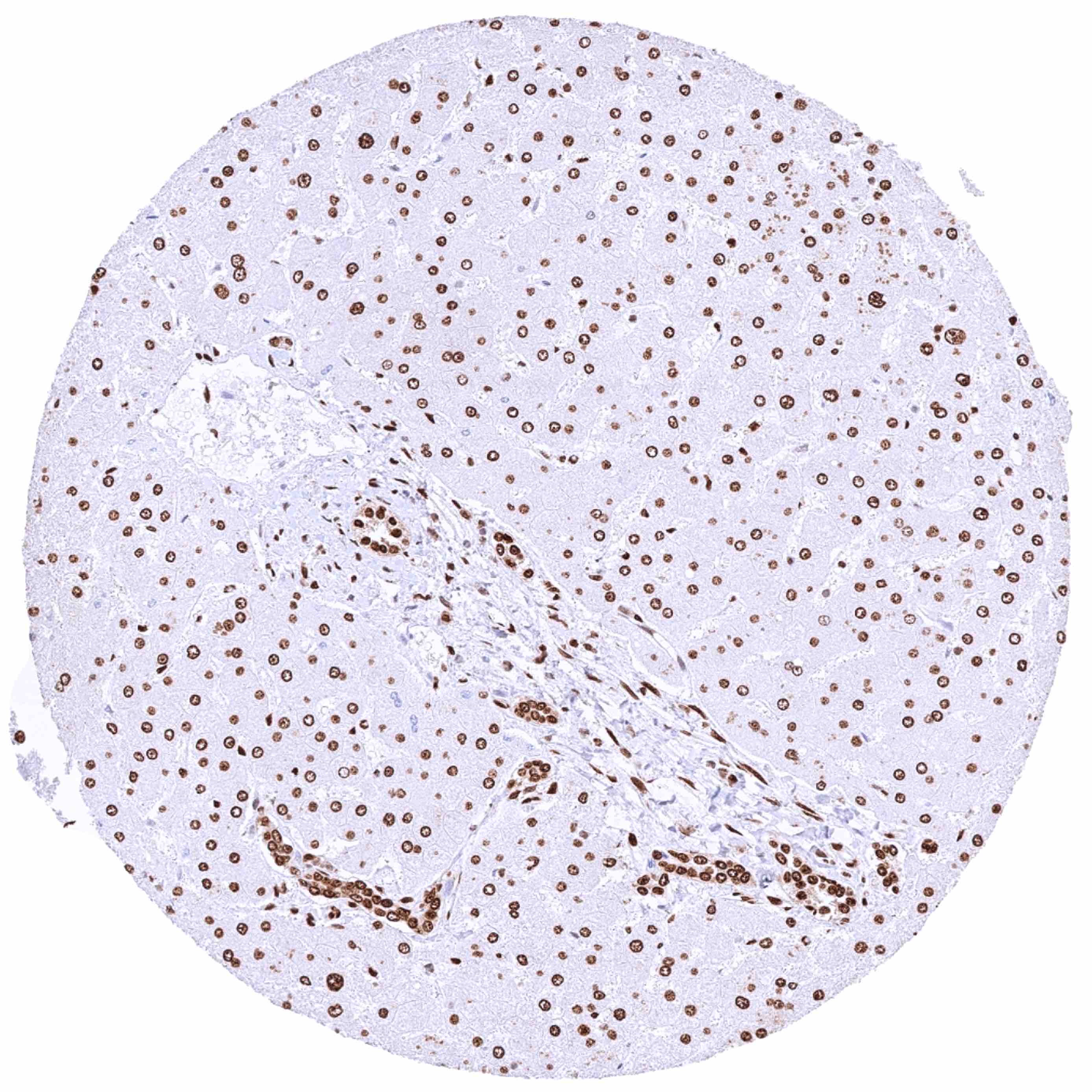
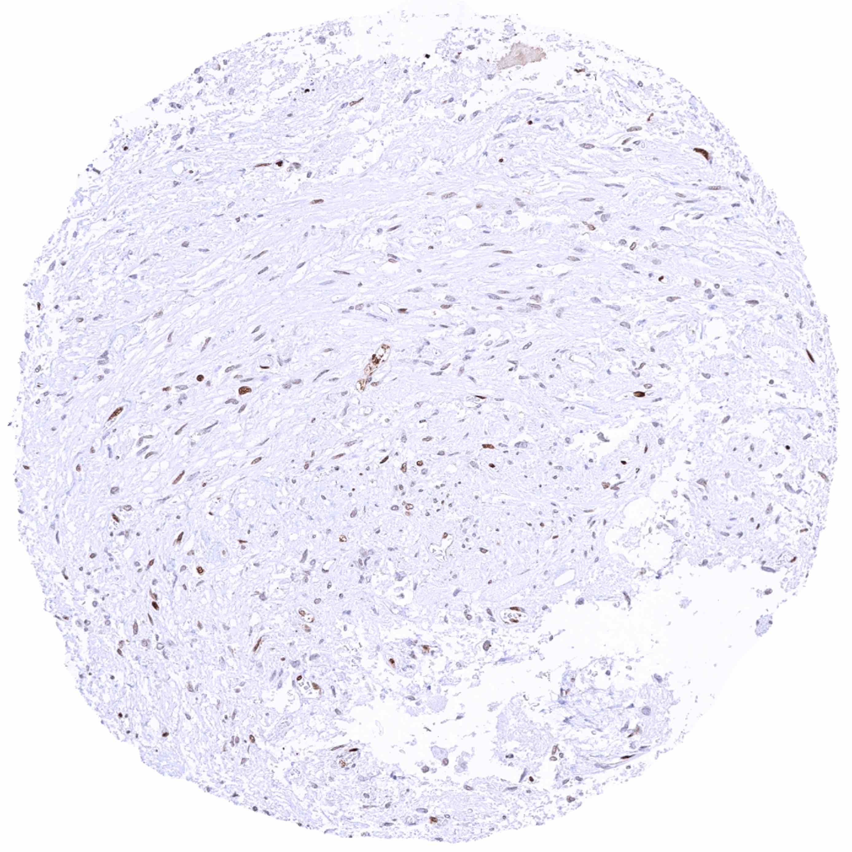
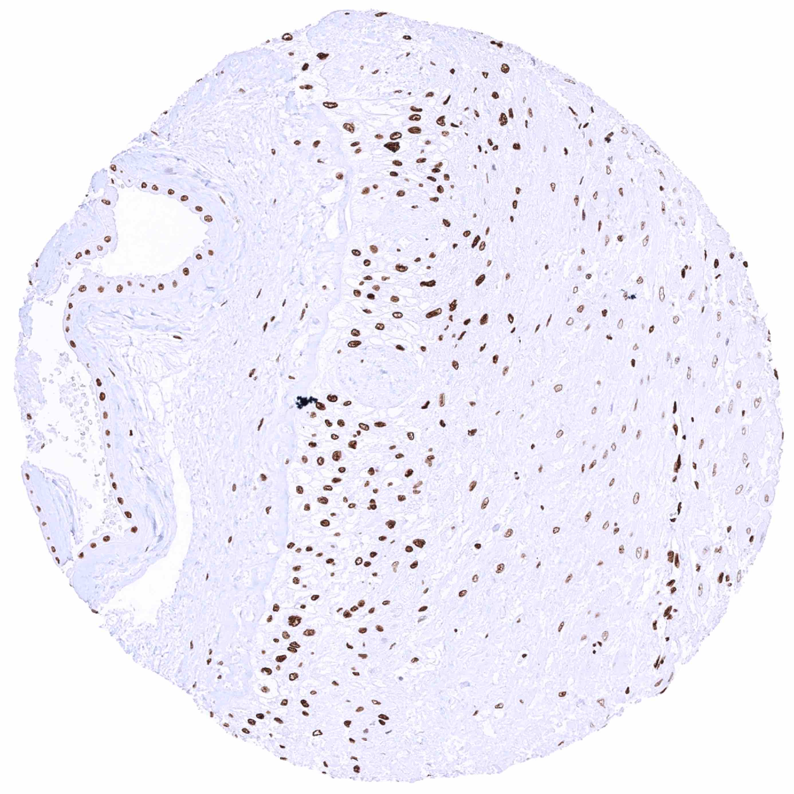
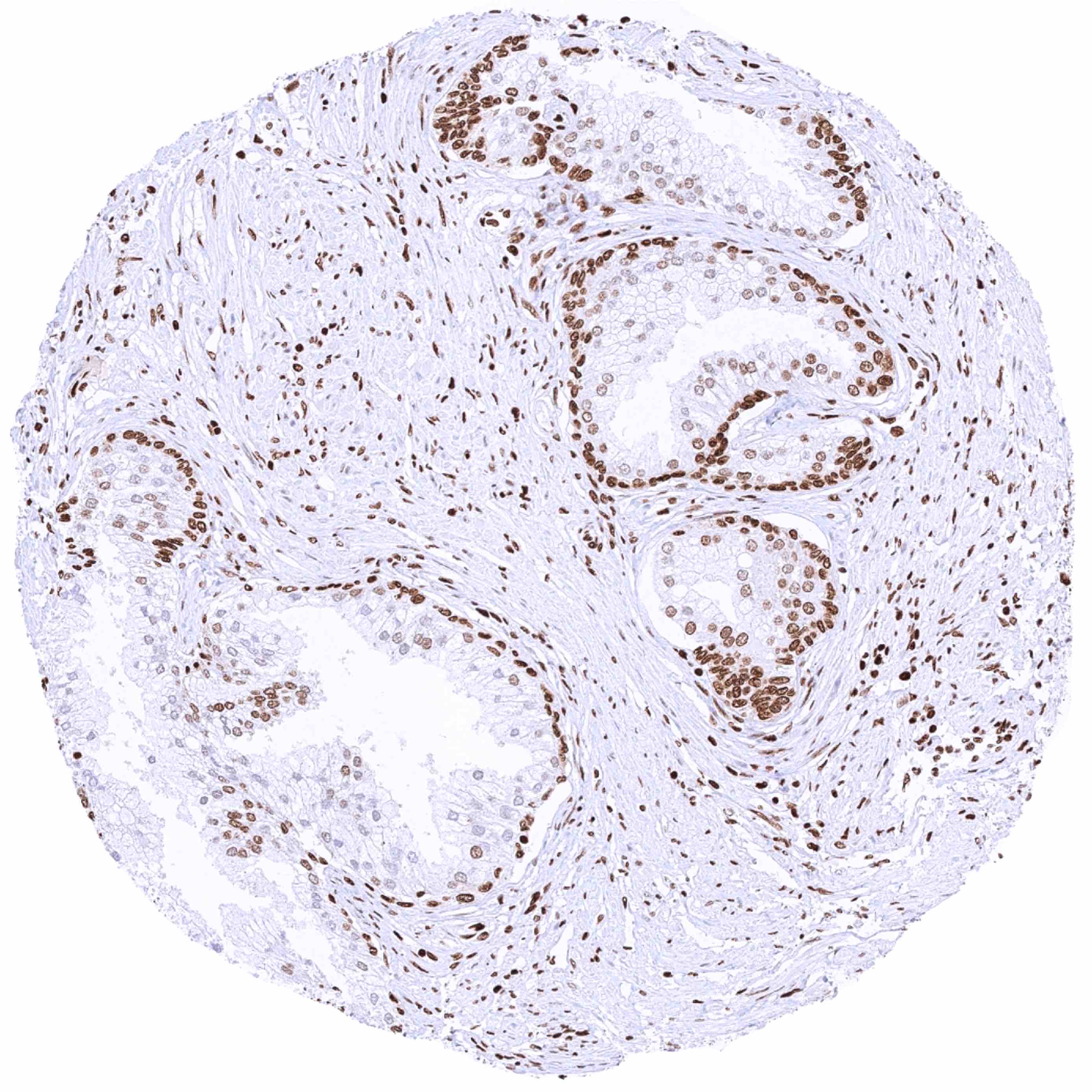
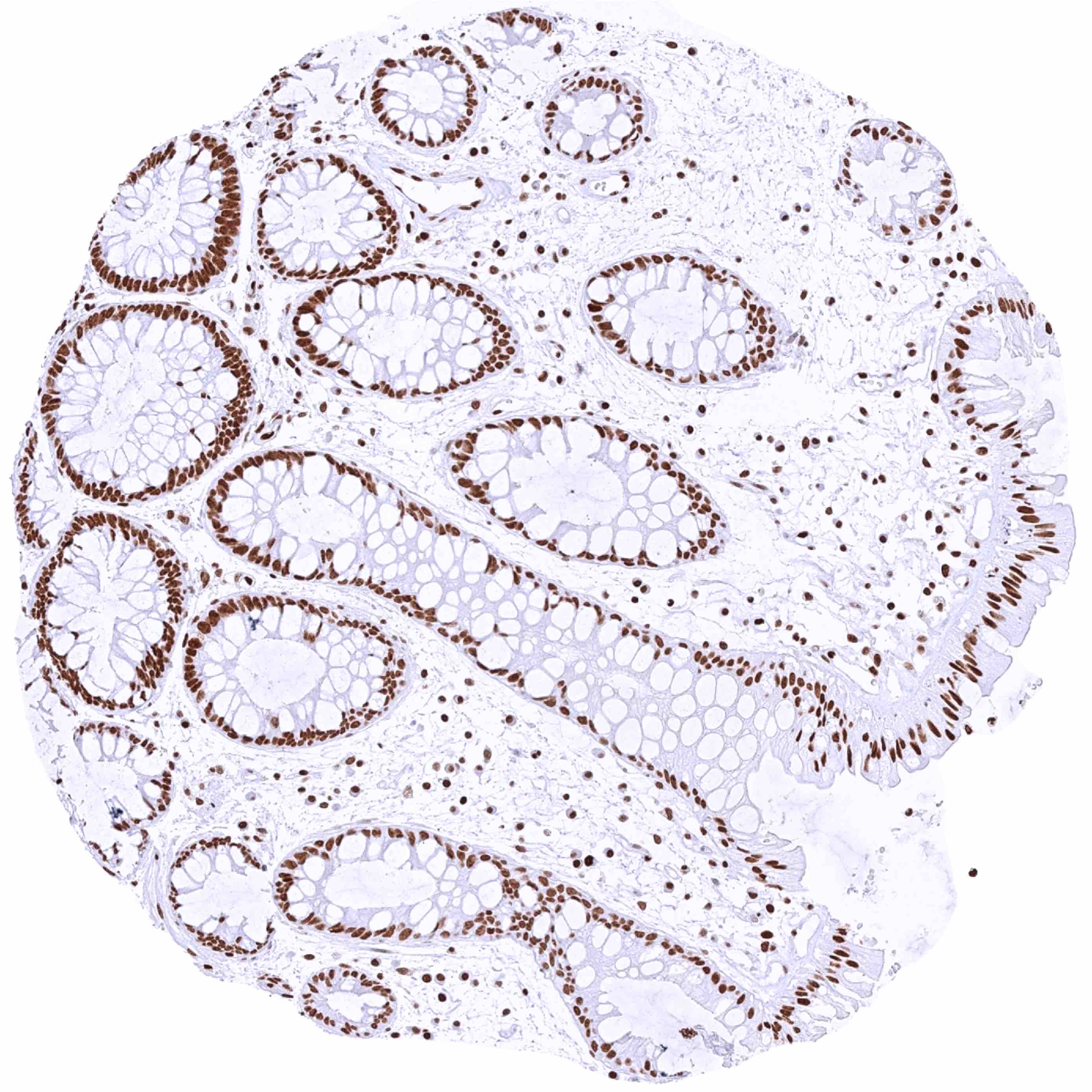
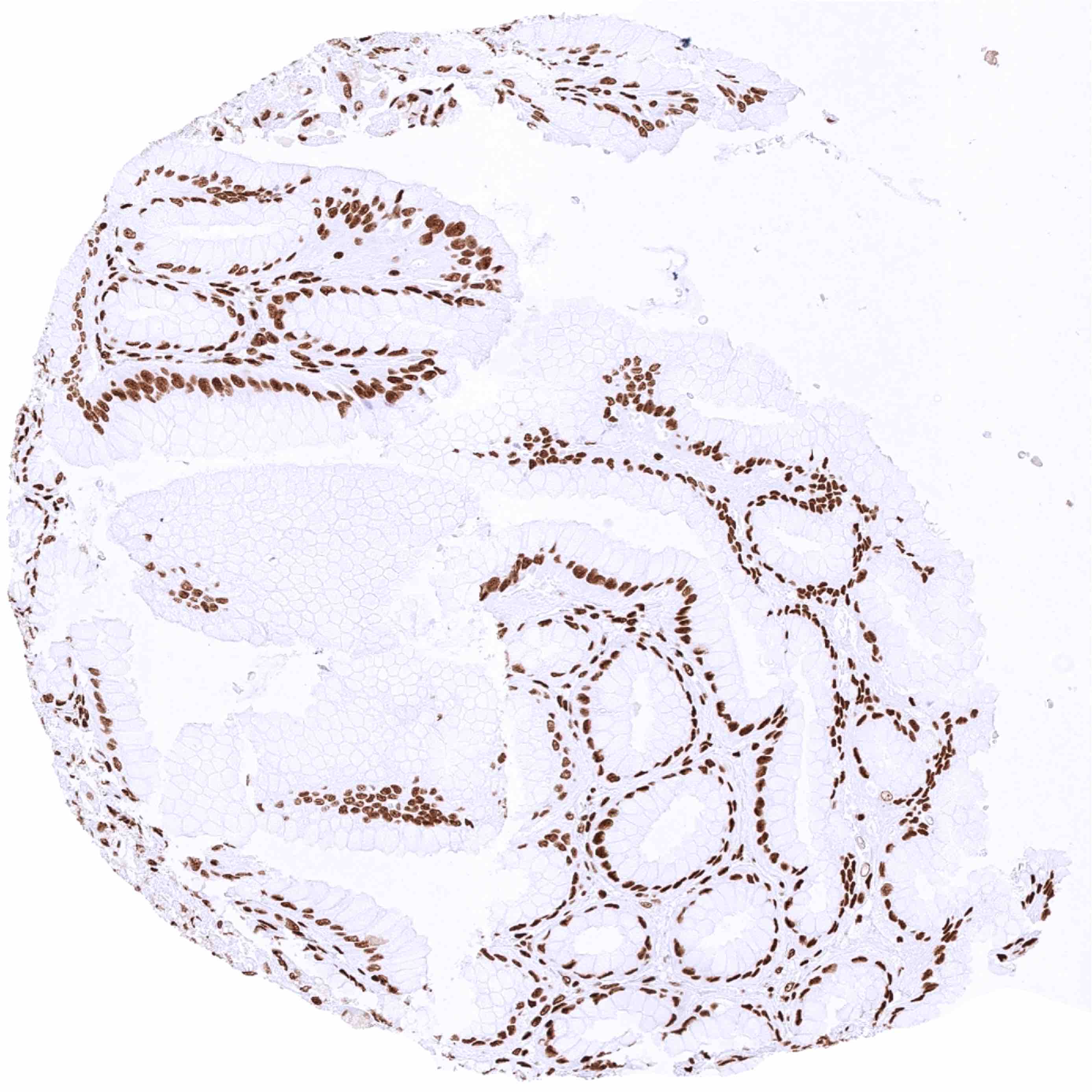
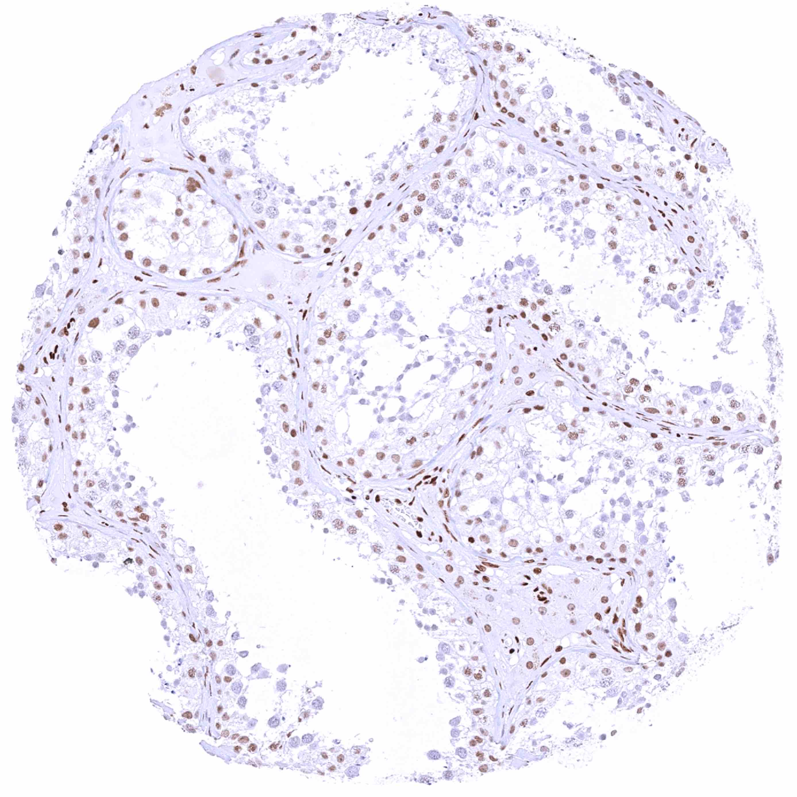
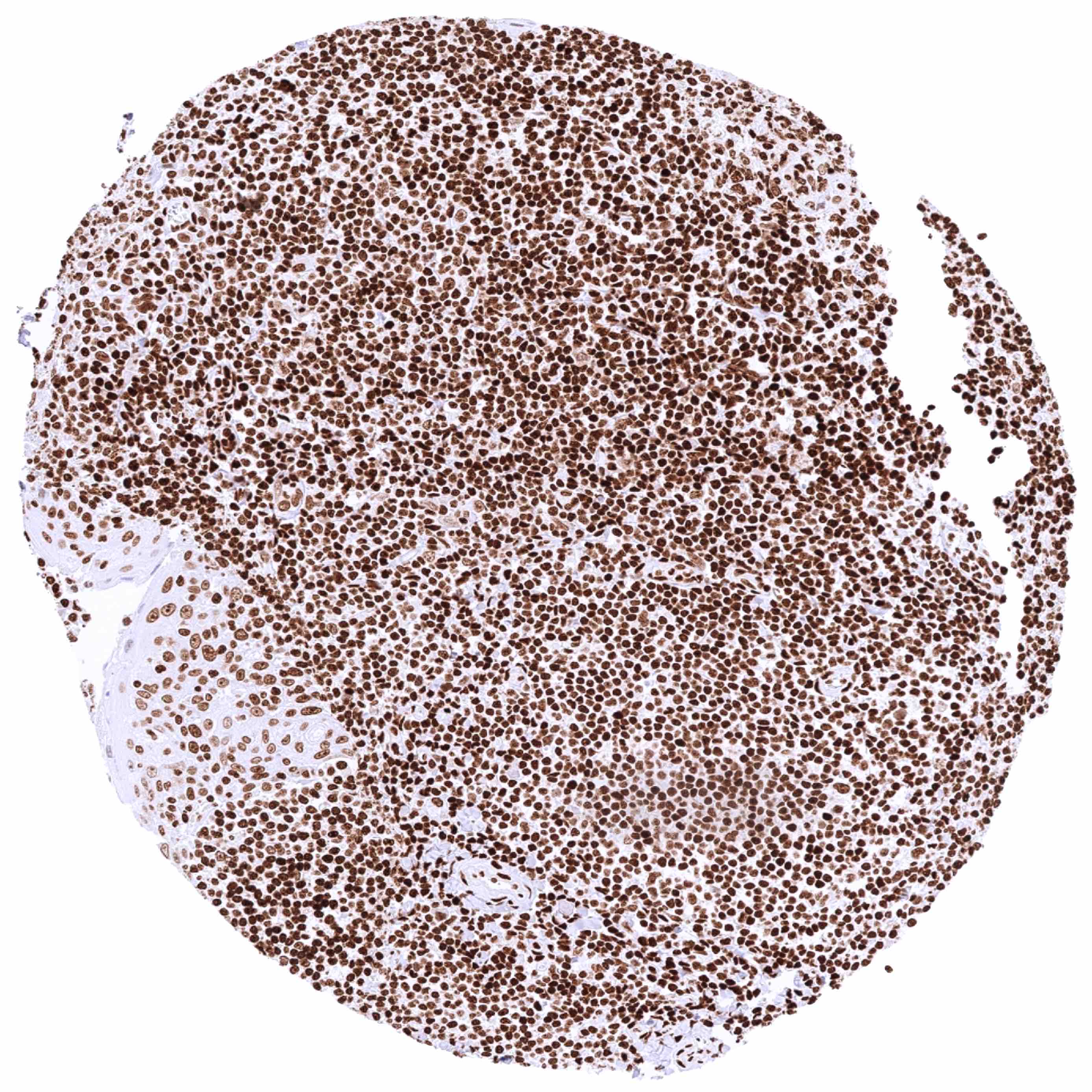
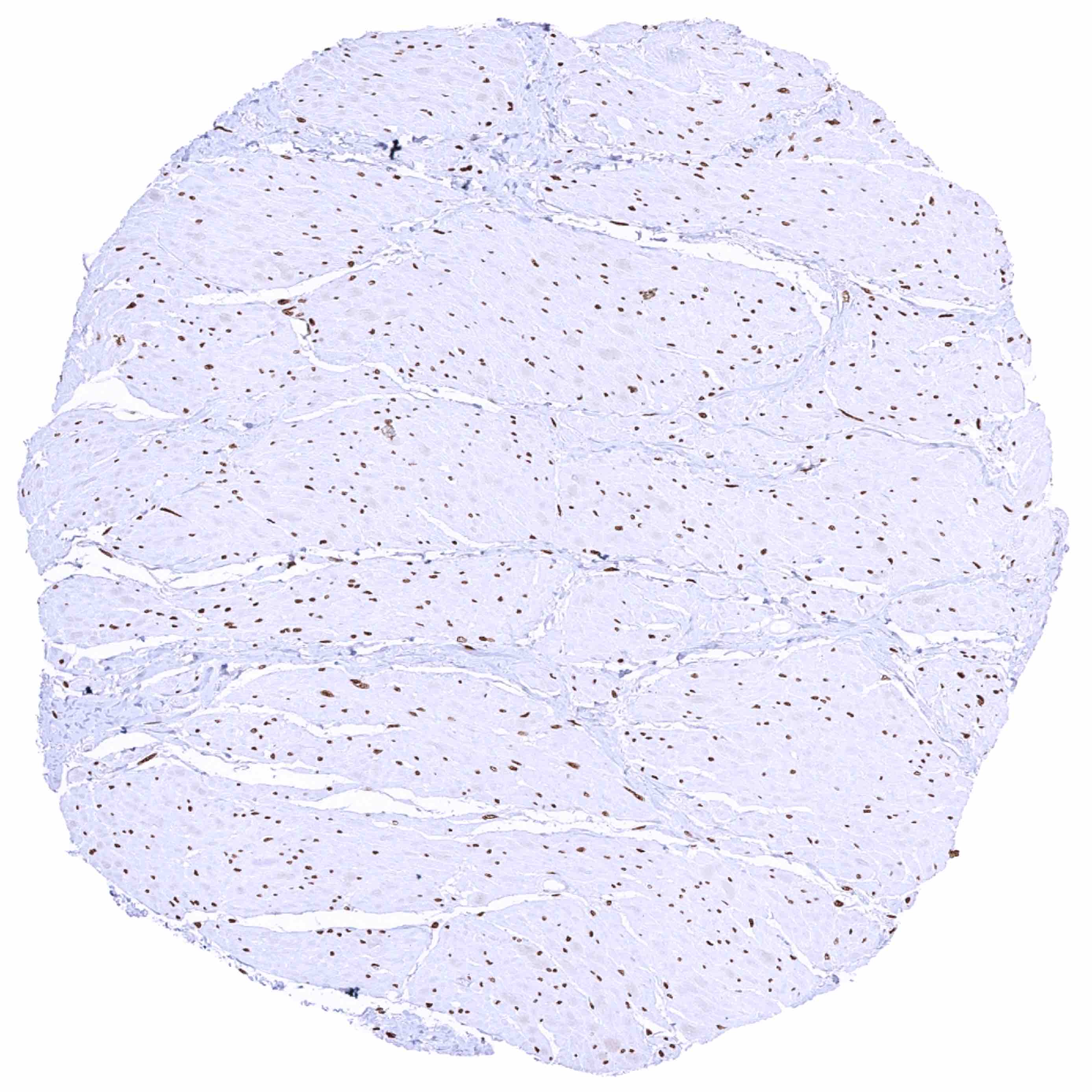
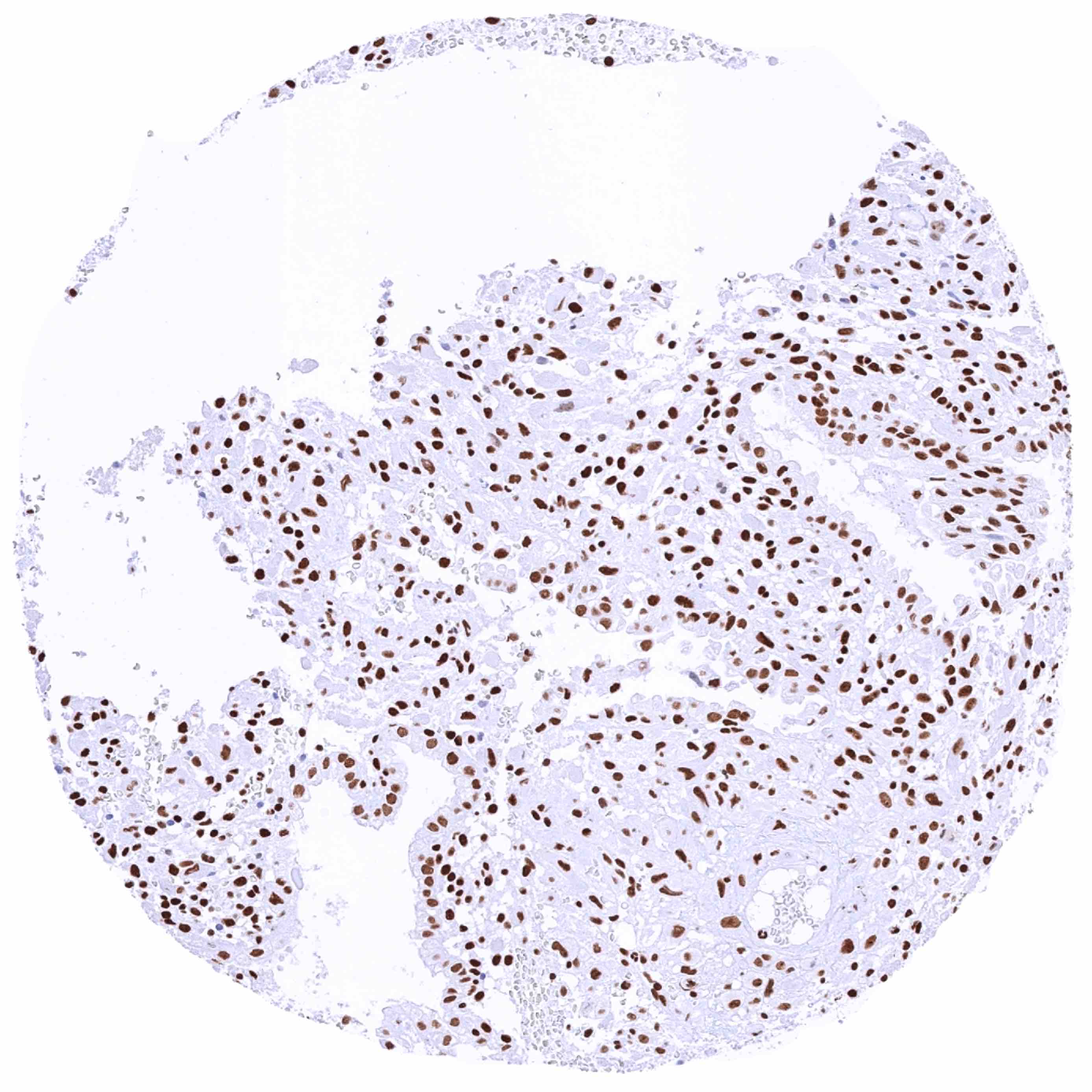
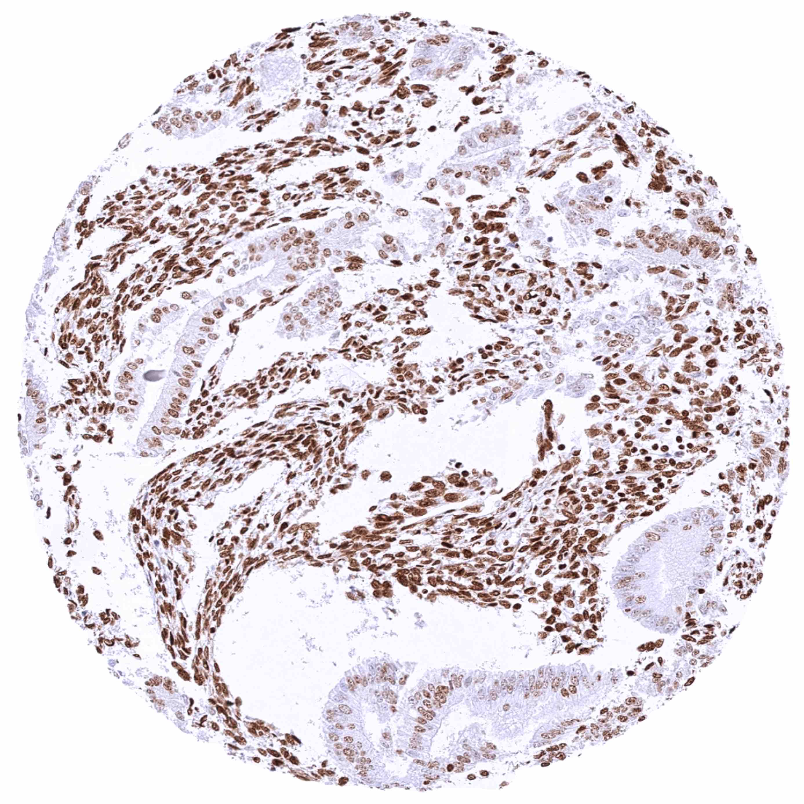
HMGB1 antibody [HMV317] HistoMAXTM


Go to Cancer Tissue Gallery
  
 Go back to product page
  

Go to HistoMAX page


Adrenal gland

Anal canal, skin – HMGB1 staining intensity is strongest in the basal and suprabasal cell layers and it countinuously decreases towards the superficial cell layers

Aorta, media

Appendix, mucosa


Appendix, muscular wall

Bone marrow – Intense nuclear HMGB1 staining of virtually all cells of the hematopesis

Breast – HMGB1 positivity is strongest in basal and luminal epithelial cells

Bronchus, mucosa – Significant nuclear HMGB1 staining of all cells. Among epithelia, HMGB1 staining is most intense in basal cells


Cerebellum (white matter)

Cerebellum, cortex (molecular layer, Purkinje cell layer, granule cell layer, white matter)

Cerebrum, grey matter

Cerebrum, white matter


Colon descendens, mucosa – Significant nuclear HMGB1 staining of all cells. Staining intensity is higher in the crypt base than in the surface epithelium

Colon descendens, muscular wall

Duodenum, Brunner gland – Significant nuclear HMGB1 staining of all epithelial and inflammatory cells. HMGB1 staining is somewhat weaker in Brunner glands

Duodenum, mucosa


Epididymis (Cauda)

Epididymis (Corpus) – HMGB1 staining is more intense in basal than in chief cells

Esophagus, squamous epithelium – HMGB1 staining intensity is strongest in the basal and suprabasal cell layers and countinuously decreases towards the superficial cell layers

Fallopian tube, mucosa


Fat

Gallbladder, epithelium

Heart muscle

Ileum, mucosa


Kidney, cortex – Significant nuclear HMGB1 staining of all cells. Staining is particularly low in tubuli (especially proximal) and highest in collecting ducts and glomeruli

Kidney, medulla – Strong nuclear HMGB1 staining of collecting duct cells

Liver – Significant nuclear HMGB1 staining of all cells. Staining is particularly high in bile ducts and particularly low in hepatocytes

Lung


Lymph node

Ovary, corpus luteum – HMGB1 staining is only faint in the corpus luteum

Ovary, stroma

Pancreas


Parathyroid gland

Parotis

Pituitary gland, anterior lobe

Pituitary gland, posterior lobe


Placenta (amnion and chorion) – Strong nuclear HMGB1 staining of chorion cells, moderate nuclear HMGB1 staining of amnion cells

Placenta, early

Placenta, mature

Prostate – Significant nuclear HMGB1 staining of all cells. Staining is particularly low in acinar cells and highest in basal cells


Rectum, mucosa – Significant nuclear HMGB1 staining of all cells. Staining intensity is higher in the crypt base than in the surface epithelium

Seminal vesicle – Significant nuclear HMGB1 staining of all cells. In the epithelium, HMGB1 staining is more intense in basal than in luminal cells

Sinus paranasales – Significant nuclear HMGB1 staining of all cells. Among epithelia, HMGB1 staining is most intense in basal cells

Skeletal muscle


Skin, hairfollicel and sebaceous glands

Spleen

Stomach, antrum

Stomach, corpus (glands)


Stomach, corpus (surface epithelium)

Testis – Significant nuclear HMGB1 staining of Sertoli cells. HMGB1 staining is weaker in the germ cells it where the staining level decreases from spermatogonia to spermatocytes and spermatides (which are HMGB1 negative)

Thymus – Strong nuclear HMGB1 staining of virtually all cells of the immune system. HMBG1 labeling is weaker in corpuscles of Hassall’s, especially in their central areas

Thyroid gland


Tonsil, surface epithelium

Tonsil

Urinary bladder, muscular wall

Urinary bladder, urothelium – Strong nuclear HMGB1 staining of all urothelial cell layers


Uterus, ectocervix – HMGB1 staining intensity is strongest in the basal and suprabasal cell layers and countinuously decreases towards the superficial cell layers

Uterus, endocervix

Uterus, endometrium (pregnancy)

Uterus, endometrium (proliferation) – Strong nuclear HMGB1 staining of epithelial and stromal cells


Uterus, endometrium (secretion) – Strong nuclear HMGB1 staining of stromal cells while HMGB1 staining is markedly less intense in epithelial cells in this sample

Uterus, myometrium


| TOP |