Normal Tissue Gallery- GTX640560

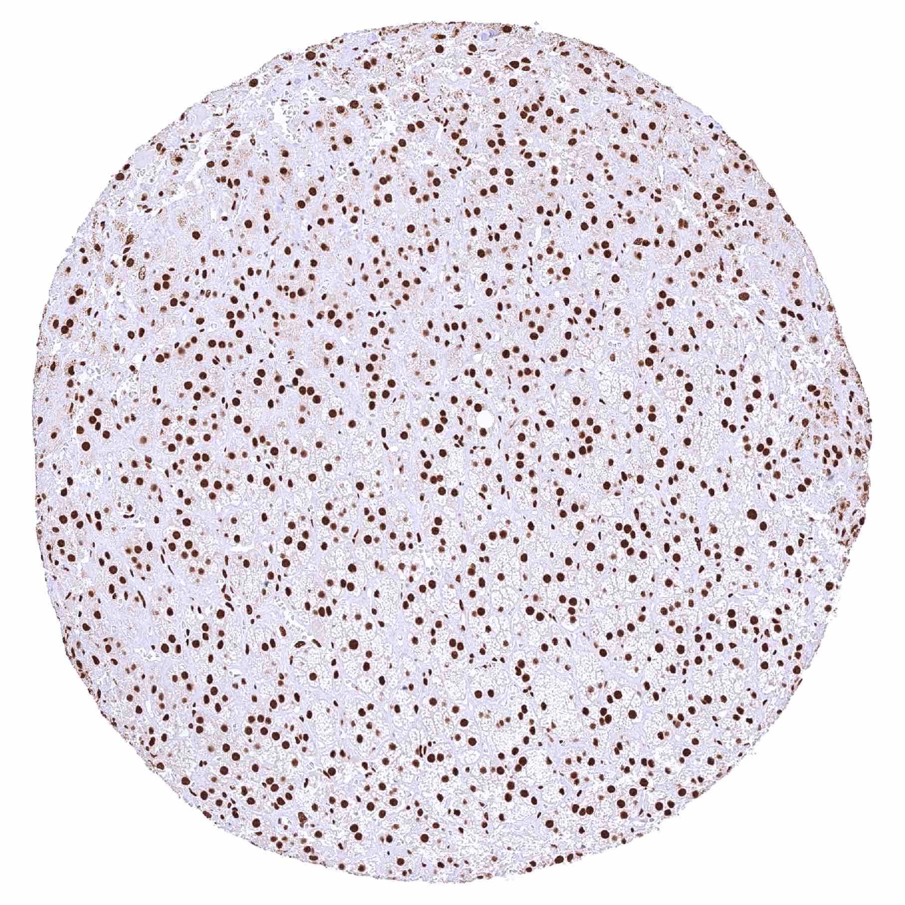
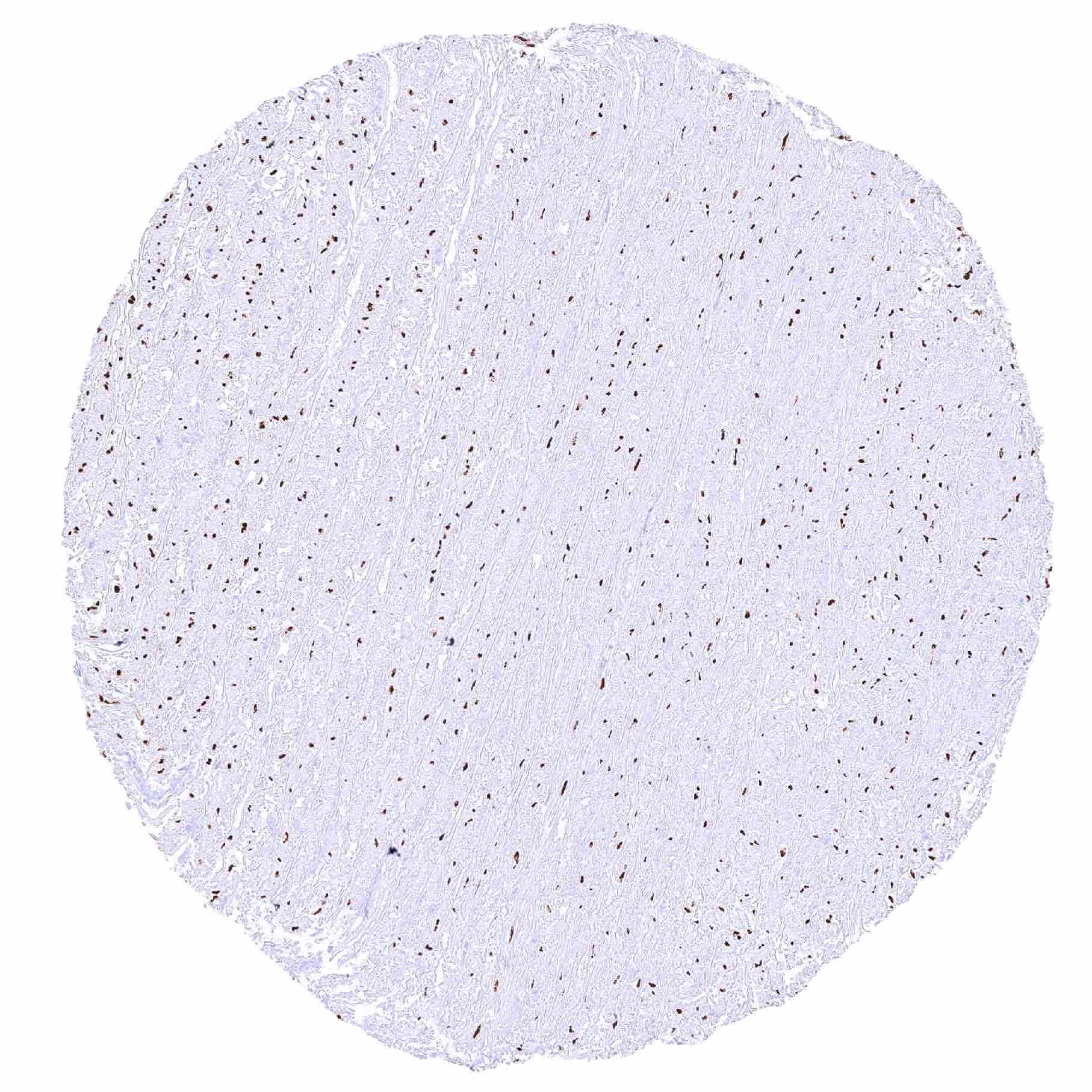
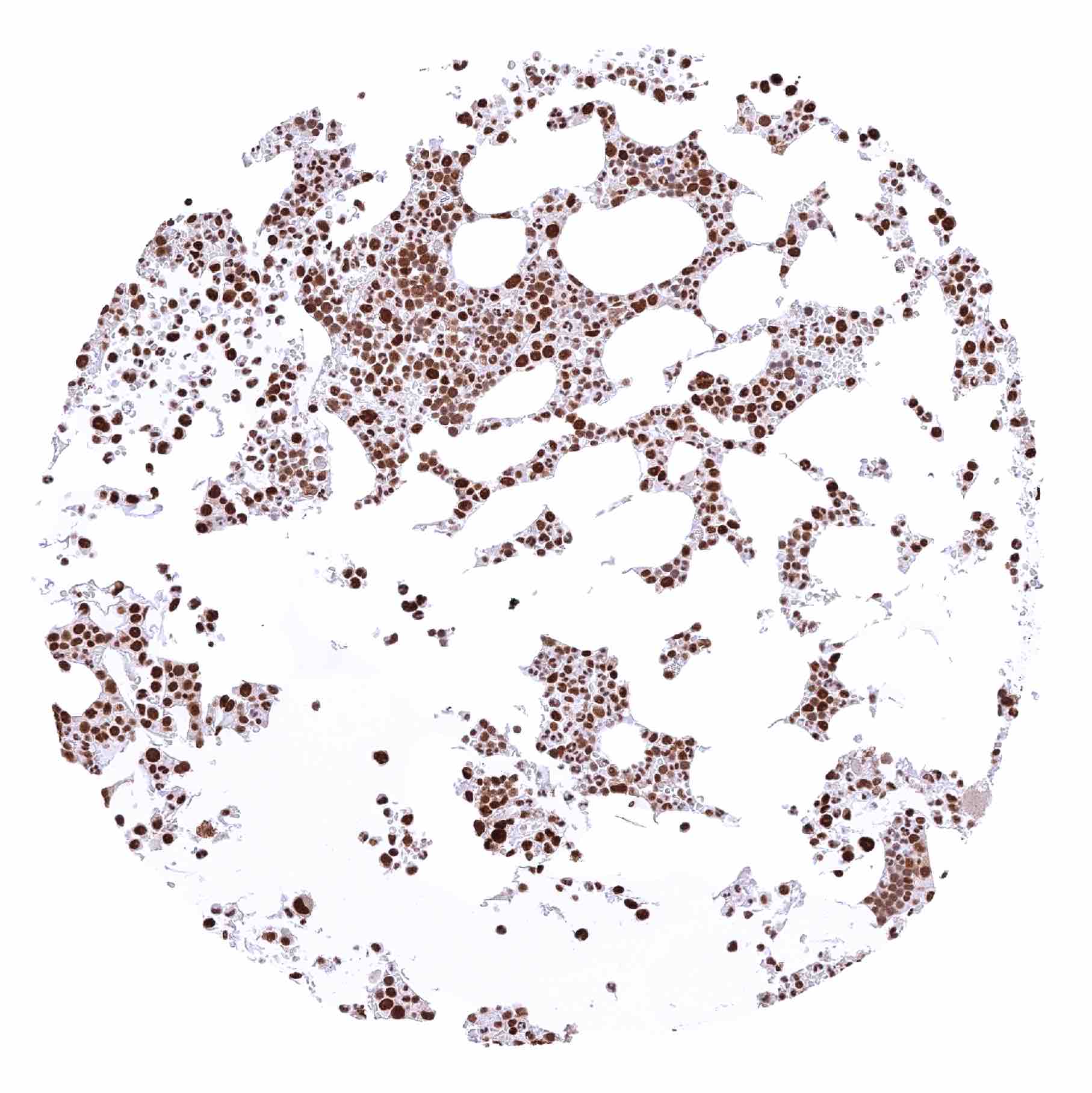
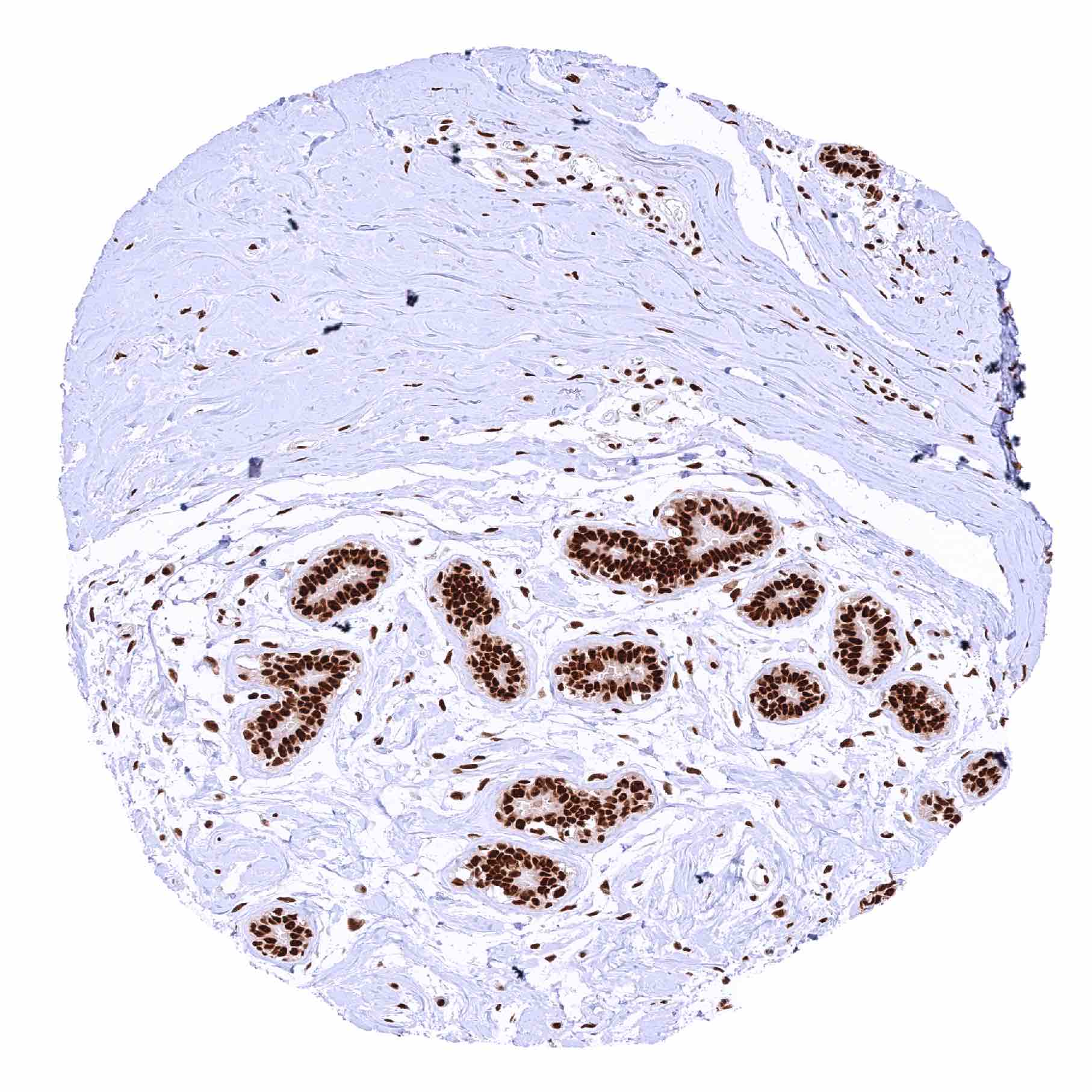
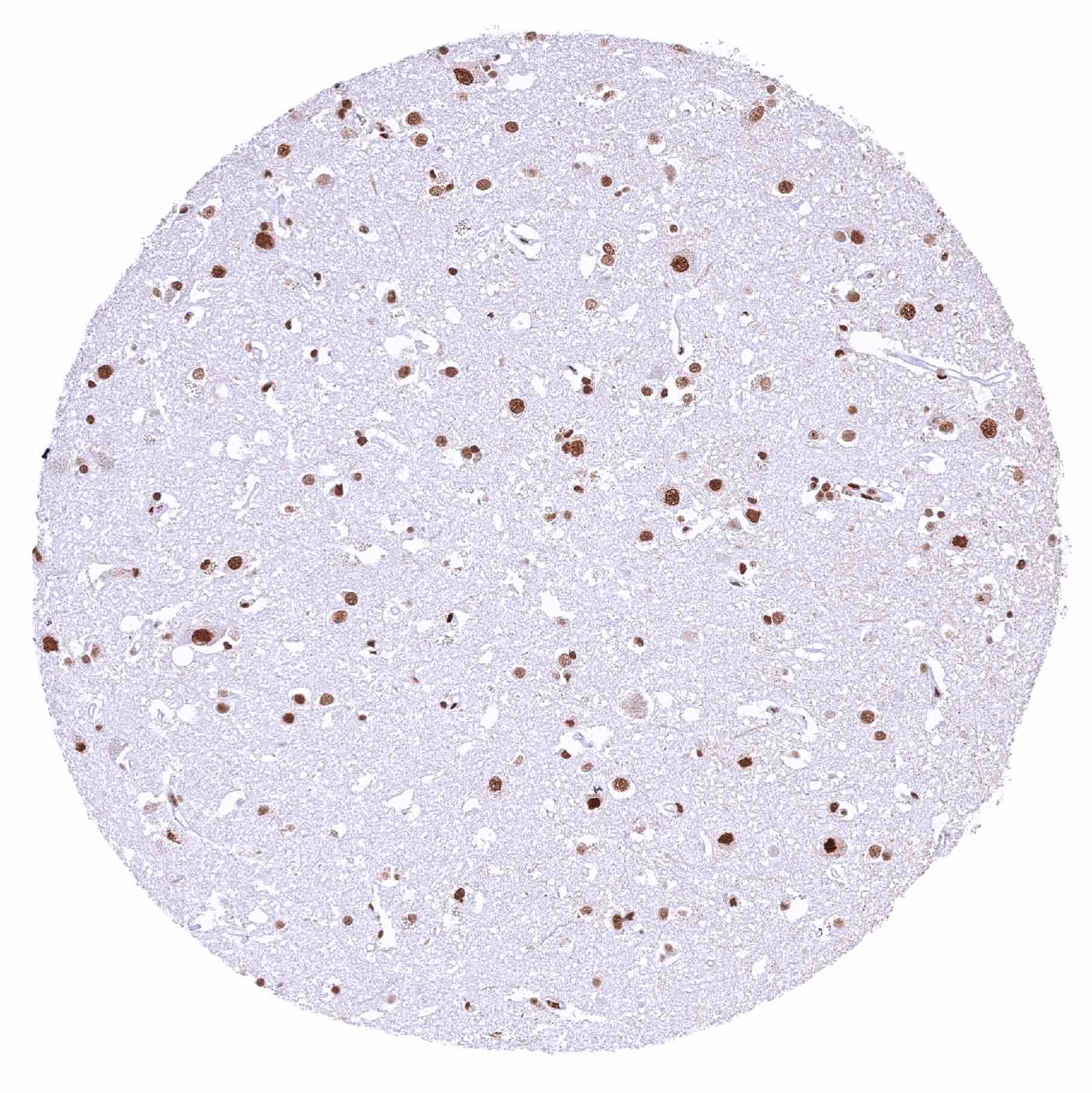
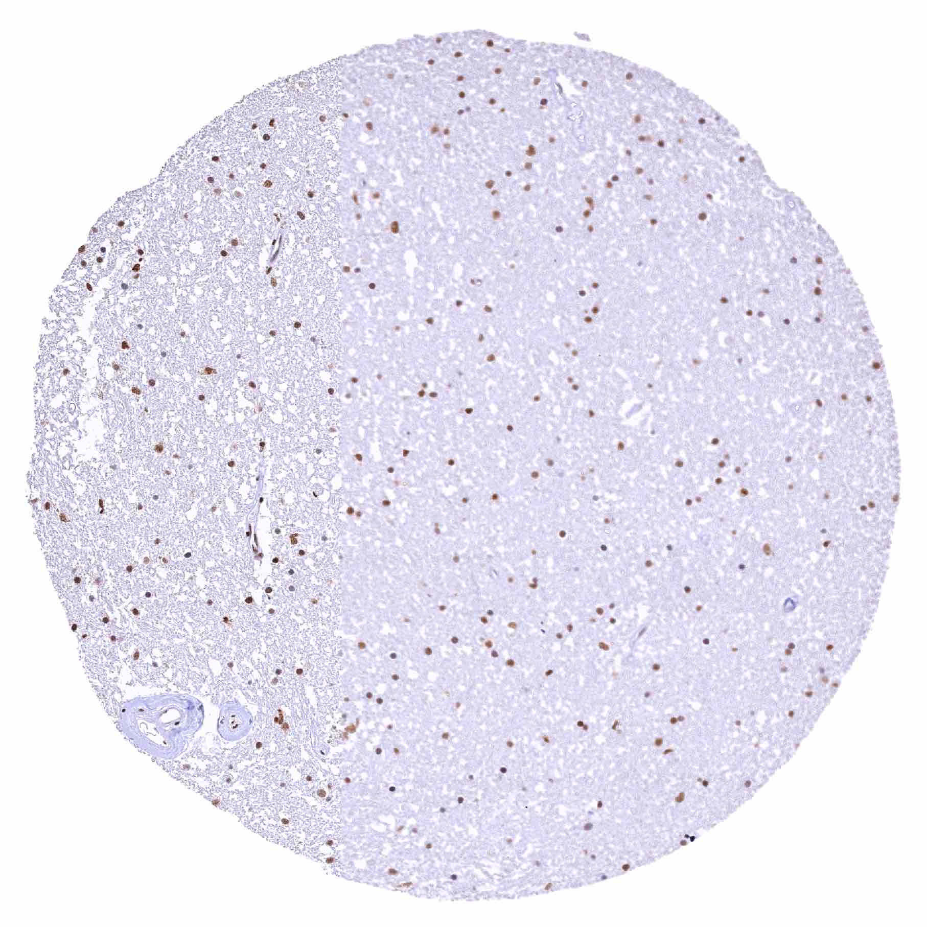
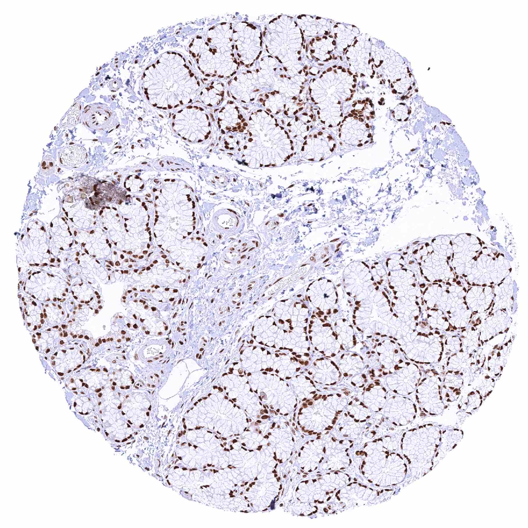
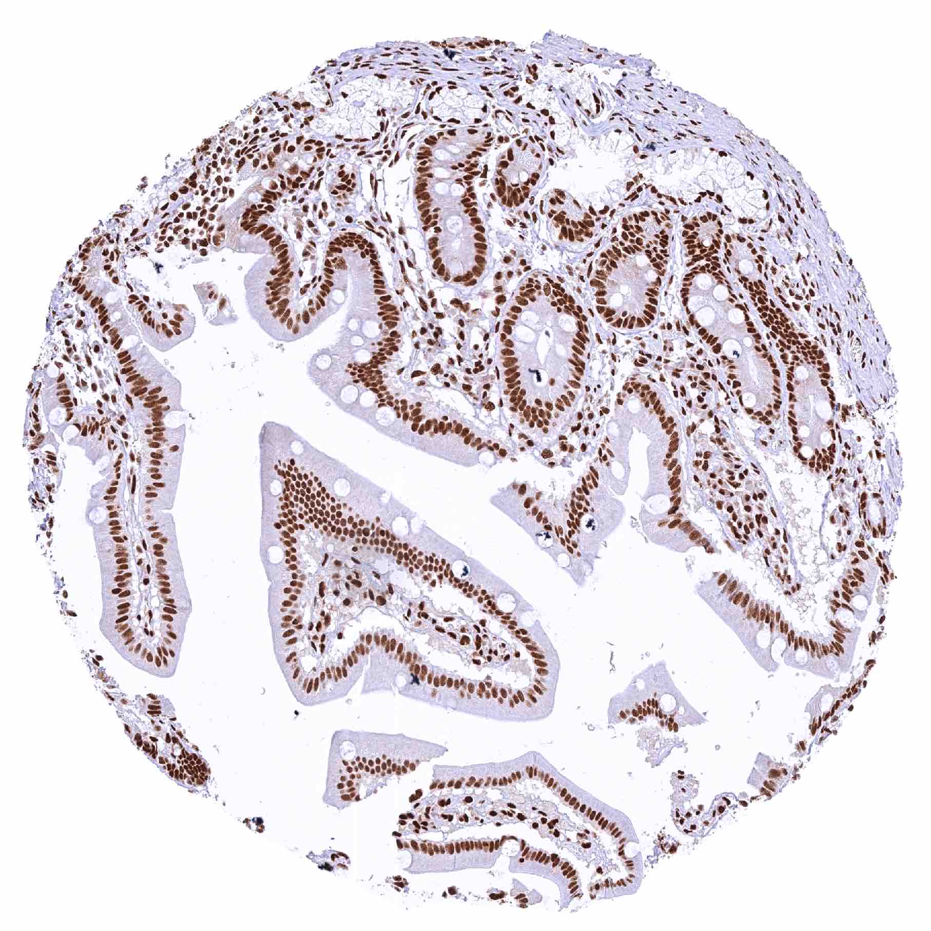
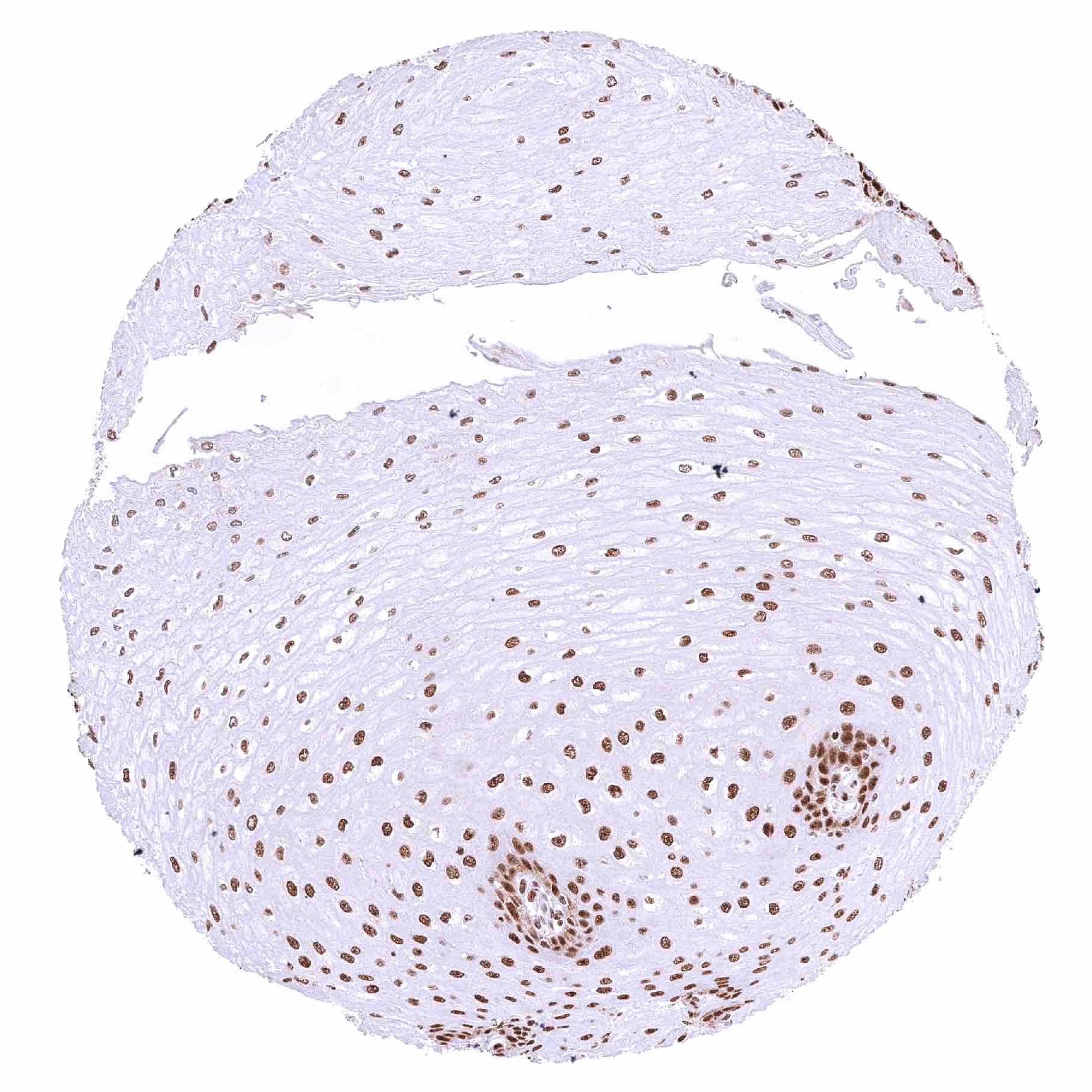
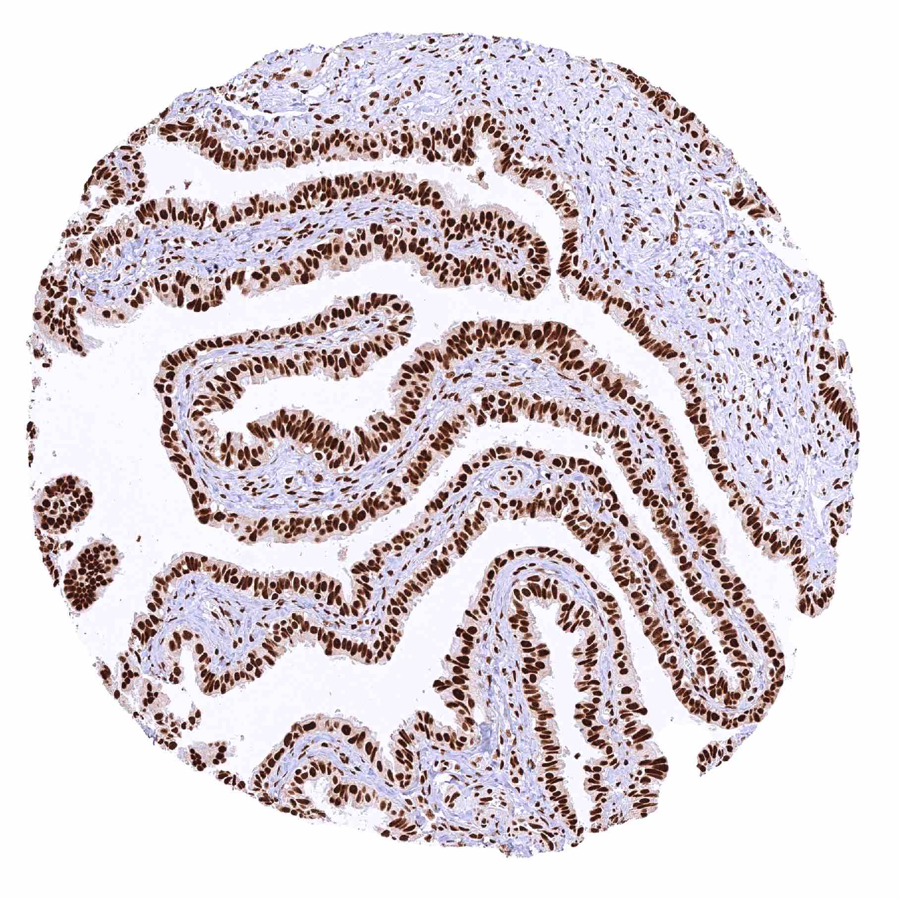
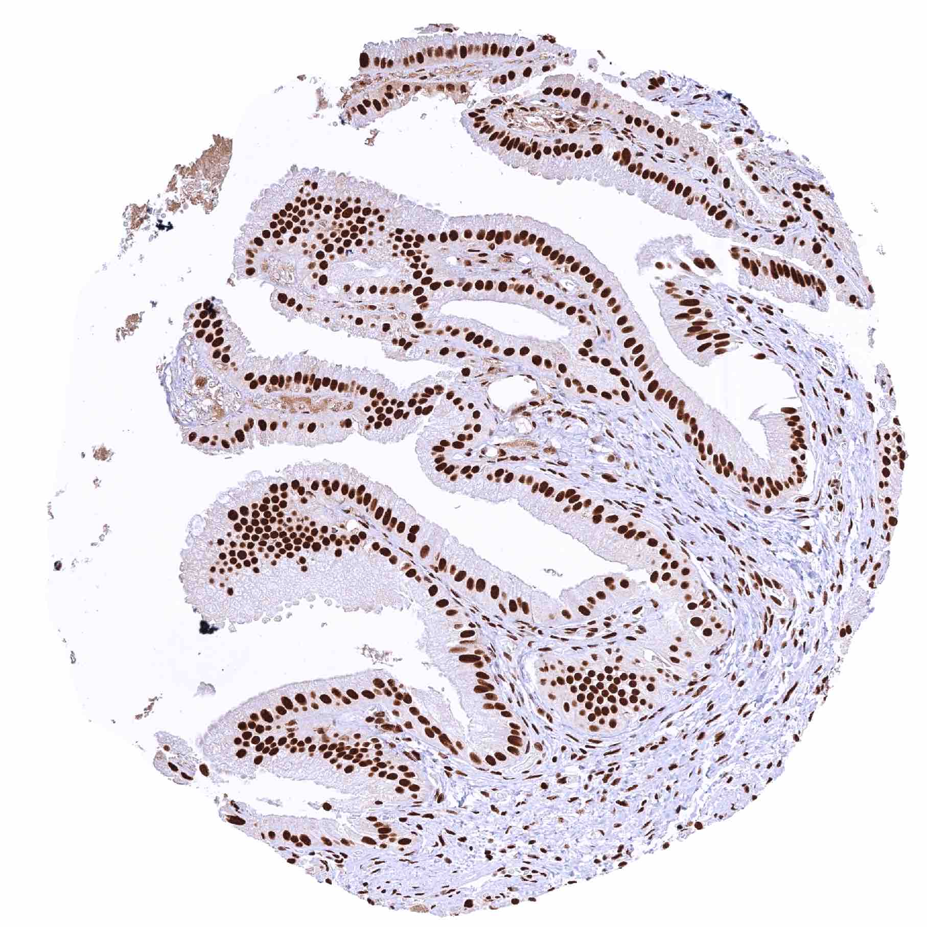
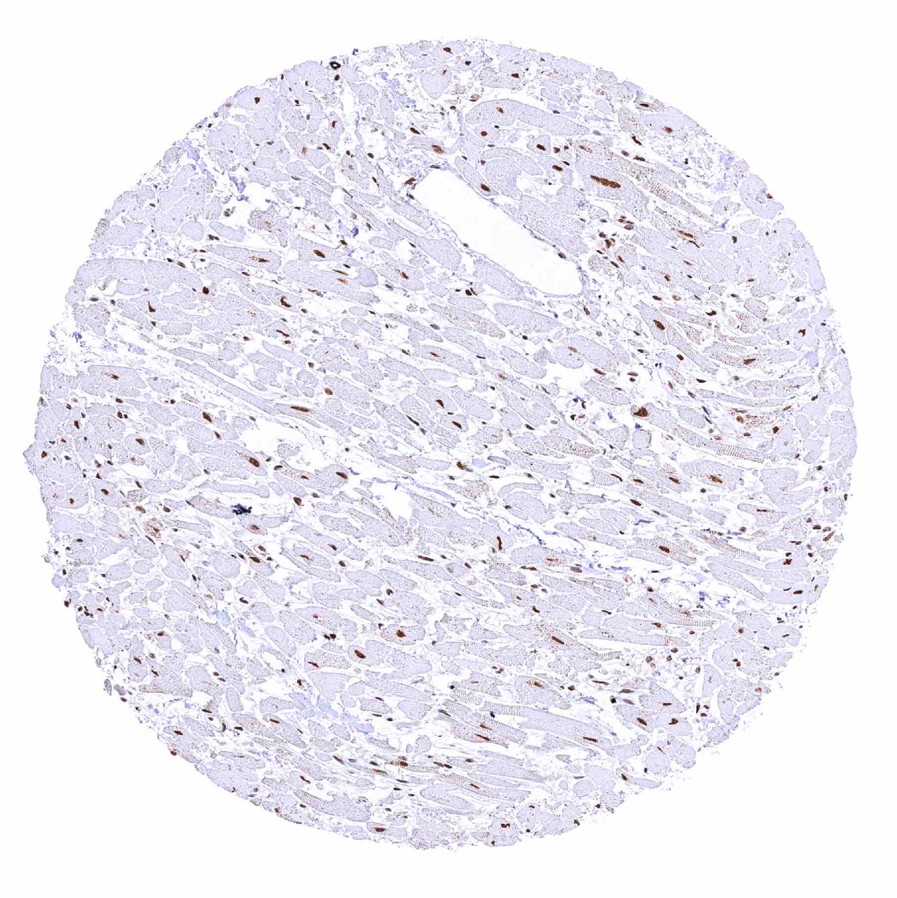
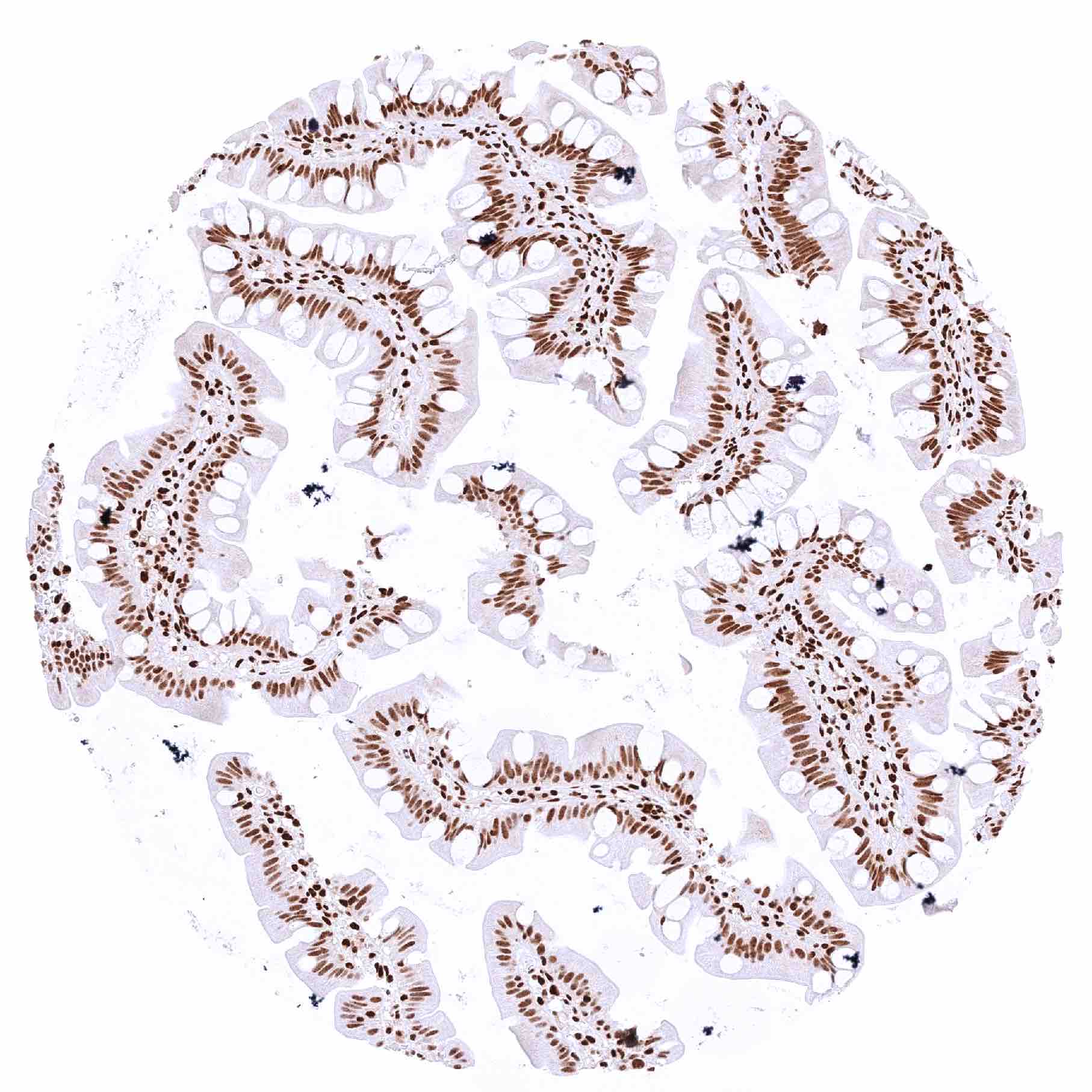
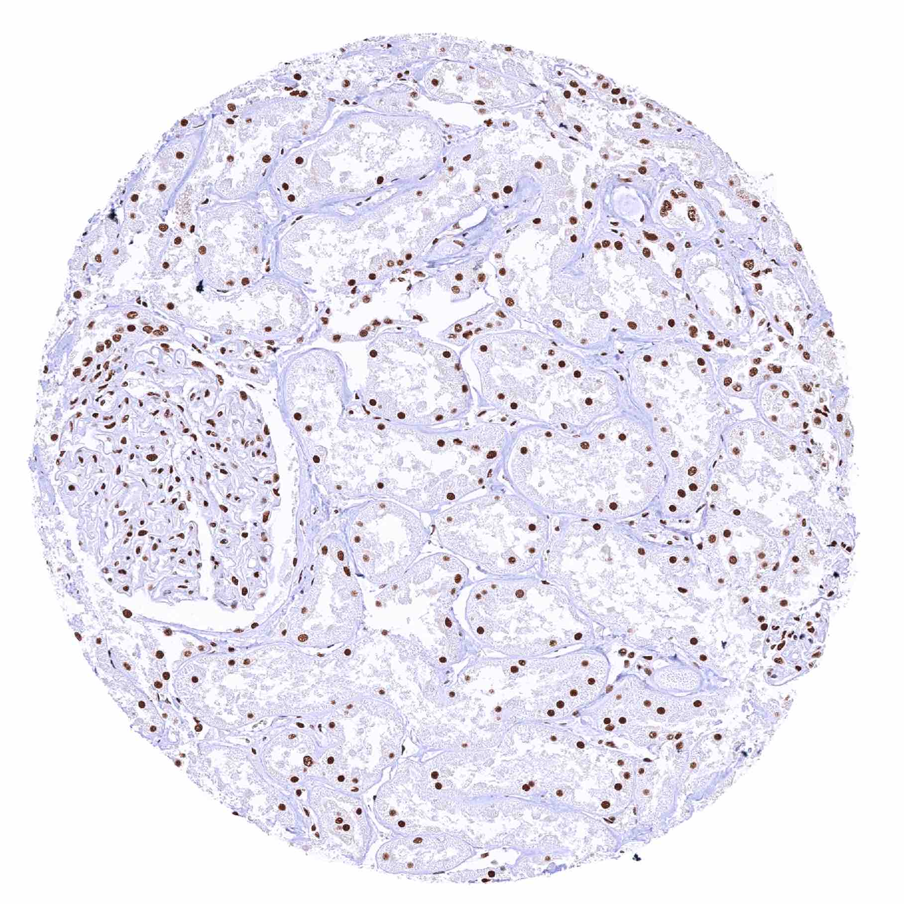
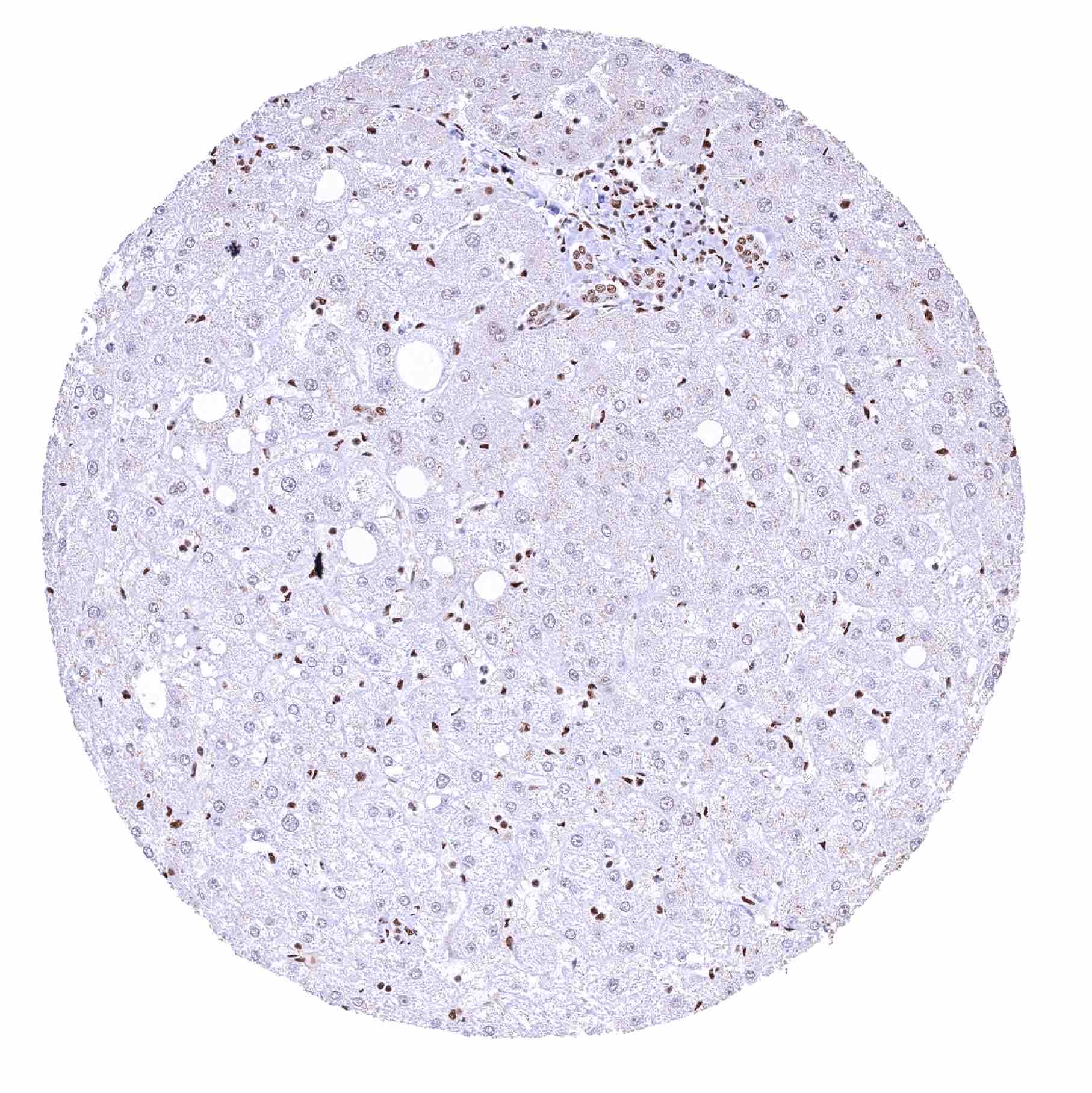
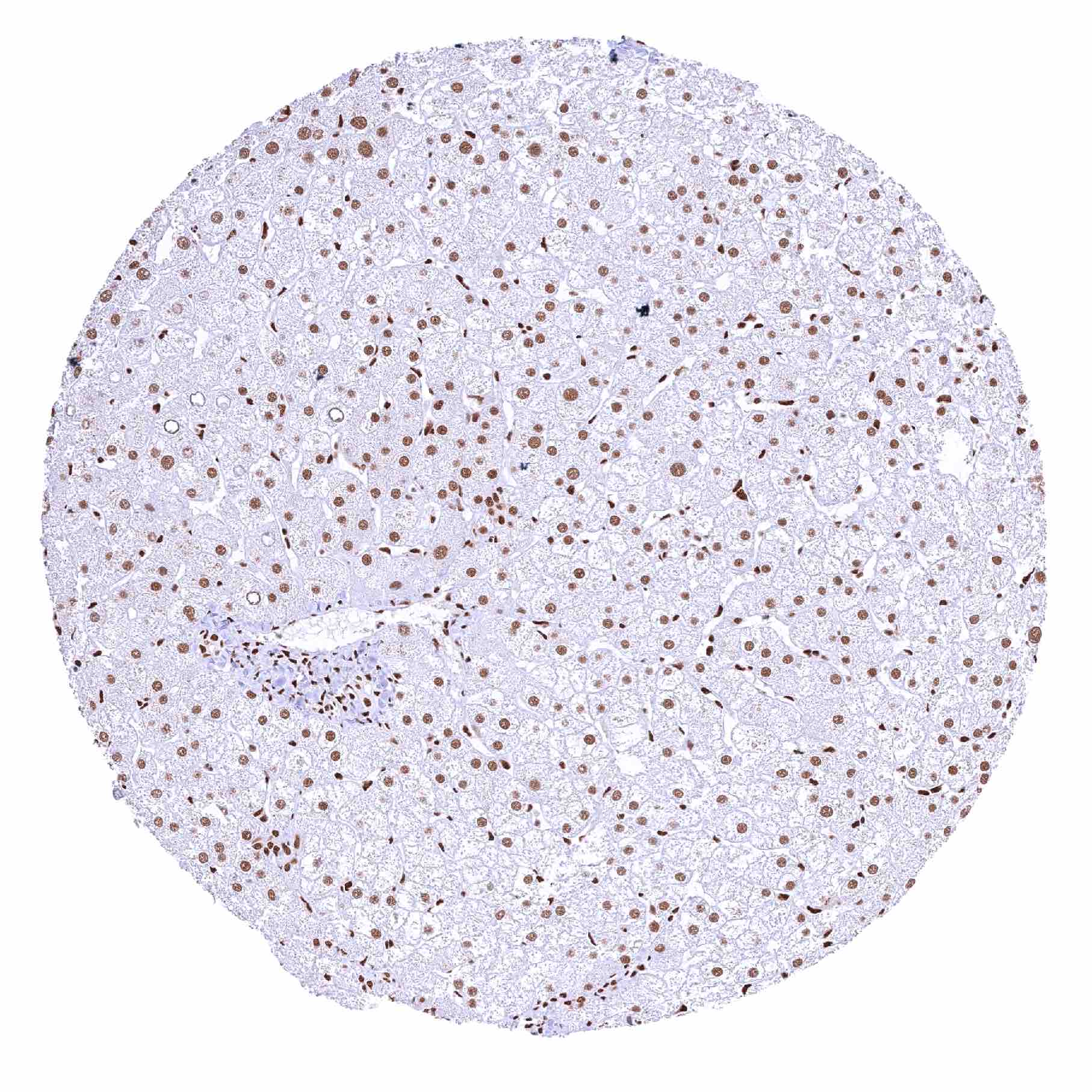
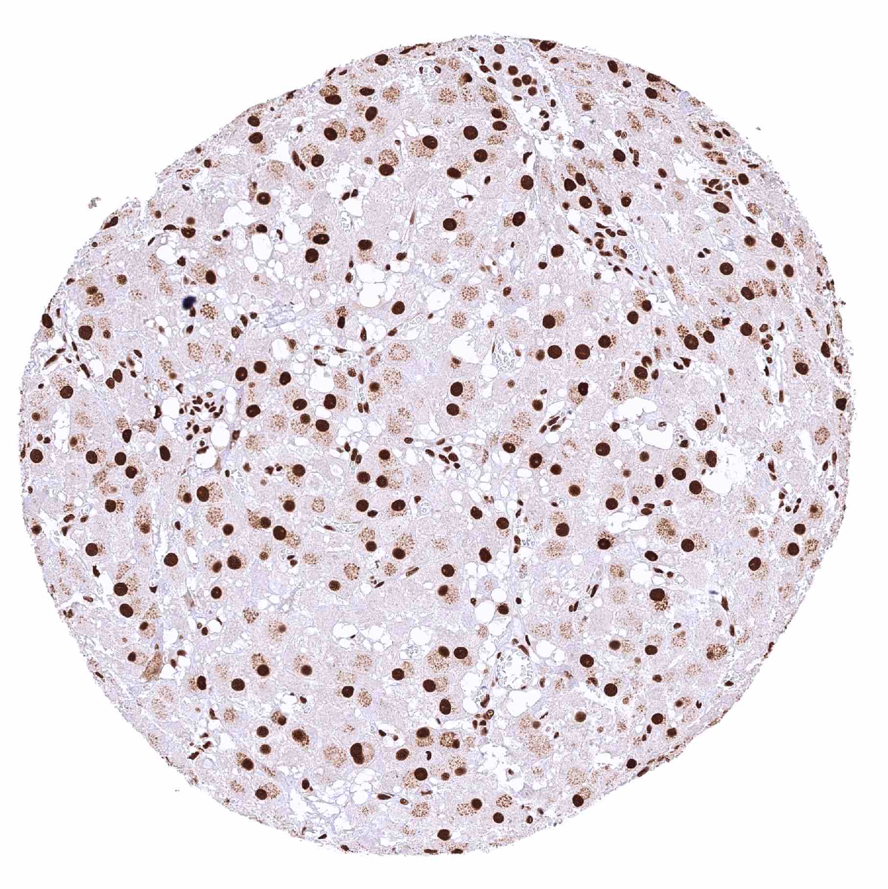
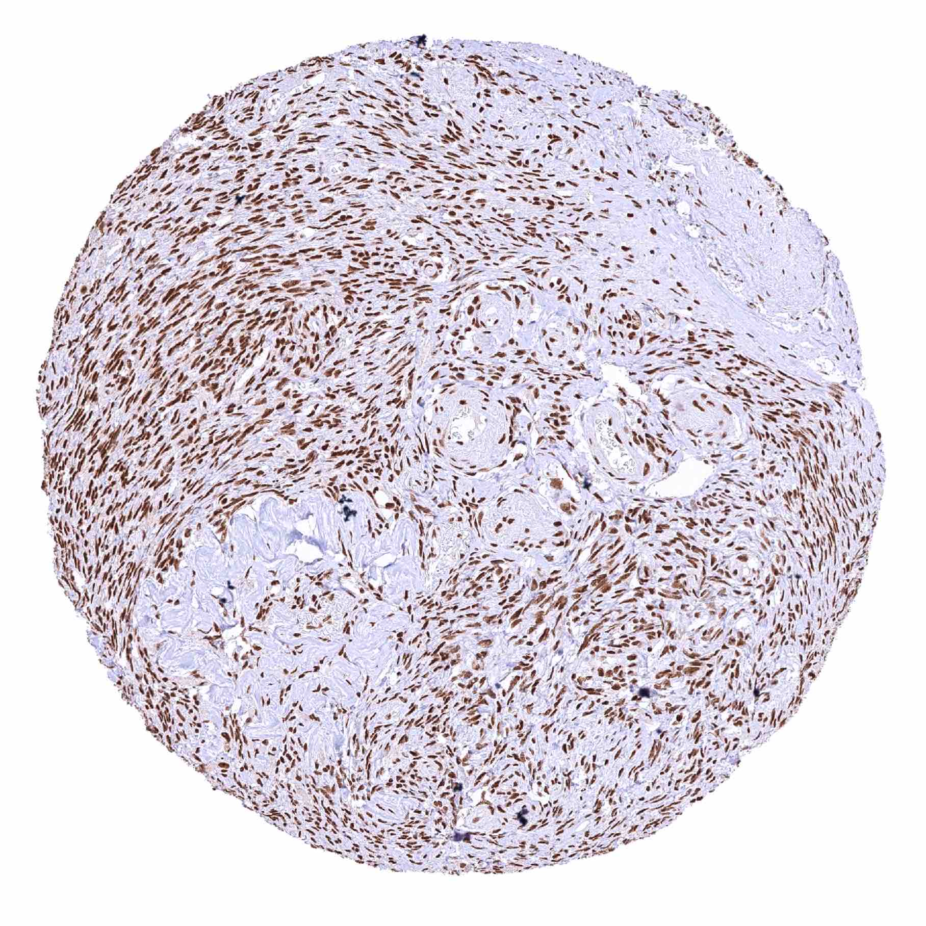
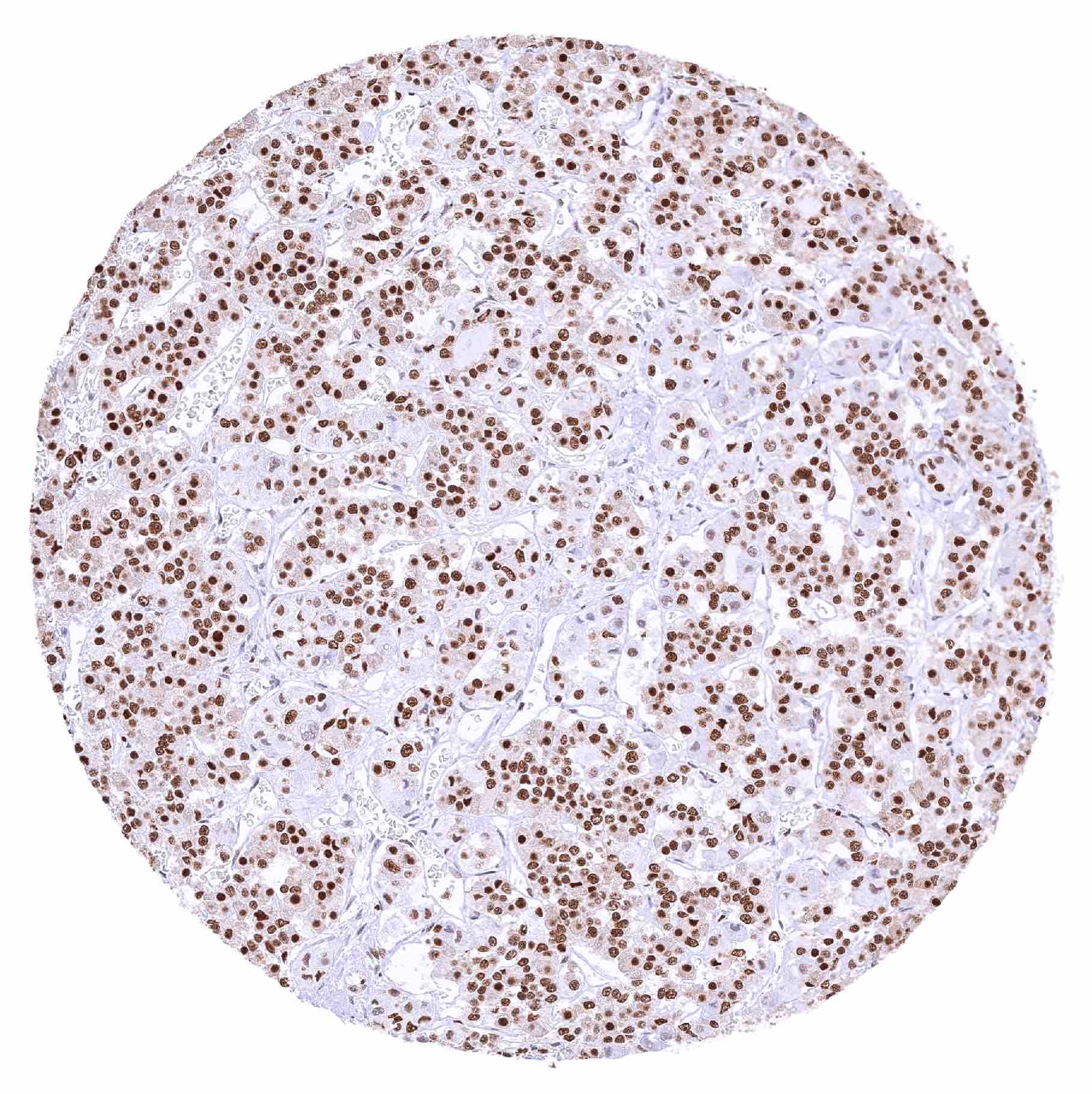
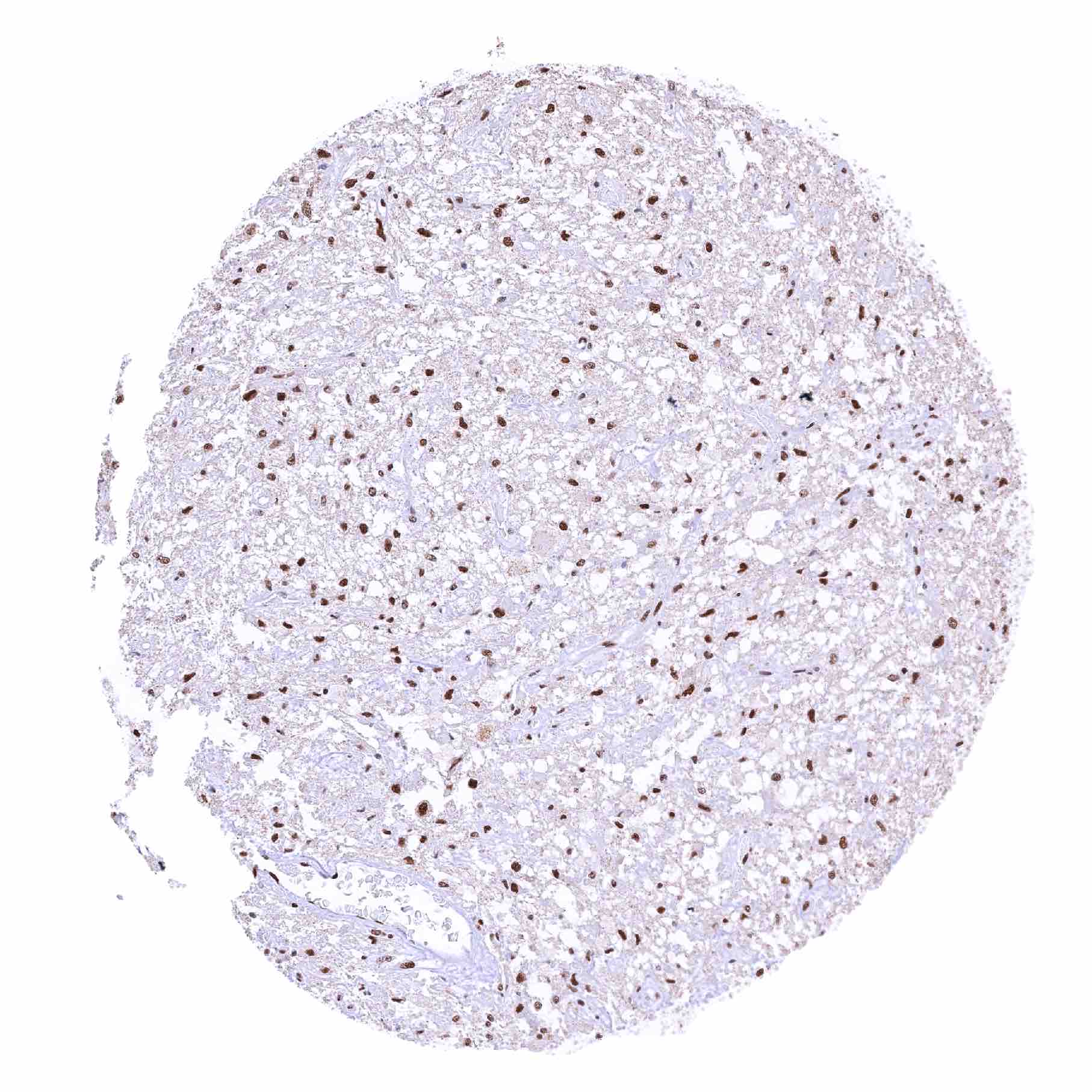
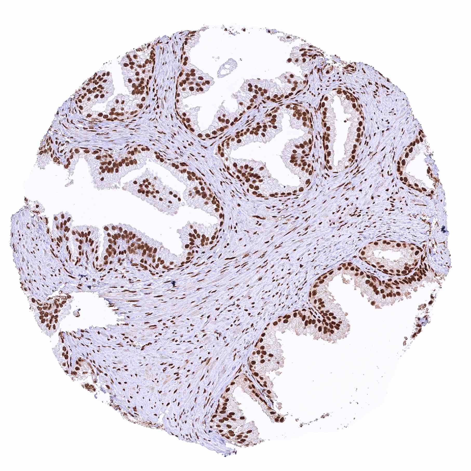
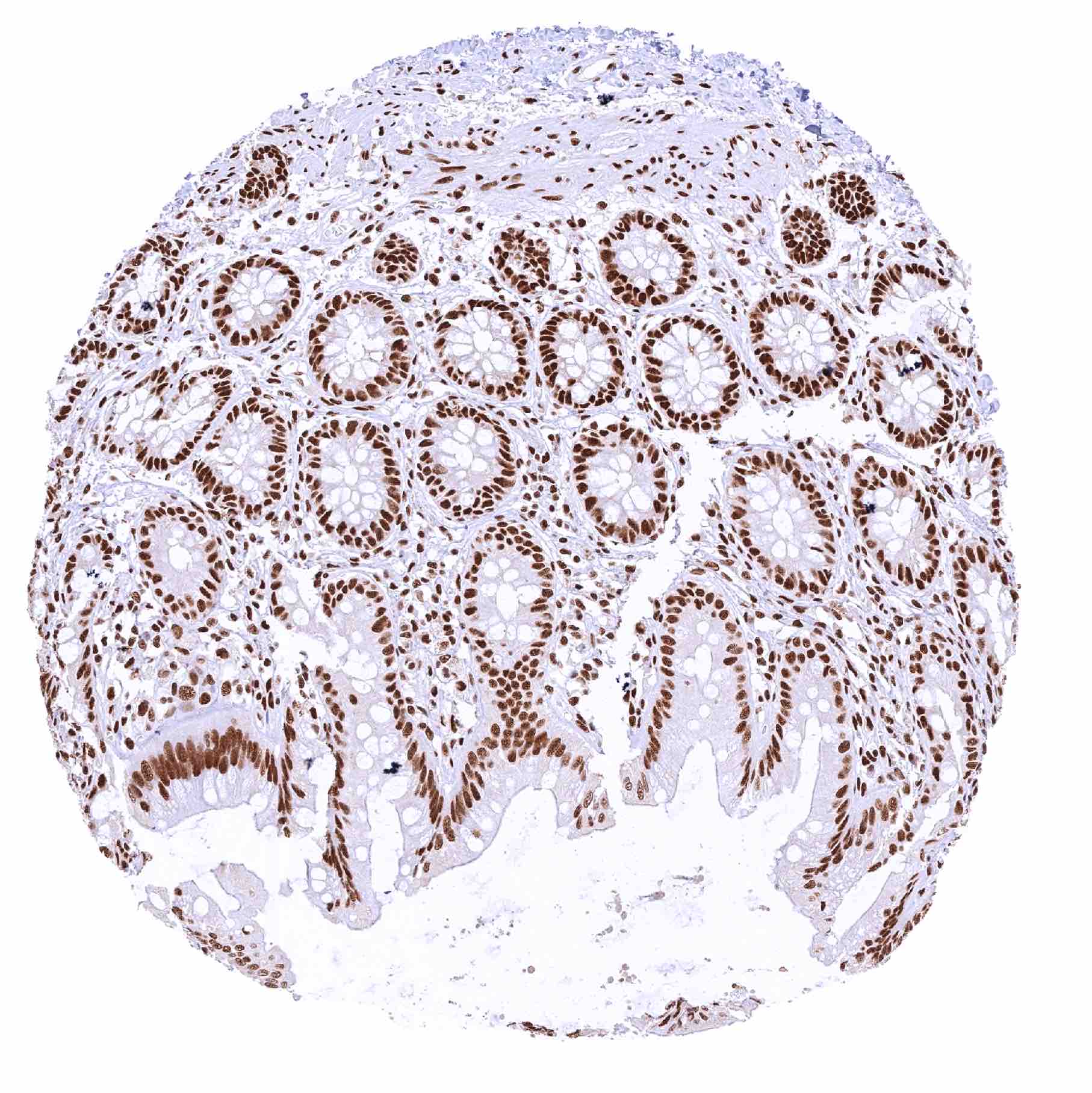
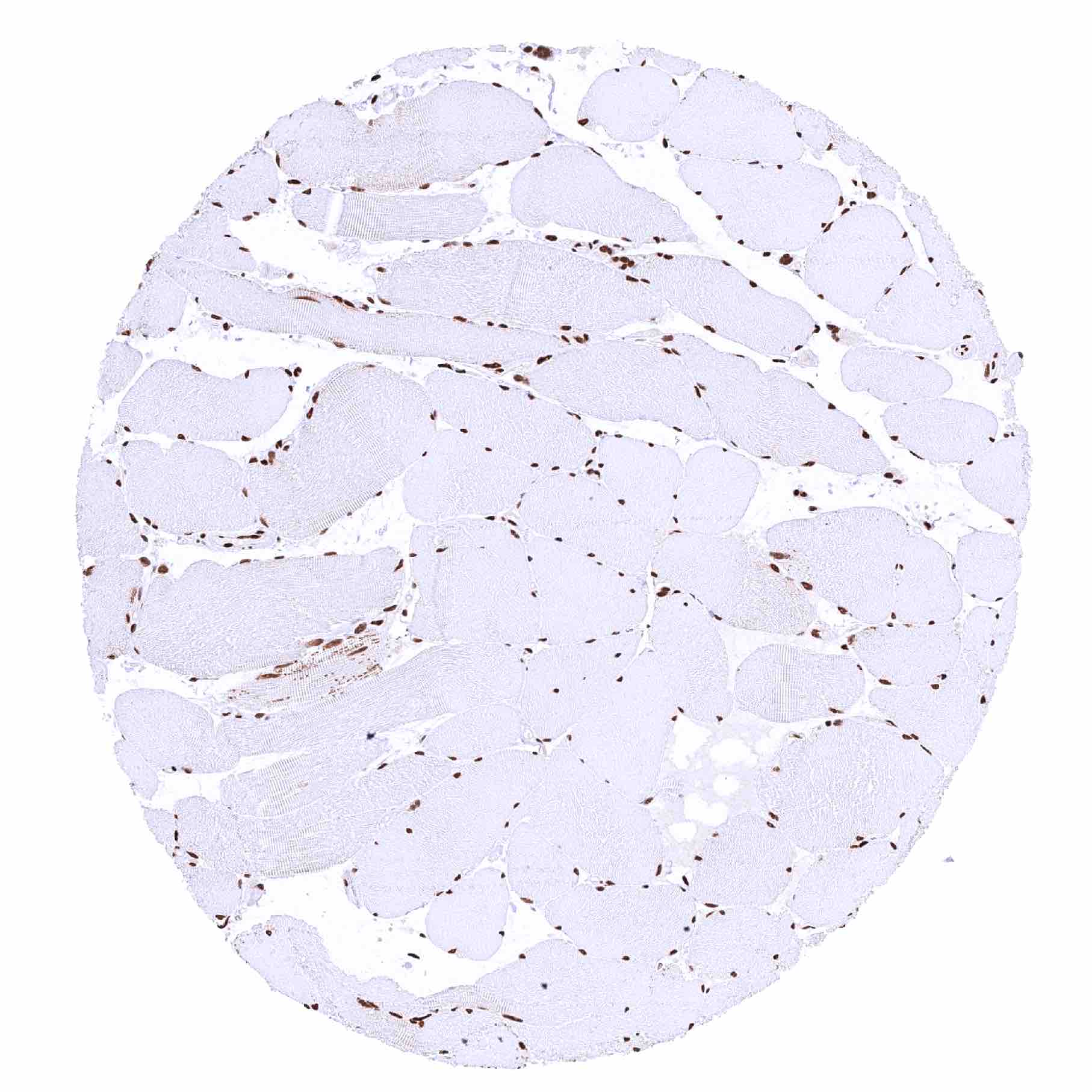
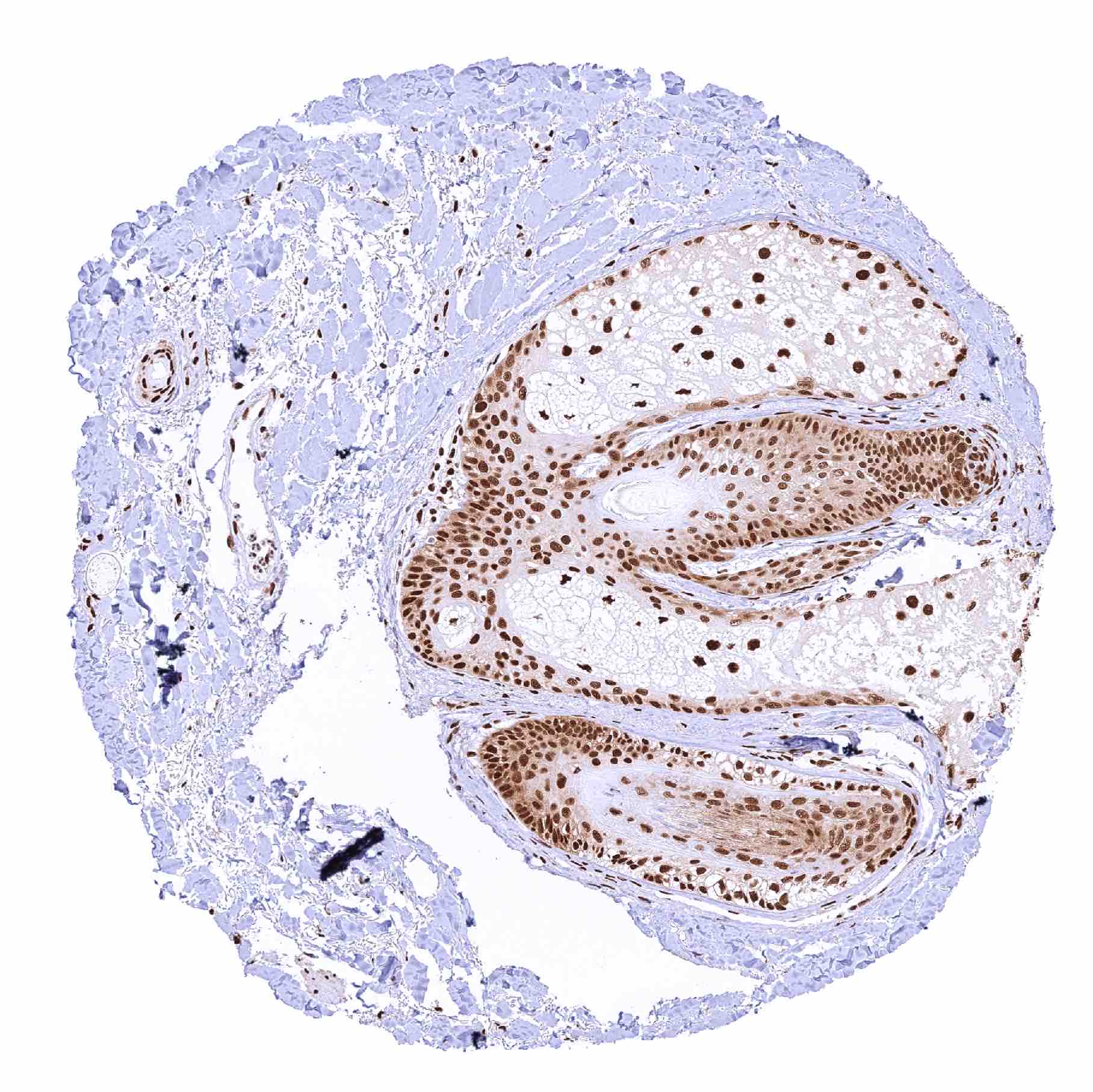
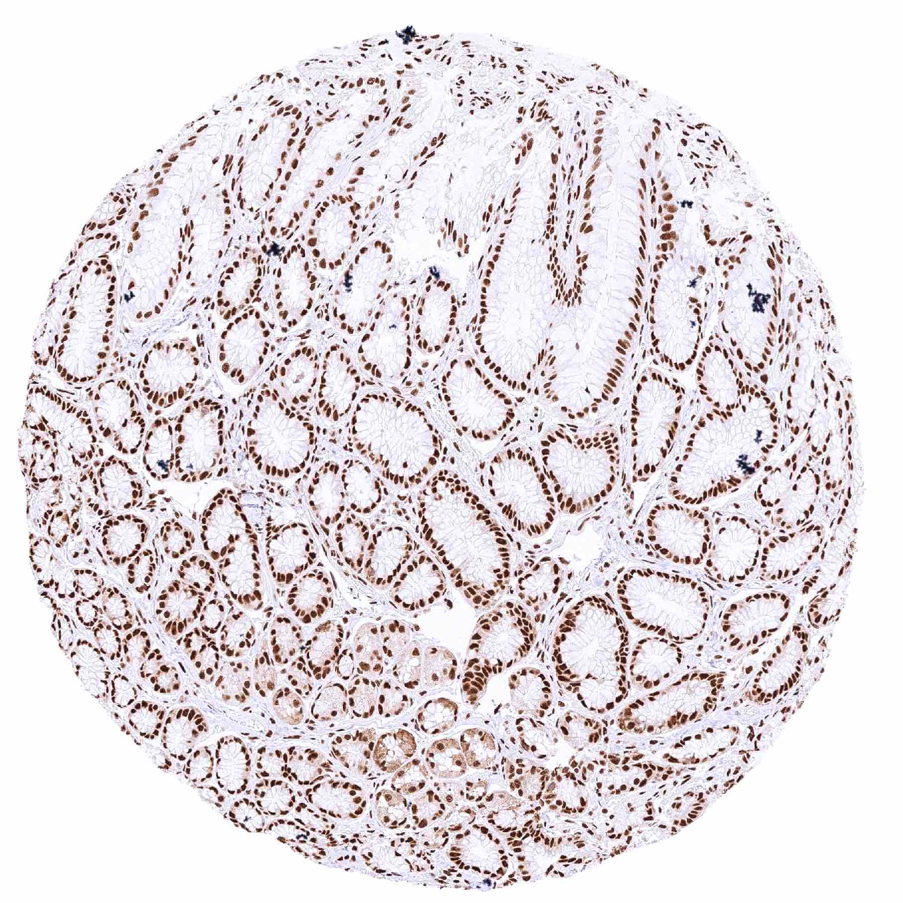
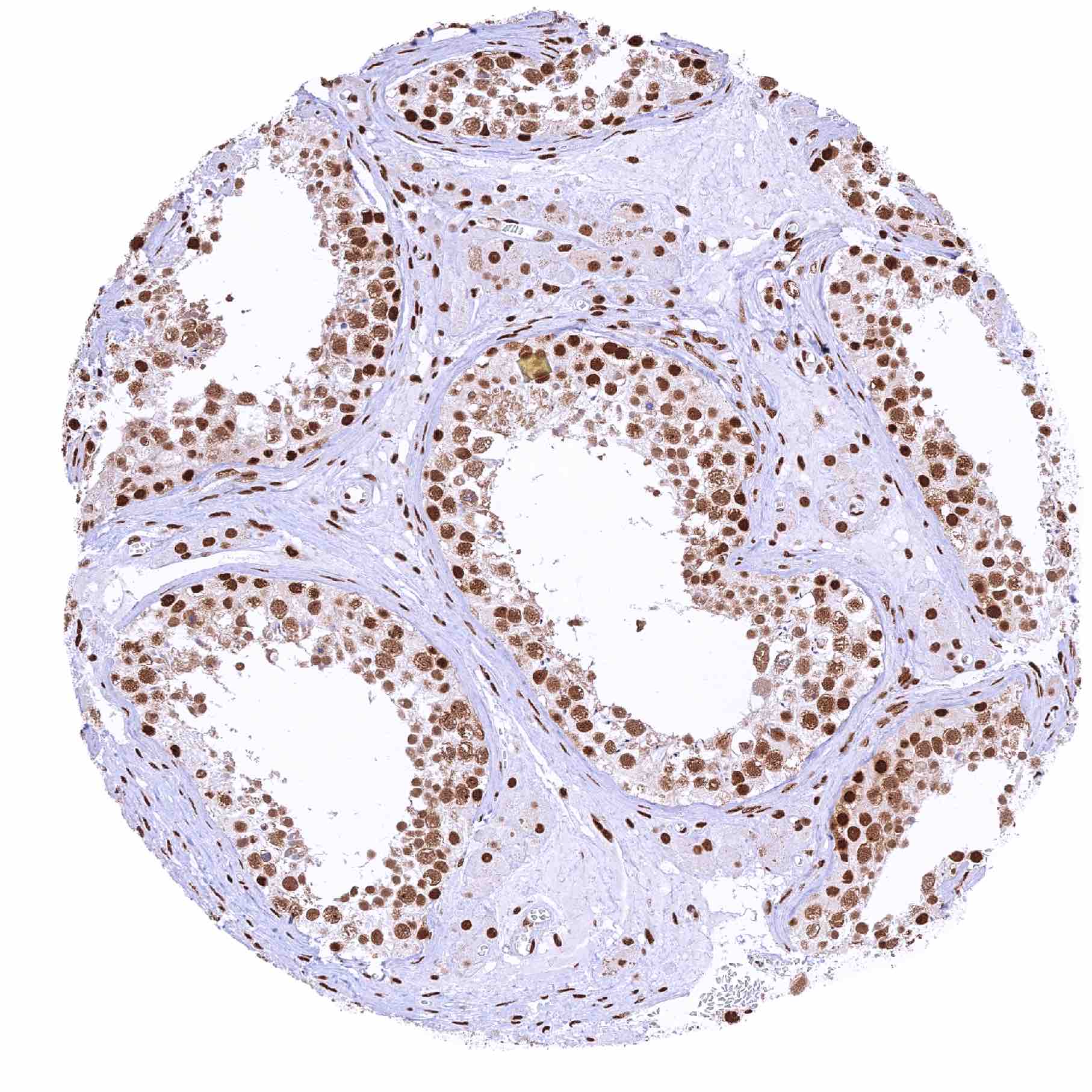
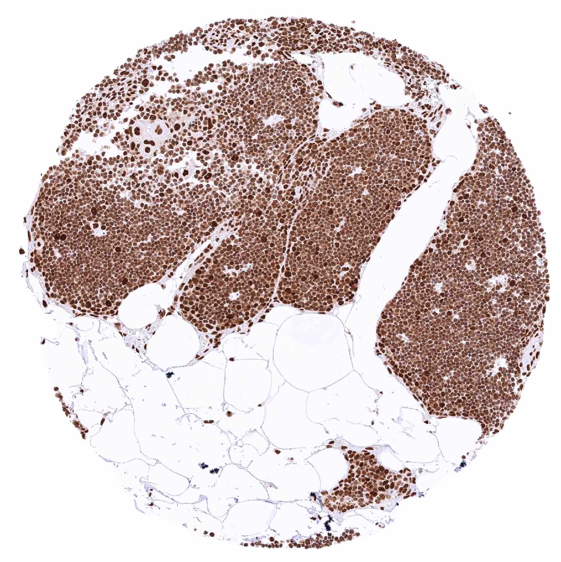
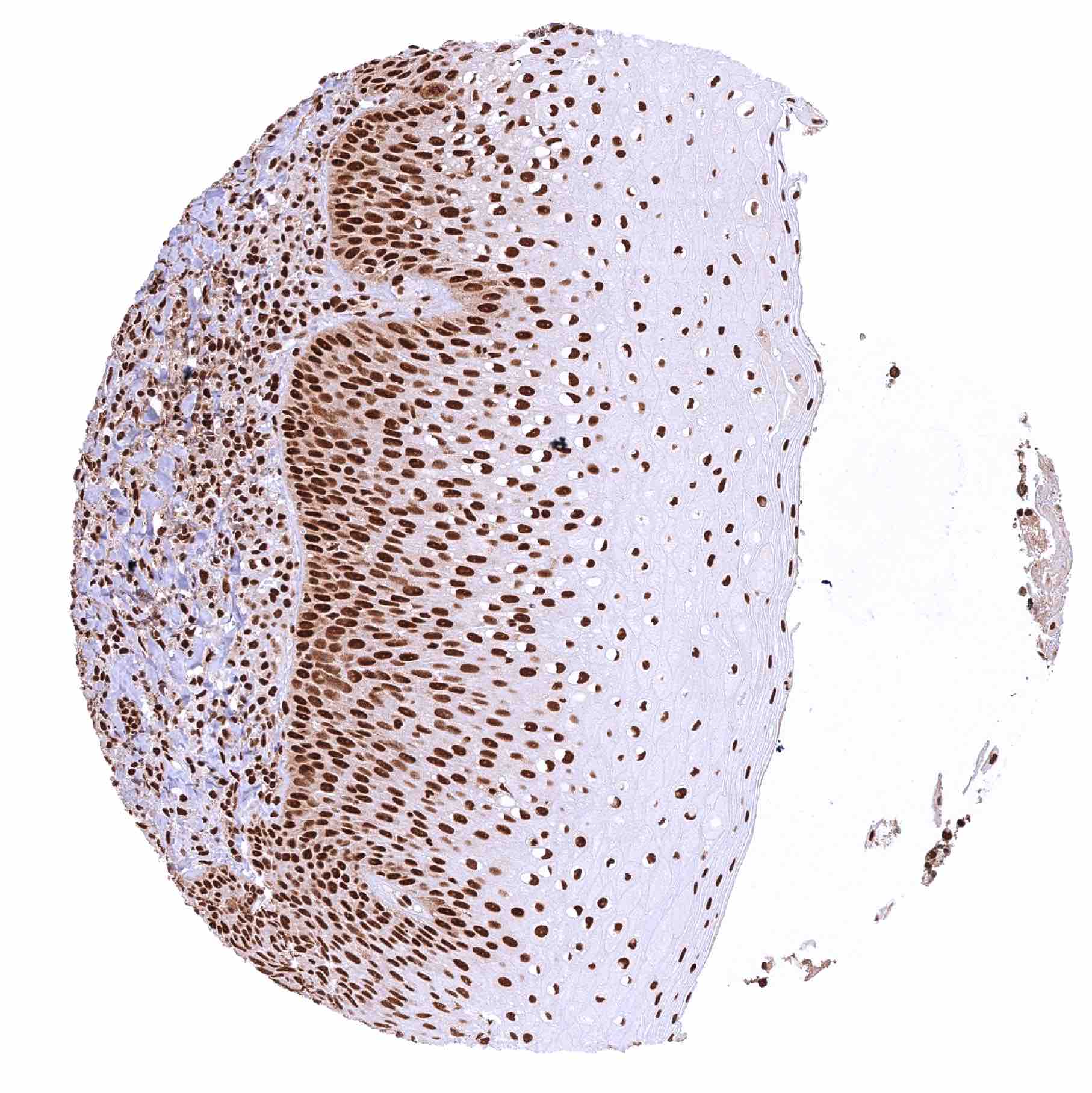
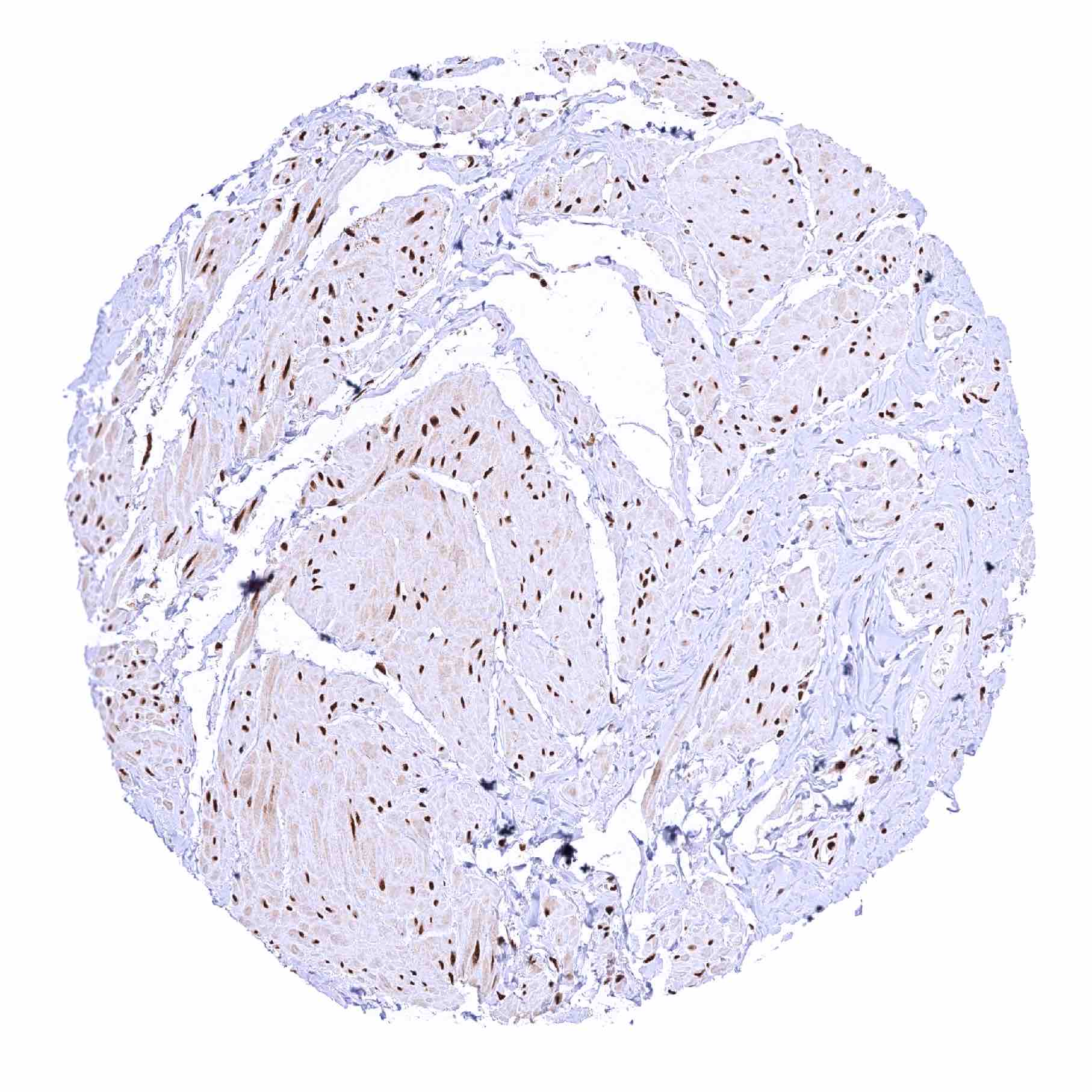
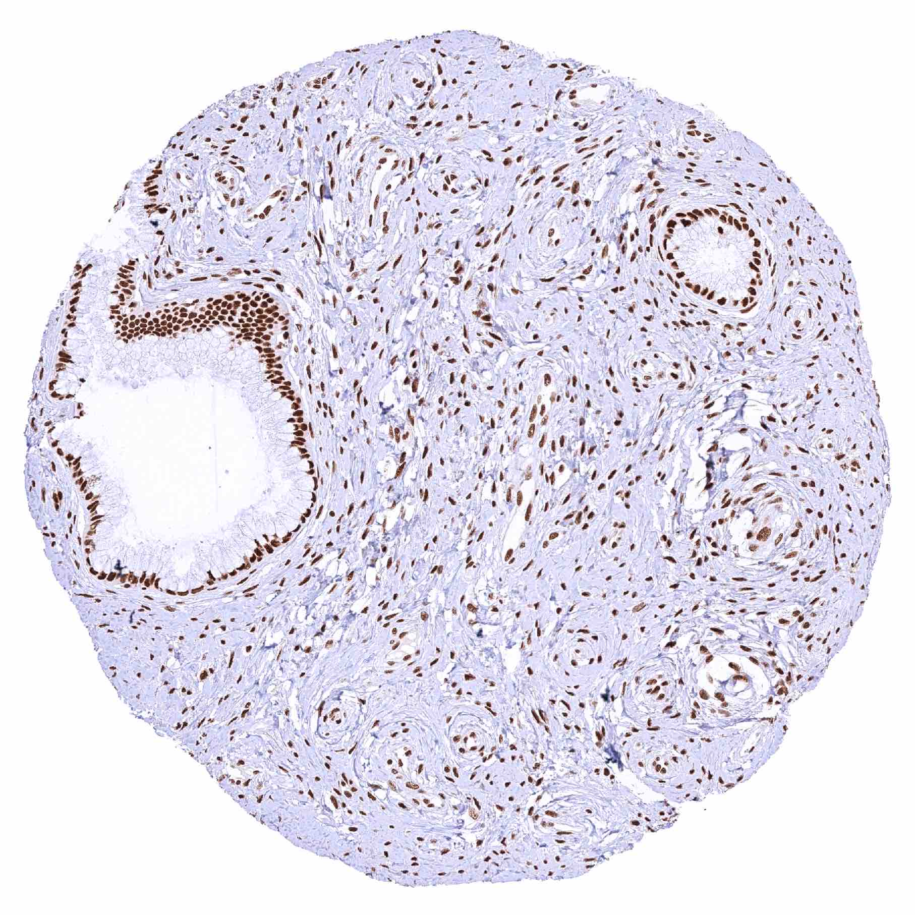
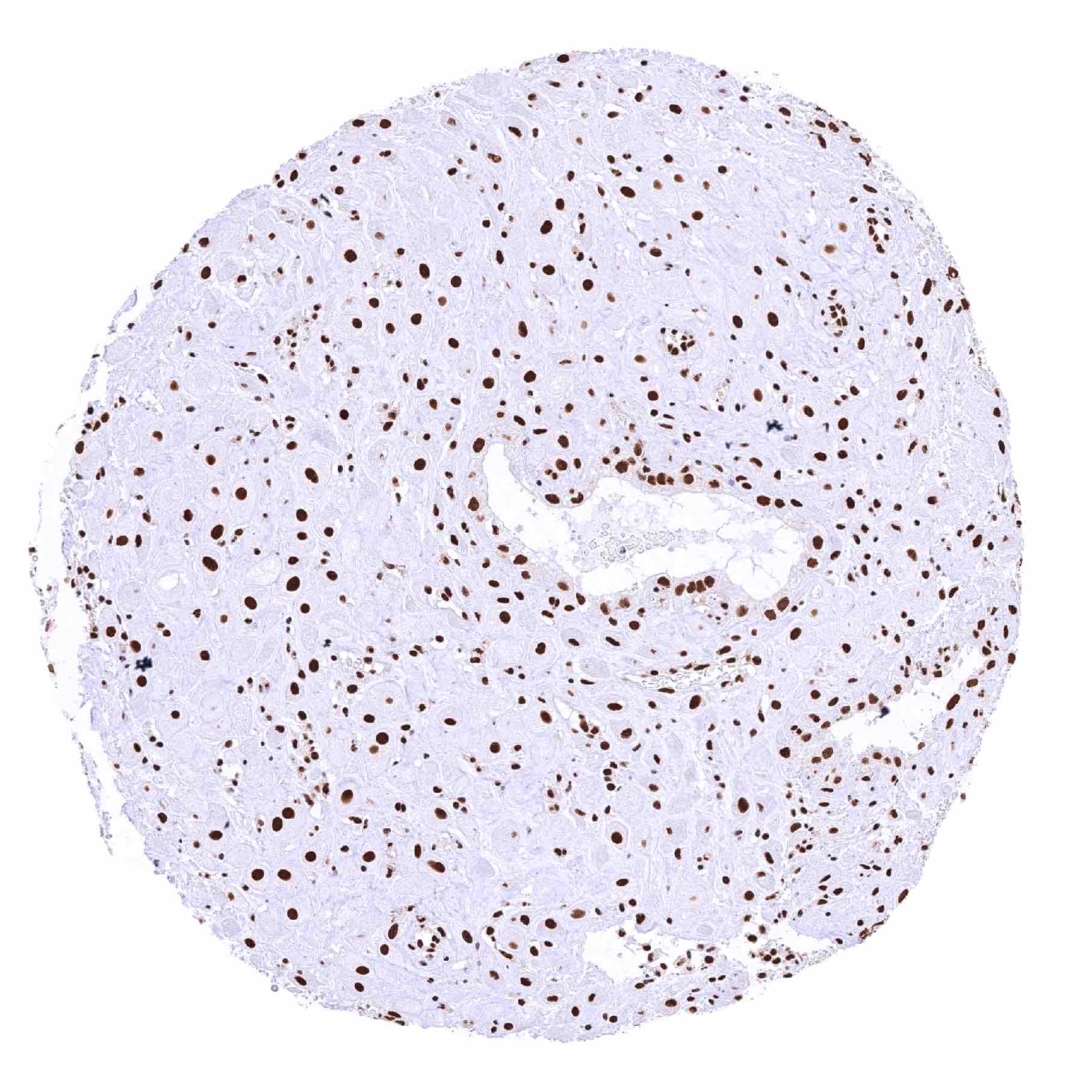
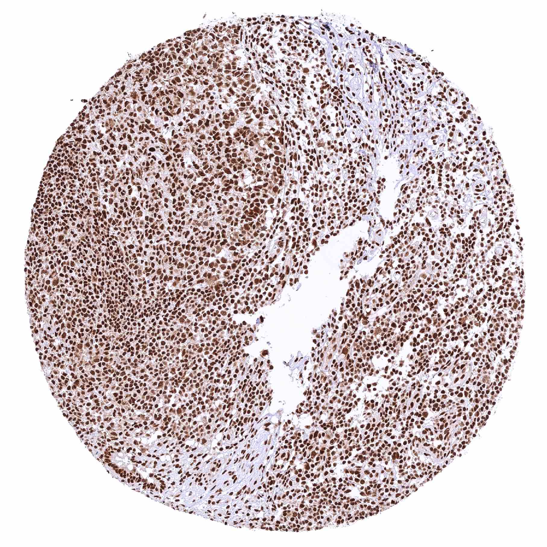
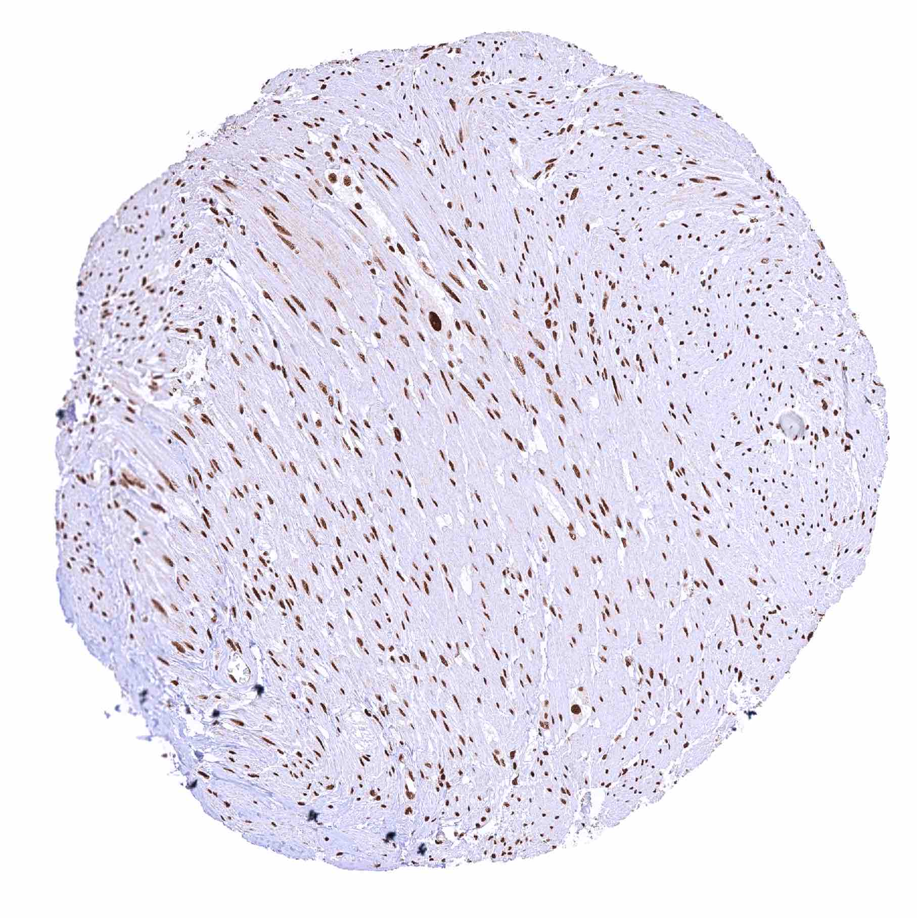
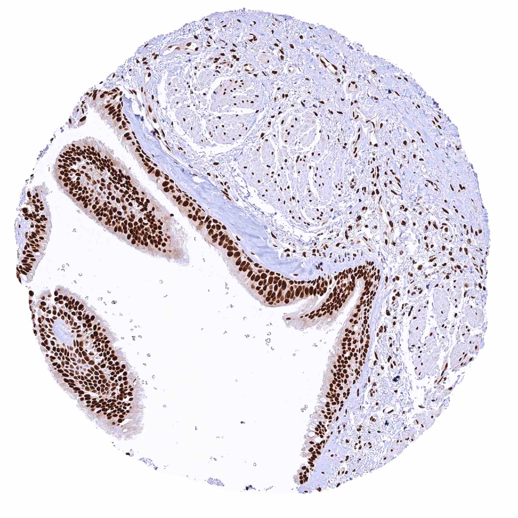
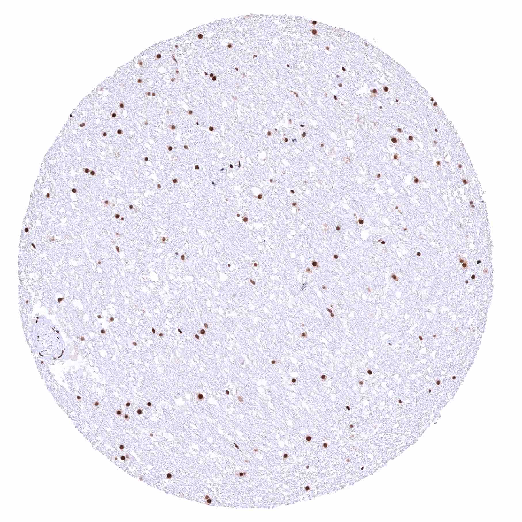
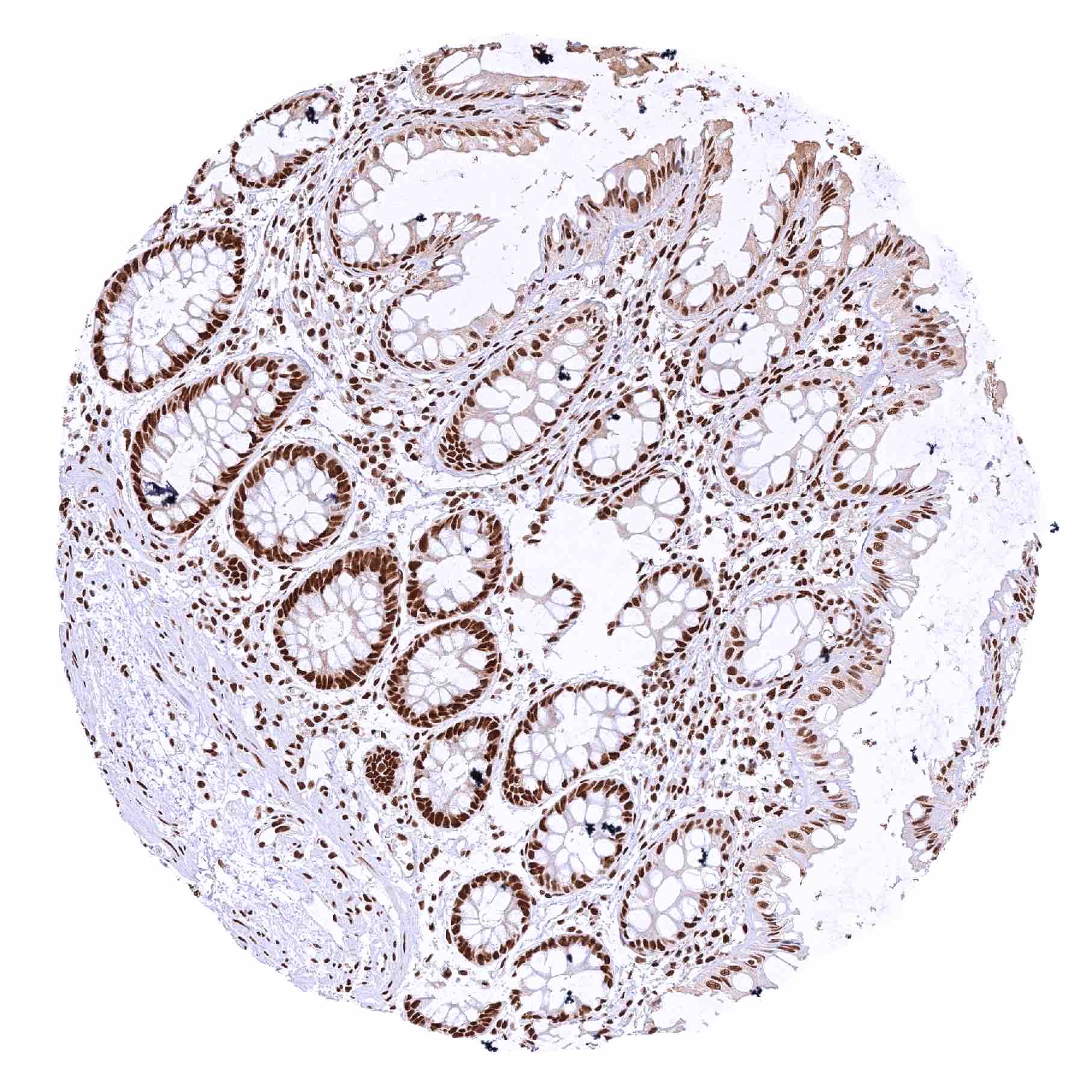
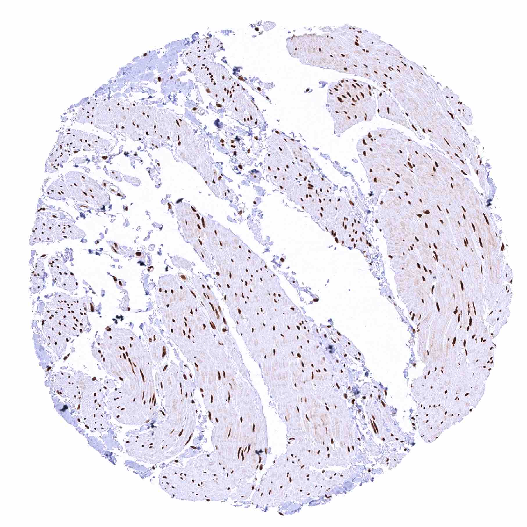
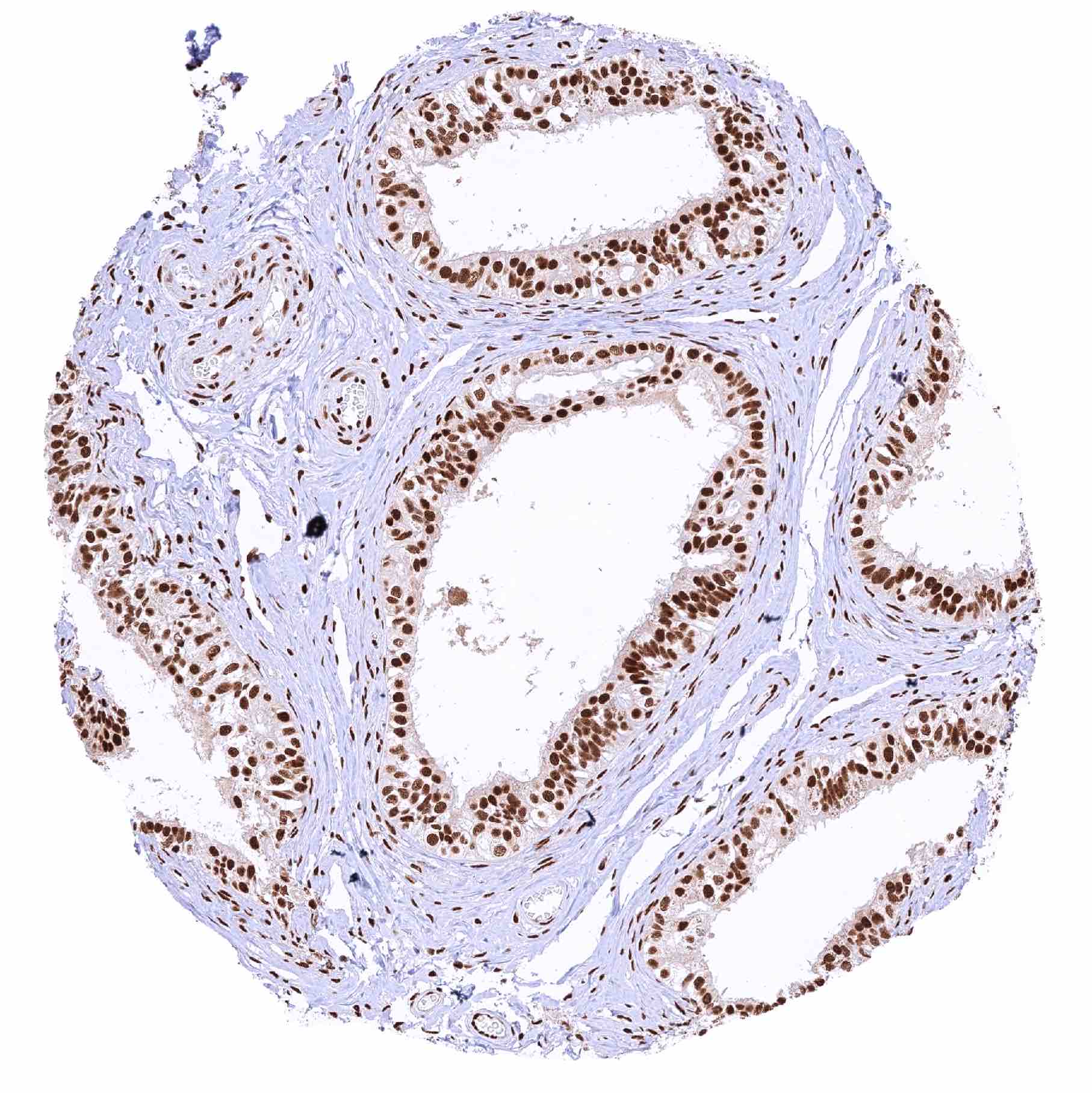
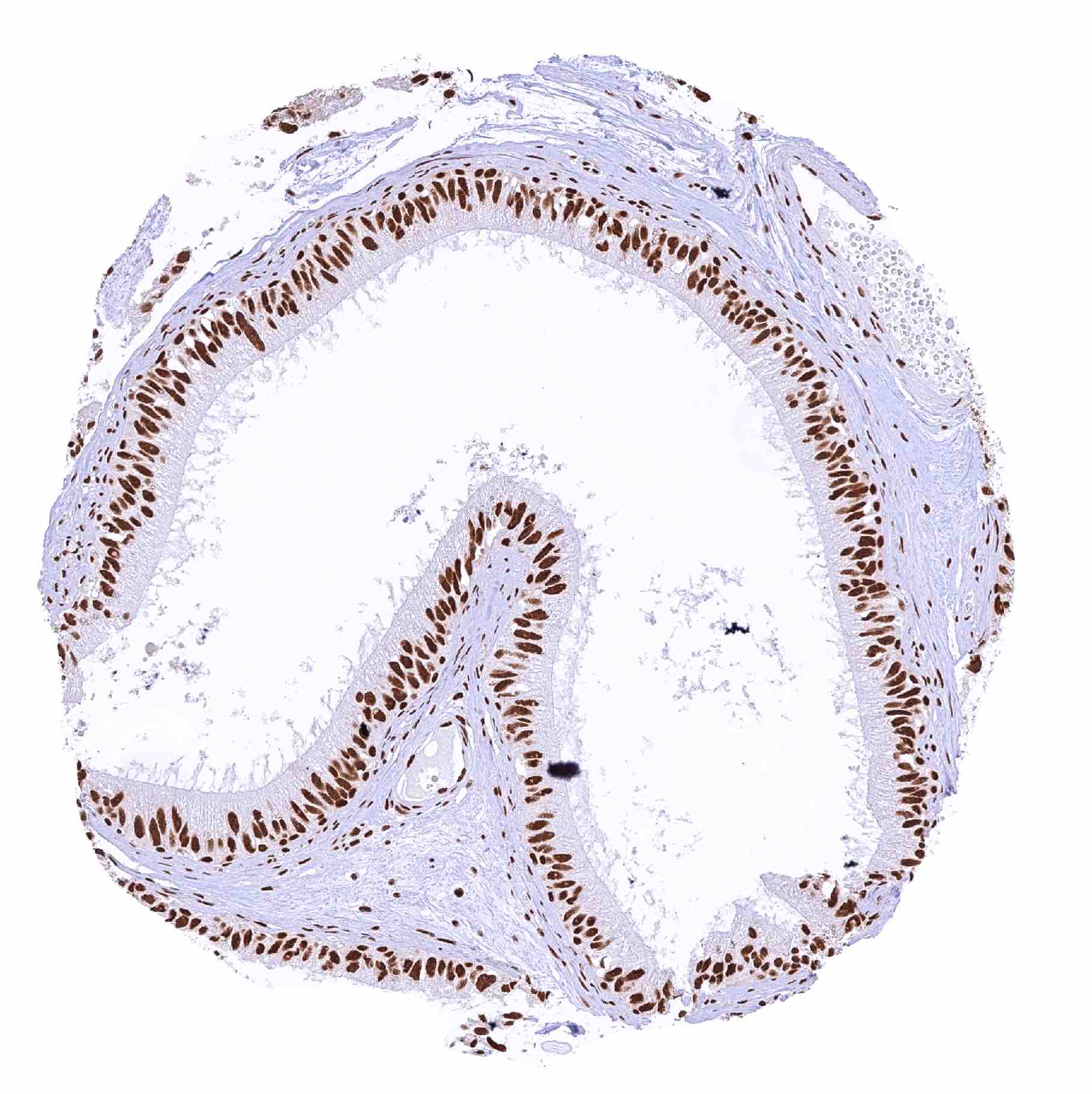
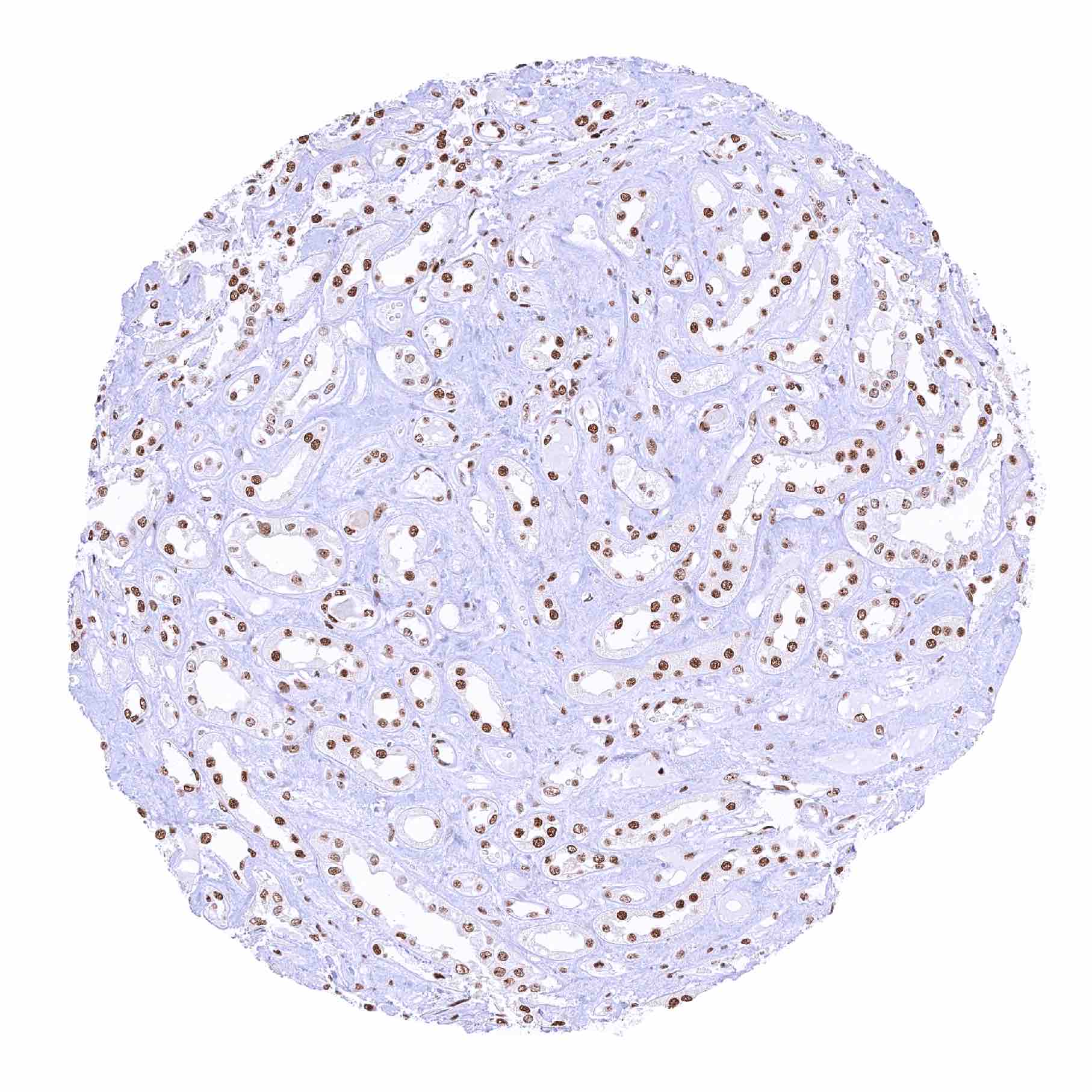
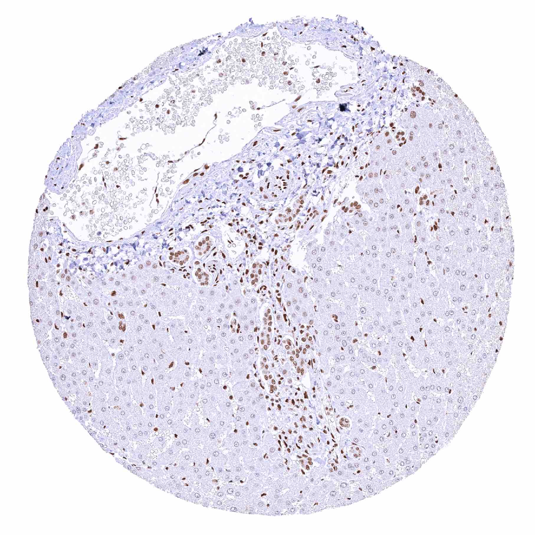
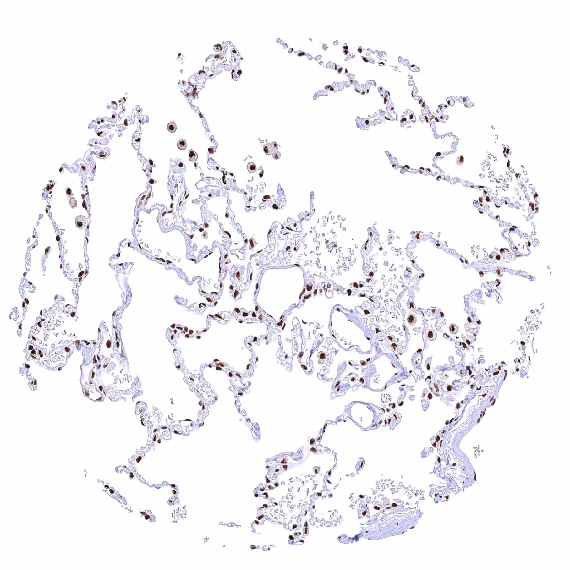
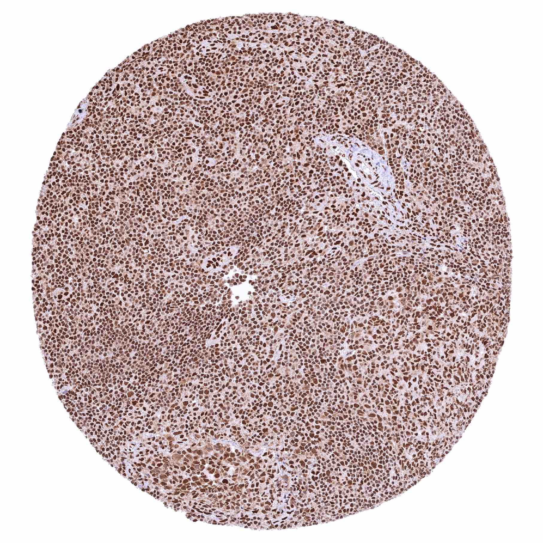
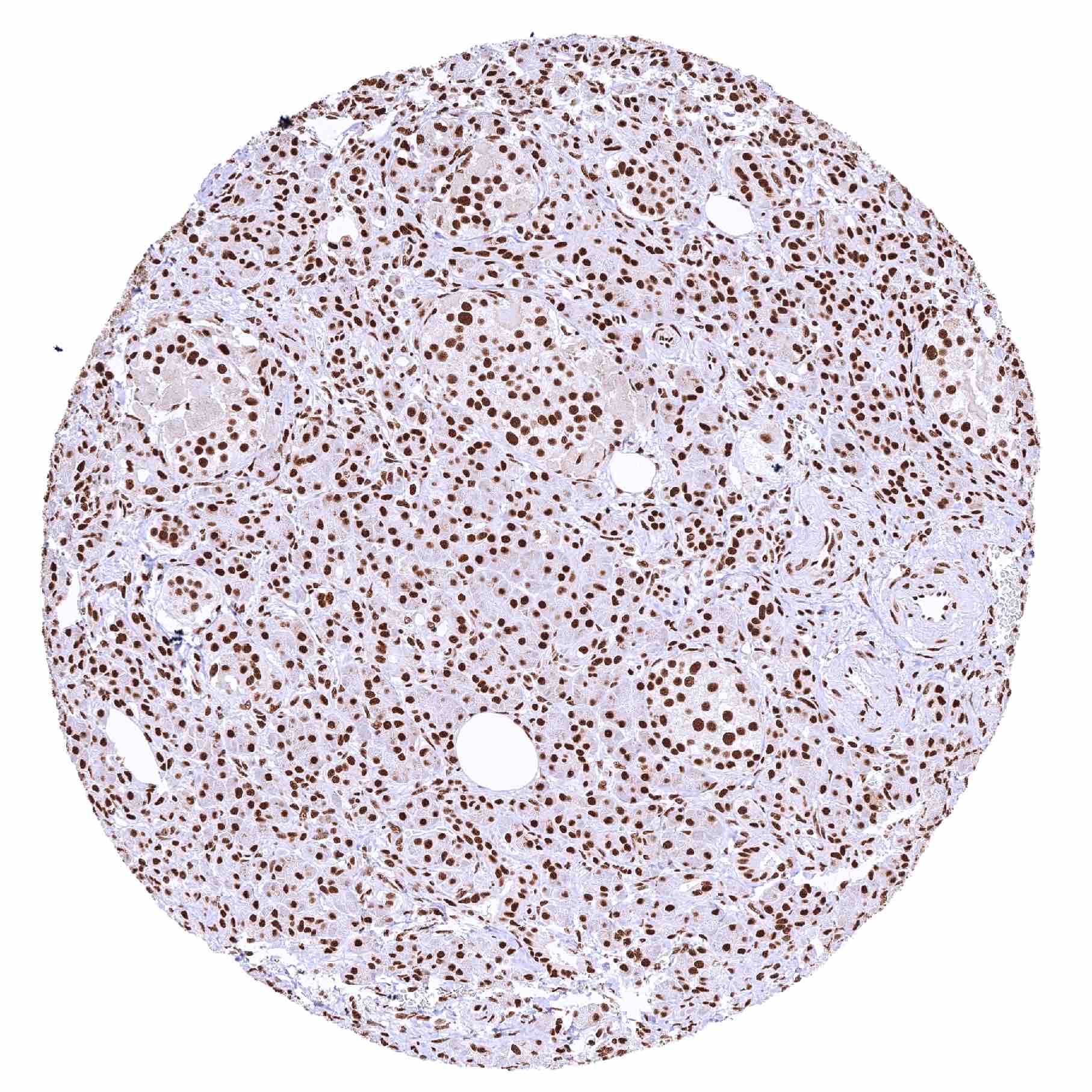
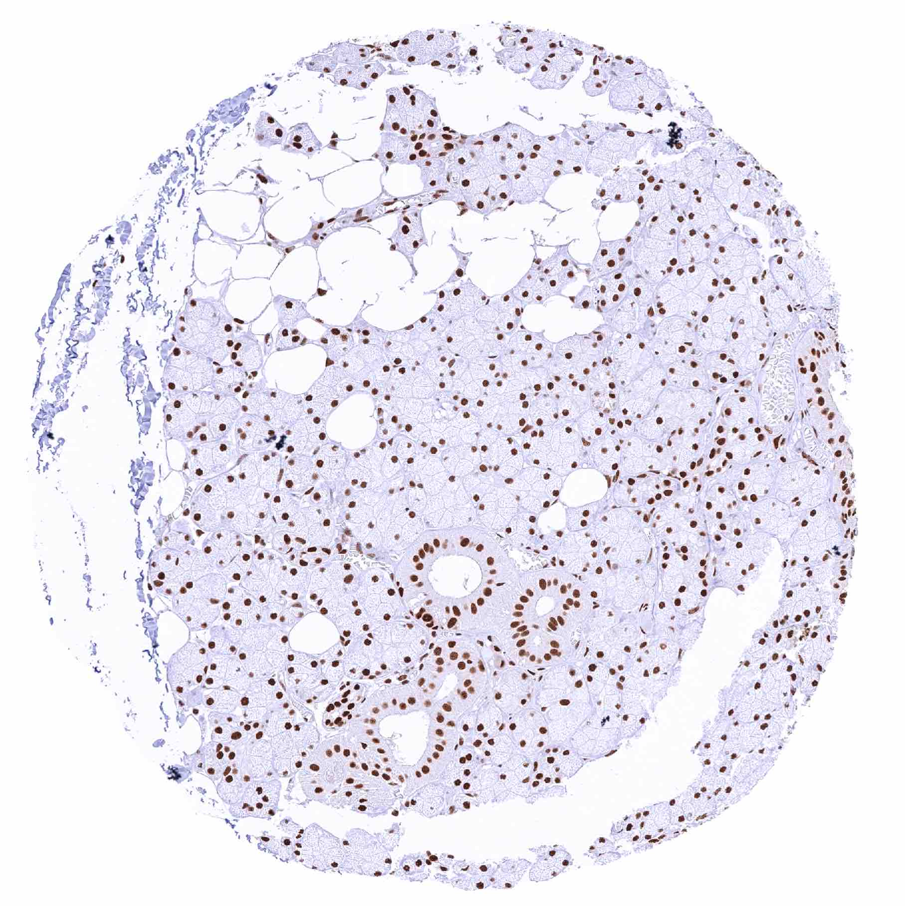
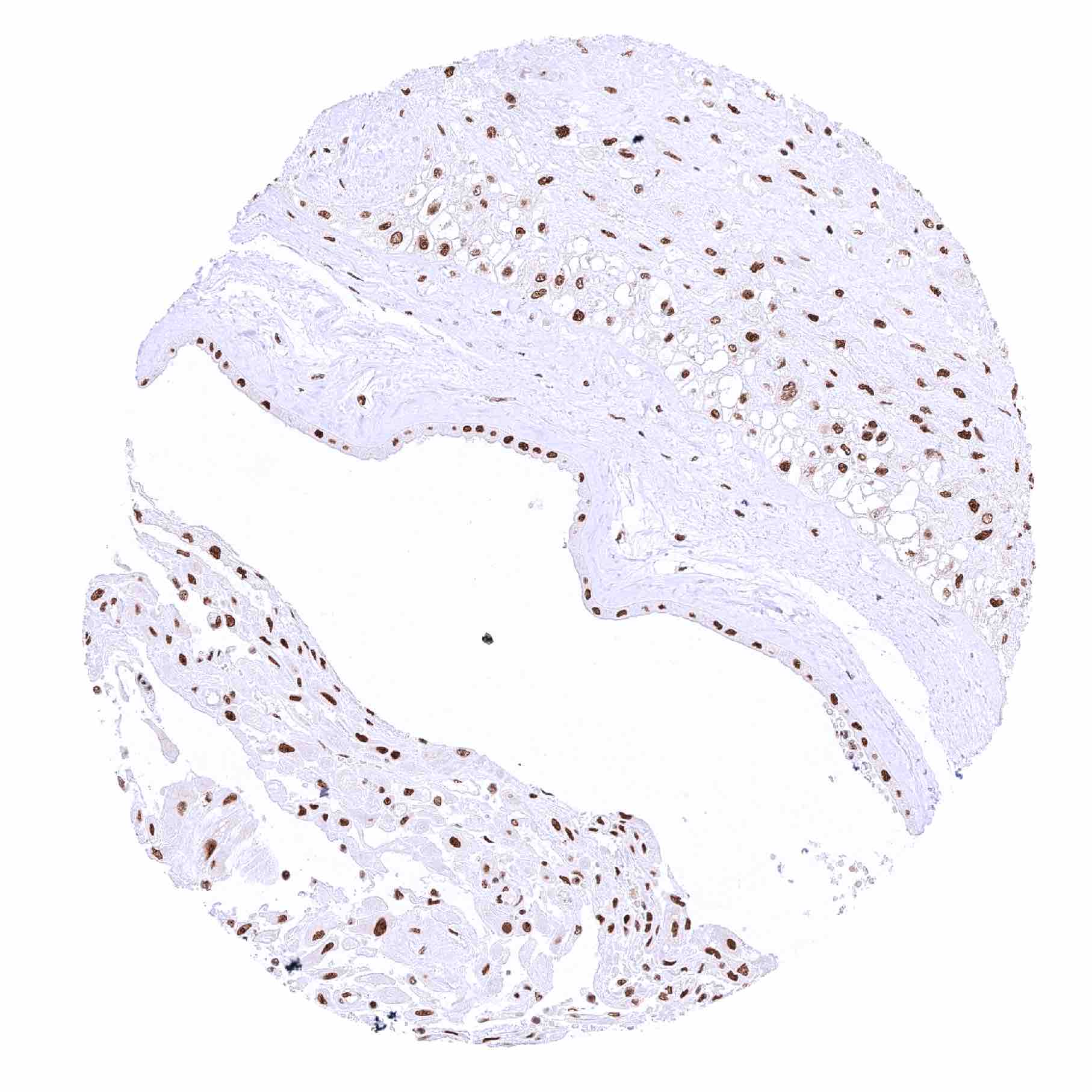
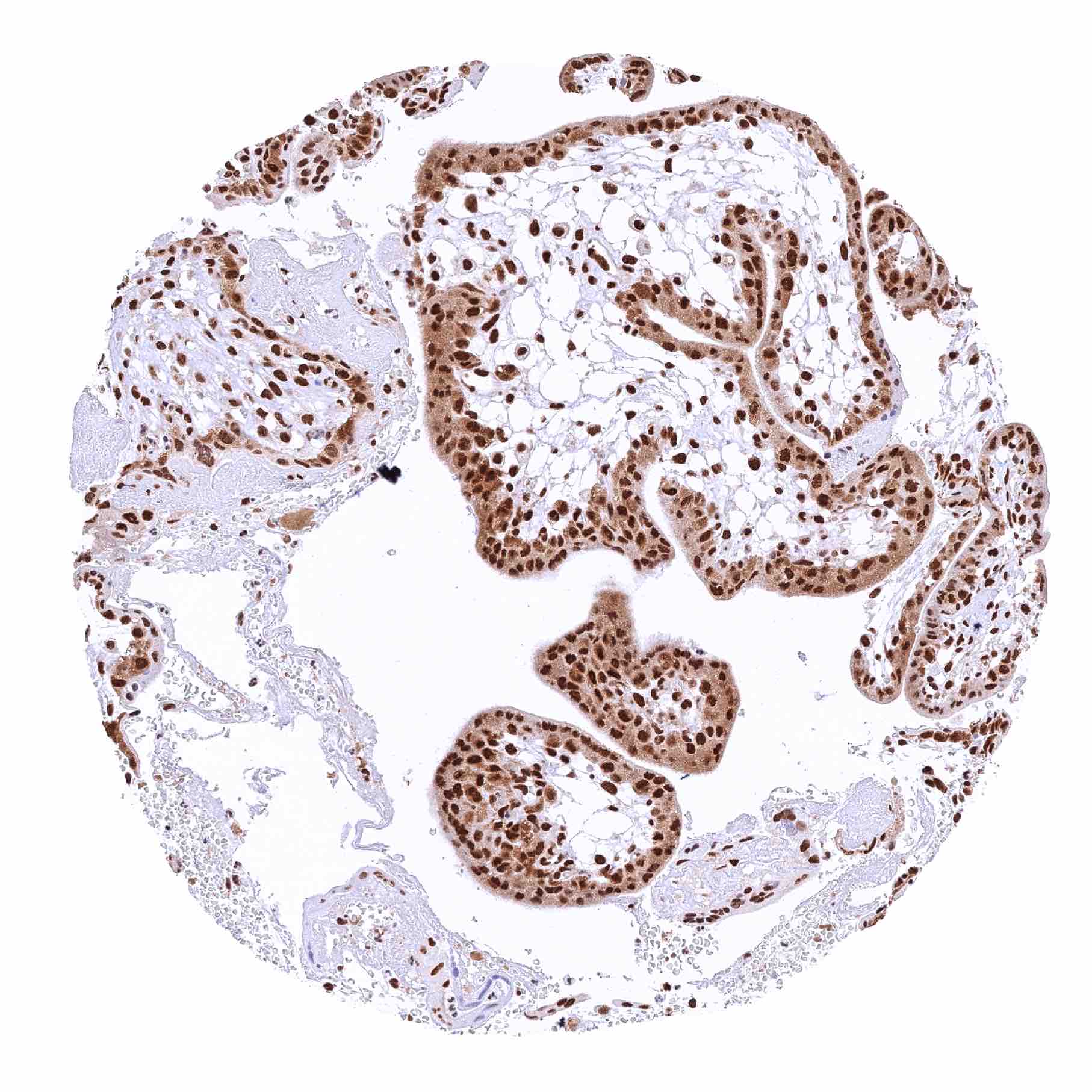
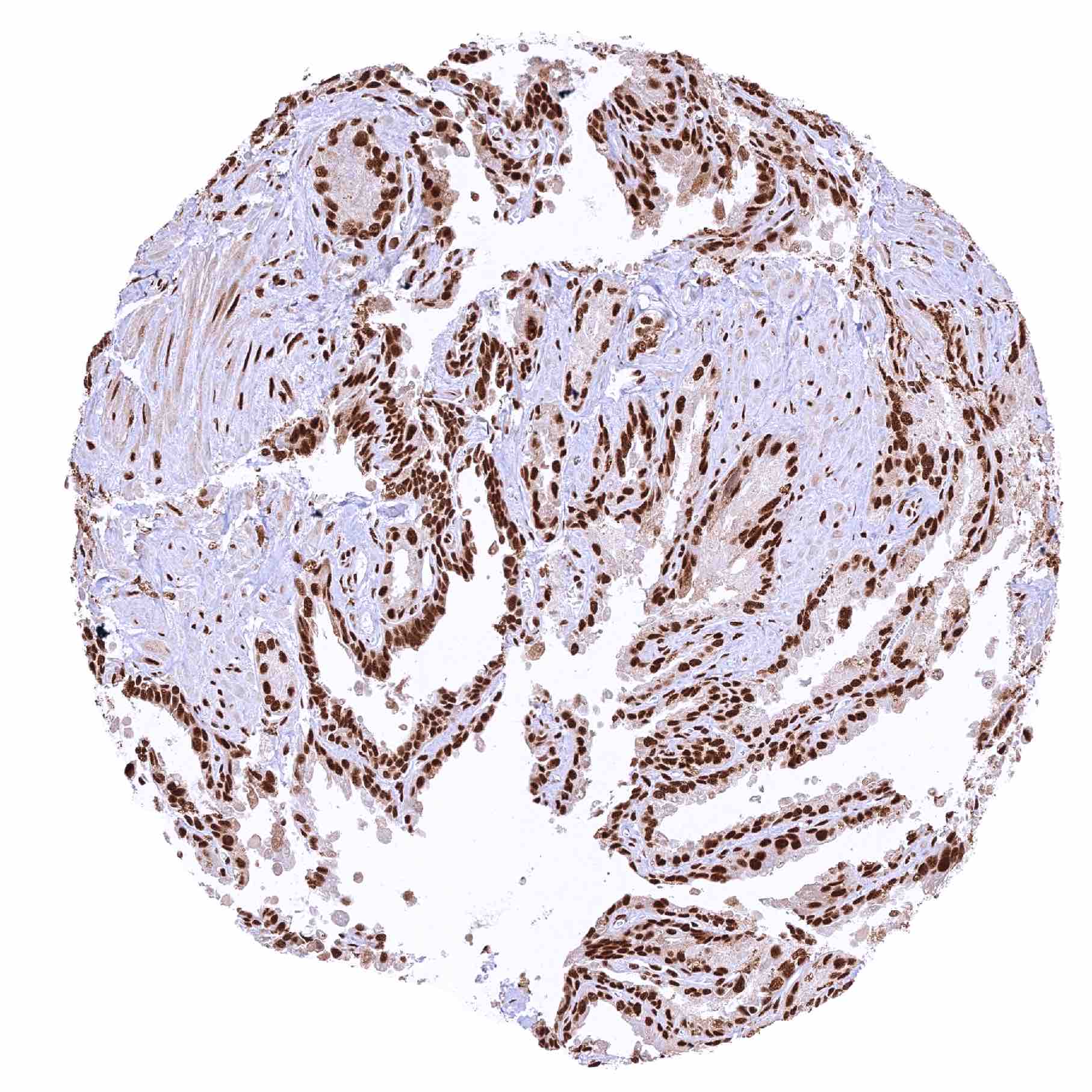
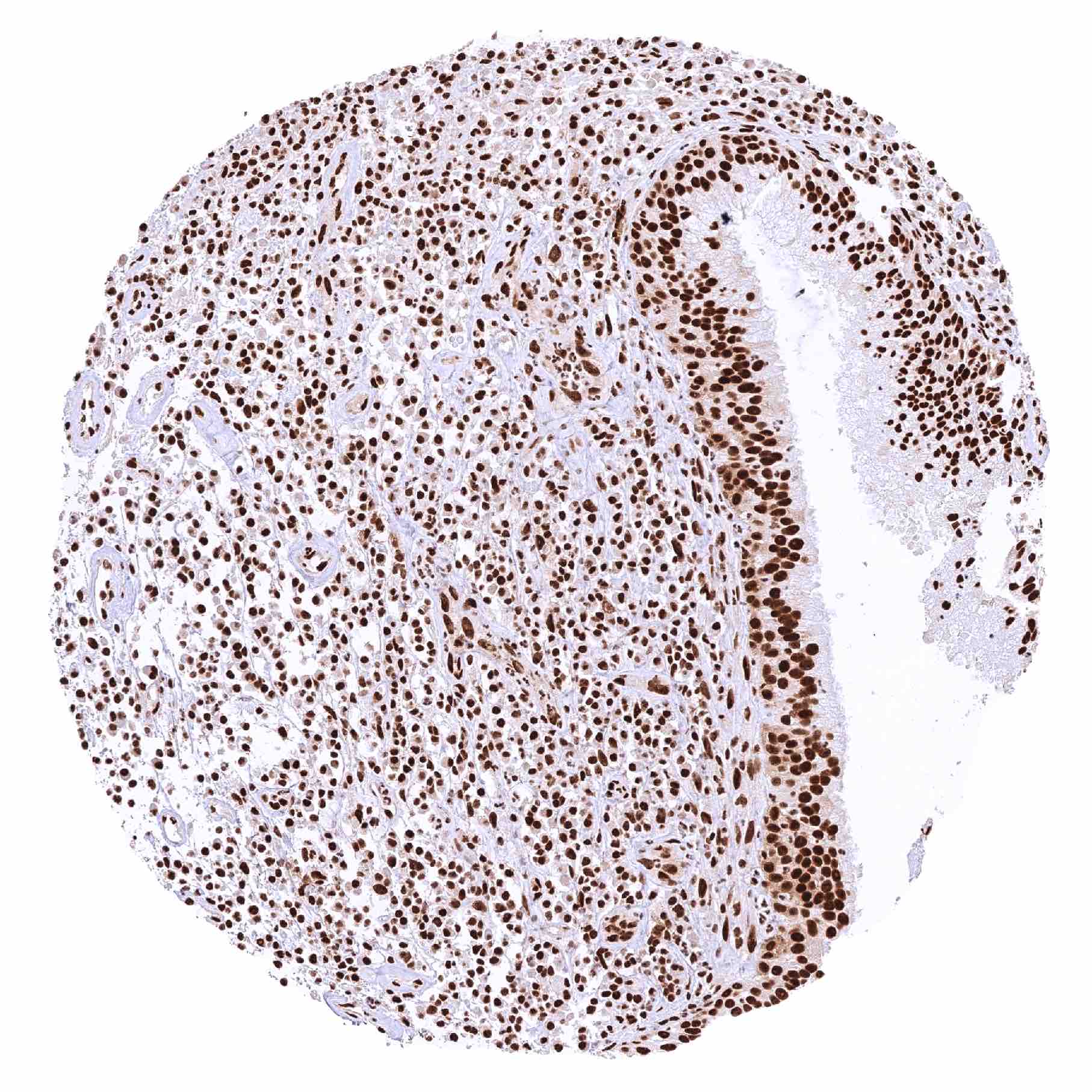
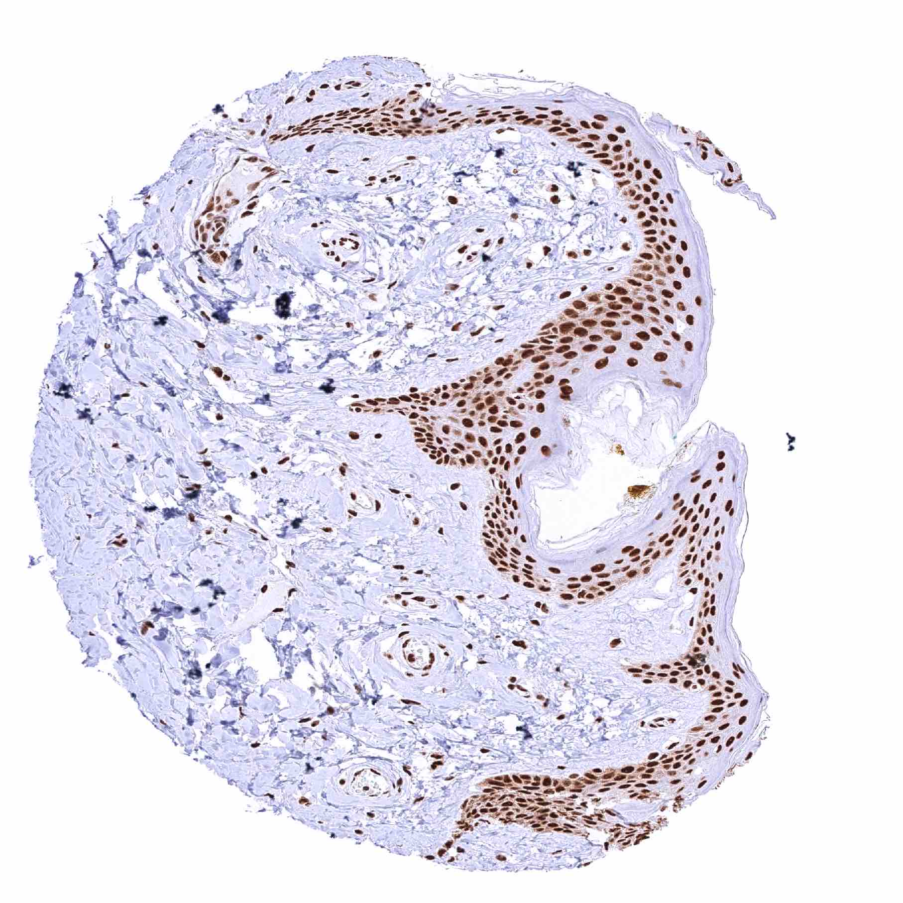
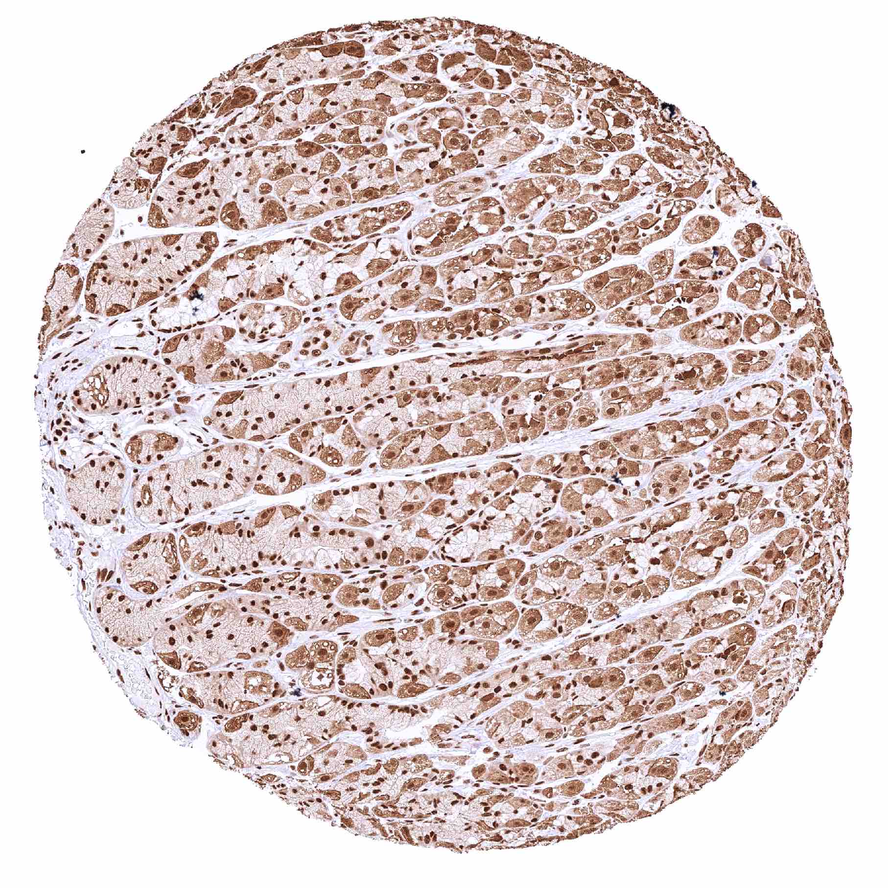
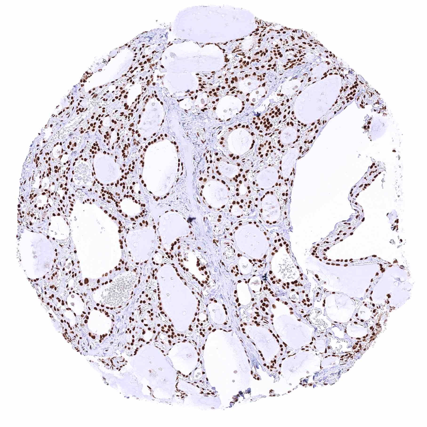
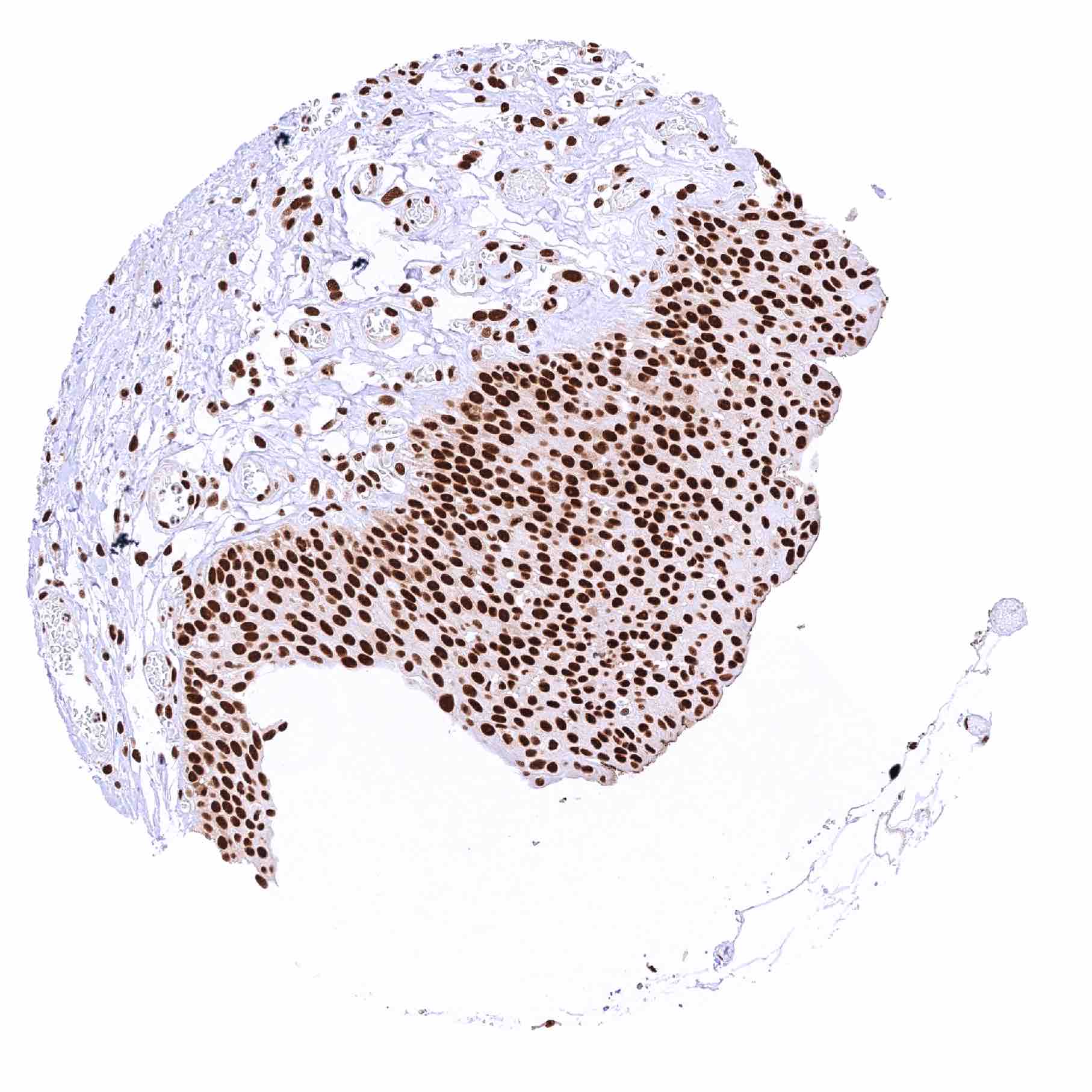
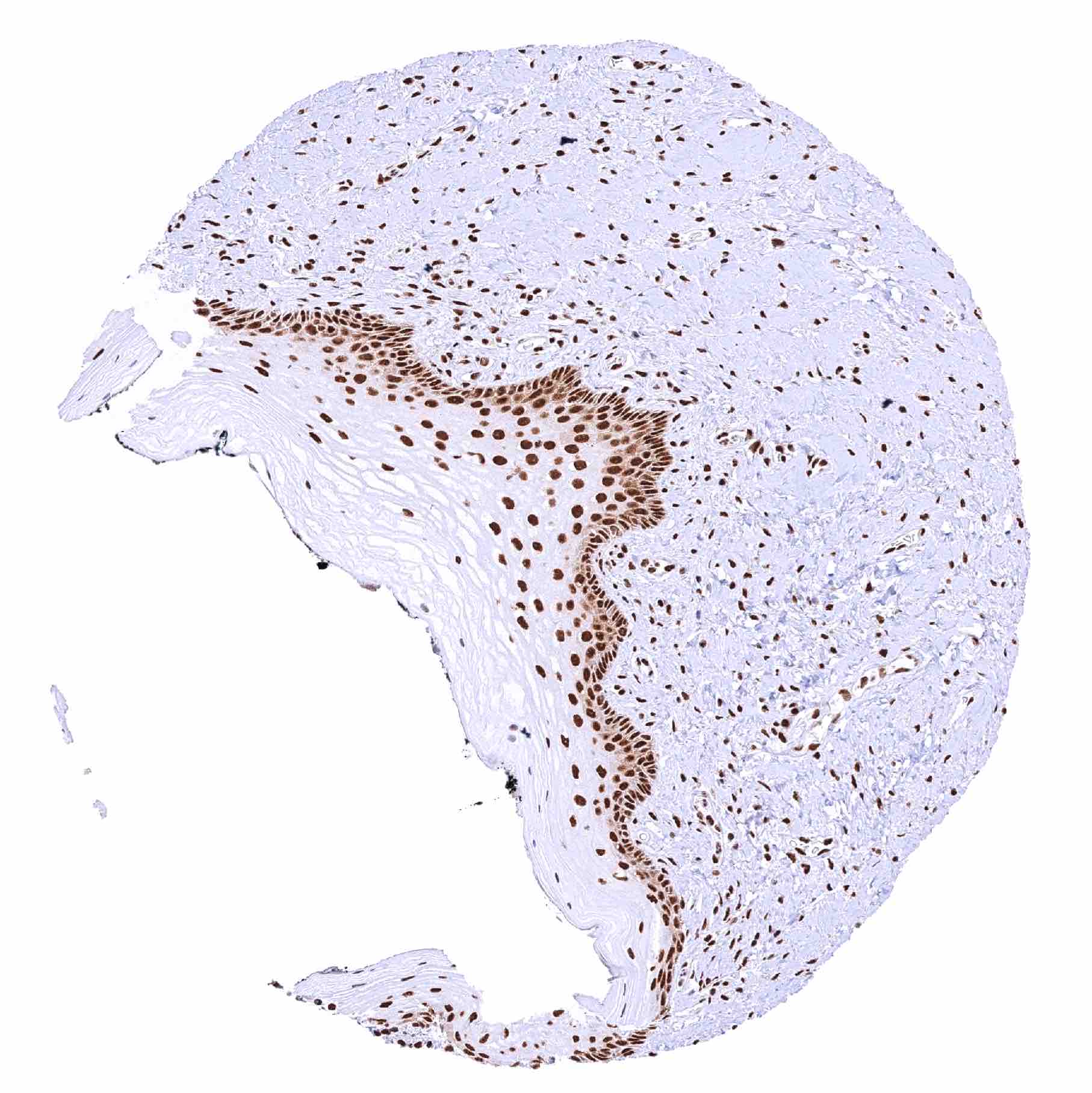
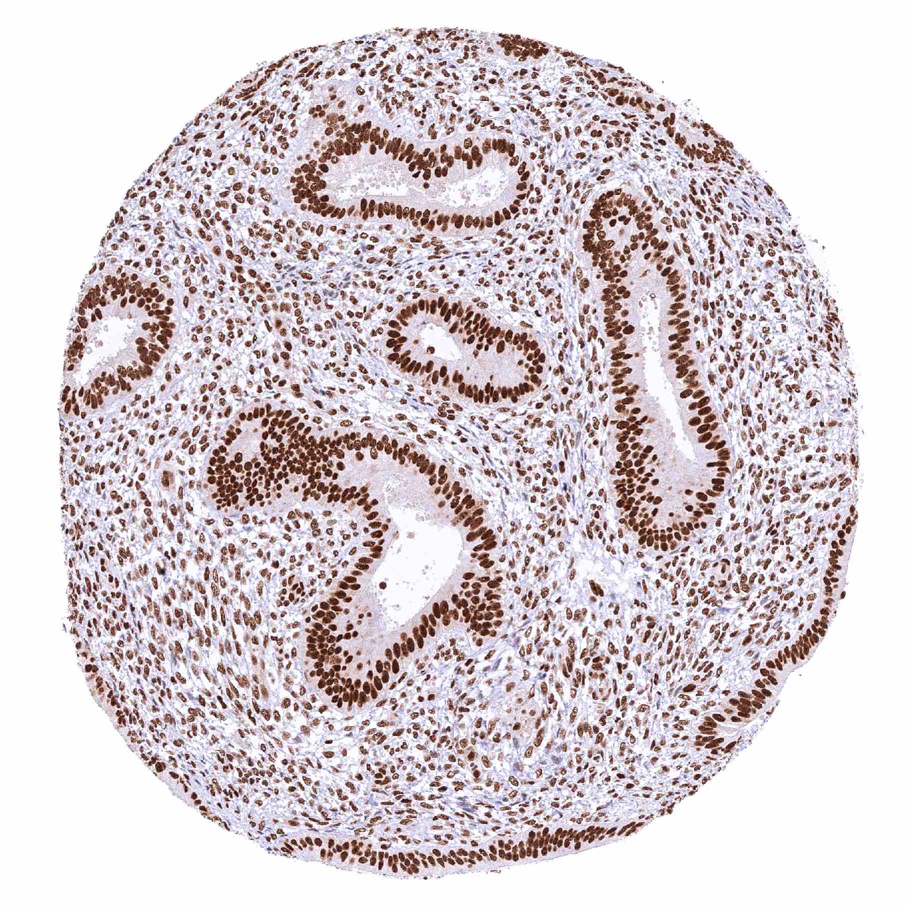
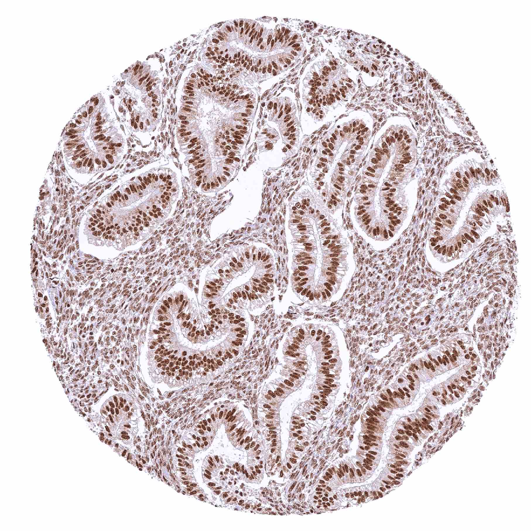
Mre11 antibody [HMV328] HistoMAX™


Go to Cancer Tissue Gallery
  
 Go back to product page
  

Go to HistoMAX page


Adrenal gland

Aorta, media

Appendix, mucosa

Appendix, muscular wall – Strong nuclear MRE11 staining of all cells


Bone marrow

Breast

Bronchus, mucosa

Cerebellum (white matter)


Cerebellum, cortex (molecular layer, Purkinje cell layer, granule cell layer)

Cerebrum, grey matter

Cerebrum, white matter

Colon descendens, mucosa – Distinct nuclear MRE11 staining of all cells. Slight but significant decrease of MRE11 staining from the crypt baseto the superficial epithelial cell layers


Colon descendens, muscular wall

Duodenum, Brunner gland

Duodenum, mucosa

Epididymis (Cauda)


Epididymis (Corpus)

Esophagus, squamous epithelium – Slight but significant decrease of MRE11 staining from the basal-suprabasal to the superficial cell layers of the squamous epithelium

Fallopian tube, mucosa

Fat


Gallbladder, epithelium

Heart muscle – Strong nuclear MRE11 staining of all cells

Ileum, mucosa

Kidney, cortex


Kidney, medulla

Kidney, pelvis, urothelium – Strong nuclear MRE11 staining of all cells. In addition, a weak to moderate cytoplasmic MRE11 staining occurs in some samples

Liver – Strong nuclear MRE11 staining of most cell types. In this sample, MRE11 staining is completely absent in hepatocytes

Liver – Strong nuclear MRE11 staining of most cells. In this sample, MRE11 staining is absent in hepatocytes


Liver – Strong nuclear MRE11 staining of most cells. MRE11 staining is weaker (weak to moderate) in hepatocytes

Lung

Lymph node

Ovary, corpus luteum


Ovary, stroma

Pancreas

Parotid gland

Pituitary gland, anterior lobe


Pituitary gland, posterior lobe

Placenta (amnion and chorion)

Placenta, early – Distinct nuclear MRE11 staining of all cells. In addition, weak to moderate cytoplasmic staining of trophoblast cells

Placenta, mature – Distinct nuclear MRE11 staining of all cells, although the nuclear staining is somewhat weaker in syncytiotrophoblast cells than in all other cell types


Prostate

Rectum, mucosa

Seminal vesicle

Sinus paranasales


Skeletal muscle – Strong nuclear MRE11 staining of all cells

Skin, hair follicle and sebaceous glands

Skin

Stomach, antrum – Strong nuclear MRE11 staining of all cells. A distinct cytoplasmic MRE11 staining of parietal cells represents a (tolerable) cross-reactivity


Stomach, antrum

Testis – Distinct nuclear MRE11 staining of all cells although the staining intensity of germ cells decreases from spermatogonia to spermatids

Thymus – Strong nuclear MRE11 staining of all cells. A few dispersed cells stand out because of a particularly strong nuclear MRE11 staining

Thyroid gland


Tonsil, surface epithelium

Urinary bladder, muscular wall

Urinary bladder, urothelium – Strong nuclear MRE11 staining of all cells

Uterus, ectocervix


Uterus, endocervix

Uterus, endometrium (pregnancy)

Uterus, endometrium (proliferation)

Uterus, endometrium (secretion)


| TOP |